Embed Size (px)
Citation preview
New Electrical Stimulation Techniques in Dynamic Myoplasty
Erik D.H. Zonnevylle
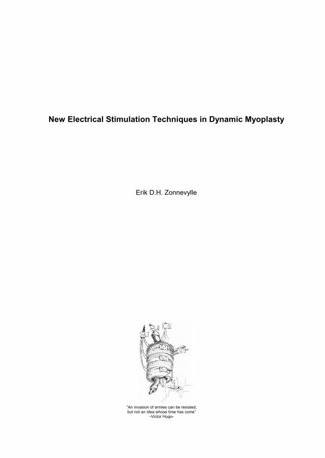
“An invasion of armies can be resisted, but not an idea whose time has come”
-Victor Hugo-
2
Zonnevylle, Erik Dirk Hendrik
New Electrical Stimulation Techniques in Dynamic Myoplasty
ISBN: 90-393-3153-7
Print: A-D Druk BV Zeist
Design and Layout: Marjo & Erik Zonnevylle
No part of this book may be reproduced in any form without written permission of the author
The studies described in this thesis were financially supported by grants from the Alliant Community
Trust Fund, the Jewish Hospital Foundation in Louisville, KY, and the Stichting Prof. Michael van
Vloten Fonds in The Netherlands.
Publication of this thesis was financially supported by:
Adriaan & Adriana Zonnevylle
Jan & Marie-Helène Bender
Johnson & Johnson Medical
Ortomed BV
AB Medical prs BV
Handen Centrum Utrecht
Smith & Nephew Hoofddorp, First Choice in Woundmanagement
Nederlandse Vereniging voor Plastische Chirurgie
Divisie Plastische, Reconstructieve & Handchirurgie, Universitair Medisch Centrum te Utrecht
3
New Electrical Stimulation Techniques in Dynamic Myoplasty
Nieuwe Elektrische Stimulatie Technieken in Dynamische Spierplastieken
(met een samenvatting in het Nederlands)
Proefschrift
ter verkrijging van de graad van doctor aan de Universiteit Utrecht op gezag van Rector Magnificus,
Prof. dr. W.H. Gispen, ingevolge het besluit van het College voor Promoties in het openbaar te
verdedigen op vrijdag 18 oktober 2002 ‘s-middags om 12.00 uur
door
Erik Dirk Hendrik Zonnevylle
geboren op 6 november 1967 te Dordrecht.

4
Promotor:
Moshe Kon, M.D., Ph.D., Professor and Head of the Division of Plastic, Reconstructive & Hand
Surgery, Department of Surgery, University Medical Center, Utrecht, The Netherlands.
Co-promotor:
John H. Barker, M.D., Ph.D., Associate Professor of Surgery, Director of Plastic & Hand Surgery
Research, University of Louisville, Louisville, Kentucky, USA.

5
to 35 mongrel dogs

6
Reading committee:
L.M.A. Akkermans, Ph.D., Professor of the Division of Experimental Surgery, Department of Surgery,
University Medical Center, Utrecht, The Netherlands
C.G.M.I. Baeten, M.D., Ph.D., Professor of the Division of Colorectal Surgery, Department of
Surgery, University Hospital Maastricht, Maastricht, The Netherlands
T.A. Boon, M.D., Ph.D., Professor of the Division of Urology, Department of Surgery, University
Medical Center, Utrecht, The Netherlands
P.J. Guelinckx, M.D., Ph.D., Professor of Plastic Surgery, General Hospital Salvator St-Ursula,
Hasselt, Belgium
S. Salmons, Ph.D., Professor of the Department of Human Anatomy and Cell Biology, University of
Liverpool, Liverpool, U.K.
Paranimfen:
Willem van Wolferen
Marjo Zonnevylle-Bender
7
Contents
Chapter 1: General introduction 9
Chapter 2: Sequential stimulation in an isometric setup 17
Chapter 3: Sequential stimulation in a non-isometric setup 33
Chapter 4: Closed-loop control: feasibility in a neo-sphincter model 45
Chapter 5: Sequential stimulation and closed-loop control: 63 application in a graciloplasty model
Chapter 6: General discussion 81
Summary 97
Samenvatting in het Nederlands 101
References 105

9
Chapter 1
General introduction
“Obstacles are those frightful things you seewhen you take your eyes off your goal”
-Henry Ford-

Chapter 1
10
Introduction
The wish for reconstruction of parts of the body is probably as old as their
mutilation. For centuries documents have appeared reporting on sometimes
brilliant efforts of creative minds to reconstruct what was lost, using surgery as a
tool. Well-known and interesting to read are the reports concerning early
reconstructions of the nose. The demand for surgical reconstruction was
increased by the vast amount of casualties in the first world war. At the same
time the need to share knowledge and experience brought together surgeons
working in this evolving field of ‘plastic surgery’. Today, plastic surgery is an
independent medical specialty, in which reconstructive surgery plays a central
role. The evolution of reconstructive surgery has had many accelerations, which
were often preceded by the development of new surgical techniques. These
new surgical techniques, almost always paved the way for an array of new
applications. A good example is the development of the microsurgical
anastomosis of small blood vessels. Rendering this technique made it possible
to transplant tissue from less critical donor sites to more precarious recipient
defects in a one-stage procedure. A multitude of flaps, consisting of all kinds of
tissues, were developed to fill an even larger multitude of recipient defects.
The mean goal of those microsurgical procedures in the early sixties and
seventies was to fill defects. After mastering the microsurgical technique,
attention was directed to restoring form. Restoration of form is challenging and
sometimes reaches artistic levels even with current surgical techniques. Today,
besides the aesthetic side of reconstructive surgery, restoration of function plays
an increasingly important role. However, in many instances its possibilities are
limited due to the restriction of applicable key (surgical) techniques.
Therefore, this thesis reports on new techniques, which are believed to be
meaningful in the development of reconstructive surgery dealing with functional
muscular deficits.
General introduction
11
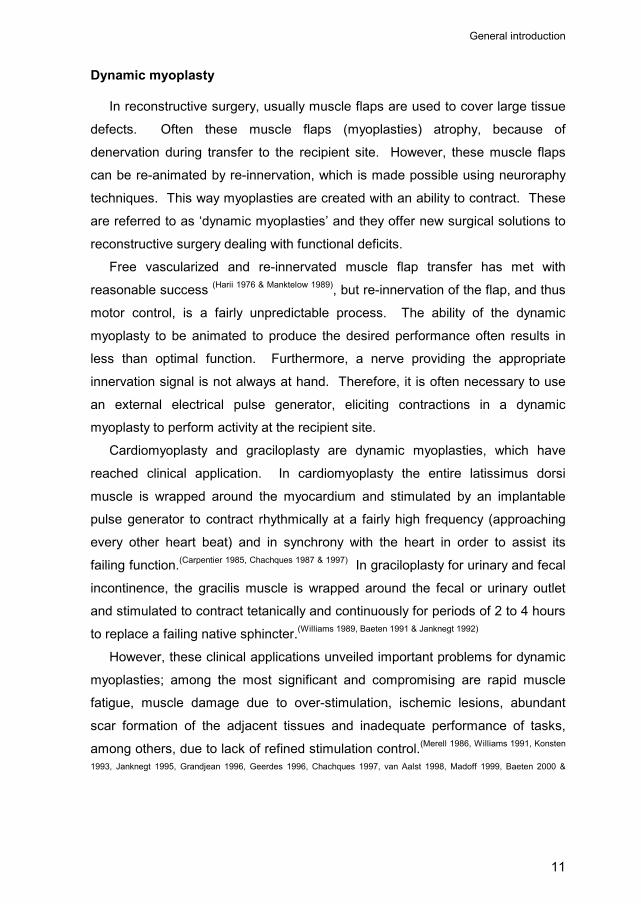
Dynamic myoplasty
In reconstructive surgery, usually muscle flaps are used to cover large tissue
defects. Often these muscle flaps (myoplasties) atrophy, because of
denervation during transfer to the recipient site. However, these muscle flaps
can be re-animated by re-innervation, which is made possible using neuroraphy
techniques. This way myoplasties are created with an ability to contract. These
are referred to as ‘dynamic myoplasties’ and they offer new surgical solutions to
reconstructive surgery dealing with functional deficits.
Free vascularized and re-innervated muscle flap transfer has met with
reasonable success (Harii 1976 & Manktelow 1989), but re-innervation of the flap, and thus
motor control, is a fairly unpredictable process. The ability of the dynamic
myoplasty to be animated to produce the desired performance often results in
less than optimal function. Furthermore, a nerve providing the appropriate
innervation signal is not always at hand. Therefore, it is often necessary to use
an external electrical pulse generator, eliciting contractions in a dynamic
myoplasty to perform activity at the recipient site.
Cardiomyoplasty and graciloplasty are dynamic myoplasties, which have
reached clinical application. In cardiomyoplasty the entire latissimus dorsi
muscle is wrapped around the myocardium and stimulated by an implantable
pulse generator to contract rhythmically at a fairly high frequency (approaching
every other heart beat) and in synchrony with the heart in order to assist its
failing function.(Carpentier 1985, Chachques 1987 & 1997) In graciloplasty for urinary and fecal
incontinence, the gracilis muscle is wrapped around the fecal or urinary outlet
and stimulated to contract tetanically and continuously for periods of 2 to 4 hours
to replace a failing native sphincter.(Williams 1989, Baeten 1991 & Janknegt 1992)
However, these clinical applications unveiled important problems for dynamic
myoplasties; among the most significant and compromising are rapid muscle
fatigue, muscle damage due to over-stimulation, ischemic lesions, abundant
scar formation of the adjacent tissues and inadequate performance of tasks,
among others, due to lack of refined stimulation control.(Merell 1986, Williams 1991, Konsten
1993, Janknegt 1995, Grandjean 1996, Geerdes 1996, Chachques 1997, van Aalst 1998, Madoff 1999, Baeten 2000 &
Chapter 1
12
Bardoel 2002) Outcomes in dynamic myoplasties therefore continue to be, to a large
degree, unpredictable.
Muscle fiber types and training protocols
An approach used in dynamic myoplasty to avoid muscle failure due to
fatigue is to train the muscle to enhance fatigue resistance.(Salmons 1969,1976, 1981 &
1994, Pette 1973 &1975, Koller 1994) Skeletal muscles are normally a mixture of various
classes of muscle fibers with different qualities, ranging from slow and relative
fatigue resistant (type 1) to fast and fatigue-prone (type 2b).(Buller 1960, Salmons 1969,
Pette 1997, Scott 2001) The innervation and the function of the muscle determine the
predominance of one fiber type over another. However, striated muscle is
plastic in nature. When the stimulation signal changes, the ratio of the muscle
fiber types changes accordingly. This change in signal can either be a physical
exercise program, re-innervation of the muscle tissue by a different nerve or
electrical stimulation.
In order to improve the endurance of dynamic myoplasties, muscle-training
programs have been designed to transform the myoplasty using an electrical
stimulation regimen. Most training regimens transform the muscle from a fatigue
prone, fast twitch, glycolytic muscle with predominantly type II fibers into a non-
fatiguing, slow twitch, oxidative muscle with predominantly type I fibers.(Salmons
1981)
The trade-off for producing fatigue resistance is a slower contracting muscle
capable of generating less power than its innate character.(Salmons 1981 & 1994, Pette
1984, Chachques 1987, Koller 1994)
Sequential segmental neuromuscular stimulation
Fatigue reduction in dynamic myoplasty is mainly achieved by electrical
stimulation training protocols at the cost of strength, responsiveness and the
chance of ischemic lesions due to constant high pressures. In this thesis a
different, more physiological approach was tested in an attempt to bypass most
of these drawbacks.
General introduction
13
Sustained contraction in skeletal muscle tissue is only possible for a brief
moment, because locally increased pressure impairs perfusion. Therefore in
physiological conditions skeletal muscle performs sustained work by recruiting
different fibers at different times. In this way a constant force is maintained,
while part of the muscle is working and part of the muscle is resting. This type of
contraction enables the muscle to be perfused constantly: while the
microcirculatory blood flow in the contracted part of the muscle is momentarily
impaired, hyperemic flow in the resting part allows metabolites to be washed out
and oxygenation to be increased.
Current electrical stimulation techniques apply a single source voltage, which
causes all of the muscle fibers to contract simultaneously, impairing perfusion
during contraction and leading to rapid fatiguing as explained above. In
sequential segmental neuromuscular stimulation the skeletal muscle is
partitioned into segments, which are alternately stimulated in a sequential order.
This approach causes only one segment of the muscle to perform the necessary
workload, thereby allowing the other non-stimulated segments to recover by
reperfusion, as is the case in physiological contraction of the muscle and leads
to a prolongation of the endurance of the muscle. Similar techniques have been
shown to reduce muscle fatigue.(Petrofsky 1979, Pournezam 1988, Thoma 1991 & Lau 1995)
Closed-loop control
A different perspective to the same problem of current non-physiological
electrical stimulation paradigms in dynamic myoplasties is their performance
control.
Under normal circumstances, a given anatomical muscle can respond to
moderate prolonged activity and brief intense activity. The exact possibility of
response, generated by the muscle, depends on the distribution of myofiber
types contained within the muscle and the pattern of activity in which they are
engaged.(Saltin 1983 & Scott 2001) In normal physiology the performance of the muscle
is tuned to the exact needs, saving performance potential and avoiding long
sustained high internal pressures. However, current applications of dynamic
Chapter 1
14
myoplasty do use tetanic stimulation, resulting in contraction of all myofibers
simultaneously leading to irreversible muscle damage, when prolonged.
Furthermore, this type of on/off control does not allow more intricate applications
of dynamic myoplasty to be developed, in which fine tuned performance control
is mandatory. Therefore, a method of electrically stimulating dynamic
myoplasties was developed, more closely mimicking physiological muscle
performance regulation.
Closed-loop performance feedback control creates the condition in which it is
possible to precisely tune the amount of force generated by the dynamic
myoplasties to the level necessary to carry out the required physiological
function.(Quintern 1997 & Lemay 1997) Economizing peak exertion to the minimum has
beneficial effects on muscle and surrounding tissues by reducing ischemic
lesions, stricture and scar formation. Moreover, this reduction augments the
endurance of the dynamic myoplasty and allows the development of dynamic
myoplasty applications in which fine-tuned performance control is obligatory.
Closed-loop control depends upon the biofeedback, which is offered by
implantable biosensors. This biofeedback can also be used to run algorithms, in
programmable units of implantable stimulators, to process decisions concerning
dynamic myoplasty performance. This offers refinement in complex tasks and
creates possibilities for more demanding implementations of dynamic
myoplasties.
General introduction
15
Hypotheses
This thesis reports on research concerning improvement of electrical
stimulation used in dynamic myoplasties. Hypotheses were formulated before
experiments were conducted. Some expectations were met and some were not.
The most important hypotheses are summarized below:
• Sequential stimulation is feasible and will result in a constant net
performance, while different segments alternate during stimulation
(supported, see chapter 2 & 3).
• Sequential stimulation will enhance blood perfusion of the stimulated
muscle, when compared to conventional stimulation (supported, see
chapter 2 & 3).
• Sequential stimulation will enhance fatigue resistance, when compared
to conventional stimulation (supported, see chapter 2 & 3).
• Sequential stimulation will prevent the need to alter muscle fiber type,
using electrical stimulation training regimens, in graciloplasty (disproved
and rejected, see chapter 3)
• Closed-loop control is feasible in a sequentially stimulated dynamic
myoplasty (supported, see chapter 4 & 5).
• Oscillation damping parameters are effective in closed-loop controlled
sequentially stimulated dynamic myoplasties (supported, see chapter 4).
• Both amplitude and frequency modulation of the stimulation signal in
closed-loop control are feasible in sequentially stimulated dynamic
myoplasties (supported, see chapter 4).
• A combination of sequential stimulation, closed-loop control and
function-controlling algorithms is feasible in dynamic myoplasty
(supported, see chapter 5).
• A combination of sequential stimulation, closed-loop control and
function-controlling algorithms improve the versatility of dynamic
myoplasties (supported, see chapter 5).
Chapter 1
16
• A combination of sequential stimulation, closed-loop control and
function-controlling algorithms in a graciloplasty-model has the potential
to function as a reasonable alternative to the native sphincter function
(supported, see chapter 5).
• The results described in this thesis justify the effort of miniaturizing
stimulating devices capable of combining sequential stimulation, closed-
loop control and function-controlling algorithms, to implantable size
(made plausible, see chapter 6).
17
Chapter 2
Sequential stimulation in an isometric setup Erik Zonnevylle, MD Naveen Somia, MD, PhD
Richard Stremel, PhD Claudio Maldonado, PhD Paul Werker, MD, PhD Moshe Kon, MD, PhD John Barker, MD, PhD Based upon the articles: Zonnevylle E, Somia N, Stremel R, Maldonado C, Werker P, Kon M and Barker J. Alternating muscle stimulation: a method to mimic motor unit recruitment to enhance fatigue resistance. Surgical Forum 48: 748-749, 1997. Zonnevylle E, Somia N, Stremel R, Maldonado C, Werker P, Kon M and Barker J. Sequential segmental neuromuscular stimulation: an effective approach to enhance fatigue resistance. Plastic & Reconstructive Surgery 105: 667-673, 2000. and presented (in part) at: the 83rd Annual Surgical Forum of the American College of Surgeons 1997, Chicago, Michigan, U.S.A.
“Do not go where the path may lead, go instead where there is no path and leave
a trail” -Ralph Waldo Emerson-
Chapter 2
18
Abstract
Electrical stimulation of skeletal muscle flaps is used clinically in applications
that require contraction of muscle and force generation at its recipient site, for
example, to assist a failing myocardium (cardiomyoplasty) or to reestablish
urinary or fecal continence as a neo-sphincter (dynamic graciloplasty). A major
problem in these applications (muscle fatigue) results from the non-physiological
manner in which most of the fibers within the muscle are recruited in a single
burst-like contraction. To circumvent this problem, current protocols call for the
muscle to be put through a rigorous training regimen in order to transform it from
a fatigue prone to a fatigue resistant state. This process takes several weeks
during which, aside from becoming fatigue resistant, the muscle looses power
and contraction speed.
In this study, we tested the feasibility of electrically stimulating a muscle flap
in a more physiologic way. Namely, by stimulating different anatomical parts of
the muscle sequentially rather than the entire muscle all at once. Sequential
Segmental Neuromuscular Stimulation allows parts of the muscle to rest while
other parts are contracting. In a paired designed acute dog study (n=7) we
compared the effects of sequential stimulation to conventional stimulation on
muscle fatigability and muscle blood perfusion in gracilis muscles: Sequential
stimulation on one side and whole muscle stimulation on the other side. In
sequential stimulation, electrodes were implanted in the muscles in such a way
that four separate segments of each muscle could be stimulated separately.
Then, each segment was stimulated in such a way that part of the muscle was
always contracted while part was always resting. This type of stimulation
permitted sequential yet continuous force generation. Muscles in both groups
maintained an equal amount of continuous force. In sequential stimulation
muscles separate segments were stimulated so that the duty cycle for any one
segment was 25%, 50%, 75% or 100%, thus varying the amount of work/rest
any one segment experiences at any one time. We found that with duty a cycle
of 25, 50 and 75%, sequential stimulation produces significantly enhanced
resistance to fatigue. In addition, we found that muscle perfusion was
Sequential stimulation in an isometric setup
19
significantly increased in these sequentially stimulated muscles compared to the
controls, receiving whole muscle stimulation.
Therefore, it is concluded that sequential stimulation reduces muscle fatigue
and enhances muscle blood flow during stimulation. These findings suggest that
using sequential stimulation in clinical myoplasty procedures could obviate the
need for prolonged training protocols and minimize muscle training associated
problems.

Chapter 2
20
Introduction
In reconstructive surgery, muscle flaps are most commonly used to cover
large tissue defects. Recent advances in implantable electrical pulse generator
technology has made it possible to electrically stimulate muscle flaps so that
they perform work at their recipient site. This new use for muscle flaps has been
given the name ‘dynamic myoplasty’. In spite of the exciting potential for this
new procedure, outcomes continue to be, to a large degree, unpredictable.
Examples of clinically applied dynamic myoplasty include cardiomyoplasty
and graciloplasty. In cardiomyoplasty, the latissimus dorsi muscle flap is passed
through the chest, wrapped around a failing heart and stimulated to contract
repeatedly, without rest, in synchrony with the heart to assist in its pumping
function.(Carpentier 1985 & Chachques 1997) In graciloplasty, the gracilis muscle flap is
wrapped around the urinary or fecal outlet and is stimulated to contract
constantly for up to 4 hours straight to replace the sphincter function.(Baeten 1991 &
2000, Williams 1991, Janknegt 1992)
In both of the above applications of dynamic myoplasty muscle fatigue of the
transferred skeletal muscle poses a major problem. Sustained contraction in
skeletal muscle tissue is only possible for a brief moment, caused by locally
increased pressure, impairing perfusion. Therefore in physiologic conditions
skeletal muscle performs sustained work by recruiting different fibers at different
times. This way a constant force is maintained, while part of the muscle is
working and part of the muscle is resting. This type of contraction enables the
muscle to be perfused constantly: while the microcirculatory blood flow in the
contracted part of the muscle is momentarily impaired, hyperemic flow in the
resting part clears metabolites and replenish ATP, necessary for contraction.
The currently used electrical stimulation paradigms require the muscle to
contract in a non-physiologic manor causing it to rapidly fatigue. Current
methods cause all the fibers within the muscle flap to contract simultaneously
and repeatedly without rest in synchrony with the heart (cardiomyoplasty) or
constantly for up to 4 hours at a time (graciloplasty). This repeated, sometimes
tetanic, muscle contraction causes a sustained increase in intra-muscular
pressure and can lead to a decrease in muscle blood perfusion.

Sequential stimulation in an isometric setup
21
To overcome muscle fatigue in the currently applied stimulation protocols the
muscle is put through a rigorous training regimen for prolonged periods before it
is made to perform its definitive work of assisting a failing heart (Koller 1994) or
restoring continence to a non-functioning sphincter. Most training regimens
transform the muscle from a fatigue prone, fast twitch, glycolytic muscle with
predominantly type II fibers into a non-fatiguing, slow twitch, oxidative muscle
with predominantly type I fibers.(Salmons 1981 & 1994) This time consuming training
regimen causes the muscle to loose power and reduces its contractile speed.
These drawbacks lead us to search for alternative methods of muscle
stimulation. We performed a paired experiment using 7 dogs to evaluate the
effects of a method we termed Sequential Segmental Neuromuscular
Stimulation. This new method was compared to conventional whole muscle
stimulation. The setup was designed to detect differences in fatigue resistance
and muscle perfusion during stimulation.
Materials and Methods
In dogs both gracilis muscles were electrically stimulated, using four-channel
sequential stimulation on one side and continuous, whole muscle stimulation on
the other side. The distal ends of the muscles were fixed to force transducers
and the muscles were stimulated to produce an equal and predetermined
amount of force. The half time to fatigue and blood perfusion during stimulation
was measured in all muscles.
Animal Care
Seven anesthetized dogs (15-20 kg; approx. 6 months of age) were used in
this experiment. Prior to the experiment, animals were housed in separate
cages at a controlled temperature (i.e., 22 °C) and with a 12-hour light/dark
cycle. The animals were fed commercial dog diet and provided with water ad
libitum. At the termination of the experiment, the dogs were euthanized with an
overdose (10 ml, IV) of Beuthanisia (390 mg sodium pentobarbital and 50 mg
sodium phenytoin per ml, Schering-Plough Animal Health Corp. Kenilworth, NJ).
Chapter 2
22
The protocol for the use of dogs in this study was approved by the Institutional
Animal Care and Use Committee (IACUC) and adhered to the NIH and APS
“Guide for the care and use of laboratory animals”. Studies were performed in
the American Association of Laboratory Animal Care (AALAC) approved
Research and Resource Center at the University of Louisville Health Sciences
Center.
Muscle Stimulation and Fatigue/Blood Flow Measurement Equipment
Stimulation of the gracilis muscle was performed using an input/output device
(CED1401plus, Cambridge Electronic Devices, Cambridge, England) in
combination with a personal computer (Gateway 2000, P5-133/16) and data
acquisition software (Spike2, version 2, Cambridge Electronic Devices,
Cambridge, England). Additional customized sequencer files and script files
were developed especially for this purpose (see appendix). The output signals
were led to a multiple channel custom-made switchbox designed to connect the
stimulation channels with the appropriate electrodes. Four pairs of Teflon
coated stainless steel wires (∅ 0.007 inch; Medwire®, Mount Vernon, NY) were
used as leads for the sequential stimulation. Denuding the last 8 mm. of the
wire created the electrode tips, while the other ends were connected to the
switch box. Conventional “whole muscle” stimulation (controls) was performed
using two single lead intra-muscular electrodes (SP-5577-30, Medtronic,
Minneapolis, MN). Muscle contractile force generated by all muscles upon
stimulation was measured using isometric force transducers (FT10C, Grass,
Quincy, Mass.). Their signals were amplified (CED 1902, Cambridge Electronic
Design, Cambridge, England) and recorded with the above mentioned
input/output device. Gracilis flap blood perfusion was measured using a flow-
probe placed on the flap’s pedicle artery and a blood flow meter (1.5RB; T206,
Transonic Systems Inc., Ithaca, NY). This signal was also amplified and
recorded.
Sequential stimulation in an isometric setup
23
Muscle Preparation and Experimental Setup:
All animals were anesthetized (Pentothal 6-12 mg/kg, Abbott Laboratories,
North Chicago, IL), intubated and ventilated (Halothane, 1.5%, oxygen 94.5%,
nitrous oxygen 4%; Halocarbon, River Edge, NY) for surgery. The animals were
positioned supine with their hind legs abducted. A medial, mid-thigh incision
was made on both legs and the gracilis muscles were identified and lifted as
single pedicled (i.e., one main vascular supply) muscle flaps. The canine
gracilis muscle consists of a bulky posterior and a slimmer, almost parallel
arranged, anterior part receiving the main pedicle. To simulate the long thin
proportions of the human gracilis we tailored the muscle by resecting the
posterior part using bipolar coagulation to divide the collateral vessels. The
tailored anterior aspect of the muscle was preserved with a width of 35 mm. The
proximal tendons (origins) were left intact, while the distal tendons (insertions)
were cut and fixed to isometric force transducers. The bony pelvis to which the
proximal gracilis muscles were left attached and the force transducers were
secured to a metal scaffolding such that the muscle resting tension could be
adjusted and fixed (see figure 1). In the gracilis flaps, receiving sequential
stimulation, four pairs of electrodes were inserted, perpendicular to the long-
axis, half way along its length, approximately 5 cm distal to where the main
neuro-vascular pedicle entered the muscle. These paired electrodes divided the
muscle into four parallel individually stimulatable segments. In control muscles
(whole muscle stimulation) two electrodes (SP 5577-30, Medtronic) were
inserted transversely from anterior to posterior, approximately 3 cm apart, into
the contralateral gracilis muscle, also half way along its length and 4 and 7 cm
distal from the neuro-vascular pedicle (see figure 2). All electrodes were
connected to the switchbox, which itself was connected to the output of the
CED1401plus. The force transducers were connected to the signal amplifiers,
which were connected to the input of the data acquisition device (CED1401plus).
After calibration, the blood flow probes (Transonic) were placed on the arteries
of the neuro-vascular pedicles of both gracilis muscles. The probes were
connected to the dual flow-measuring device, which was connected to the
Chapter 2
24
CED1401plus input/output device in order to record the arterial blood flow to both
muscles (see figure 3).
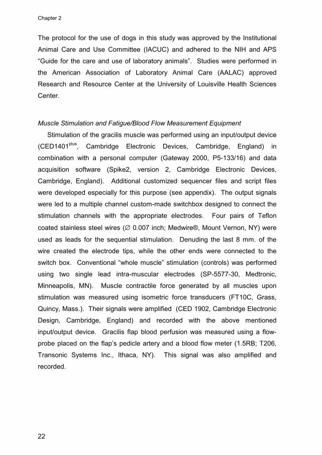
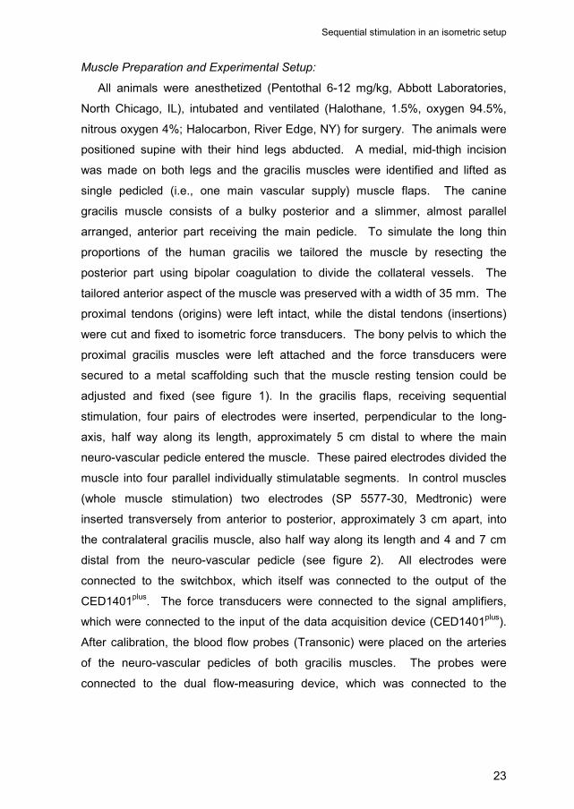
Figure 1: Simplified overview of the setup. Medial insertions of the gracilis muscles (GM) were left intact while the lateral insertions were attached to force transducers (FT). A metal scaffolding (SC) kept distances fixed. Flow-probes (FP) were attached to the single supplying arteries. Dashed lines outline the muscle parts, which were cut off during tailoring. Details concerning electrodes are depicted in Figures 2a and 2b.
Figure 2a: Four pairs of Teflon coated stainless steel wires were inserted into the sequentially stimulated muscle around 5 cm. distal to the neuro-vascular pedicle. These were repositioned until only the intended isolated selection of the muscle (S1-4) contracted on electrical stimulation.
Figure 2b: Two single lead intra-muscular electrodes(E1-2) were inserted into the control muscle 4 and 7 cmdistal from the neuro-vascular pedicle.
Sequential stimulation in an isometric setup
25
Pilot studies were conducted to determine the values for frequency and
pulse-width, which would generate a complete tetanic contraction with the lowest
possible stimulus intensity. This was found to occur at a frequency of 30 Hz and
a pulse width of 500 µsec. The pulse shape consisted of a mono-phasic block.
The force generated by the muscle was fixed and the stimulus intensity adjusted
for every experiment. Generated force was chosen being of sufficient
magnitude to fatigue the muscle, but not so large as to damage the muscle and
prevent repeated tests. The four muscle segments were sequentially stimulated
in 1.0 sec. steps.
Fatigue/Blood Flow Measurements
In both groups the distal ends of the muscles (sequential stimulation and
whole muscle stimulation) were attached to force transducers and were
stimulated at an intensity that generated an equal and predetermined starting
force. Fatigue was determined by measuring the amount of time it took for a
muscle to decrease the generated force to one-half of the starting force, i.e., half
time to fatigue. Measurements were performed four times, alternating back and
forth between each muscle, with 15-minutes resting periods between
measurements.
The number of simultaneously stimulated muscle segments in the
sequentially stimulated group determined the duty cycle of the segments and
FD
SC
SC
SB
IO PCFPFT
M
FD
SC
SC
SB
IO PCFPFT
M
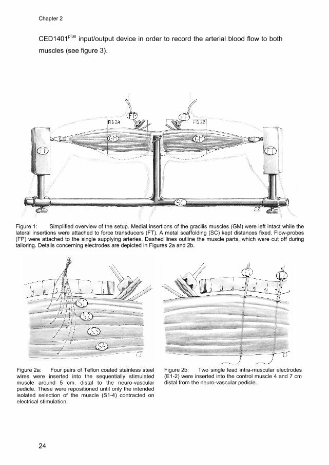
Figure 3: Schematic representation of the connections: The controlling personal computer (PC) was connected to the input/output (IO) device, which generated the electrical stimulation signal. This signal was led to the muscles (M) via the switchbox (SB) and the electrodes. The flow-probes (FP) were connected to the dual flow measurement device (FD), which delivered the arterial blood flow values via signal conditioners (SC) to the input/output device (IO). The force-transducers (FT) were also connected to the input/output device (IO) via signal conditioners (SC). All incoming signals were analogue-to-digitally converted and delivered to the controlling personal computer (PC).
Chapter 2
26
also influenced the force generated. For example, the sequentially stimulated
muscle was stimulated so that only one of the four segments was contracting at
a time; this constituted a duty cycle of 25% and generated initially a constant
force of 0.35 kiloponds. When two segments were stimulated simultaneously, a
50% duty cycle and 0.65 kiloponds of force were created. Three simultaneously
stimulated segments gave a 75% duty cycle and produced a generated 1.0
kilopond of force. Finally, when all four segments of the sequentially stimulated
muscle were simultaneously contracted a 100% duty cycle resulted, producing a
1.2 kiloponds force generation. The conventional (whole muscle) stimulation
and the sequential stimulation were always compared at the same generated
force. All measurements were terminated after 20 minutes, regardless the
amount of occurred fatigue. The arterial resting flow and the mean of the flow,
between 16-32 seconds after starting stimulation were measured for every
muscle.
Statistics
To compare fatigue rates between sequential and conventional stimulation
the ratio’s of the half times to fatigue between both stimulation regimens were
calculated for all four steps in the measurement protocol. Differences were also
statistically analyzed using the paired t-test after checking the distribution of the
values for normality using the Kolmogorov-Smirnov method. To compare
perfusion in the muscles exposed to the two different stimulation regimens, the
averaged gain of arterial blood-flow between 16 sec and 32 sec after start of
stimulation was expressed as a percentage of resting flow. The percentages of
both stimulation approaches were compared using the paired t-test for means
after checking the distribution of the values for normality using the Kolmogorov-
Smirnov method.
Sequential stimulation in an isometric setup
27
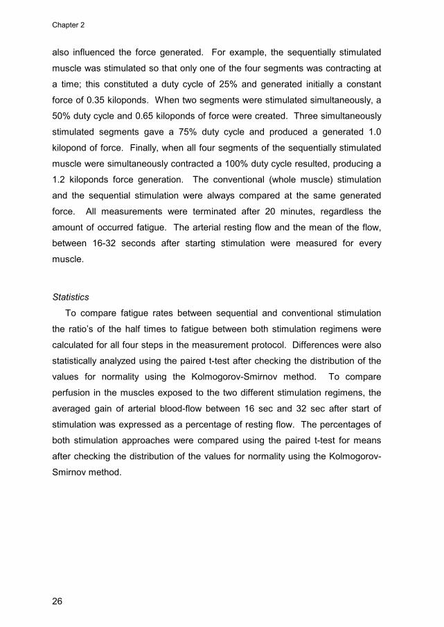
Results
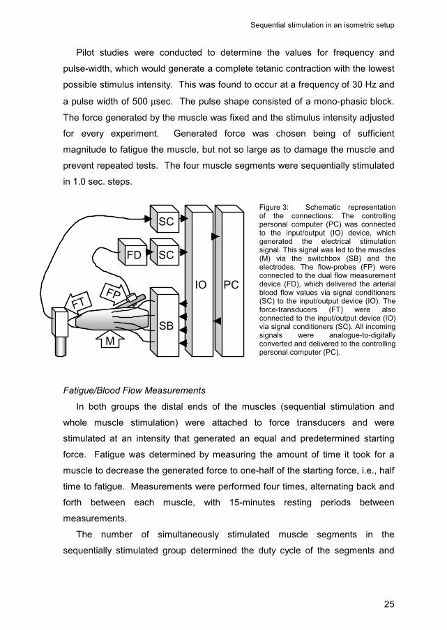
The half times to fatigue (mean ± SEM) of sequential stimulation and whole
muscle stimulated muscles are depicted in figure 4. During whole muscle
stimulation, the half times to fatigue did not significantly vary for the various
loads (51 ± 5 sec for the 0.35 kp load to 64 ± 13 sec for the 1.2 kp load). This
was in contrast to sequential stimulation where a decreasing duty cycle
markedly increased the half time to fatigue: 100%: 65 ± 6 sec.; 75%: 119 ± 7
sec; 50%: 262 ± 29 sec and 25% : 1,133 ± 33 sec. As the duty cycle decreased,
the load on any individual muscle segment stayed relatively constant while the
period of rest and recovery increased. When expressed as a ratio, sequential
stimulation with a duty cycle of 25% performed 22.2 times better than
conventional whole muscle stimulation. For 50% the ratio was 4.8; for 75% the
ratio was 2.2 and for 100% there were no significant differences and thus the
ratio was 1.0.
half times to fatigue
262
55 55
1133
11965
6451
0
200
400
600
800
1000
1200
1400
25% 50% 75% 100%
duty cycle
time
in s
econ
ds
sequential
conventional*
* *
*p<0.001
Figure 4: Half times to fatigue (mean and S.E.M. inseconds) of sequential and conventional stimulation for all fourgroups of duty cycles. Differences were statistically significantfor 25%, 50% and 75%.
Chapter 2
28
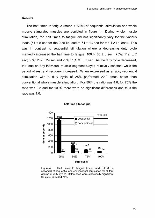
In figure 5 the averaged increase in blood-flow during stimulation is
represented for the four measured duty cycles of sequential stimulation and their
respective controls. At a duty cycle of 25% (and corresponding resting cycle of
75%) and a generated force of 0.35 kiloponds, blood-flow increased to an
average of 257 ± 20% of the resting flow. For the duty cycle of 50% (and
corresponding resting cycle of 50%), and 0.65 kiloponds of force, the averaged
blood-flow increased to 302 ± 44%. For a 75% duty cycle (and corresponding
resting cycle of 25%), and 0.90 kiloponds of force, blood flow increased to 272 ±
30%. Finally at a 100% duty cycle (and no resting cycle) and 1.2 kiloponds of
force, blood flow increased to 145 ± 5%. The same forces generated with whole
muscle stimulation showed minor increases in arterial blood-flow (134 ± 8% for
the 0.35 kp. load to 133 ± 11% for the 1.2 kp. load).
Figure 5: Increases in blood-flow (mean and S.E.M.)are expressed as percentages of the blood-flow in rest.Differences between sequential and conventionalstimulation were statistically significant for 25%, 50% and75%.
increase in arterial bloodflow during stimulation
257%
302%
272%
145% 133%136%
115%
134%
100%
150%
200%
250%
300%
350%
400%
25% 50% 75% 100%duty cycle
perc
enta
ge o
f res
ing
flow sequential
conventional
*
*
*
*p<0.005
Sequential stimulation in an isometric setup
29
Discussion
In clinical cardiomyoplasty and graciloplasty procedures, latissimus dorsi and
gracilis muscle fatigue is overcome by putting the muscles through a prolonged
training regimen. This training transforms the normally fatigue prone, fast twitch,
glycolytic, predominantly fiber type II skeletal muscle into a fatigue resistant,
slow twitch, oxidative, predominantly fiber type I muscle.
While this rigorous training protocol transforms the muscle to fatigue
resistant, it also causes the muscle to loose power and decreases its velocity of
contraction. Other drawbacks associated with transforming skeletal muscle are
the prolonged time required before the muscle can be used to assist the failing
heart or replace sphincter function.
All or part of these drawbacks could contribute to the inconsistent outcomes
reported in clinical cardiomyoplasty and graciloplasty.
Based on the above drawbacks we proposed a different approach to
minimizing skeletal muscle fatigue, using a more physiologic approach. Rather
than stimulating the entire muscle in one electrical burst and thus recruiting the
same fibers simultaneously, we studied the feasibility of stimulating different
muscle segments sequentially. The latter allows parts of the muscle to rest,
while other parts work. To produce a precisely tuned amount of force, it was
necessary to locate electrodes distant from the main neural pedicle, which is in
contrast with current methods. Thus, small nerve branches were activated,
rather than large sections of the muscle.
We compared both approaches in 7 pairs of dog gracilis muscles and found
that sequential stimulation significantly reduced muscle fatigue compared to
controls. In addition we found that smaller duty cycles correlate with greater
fatigue reduction. Some care should be taken though, while interpreting the
exact relation between the duty cycles and fatigue, since force generation was
arbitrary selected and the order of measurements was not randomized because
of small numbers (n=7).
These findings are in agreement with literature dealing with endurance
enhancement in neuromuscular prosthesis research for gait, in which alternation
between agonistic muscles proved to be beneficial.(Peckham 1970, Pournezam 1988) It is

Chapter 2
30
also in agreement with observations described in the literature concerning the
indirect or neural multi-channel stimulation, which was also developed to
sequentially recruit separate parts of a muscle.(Petrofsky 1979, Baer 1990, Thoma 1991)
Unfortunately, it was also reported that this type of peripheral nerve stimulation
causes severe damage to the nerve and therefore never achieved widespread
clinical application.
The significant improvement in muscle function observed in this acute model
could certainly be attributed to the significant improvement in muscle blood flow
during sequential stimulation as compared to whole muscle stimulation. Blood
flow in the main pedicle artery, solely feeding the gracilis flap, was measured in
a time window from 16 to 32 seconds in order to measure muscle perfusion
during both stimulation regimens. This measuring time was pragmatically
chosen, because we noted that after 16 seconds (four cycles of sequential
stimulation) blood flow values had stabilized. Measurements were stopped after
32 seconds, because the subsequent rapid drop in force in the conventionally
stimulated muscle caused increasing interference. This drop allowed partial
hyperemic perfusion of the abandoned fatigued muscle fibers, causing the blood
flow to slowly rise after the (arbitrary) measurement ending time of 32 seconds.
The work described in this report studied muscle fatigue and blood flow in
sequential stimulation muscles prepared acutely. The question arises whether
in long-term studies a sequentially stimulated muscle will experience
transformation and how this will affect the long-term function.
From the results of this experiment it can be stated that sequential stimulation
provides a substantial gain in fatigue resistance over conventional stimulation,
which is inversely progressive to the duty cycle. Thus, if a muscle flap used in a
given myoplasty procedure is not required to contract maximally, as in
graciloplasty for the treatment of urinary incontinence or fecal incontinence,
sequential stimulation could improve endurance acutely. This could result in
shorter training regimens and earlier full functioning of the myoplasty.
Sequential stimulation in an isometric setup
31
Conclusions
Sequential Segmental Neuromuscular Stimulation significantly enhances
fatigue resistance in an inverse progressive ratio to the applied duty cycle.
Reperfusion during sequential contraction is demonstrated and is assumed to
contribute to the enhanced fatigue resistance.
33
Chapter 3
Sequential stimulation in a non-isometric setup
Erik Zonnevylle, MD Naveen Somia, MD, PhD Gustavo Perez Abadia, MD Richard Stremel, PhD Claudio Maldonado, PhD Paul Werker, MD, PhD Moshe Kon, MD, PhD John Barker, MD, PhD Based upon the articles: Perez Abadia G, Zonnevylle E, Somia N, Stremel R, Koenig S, Palacio M, , Werker P, Kon M, Maldonado C, Tobin R and Barker J. Dynamic graciloplasty: sequential segmental neuromuscular stimulation (SSNS) improves neo-sphincter performance. Surgical Forum 49: 669-671, 1998. Zonnevylle E, Somia N, Perez Abadia G, Stremel R, Maldonado C, Werker P, Kon M and Barker J. Sequential segmental neuromuscular stimulation reduces fatigue and improves perfusion in dynamic graciloplasty. Annals of Plastic Surgery 45: 292-297, 2000. and presented (in part) at: the Annual Meeting of the Plastic Surgery Research Council 1998, Loma Linda, California, U.S.A. the 84th Annual Surgical Forum of the American College of Surgeons 1998, Orlando, Florida, U.S.A.
“The great tragedy of science: the slaying of a beautiful hypothesis by
an ugly fact” -Thomas Huxley-
Chapter 3
34
Abstract
Dynamic graciloplasty is used as a treatment modality for total urinary
incontinence caused by a paralyzed sphincter. A problem in this application is
undesirable fatigue of the muscle caused by continuous electrical stimulation.
Therefore, the neo-sphincter must be trained via a rigorous regimen in order to
transform it from a fatigue prone to a fatigue resistant state. To avoid or shorten
this training period, the application of sequential segmental neuromuscular
stimulation was examined. This form of stimulation proved to be highly effective
in acutely reducing fatigue caused by electrical stimulation.
The contractile function and perfusion of gracilis muscles employed as neo-
sphincters were compared between conventional, single-channel, continuous
stimulation and multi-channel, sequential stimulation in 8 dogs. The sequentially
stimulated neo-sphincter proved to have a 2.9 times longer endurance (as
measured by time to half-fatigue) and a better blood perfusion during stimulation
and both differences were statistically significant. Clinically this will not outdate
training of the muscle, however, sequential stimulation is likely to reduce the
need for long and rigorous training protocols making dynamic graciloplasty more
attractive as a method for treating urinary or fecal incontinence.
Sequential stimulation in a non-isometric setup
35
Introduction
Dynamic Graciloplasty is the technique in which the gracilis muscle flap is
wrapped around the urinary or fecal outlets and used to function as a neo-
sphincter. In currently used procedures, the gracilis muscle is incompletely
raised and transposed with its distal end around the target outlet.(Williams 1989, Baeten
1991, Janknegt 1992, Williams 1993) Because of ischemia and poor function of this distal
end, it was recently proposed to raise the gracilis muscle as an innervated free
flap and use the most suitable part of the muscle for the wrap.(van Aalst 1996, 1998)
Although this seems to be a major step forward in the field of custom made neo-
sphincter construction, undesirable sphincter fatigue caused by the necessary
electrical stimulation continues to be a major drawback.
In the currently applied procedures, fatigue is overcome by putting the
muscle through a rigorous training regimen for prolonged periods. The muscle
is transformed from a fatigue prone, fast twitch, glycolytic muscle with
predominantly type II fibers into a fatigue resistant, slow twitch, oxidative muscle
with predominantly type I fibers.(Salmons 1969, 1976, 1981 & 1994, Pette 1973 & 1997) The long
training period required to transform the muscle and the subsequent prolonged
period of incontinence, motivated searches for a better alternative to the
currently used protocols for training skeletal muscle in dynamic graciloplasty.
In the previous chapter, the feasibility of sequential segmental neuromuscular
stimulation was tested: electrically stimulating different segments of a muscle in
an alternating fashion rather than the entire muscle all at once in order to
increase acute endurance. In this chapter it is suggested that muscle fatigue
during continuous electrical stimulation is mainly due to the fact that the same
muscle fibers are being stimulated to contract without rest. Sequential /
alternating fiber recruitment during stimulation provides interim rest of some
segments, permitting temporary hyperemia of segments, while other segments
are contracting. The periodic, temporary hyperemia allows metabolites to be
washed out and oxygenation to be increased in the non-contractile muscle
segments. This causes the endurance of the muscle to increase. Although the
results of sequential stimulation were only known for acute isometric contraction,
it was predicted that in the non-isometric case of the modified dynamic
Chapter 3
36
graciloplasty the endurance could be prolonged using sequential stimulation.
Theoretically, this could benefit patients by decreasing the period of electrical
training of the neo-sphincter and therefore more rapidly provide continence post-
operatively.
In this study, fatigue rates and perfusion of sequentially stimulated neo-
sphincters were compared with conventional continuously stimulated neo-
sphincters. Sequential segmental neuromuscular stimulation significantly
decreased fatigue compared to the currently used method of continuous
stimulation in dynamic graciloplasty.
Materials and Methods
In dogs both gracilis muscles were converted into neo-sphincters and
electrically stimulated, using three-channel sequential stimulation on one side
and conventional single channel continuous stimulation on the other side. The
neo-sphincters were wrapped around balloon catheters attached to pressure-
transducers and stimulated to produce an equal and predetermined amount of
pressure. The time to decline to half the value of the initial pressure and the
arterial blood flow were measured in all muscles.
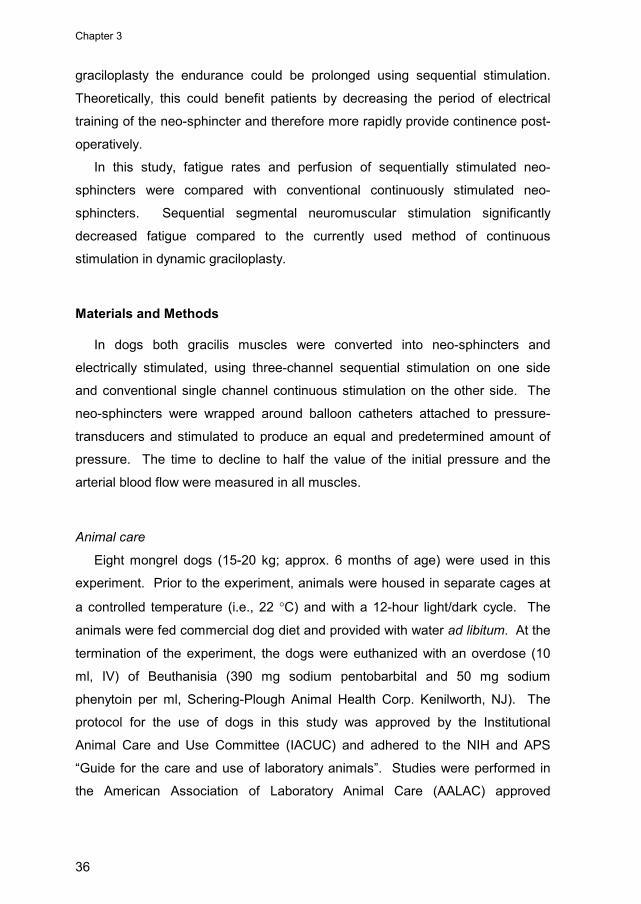
Animal care
Eight mongrel dogs (15-20 kg; approx. 6 months of age) were used in this
experiment. Prior to the experiment, animals were housed in separate cages at
a controlled temperature (i.e., 22 °C) and with a 12-hour light/dark cycle. The
animals were fed commercial dog diet and provided with water ad libitum. At the
termination of the experiment, the dogs were euthanized with an overdose (10
ml, IV) of Beuthanisia (390 mg sodium pentobarbital and 50 mg sodium
phenytoin per ml, Schering-Plough Animal Health Corp. Kenilworth, NJ). The
protocol for the use of dogs in this study was approved by the Institutional
Animal Care and Use Committee (IACUC) and adhered to the NIH and APS
“Guide for the care and use of laboratory animals”. Studies were performed in
the American Association of Laboratory Animal Care (AALAC) approved
Sequential stimulation in a non-isometric setup
37
Research and Resource Center at the University of Louisville Health Sciences
Center.
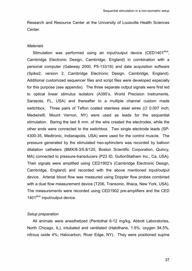
Materials
Stimulation was performed using an input/output device (CED1401plus,
Cambridge Electronic Design, Cambridge, England) in combination with a
personal computer (Gateway 2000, P5-133/16) and data acquisition software
(Spike2, version 2, Cambridge Electronic Design, Cambridge, England).
Additional customized sequencer files and script files were developed especially
for this purpose (see appendix). The three separate output signals were first led
to optical linear stimulus isolators (A395’s, World Precision Instruments,
Sarasota, FL, USA) and thereafter to a multiple channel custom made
switchbox. Three pairs of Teflon coated stainless steel wires (∅ 0.007 inch;
Medwire®, Mount Vernon, NY) were used as leads for the sequential
stimulation. Baring the last 8 mm. of the wire created the electrodes, while the
other ends were connected to the switchbox. Two single electrode leads (SP-
4300-35, Medtronic, Indianapolis, USA) were used for the control muscle. The
pressure generated by the stimulated neo-sphincters was recorded by balloon
dilatation catheters (BMX/8-3/5.8/120, Boston Scientific Corporation, Quincy,
MA) connected to pressure-transducers (P23 ID, GultonStatham Inc., Ca, USA).
Their signals were amplified using CED1902’s (Cambridge Electronic Design,
Cambridge, England) and recorded with the above mentioned input/output
device. Arterial blood flow was measured using Doppler flow probes combined
with a dual flow measurement device (T206, Transonic, Ithaca, New York, USA).
The measurements were recorded using CED1902 pre-amplifiers and the CED
1401plus input/output device.
Setup preparation
All animals were anesthetized (Pentothal 6-12 mg/kg, Abbott Laboratories,
North Chicago, IL), intubated and ventilated (Halothane, 1.5%, oxygen 94.5%,
nitrous oxide 4%; Halocarbon, River Edge, NY). They were positioned supine
Chapter 3
38
with their hind legs abducted. Bilateral medial incisions were made through
which both gracilis muscles were identified and lifted as single pedicled neuro-
vascular flaps. The muscles were reduced to a width of 35 mm by removing the
caudal aspect. At this point, two single leads were inserted into the
conventionally stimulated muscle, four and seven cm. distal from the point of
entry of the main pedicle (which includes the nerve at entry point). Starting from
the proximal tendo-muscular transition, 12 cm. length was marked. These
marked muscle-parts were cut from their origins and insertions. Each muscle
was tensionless but tightly wrapped around a latex tube (∅ 8mm, wall thickness:
0.5 mm.) and a balloon catheter. Three pairs of Teflon coated leads were
transversely inserted into the sequentially stimulated neo-sphincter, at an angle
of 45 degrees to the direction of the fibers, 5 to 6 cm. distal from the main
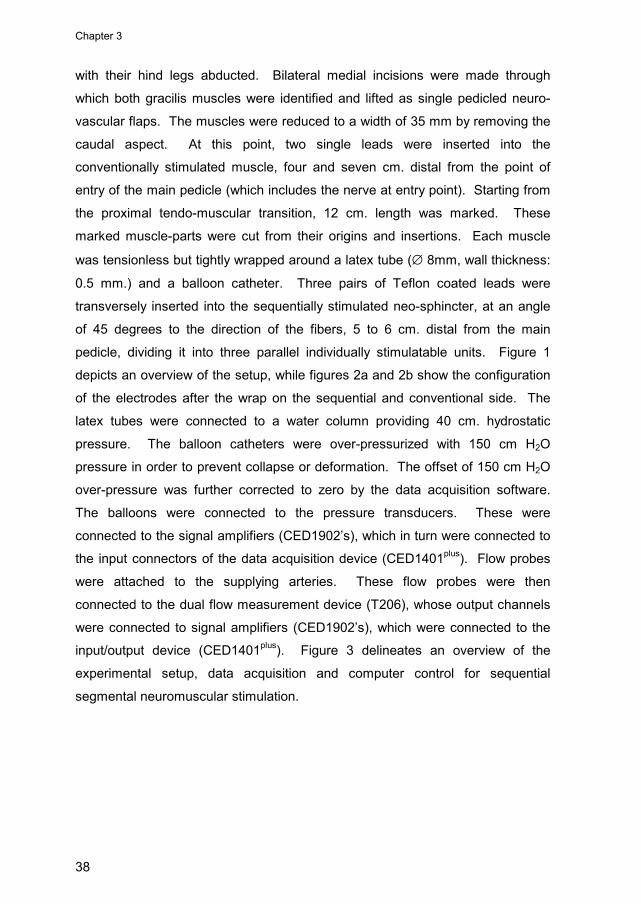
pedicle, dividing it into three parallel individually stimulatable units. Figure 1
depicts an overview of the setup, while figures 2a and 2b show the configuration
of the electrodes after the wrap on the sequential and conventional side. The
latex tubes were connected to a water column providing 40 cm. hydrostatic
pressure. The balloon catheters were over-pressurized with 150 cm H2O
pressure in order to prevent collapse or deformation. The offset of 150 cm H2O
over-pressure was further corrected to zero by the data acquisition software.
The balloons were connected to the pressure transducers. These were
connected to the signal amplifiers (CED1902’s), which in turn were connected to
the input connectors of the data acquisition device (CED1401plus). Flow probes
were attached to the supplying arteries. These flow probes were then
connected to the dual flow measurement device (T206), whose output channels
were connected to signal amplifiers (CED1902’s), which were connected to the
input/output device (CED1401plus). Figure 3 delineates an overview of the
experimental setup, data acquisition and computer control for sequential
segmental neuromuscular stimulation.
Sequential stimulation in a non-isometric setup
39
Figure 1: Surgical preparation. Gracilis muscles were turned into neo-sphincters (NS), wrapped aroundartificial urethrae (AU) and catheter balloons (CB). Flow probes (FP) were attached to the arteries of thesingle neurovascular pedicles (NP).
Figure 2a: Sequentially stimulated neo-sphincter.This neo-sphincter was sequentially stimulated usingthree segments (S1,S2,S3). Three pairs of wire-electrodes (WE) were transversely inserted into themuscle approximately 5 cm. distal to the pedicle.Electrodes were inserted in that way that eachsegment could be independently recruited andproportionally controlled modulating the amplitude ofthe signal.
Figure 2b: Conventionally stimulated neo-sphincter. This neo-sphincter was conventionally stimulated acting as control. Two electrodes (E1,E2) were transversely inserted into the muscle 4 and 7 cm. distal to the pedicle. These positions ensured proportional control over the generated pressure, by modulating amplitude.
Chapter 3
40
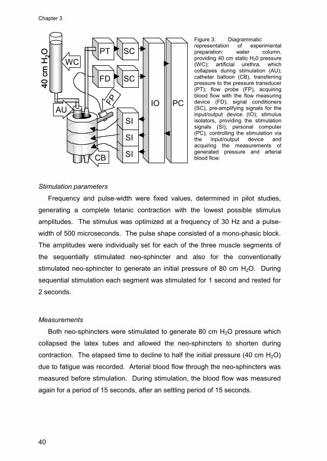
Stimulation parameters
Frequency and pulse-width were fixed values, determined in pilot studies,
generating a complete tetanic contraction with the lowest possible stimulus
amplitudes. The stimulus was optimized at a frequency of 30 Hz and a pulse-
width of 500 microseconds. The pulse shape consisted of a mono-phasic block.
The amplitudes were individually set for each of the three muscle segments of
the sequentially stimulated neo-sphincter and also for the conventionally
stimulated neo-sphincter to generate an initial pressure of 80 cm H2O. During
sequential stimulation each segment was stimulated for 1 second and rested for
2 seconds.
Measurements
Both neo-sphincters were stimulated to generate 80 cm H2O pressure which
collapsed the latex tubes and allowed the neo-sphincters to shorten during
contraction. The elapsed time to decline to half the initial pressure (40 cm H2O)
due to fatigue was recorded. Arterial blood flow through the neo-sphincters was
measured before stimulation. During stimulation, the blood flow was measured
again for a period of 15 seconds, after an settling period of 15 seconds.
40 c
m H
2O PT
FD
SC
SC
SI
SI
SI
IO PC
WC
FPAU
CB
40 c
m H
2O PT
FD
SC
SC
SI
SI
SI
IO PC
WC
FPAU
CB
Figure 3: Diagrammatic representation of experimental preparation: water column, providing 40 cm static H20 pressure (WC); artificial urethra, which collapses during stimulation (AU); catheter balloon (CB), transferring pressure to the pressure transducer (PT); flow probe (FP), acquiring blood flow with the flow measuring device (FD); signal conditioners (SC), pre-amplifying signals for the input/output device (IO); stimulus isolators, providing the stimulation signals (SI); personal computer (PC), controlling the stimulation via the input/output device and acquiring the measurements of generated pressure and arterial blood flow.

Sequential stimulation in a non-isometric setup
41
Analysis
Differences in endurance were quantified by comparing the elapsed times to
fatigue between sequential stimulation and continuous stimulation. Statistical
significance was demonstrated using the paired t-test for means, after checking
the distribution of the values for normality using the Kolmogorov-Smirnov
method.
The mean value of the arterial flow, measured between 15 and 30 seconds
after stimulation started, was expressed as a percentage of the flow before
stimulation. The differences between the relative changes in arterial blood flow
of sequential and continuous stimulation were tested for statistical significance
using a paired t-test for means after checking the distribution of the values for
normality using the Kolmogorov-Smirnov method.
Results
During sequential stimulation, the averaged elapsed time to fatigue was 143
± 17 seconds (mean ± sem). This is in contrast to conventional stimulation
where the averaged elapsed time to fatigue was only 51 ± 6 seconds (see figure
4). Therefore, the ratio of the elapsed times to fatigue of the sequential and
conventional stimulation was 2.9 ± 0.2. The difference is statistically significant
(p<0.05).
half times to fatigue (n=8)
143
510
4080
120160200
sequential conventional
seco
nds
p<0.05
Figure 4: Averaged half times to fatigue (time for the stimulated neo-sphincter pressure to decrease from 80 to 40 cm H20) for sequential and conventional stimulation. The difference between sequential and conventional stimulation is statistically significant.

Chapter 3
42
In figure 5, the averaged percentage changes in arterial blood-flow during
stimulation are represented for the sequential and conventional stimulation. The
sequentially stimulated neo-sphincters showed an averaged blood-flow increase
of 86 % above control. The same work achieved with conventional stimulation
resulted in a decrease in arterial blood-flow below control of 37%. The
difference is statistically significant (p<0.05).
Discussion
Dynamic graciloplasty is an exciting approach to the treatment of urine or
fecal incontinence, but clinical trials have shown that the concept needs some
refinement.(Konsten 1993, Baeten 1995, Janknegt 1995) A promising improvement is the
innervated free flap approach developed by van Aalst et al. reported in 1996 and
1998. Although this approach might decrease scarring and improve
effectiveness of the neo-sphincter in generating pressure, it still requires an
eight-week training period to produce a fatigue resistant neo-sphincter at the
Figure 5: Relative changes in arterial blood flow during sequential and conventional stimulation compared to resting arterial flow. Resting blood flow was normalized as 100%. The difference between the flow changes during sequential and conventional stimulation is statistically significant.
relative arterial flowbefore and during stimulation
100100
63
186
0%
50%
100%
150%
200%
250%
sequential conventional
perc
enta
ge o
f res
ting
flow before
during*
*
*p<0.05
Sequential stimulation in a non-isometric setup
43
cost of strength and reaction speed. The importance of the latter is discussed in
chapter 4 and 5 reporting the combination of sequential segmental
neuromuscular stimulation with closed-loop control.
The previous chapter demonstrated the poor acute performance of dynamic
myoplasties, when whole muscle recruiting electrical stimulation is applied. This
stimulation continuously paces the same muscle fibers over and over again.
This impedes local perfusion and results in metabolic acidosis, followed by a
decrease in performance. Currently, rigorous training programs are used to alter
the metabolic pathway of the involved skeletal muscle. The fast-twitch type II
glycolytic fibers are replaced by slow-twitch type I oxidative fibers. In this way
the muscle can better deal with the impeded perfusion, again, at the cost of
strength and responsiveness. Furthermore, during the months of training
following a graciloplasty procedure, the patient cannot enjoy the benefits of the
procedure.
It is clear from the results that sequential stimulation of the neo-sphincter
does acutely improve endurance substantially over continuous stimulation. The
increase in arterial blood flow during sequential stimulation is in sharp contrast
with the decrease measured in conventional stimulation. This supports the
theoretical explanation of the prolonged endurance in sequential stimulation
caused by temporary hyperemia of the intermittently resting muscle segments.
Because of practical reasons, the number of individually stimulated segments
was chosen to be three in the experiment described in this chapter. This
resulted in a duty cycle of 33%. Based on the results in the isometric gracilis
preparation, described in chapter 2, it was anticipated that acute endurance
could be prolonged 10-15 times, by extrapolating a duty cycle of 33% in the
results graph of figure 4 in chapter 2. This proved not to be the case, since the
difference was actually, only three times. Difference in experimental setup could
be an explanation for this lower gain in endurance. The experiment above was
deliberately chosen not to be isometric in order to resemble the actual procedure
of dynamic graciloplasty. In the isometric preparation, sarcomeres in the muscle
are kept in an ideal position to generate force, while the compressible artificial
urethra allowed, at least for the inner part of the neo-sphincter, the more

Chapter 3
44
unfavorable shortened position of the sarcomeres in the non-isometric
preparation of this research. Furthermore, the vessel bed of the muscles in the
isometric preparation was slightly stretched and kept stretched during
stimulation, while the wrap around the artificial urethra and the balloon catheter
caused hitching and torsion of the intra-muscular vessel bed, which was
increased during stimulation. The latter caused higher resistance and
subsequent lower perfusion. It is most likely that these differences at least partly
caused the lower endurance in the experiment described in this chapter.
Conclusion
Sequential segmental neuromuscular stimulation does not outdate a training
regimen in the clinical setting of dynamic graciloplasty. However, it acutely
prolongs endurance and enhances perfusion. It is anticipated that it will
substantially shorten the necessary training protocol, leading to an earlier state
of acceptable continence for patients undergoing dynamic graciloplasty.
Furthermore it is postulated that this form of stimulation makes it less necessary
for the muscle to replace fast twitch, type II, glycolytic fibers by slow twitch, type
I, oxidative fibers, preserving strength and responsiveness, which becomes
important in the next two chapters.
45
Chapter 4
Closed-loop Control:
feasibility in a neo-sphincter model
Erik Zonnevylle, MD Gustavo Perez Abadia, MD Naveen Somia, MD, PhD Moshe Kon, MD, PhD John Barker, MD, PhD Steven Koenig, PhD Daniel Ewert, PhD Richard Stremel, PhD Based upon the articles: Zonnevylle E, Perez Abadia G, Somia N, Stremel R, Maldonado C, Koenig S, Palacio M, Werker P, Kon M and Barker J. Feedback (closed-loop) control of a urinary graciloplasty neo-sphincter. Surgical Forum 49: 314-316, 1998. Zonnevylle E, Somia N, Perez Abadia G, Stremel R, Maldonado C, Werker P, Kon M and Barker J. Three parameters optimizing closed-loop control in sequential segmental neuromuscular stimulation. Artificial Organs 23: 388-391, 1999. Zonnevylle E, Perez Abadia G, Somia N, Kon M, Barker J, Koenig S, Ewert D and Stremel R. A technique for sequential segmental neuromuscular stimulation with closed-loop feedback control. Journal of Investigative Surgery 15(2): 91-99, 2002. and presented (in part) at: the 6th Vienna International Workshop on FES 1998, Vienna, Austria. the 84th Annual Surgical Forum at the American College of Surgeons 1998, Orlando, Florida. U.S.A.
“Minds are like parachutes; they work best when open” -Lord Thomas Dewar-
Chapter 4
46
Abstract
In dynamic myoplasty, dysfunctional muscle is assisted or replaced with
skeletal muscle from a donor site. Electrical stimulation is commonly used to
train and animate the skeletal muscle to perform its new task. Owing to
simultaneous, tetanic contractions of the entire myoplasty, muscles are deprived
of perfusion and fatigue rapidly causing long-term problems such as excessive
scarring and muscle ischemia. Sequential segmental neuromuscular stimulation
contracts part of the muscle while other parts rest, thus significantly improving
blood perfusion. Nevertheless, the muscle still fatigues. In this chapter, we
report the feasibility of using closed-loop control to economize the contractions
of the sequentially stimulated myoplasty. A simple stimulation algorithm was
developed and tested on sequentially stimulated neo-sphincters designed from a
canine gracilis muscle. Pressure generated in the lumen of the myoplasty neo-
sphincters was used as feedback to regulate the stimulation signal via three
control parameters, thereby optimizing the performance of the myoplasty.
Additionally, we investigated and compared the efficiency of amplitude and
frequency modulation techniques. Closed-loop control enabled the maintenance
of target pressures within 10% deviation, using amplitude modulation and
optimized control parameters. The large-scale stimulation/feedback setup was
unsuitable for chronic experimentation, but can be used as a blueprint for a
small-scale version to reveal the theoretical benefits of closed-loop control in
chronic experimentation.
Closed-loop control: feasibility in a neo-sphincter model
47
Introduction
Dynamic myoplasty is an evolving surgical procedure in which dysfunctional
muscle tissue is assisted or replaced with skeletal muscle from a less critical
donor site. Cardiomyoplasty and graciloplasty have been the most promising
clinical applications of this technique to date. In cardiomyoplasty, the latissimus
dorsi muscle is wrapped around the heart and electrically paced in synchrony
with the cardiac rhythm, assisting the heart to contract more effectively.(Carpentier
1985, Chachques 1989 & 1997) In graciloplasty, the gracilis muscle is wrapped around
either the urethra or anal canal and is continuously electrically stimulated to
prevent incontinence.(Williams 1989, Baeten 1991& Janknegt 1992) Electrical stimulation has
been most commonly accomplished by delivering a voltage pulse train to the
latissimus and gracilis muscles.(Hallan 1990, Lucas 1991 & Grandjean 1996) More recently,
biphasic pulse train input(Scheiner 1990) and differential electrode configurations
have been employed. These techniques apply a single source voltage, which
causes all of the muscle fibers to contract simultaneously. Continuous
contraction of all of the muscle fibers is a non-physiological approach and
consequently, the muscle fatigues quickly.
In order to improve endurance, muscle-training programs have been
designed to transform the myoplasty from a fast twitch, fatigable muscle to a
slow twitch, fatigue resistant muscle at a cost of reduced power.(Salmons 1969, 1976
1981 & 1994, Pette 1973 & 1975, 1984 & Chachques 1987) This training protocol generally takes up
to 8 weeks, delaying the assistance from the myoplasty. Furthermore, in
graciloplasty for urinary incontinence, continuous contraction of the myoplasty at
the high pressures required maintaining continence during peak bladder
pressure and (short) periods of extraneous pressure (e.g., coughing or lifting
heavy objects) can cause muscle scarring, stricture, and ischemic lesions.(Williams
1993, Konsten 1993, Baeten 1995, Janknegt 1995, Geerdes 1996, van Aalst 1998 & Madoff 1999)
In chapters 2 and 3, sequential stimulation was introduced as an alternative
methodology to single source electrical stimulation. In this technique the
skeletal muscle is partitioned into segments, which are alternately stimulated in
a sequential order. This approach causes only one segment of the muscle to
perform the necessary workload, thereby allowing the other non-stimulated

Chapter 4
48
segments to recover. This and similar techniques have been shown to reduce
muscle fatigue(Petrofsky 1979, Pournezam 1988 & Thoma 1991); however, the muscle is still
susceptible to fatigue and eventually fails.
Ideally, a dynamic myoplasty should perform the required physiological
function indefinitely. In this chapter, sequential segmental neuromuscular
stimulation is combined with a new closed-loop pressure feedback control
scheme. This created the condition in which it is possible to reduce the amount
of work the myoplasty performs to the minimal level necessary to carry out the
required physiological function for prolonged periods. As a model a neo-
sphincter was chosen, created using a gracilis muscle as used in dynamic
graciloplasty for treatment of incontinence.
Materials and Methods
In a dog experimental model (n=8), triple partitioned neo-sphincters were
created around pressure measuring devices and sequentially stimulated. Via an
input/output device, a computer acquired the generated pressures and provided
the stimulation signals to the neo-sphincters. The measured pressures were
compared to target pressures and deviations were corrected by adjusting the
stimulation signals using a closed-loop pressure feedback control algorithm.
Constants and parameters of this algorithm were evaluated for their ability to
generate target pressures and minimize pressure fluctuation.
Surgical technique
After a pilot involving three dogs a final study design was composed in which
both gracilis muscles of mongrel dogs (15-20 kg; approx. 6 months of age) were
dissected from their surrounding tissue and left attached only by their primary
blood/nerve supply. The muscles were then tailored into 3.5 cm X 12 cm size
and were wrapped around artificial urethras and catheter balloons in the shape
of neo-sphincters. Pressure in the artificial urethras was regulated using
connected water columns, providing approximately 5 cm H2O less pressure than
the target pressure. The muscles were functionally divided into three separately
Closed-loop control: feasibility in a neo-sphincter model
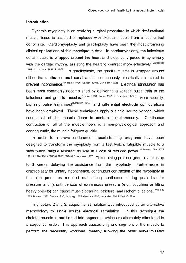
49
stimulatable units using three pairs of stainless steel wire electrodes connected
to three isolated sources of electrical stimulation (see figure 1).
Sequential stimulation and pressure feedback control instrumentation
The stimulation setup consisted of a desktop computer (Gateway 2000
Pentium 200/80, Sioux Falls, SD), an input/output data acquisition (I/O) device
(CED 1401plus, Cambridge Electronic Design, Cambridge, England), three linear
stimulus isolators (World Precision Instruments, Sarasota, FL), and Teflon-
coated stainless steel wire stimulation leads (0.007 inch in diameter, Medwire®,
Mount Vernon, NY). The desktop computer and I/O device, which generated
digital-to-analog waveforms and/or analog-to-digital data files, were used to
Figure 1. Surgical procedure:Neo-sphincters were constructedusing canine gracilis musclesisolated on their main neurovascularpedicles (NP). Each neo-sphincterwas divided into three individuallystimulatable segments (S1, S2, S3,arbitrary borders by dashed lines),using three pairs of wire electrodes(WE). Neo-sphincters were wrappedaround artificial urethras (AU) andpressure measuring catheterballoons (CB).
Chapter 4
50
produce three independent segmental stimulation voltage signals. These
signals had user-selectable characteristics (i.e. amplitude, pulse width, etc.) that
were programmed using custom written scripts (see appendix) in data
acquisition software (Spike2, v2.21, Cambridge Electronic Design, Cambridge,
England). The stimulation profiles that were generated were applied to the
individual segments through isolation amplifiers via the stimulation leads. The
isolation amplifiers eliminated potential conduction pathways between stimulated
and non-stimulated leads, thereby preventing non-stimulated muscle segments
from inadvertently contracting. Balloon dilatation catheters (BMX/8-3/5.8/120,
Boston Scientific Corporation, Quincy, MA) were used to measure the pressures
generated by artificial neo-sphincters, as described in the surgical technique
section. The pressure in the balloon was measured using a pressure-transducer
(P23 ID, GultonStatham Inc., CA, USA), which was amplified using a CED1902
signal conditioner (Cambridge Electronic Design, Cambridge, England). The
measured pressure was converted from analog-to-digital format by the I/O
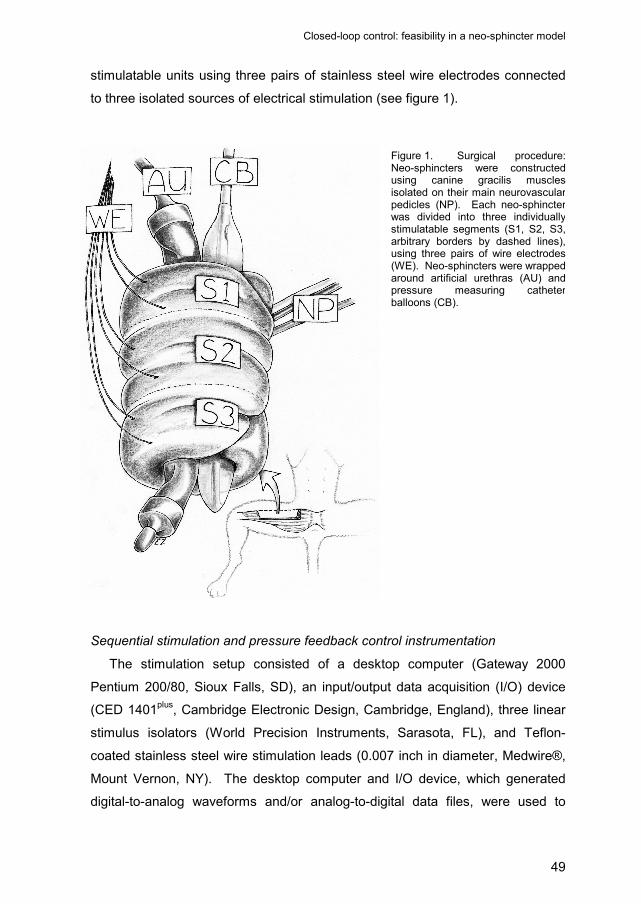
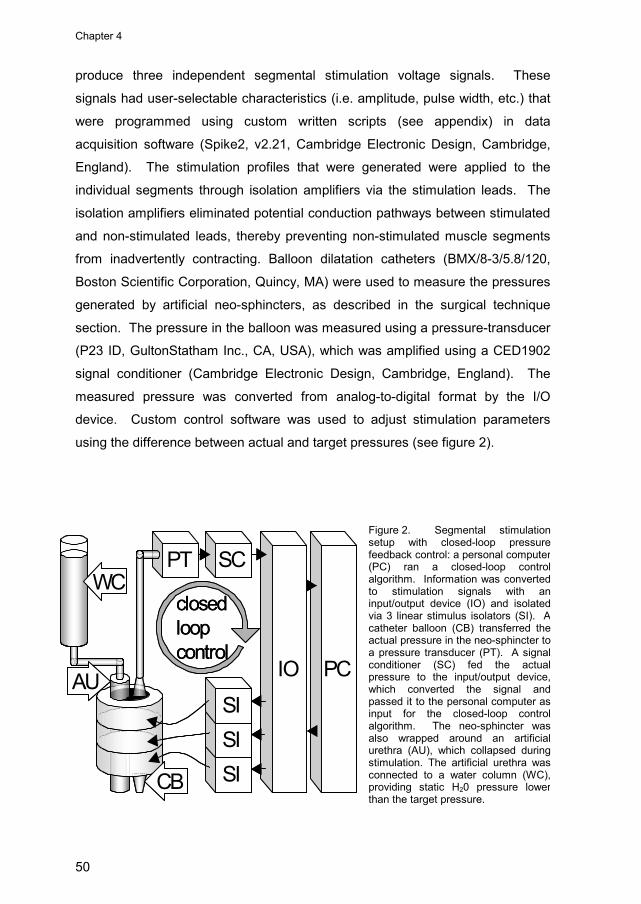
device. Custom control software was used to adjust stimulation parameters
using the difference between actual and target pressures (see figure 2).
PT SC
SISISI
IO PC
WC
AU
CB
closedloopcontrol
PT SC
SISISI
IO PC
WC
AU
CB
closedloopcontrol
Figure 2. Segmental stimulationsetup with closed-loop pressurefeedback control: a personal computer(PC) ran a closed-loop controlalgorithm. Information was convertedto stimulation signals with aninput/output device (IO) and isolatedvia 3 linear stimulus isolators (SI). Acatheter balloon (CB) transferred theactual pressure in the neo-sphincter toa pressure transducer (PT). A signalconditioner (SC) fed the actualpressure to the input/output device,which converted the signal andpassed it to the personal computer asinput for the closed-loop controlalgorithm. The neo-sphincter wasalso wrapped around an artificialurethra (AU), which collapsed duringstimulation. The artificial urethra wasconnected to a water column (WC),providing static H20 pressure lowerthan the target pressure.

Closed-loop control: feasibility in a neo-sphincter model
51
Closed-loop control algorithm
Closed-loop control was accomplished using amplitude or frequency
modulation. When amplitude modulation was selected, the output of the I/O
device was a monophasic pulse train with variable amplitude, a pulse width of
500 µs, and pulse trains generated at 30 Hz. In the case of frequency
modulation, the frequency was varied while the amplitude of each individual
segment was fixed at 2.5 times its twitch threshold defined as the minimal value
of constant current to produce a measurable twitch. A ‘multiplication factor’ was
used to convert the pressure error to the stimulation parameter adjustment. The
multiplication factor (K) was set at K = 2.0 for amplitude modulation and K = 0.5
for frequency modulation. When the difference between the actual pressure (PA)
and the target pressure (PT) exceeded the ‘correction threshold’ the stimulation
signal was adjusted at the rate of the ‘correction frequency’. The stimulation
signal was not adjusted during the interval after the start of stimulation of a new
segment, which was defined as the ‘transition time’. Segment stimulation
periods were set to 1-second time intervals with individual segments being
stimulated in a sequential order. The control algorithm equations for amplitude
and frequency modulation (Equation 1) are shown below.
Equation 1
( )
∑∞
=
+
∆=
−=∆
1
1 )()(
nn
ATn
FactorCorrection
nPnPK
Study parameters and data acquisition
As described in the surgical technique section, an artificial neo-sphincter
(graciloplasty) was created. The balloon pressure catheter was inflated to150
cm H2O, in order to minimize its compliance. Subsequently, any pressure
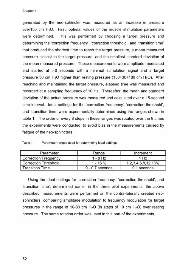
Chapter 4
52
generated by the neo-sphincter was measured as an increase in pressure
over150 cm H2O. First, optimal values of the muscle stimulation parameters
were determined. This was performed by choosing a target pressure and
determining the ‘correction frequency’, ‘correction threshold’, and ‘transition time’
that produced the shortest time to reach the target pressure, a mean measured
pressure closest to the target pressure, and the smallest standard deviation of
the mean measured pressure. These measurements were amplitude modulated
and started at t=0 seconds with a minimal stimulation signal and a target
pressure 30 cm H2O higher than resting pressure (150+30=180 cm H2O). After
reaching and maintaining the target pressure, elapsed time was measured and
recorded at a sampling frequency of 10 Hz. Thereafter, the mean and standard
deviation of the actual pressure was measured and calculated over a 15-second
time interval. Ideal settings for the ‘correction frequency’, ‘correction threshold’,
and ‘transition time’ were experimentally determined using the ranges shown in
table 1. The order of every 8 steps in these ranges was rotated over the 8 times
the experiments were conducted, to avoid bias in the measurements caused by
fatigue of the neo-sphincters.
Parameter Range Increment Correction Frequency 1 - 8 Hz 1 Hz Correction Threshold 1 - 16 % 1,2,3,4,6,8,12,16% Transition Time 0 - 0.7 seconds 0.1 seconds
Using the ideal settings for ‘correction frequency’, ‘correction threshold’, and
‘transition time’, determined earlier in the three pilot experiments, the above
described measurements were performed on the contra-laterally created neo-
sphincters, comparing amplitude modulation to frequency modulation for target
pressures in the range of 10-80 cm H2O (in steps of 10 cm H2O) over resting
pressure. The same rotation order was used in this part of the experiments.
Table 1. Parameter ranges used for determining ideal settings.
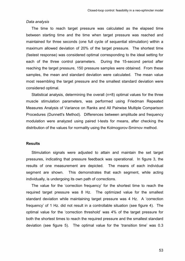
Closed-loop control: feasibility in a neo-sphincter model
53
Data analysis
The time to reach target pressure was calculated as the elapsed time
between starting time and the time when target pressure was reached and
maintained for three seconds (one full cycle of sequential stimulation) within a
maximum allowed deviation of 20% of the target pressure. The shortest time
(fastest response) was considered optimal corresponding to the ideal setting for
each of the three control parameters. During the 15-second period after
reaching the target pressure, 150 pressure samples were obtained. From these
samples, the mean and standard deviation were calculated. The mean value
most resembling the target pressure and the smallest standard deviation were
considered optimal.
Statistical analysis, determining the overall (n=8) optimal values for the three
muscle stimulation parameters, was performed using Friedman Repeated
Measures Analysis of Variance on Ranks and All Pairwise Multiple Comparison
Procedures (Dunnett's Method). Differences between amplitude and frequency
modulation were analyzed using paired t-tests for means, after checking the
distribution of the values for normality using the Kolmogorov-Smirnov method.
Results
Stimulation signals were adjusted to attain and maintain the set target
pressures, indicating that pressure feedback was operational. In figure 3, the
results of one measurement are depicted. The means of each individual
segment are shown. This demonstrates that each segment, while acting
individually, is undergoing its own path of corrections.
The value for the ‘correction frequency’ for the shortest time to reach the
required target pressure was 8 Hz. The optimized value for the smallest
standard deviation while maintaining target pressure was 4 Hz. A ‘correction
frequency’ of 1 Hz. did not result in a controllable situation (see figure 4). The
optimal value for the ‘correction threshold’ was 4% of the target pressure for
both the shortest times to reach the required pressure and the smallest standard
deviation (see figure 5). The optimal value for the ‘transition time’ was 0.3
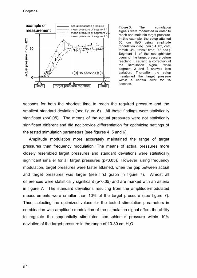
Chapter 4
54
seconds for both the shortest time to reach the required pressure and the
smallest standard deviation (see figure 6). All these findings were statistically
significant (p<0.05). The means of the actual pressures were not statistically
significant different and did not provide differentiation for optimizing settings of
the tested stimulation parameters (see figures 4, 5 and 6).
Amplitude modulation more accurately maintained the range of target
pressures than frequency modulation: The means of actual pressures more
closely resembled target pressures and standard deviations were statistically
significant smaller for all target pressures (p<0.05). However, using frequency
modulation, target pressures were faster attained, when the gap between actual
and target pressures was larger (see first graph in figure 7). Almost all
differences were statistically significant (p<0.05) and are marked with an asterix
in figure 7. The standard deviations resulting from the amplitude-modulated
measurements were smaller than 10% of the target pressure (see figure 7).
Thus, selecting the optimized values for the tested stimulation parameters in
combination with amplitude modulation of the stimulation signal offers the ability
to regulate the sequentially stimulated neo-sphincter pressure within 10%
deviation of the target pressure in the range of 10-80 cm H2O.
0
60
actu
al p
ress
ure
in c
m H
2O
15 seconds
actual measured pressuremean pressure of segment 1mean pressure of segment 2mean pressure of segment 3
example ofmeasurement
start stoptarget pressure reached
0
60
actu
al p
ress
ure
in c
m H
2O
15 seconds
actual measured pressuremean pressure of segment 1mean pressure of segment 2mean pressure of segment 3
example ofmeasurement
start stoptarget pressure reached
Figure 3. The stimulationsignals were modulated in order toreach and maintain target pressure.In this example, the setup attained60 cm H2O using amplitudemodulation (freq. corr.: 4 Hz, corr.thresh. 4%, transit time: 0.3 sec.).Segment 1 of the neo-sphincterovershot the target pressure beforereaching it causing a correction ofthe stimulation signal, whilesegment 2 and 3 showed lessvariation. Thereafter the setupmaintained the target pressurewithin a certain error for 15seconds.

Closed-loop control: feasibility in a neo-sphincter model
55
Figure 5. The optimal‘correction threshold’ showedfastest time to reach targetpressure and smallest standarddeviation while allowing theactual pressure to deviate up to4% of the target pressure beforecorrection of the stimulationsignal was executed. Nodifferentiation could be madeconcerning the mean of theactual pressures.
times to reach target pressure,actual pressures and standard deviations
for different correction thresholds
0
10
20
30
40
50
1 2 3 4 6 8 12 16
seco
nds
25
27
29
31
33
35
1 2 3 4 6 8 12 16
cm H
2O
3.5
4
4.5
5
5.5
6
1 2 3 4 6 8 12 16
stan
dard
de
viat
ion
correction threshold in % of target pressure
*
*****
*p<0.05
**** **
Figure 4. The ‘correctionfrequencies’ showed fastest timeto reach target pressure at 8 Hz.and the optimized value forsmallest standard deviationwhile maintaining targetpressure was 4 Hz. Nodifferentiation could be madeconcerning the mean of theactual pressures. A correctionfrequency of 1 Hz. did not resultin a controlled situation.
05
1015202530
1 2 3 4 5 6 7 8
seco
nds
25
27
29
31
33
35
1 2 3 4 5 6 7 8
cm H
2O
2
2.5
3
3.5
4
4.5
1 2 3 4 5 6 7 8
stan
dard
de
viat
ion
correction frequency in Hz.
times to reach target pressure,actual pressures and standard deviations
for different correction frequencies
**
**
*
** * * *
*p<0.05
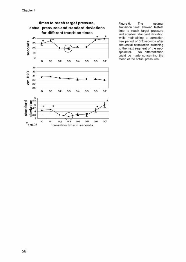
Chapter 4
56
Figure 6. The optimal‘transition time’ showed fastesttime to reach target pressureand smallest standard deviationwhile maintaining a correctionfree period of 0.3 seconds aftersequential stimulation switchingto the next segment of the neo-sphincter. No differentiationcould be made concerning themean of the actual pressures. 0
10
20
30
40
0 0.1 0.2 0.3 0.4 0.5 0.6 0.7
seco
nds
25
27
29
31
33
35
0 0.1 0.2 0.3 0.4 0.5 0.6 0.7
cm H
2O
33.54
4.55
5.56
0 0.1 0.2 0.3 0.4 0.5 0.6 0.7
stan
dard
de
viat
ion
transition time in seconds
times to reach target pressure,actual pressures and standard deviations
for different transition times
*p<0.05
*** *
* **
*

Closed-loop control: feasibility in a neo-sphincter model
57
Figure 7. Target pressures were reached quicker using frequency modulation. However amplitude modulation was more accurate than frequency modulation in maintaining the range of target pressures: the standard deviations of the actual pressures were lower and the means more closely resembled target pressures.
0
10
20
30
40
50
10 20 30 40 50 60 70 80
sec
onds
amplitude
frequency
0
20
40
60
80
100
10 20 30 40 50 60 70 80
actu
al p
ress
ure
cm H
2O
amplitude
frequency
02468
101214
10 20 30 40 50 60 70 80
stan
dard
dev
iatio
n
amplitude
frequency
times to reach target pressures,actual pressures and standard deviations
comparingamplitude versus frequency modulation
target pressure in cmH2O*p<0.05
* *****
***
*
**
***
*****
*
**

Chapter 4
58
Discussion
Physiological muscle fatigue is primarily due to depletion of local energy.
This local energy is stored chemically in the muscle tissue as adenosine
triphosphate (ATP). In the process of contraction, ATP is chemically
downgraded into adenosine diphosphate (ADP). This breakdown of ATP
releases the energy necessary for myofilament interaction and contraction.
Metabolism of nutrients and the consumption of oxygen constantly regenerate
ATP. The oxygen and nutrients are replenished by blood flowing through the
muscle tissue. Therefore, a given amount of maintained muscle contraction
demands a certain amount of metabolism that in turn requires a given amount of
blood flow in the muscle tissue. When the demand exceeds the amount of
replenishment (via the blood flow), ATP concentrations can only be maintained
through non-oxygen requiring metabolism. The result is lactic acid production
and muscle fatigue. Furthermore, contraction of muscle tissue increases tissue
pressure, which is associated with vessel occlusion and reduced blood flow.
Therefore, during electrical stimulation of the muscle, the force of the contraction
can result in reduced regional blood flow, leading to fatigue.
Conventional stimulation techniques usually stimulate all muscle fibers
simultaneously by applying a single stimulus that results in rapid muscle fatigue.
An advance to the conventional technique is to stimulate segments of a muscle
independently in a sequential fashion, such that one segment produces the
required force for a short time interval, while the non-stimulated segments relax
and recover by receiving adequate local reperfusion. The number of segments
and stimulation time intervals can be optimized to further delay the onset of
muscle fatigue, but cannot eliminate it entirely. Muscle fatigue is still caused by
full contraction over prolonged periods, while maximum contractile force is not
always necessary. A method of preventing long-term muscle fatigue would be to
require the muscle to economically perform in proportion with the actual needs.
Therefore, some form of performance regulating control needs to be established.
In this chapter, the concept of closed-loop control was used to regulate the
performance of a sequentially stimulated myoplasty, proving its feasibility.
However, closed-loop control has a tendency to oscillate. This is caused by the
Closed-loop control: feasibility in a neo-sphincter model
59
constant cyclic repetition of evaluation of the current performance, followed by
correction. Several parameters were introduced in the algorithm to damp
oscillations and improve effectiveness.
Corrections have a latency before they are effective, which is system specific.
If the corrections overtake their own application, caused by system latency, the
closed-loop control starts to oscillate. Meanwhile, the performance directed with
this closed-loop control has a tendency to diverge within a certain time, which
needs correction. Therefore, the frequency of corrections was evaluated on
efficiency and showed an optimum value at 4Hz, while maintaining target
pressure. At lower frequencies, corrections were relatively late allowing greater
deviations. At higher frequencies, adaptation of the neo-sphincter to corrected
stimulation amplitudes was too slow to efficiently establish a new pressure
before another round of evaluation resulted in another correction. This led to
over-correction of the amplitudes and overshooting of the target pressure, which
augmented oscillations. Using 8 Hz. as the ‘correction frequency’, target
pressure was reached fastest, due to the rapid successive corrections.
However, after reaching target pressure, larger oscillations occurred during
maintenance of the target pressure, making 8 Hz. less favorable.
The control over performance output is not infinitely precise. Correcting for
small deviations of performance output, while the corrected signal will not apply
this intended correction promptly, will be repeated cycle by cycle until a
threshold is passed and the performance output reacts with an overshoot of the
intention. To damp these overshoots, a certain deviation must be allowed, while
at the same time the threshold for corrections should be as low as the system
allows, preventing gross deviations. The threshold for corrections showed an
optimum in speed and efficiency at a value of 4% of the target pressure. With
lower thresholds the algorithm also compensated for minimal differences
between actual and target pressure. Being insufficient in this range under 4% of
the target pressure, over-correction augmented oscillations. With higher
thresholds the system was more indifferent to deviations, allowing unnecessary
inefficiency.
Chapter 4
60
The algorithm controlled all three segments of the sequentially stimulated
neo-sphincter separately, each having its own amplitude and individual
correction of this amplitude. When switching from one segment to the next, the
muscle and thus the pressure feedback did show a delay. Therefore, it was
possible that a correction in amplitude was executed on a just starting segment,
based on pressure feedback information actually referring to the previously
stimulated segment. This faulty information led to poor corrections in amplitude
of the stimulation signal. Therefore, a transition time in which no corrections
were fulfilled was introduced. An optimum was found around 0.3 seconds.
Shorter transition times showed the above-described interference of different
segments and their pressure feedback, resulting in higher standard deviations.
Longer transition times also resulted in less efficiency, simply caused by the lack
of corrections during stimulation.
Further refinements in accuracy can be achieved by further development of
the controlling algorithms and by including additional parameters. Even greater
accuracy can be expected from so called ‘intelligent/self-learning’ algorithms.
Amplitude modulation of the stimulation signal delivered more accurate
performance regulation than frequency modulation, although, in our model,
frequency modulation attained target pressure faster at start up, when larger
differences between actual pressure and target pressure had to be bridged.
These findings suggest that frequency modulation produces grosser neo-
sphincter reaction. During experimentation it proved difficult to balance the size
of change in the frequency and the resulting change in performance. On the
other hand, modulation of the amplitude resulted in predictable reactions of the
performance.
A major advantage of closed-loop control is that it is self-regulating under
changing conditions. Short-term changes like pressure fluctuations in the
abdomen (e.g. during lifting heavy objects) can be met while maintaining
continence in graciloplasty. Adaptation to long-term changes is automatically
executed. For instance, the stimulation signal can be turned up, while
effectiveness of the electrodes decreases due to scar formation in the first
months of implantation.
Closed-loop control: feasibility in a neo-sphincter model
61
Ultimately, combining sequential stimulation and closed-loop control has the
potential for fine-tuning the performance of a myoplasty so that it performs the
precise function required, continuously and dynamically adapting according to
need. In the case of dynamic graciloplasty to treat urinary or fecal incontinence
this type of self-controlling stimulation would provide advantages over existing
treatments. This acute experiment has laid the theoretical groundwork for
further experiments that will develop and test the necessary devices and
principles that will enable this concept to be applied in a chronic protocol.
Limitations
Some difficulties need to be overcome before the presented concept can be
applied in chronic experiments. Among others, the methods used to gather or
sense the feedback information must be improved. The currently used sensor
setup is completely unsuitable for chronic experimentation, because of the size,
immobility and constant need for re-calibration. However, sensor technique is
rapidly evolving and the latest miniature pressure sensors are well suited to
perform in downsized chronic closed-loop control setups. Other new sensors
measuring stress, strain or tension could also be used in a whole new variety of
applications of closed-loop controlled myoplasties, providing accurately
regulated performance.
Another difficulty will be electrode design. Both sequential stimulation and
closed-loop control require delicate and very precise placement of the electrode
tips. The Teflon coated stainless steel wires with bared tips used in the current
setup could be precisely placed after numerous attempts without inflicting too
much damage to the muscle tissue. However, these electrodes will break during
chronic experimentation, requiring more solid, probably helix-formed, versions.
Insertion of more solid, thicker electrodes in muscle tissue inflicts much more
damage and requires a perfect placement in one attempt. Therefore, more
knowledge is required about electrode placement and design in closed-loop
controlled sequentially stimulated dynamic myoplasty.
Chapter 4
62
Conclusion
The concept of closed-loop control was explored to economize the
performance of a sequentially stimulated dynamic myoplasty, in order to
minimize muscle fatigue and long-term scar formation, when applied chronically.
Based on our findings, this concept appears to be feasible and should be further
developed toward application in a chronic setting. These findings contribute to
the development of improved methods for applying dynamic myoplasty in the
clinical setting.
63
Chapter 5
Sequential stimulation and closed-loop control:
application in a graciloplasty model
Erik Zonnevylle, MD Gustavo Perez Abadia, MD Richard W. Stremel, PhD Claudio J. Maldonado, PhD Moshe Kon, MD, PhD John H. Barker, MD, PhD Based upon the article: Zonnevylle E, Perez Abadia G, Stremel R, Maldonado C, Kon M and Barker J. Seqential closed-loop electrical stimulation of a urinary neo-sphincter. (submitted).
“Whenever you find yourself on the side of the majority, it’s time to pause and
reflect” -Mark Twain-

Chapter 5
64
Abstract
Muscle tissue transplantation applied to regain or dynamically assist
contractile functions is known as “dynamic myoplasty”. Success rates of clinical
applications are unpredictable, because of lack of endurance, ischemic lesions,
abundant scar formation and inadequate performance of tasks due to lack of
refined control. Electrical stimulation is used to control dynamic myoplasties and
should be improved to reduce some of these drawbacks. Sequential segmental
neuromuscular stimulation improves the endurance and closed-loop control
offers refinement in rate of contraction of the muscle, while function-controlling
stimulator algorithms present the possibility of performing more complex tasks.
An acute feasibility study was performed in anesthetized dogs combining
these techniques. Electrically stimulated gracilis-based neo-sphincters were
compared to native sphincters with regard to their ability to maintain continence.
Measurements were made during fast bladder pressure changes, static high
bladder pressure and slow filling of the bladder, mimicking among others posture
changes, lifting heavy objects and diuresis.
In general, neo-sphincter and native sphincter performance showed no
significant difference during these measurements. However, during high bladder
pressures reaching 40 cm H2O the neo-sphincters maintained positive pressure
gradients, whereas most native sphincters relaxed. During slow filling of the
bladder the neo-sphincters maintained a controlled positive pressure gradient for
a prolonged time without any form of training. Furthermore, the accuracy of
these maintained pressure gradients proved to be within the limits setup by the
native sphincters. Refinements using more complicated self-learning function-
controlling algorithms proved to be effective also and are briefly discussed.
In conclusion, a combination of sequential stimulation, closed-loop control
and function-controlling algorithms proved feasible in this dynamic graciloplasty-
model. Neo-sphincters were created, which would probably provide an
acceptable performance, when the stimulation system could be implanted and
further tested. Sizing this technique down to implantable proportions seems to
be justified and will enable exploration of the possible benefits.

Sequential stimulation and closed-loop control: application in a graciloplasty model
65
Introduction
In reconstructive surgery, dynamic myoplasty is a challenging but limited area
of investigation with few clinical applications. Muscle tissue transplantation
aiming to regain or dynamically assist contractile functions could theoretically fit
a large number of purposes, but still remains futuristic for the most part.
However, one example of such a clinical application is the dynamic graciloplasty.
In graciloplasty, the gracilis muscle flap is wrapped around the urinary or fecal
outlet and is stimulated to contract continuously to replace the native sphincter
function.(Baeten 1991, Janknegt 1992, Williams 1991 & 1993) Nevertheless, success rates of
clinical applications of dynamic myoplasties vary.(Grandjean 1996, Chachques 1997, Konsten
1993, Janknegt 1995, Geerdes 1996 & Baeten 2000) Some drawbacks hindering dynamic
myoplasties are fatigue or lack of endurance of the muscle tissues, ischemic
lesions, abundant scar formation of the adjacent tissues and inadequate
performance of tasks due to lack of refined stimulation control.(Merell 1986, van Aalst
1998 & Bardoel 2002)
Most commonly a single source voltage pulse-train accomplishes stimulation
of the entire dynamic myoplasty, resulting in non-physiologic complete, spasm-
like muscle contraction and thus intra-muscular ischemia due to constant
pressure on the microcirculation. As described in chapter 2 and 3, sequential
stimulation provides an alternative methodology to single source electrical
stimulation by partitioning the myoplasty into segments. These segments are
alternately stimulated in a sequential order, allowing some segments to have
temporary hyperemia, while other segments are contracting. This form of
stimulation and similar techniques have shown to enhance intra-muscular
perfusion and prolong endurance,(Peckham 1970, Pournezam 1988, Thoma 1991 & Lau 1995)
meanwhile lessening the need for a training regimen, which builds endurance at
the cost of responsiveness and power of the dynamic myoplasty by fiber
transformation.(Salmons 1969, 1976, 1981 & 1994, Sréter 1973, Pette 1975 & 1984)
In chapter 4 the feasibility and effectiveness of closed-loop control is
described. It offers the possibility for dynamic myoplasties to precisely tune the
amount of force generated to the level necessary to carry out the required
physiological function.(Quintern 1997 & Lemay 1997) Implantable sensors offer
Chapter 5
66
biofeedback, which is used in closed-loop control. However, biofeedback can
also provide input for computing devices running algorithms, which process
decisions concerning dynamic myoplasty performance.
In this chapter the feasibility of a dynamic myoplasty combining sequential
stimulation, closed-loop control and algorithm based function control is reported.
While designing this study, complexity was kept to an absolute minimum and
potential clinical application was not discarded. Therefore, dynamic
graciloplasty to treat urine incontinence was selected. The basic design of this
dynamic myoplasty was further elaborated with the above-described
enhancements. In this acute study on dogs, gracilis based neo-sphincters were
sequentially stimulated and the generated pressures were controlled in order to
maintain positive pressure gradients with the bladder-pressure. Performance
was compared to the native sphincters, while bladder-pressures were varied.
Materials and Methods
In dogs, both gracilis muscles were converted into neo-sphincters and
electrically stimulated, using three-channel sequential stimulation with closed-
loop pressure feedback control. A computer, combined with an input/output
device, recorded pressures of neo-sphincters, native sphincters and the bladder.
An algorithm calculated the actual pressure gradients between sphincters and
the bladder, comparing it to a predetermined standard and provided modulated
stimulation signals to the neo-sphincters. Performance of the neo-sphincters
and the native sphincters was recorded and compared while slow and rapid
bladder pressure changes were induced.
Animal care
Eight mongrel dogs (15-20 kg; approx. 6 months of age) were studied in this
experiment. Prior to the experiment, animals were housed in separate cages at
a controlled temperature (i.e., 22 °C) and with a 12-hour light/dark cycle. The
animals were fed commercial dog diet and provided with water ad libitum. At the
termination of the experiment, the dogs were euthanized with an overdose (10
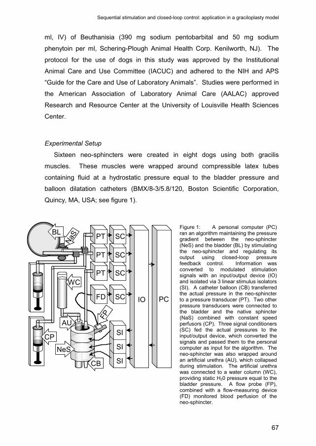
Sequential stimulation and closed-loop control: application in a graciloplasty model
67
ml, IV) of Beuthanisia (390 mg sodium pentobarbital and 50 mg sodium
phenytoin per ml, Schering-Plough Animal Health Corp. Kenilworth, NJ). The
protocol for the use of dogs in this study was approved by the Institutional
Animal Care and Use Committee (IACUC) and adhered to the NIH and APS
“Guide for the Care and Use of Laboratory Animals”. Studies were performed in
the American Association of Laboratory Animal Care (AALAC) approved
Research and Resource Center at the University of Louisville Health Sciences
Center.
Experimental Setup
Sixteen neo-sphincters were created in eight dogs using both gracilis
muscles. These muscles were wrapped around compressible latex tubes
containing fluid at a hydrostatic pressure equal to the bladder pressure and
balloon dilatation catheters (BMX/8-3/5.8/120, Boston Scientific Corporation,
Quincy, MA, USA; see figure 1).
WC
FP
AU
CB
CP
NaS
BL PT
PT
PT
FD
SC
SC
SC
SC IO PC
SI
SI
SINeS
WC
FP
AU
CB
CP
NaS
BL PT
PT
PT
FD
SC
SC
SC
SC IO PC
SI
SI
SINeS
Figure 1: A personal computer (PC)ran an algorithm maintaining the pressuregradient between the neo-sphincter(NeS) and the bladder (BL) by stimulatingthe neo-sphincter and regulating itsoutput using closed-loop pressurefeedback control. Information wasconverted to modulated stimulationsignals with an input/output device (IO)and isolated via 3 linear stimulus isolators(SI). A catheter balloon (CB) transferredthe actual pressure in the neo-sphincterto a pressure transducer (PT). Two otherpressure transducers were connected tothe bladder and the native sphincter(NaS) combined with constant speedperfusors (CP). Three signal conditioners(SC) fed the actual pressures to theinput/output device, which converted thesignals and passed them to the personalcomputer as input for the algorithm. Theneo-sphincter was also wrapped aroundan artificial urethra (AU), which collapsedduring stimulation. The artificial urethrawas connected to a water column (WC),providing static H20 pressure equal to thebladder pressure. A flow probe (FP),combined with a flow-measuring device(FD) monitored blood perfusion of theneo-sphincter.
Chapter 5
68
Stimulation of the neo-sphincters was performed using an input/output device
(CED1401plus, Cambridge Electronic Design, Cambridge, England) in
combination with a personal computer (Gateway 2000, Pentium-200/80) and
data acquisition software (Spike2, version 2.21, Cambridge Electronic Design,
Cambridge, England). Additional customized sequencer files and script files
were developed providing closed-loop control to the neo-sphincter (see
appendix). Details about the closed-loop control are reported in chapter 4. In
addition to the oscillation damping parameters described in chapter 4, an
algorithm controlling the neo-sphincter bladder pressure gradient was
incorporated in the present work. To create the stimulus, three separate output
signals were first led to optical linear stimulus isolators (A395’s, World Precision
Instruments, Sarasota, FL, USA) and thereafter to a multiple channel custom
made switchbox. A three-channel stimulation signal was provided to each neo-
sphincter, using three pairs of Teflon coated stainless steel wires (∅ 0.007 inch;
Medwire®, Mount Vernon, NY, USA) as leads. Baring the last 8 mm. of the wire
created the electrodes, while the other ends were connected to the switchbox.
Pressures generated by the stimulated neo-sphincters were recorded using the
balloon dilatation catheters connected to pressure-transducers (P23 ID,
GultonStatham Inc., CA, USA). Native sphincter pressures and bladder
pressures were measured using urine catheters (8 french) combined with
constant speed perfusors (model 600-900, Harvard Apparatus, Inc., Holliston,
MA, USA) connected to pressure transducers (P23 ID, GultonStatham Inc., CA,
USA). Doppler flow probes combined with a dual flow measurement device
(T206, Transonic, Ithaca, NY, USA) measured arterial blood flow. Signals of all
pressure transducers and flow probes were amplified by CED1902s (Cambridge
Electronic Design, Cambridge, England) and recorded with the above mentioned
input/output device.
Stimulation
The input/output device (CED 1401plus, Cambridge Electronic Design,
Cambridge, England) was used to generate a three-channel sequential
stimulation signal (mono-phasic block-shaped pulse-trains; frequency 30 Hz;

Sequential stimulation and closed-loop control: application in a graciloplasty model
69
pulse-width 500 µ-seconds; transition to next segment after 1.0 second). The
customized sequencer and script files controlled the amplitudes of the
stimulation signals for each individual segment.
Surgical preparation
All animals were anesthetized (Pentothal 6-12 mg/kg, Abbott Laboratories,
North Chicago, IL, USA), intubated and ventilated (Halothane, 1.5%, oxygen
94.5%, nitrous oxide 4%; Halocarbon, River Edge, NY, USA). Animals were
placed in the supine position with their hind legs abducted. Bilateral medial
incisions were made through which both gracilis muscles were identified and
lifted as single pedicled neuro-vascular flaps. The muscles were reduced to a
35 mm width by removing the caudal aspect. Starting from the proximal
musculo-tendinious junction, 12 cm. length was marked. These marked muscle-
parts were cut from their origins and insertions. Each muscle was tensionless
but closely wrapped around a latex tube (∅ 8mm, wall thickness: 0.5 mm.) and a
balloon catheter. Three pairs of Teflon coated electrodes were transversely
inserted into the neo-sphincters, at an angle of 45 degrees to the direction of the
fibers, 5 to 6 cm. distal from the main pedicle, dividing them into three parallel
individually stimulatable units (see also figure 2). The latex tubes were
connected to a water column providing the hydrostatic pressure equivalent of the
bladder pressure. The balloon catheter was inflated to150 cm H2O, in order to
minimize its compliance. Subsequently, any pressure generated by the neo-
sphincter was measured as an increase in pressure over 150 cm H2O. The tips
of urine catheters were positioned in the urethra at the level of the native
sphincter and in the bladder. The connected constant speed perfusors provided
saline at 1 ml.min.-1. Flow probes were connected to the arteries of the neuro-
vascular pedicles.
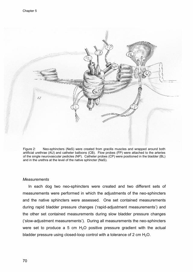
Chapter 5
70
Measurements
In each dog two neo-sphincters were created and two different sets of
measurements were performed in which the adjustments of the neo-sphincters
and the native sphincters were assessed. One set contained measurements
during rapid bladder pressure changes (‘rapid-adjustment measurements’) and
the other set contained measurements during slow bladder pressure changes
(‘slow-adjustment measurements’). During all measurements the neo-sphincters
were set to produce a 5 cm H2O positive pressure gradient with the actual
bladder pressure using closed-loop control with a tolerance of 2 cm H2O.
Figure 2: Neo-sphincters (NeS) were created from gracilis muscles and wrapped around bothartificial urethrae (AU) and catheter balloons (CB). Flow probes (FP) were attached to the arteriesof the single neurovascular pedicles (NP). Catheter probes (CP) were positioned in the bladder (BL)and in the urethra at the level of the native sphincter (NaS).
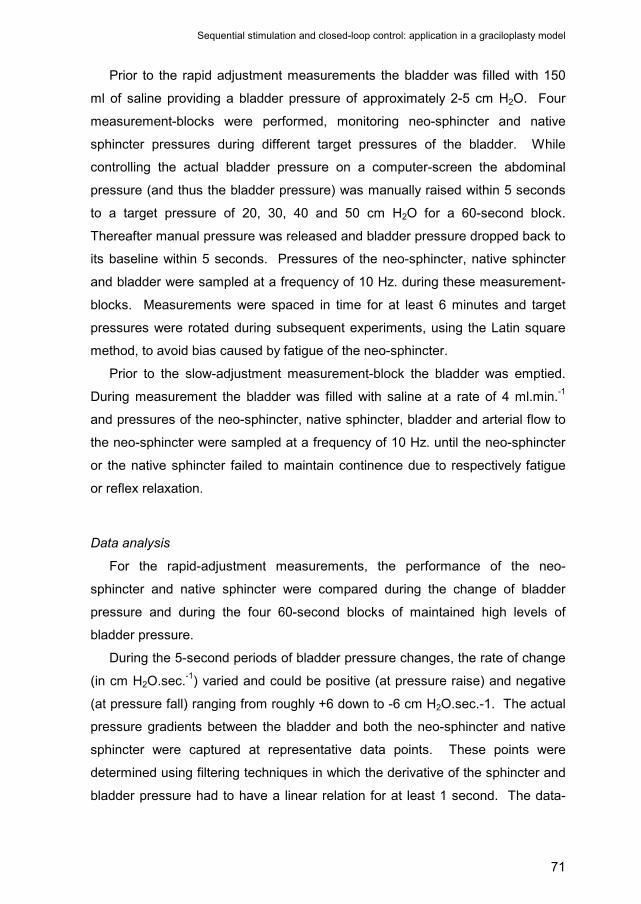
Sequential stimulation and closed-loop control: application in a graciloplasty model
71
Prior to the rapid adjustment measurements the bladder was filled with 150
ml of saline providing a bladder pressure of approximately 2-5 cm H2O. Four
measurement-blocks were performed, monitoring neo-sphincter and native
sphincter pressures during different target pressures of the bladder. While
controlling the actual bladder pressure on a computer-screen the abdominal
pressure (and thus the bladder pressure) was manually raised within 5 seconds
to a target pressure of 20, 30, 40 and 50 cm H2O for a 60-second block.
Thereafter manual pressure was released and bladder pressure dropped back to
its baseline within 5 seconds. Pressures of the neo-sphincter, native sphincter
and bladder were sampled at a frequency of 10 Hz. during these measurement-
blocks. Measurements were spaced in time for at least 6 minutes and target
pressures were rotated during subsequent experiments, using the Latin square
method, to avoid bias caused by fatigue of the neo-sphincter.
Prior to the slow-adjustment measurement-block the bladder was emptied.
During measurement the bladder was filled with saline at a rate of 4 ml.min.-1
and pressures of the neo-sphincter, native sphincter, bladder and arterial flow to
the neo-sphincter were sampled at a frequency of 10 Hz. until the neo-sphincter
or the native sphincter failed to maintain continence due to respectively fatigue
or reflex relaxation.
Data analysis
For the rapid-adjustment measurements, the performance of the neo-
sphincter and native sphincter were compared during the change of bladder
pressure and during the four 60-second blocks of maintained high levels of
bladder pressure.
During the 5-second periods of bladder pressure changes, the rate of change
(in cm H2O.sec.-1) varied and could be positive (at pressure raise) and negative
(at pressure fall) ranging from roughly +6 down to -6 cm H2O.sec.-1. The actual
pressure gradients between the bladder and both the neo-sphincter and native
sphincter were captured at representative data points. These points were
determined using filtering techniques in which the derivative of the sphincter and
bladder pressure had to have a linear relation for at least 1 second. The data-

Chapter 5
72
points were plotted against the rate of bladder pressure change (δPb/δt; see also
figure 3). Cubic regression lines were calculated for both the neo-sphincter and
native sphincter adjustment performance (SPSS 9.0, SPSS Inc, Chicago, IL,
USA).
During the four 60-second blocks of maintained high bladder pressure, the
means and the standard deviation of the pressure gradients between the
bladder and both the neo-sphincters and native sphincters were calculated and
compared using paired t-tests for each level of bladder pressure (see also figure
3). Also during the periods of continence, while performing the slow adjustment
measurements, the means and the standard deviation of the pressure gradients
between the bladder and both the neo-sphincters and native sphincters were
calculated and compared using paired t-tests.
pres
sure
Pbla meanPsph mean
∆P mean
standard deviation
calculations during calculations during calculations during calculations during rapid rapid rapid rapid adjustment measurements adjustment measurements adjustment measurements adjustment measurements rate of bladder pressure change andaccompanying pressure gradient
bb
rapidly descending pressuremaintained high pressure
rapidly ascending pressure
Pbla:Psph:∆P:
bladder pressuresphincter pressurepressure gradient
∆P
δPblaδt
Psph minus PblaPbla
Psph
time
pres
sure
Pbla meanPsph mean
∆P mean
standard deviation
calculations during calculations during calculations during calculations during rapid rapid rapid rapid adjustment measurements adjustment measurements adjustment measurements adjustment measurements rate of bladder pressure change andaccompanying pressure gradient
bb
rapidly descending pressuremaintained high pressure
rapidly ascending pressure
Pbla:Psph:∆P:
bladder pressuresphincter pressurepressure gradient
Pbla:Psph:∆P:
bladder pressuresphincter pressurepressure gradient
∆P
δPblaδt
Psph minus PblaPblaPbla
PsphPsph
time
Figure 3: Throughout rapid-adjustment measurements data pairs of the first derivative of the bladder pressure (δPbla/δt) and the pressure gradient of the sphincters (∆P) were collected during rapid ascending and descending bladder pressures. Furthermore, during maintained high bladder pressure the mean of the sphincters and bladder pressures (Psphmean, Pblamean) as well as the mean and standard deviation of the pressure gradients between sphincters and bladder were calculated from 600 samples collected in 60 seconds.

Sequential stimulation and closed-loop control: application in a graciloplasty model
73
Results
During the rapid-adjustment measurements, 5-second periods of bladder
pressure change both preceded and followed the 60 second-blocks of
maintained high bladder pressure. From the recorded data it was possible to
assess 158 relevant data-points linking the rate of bladder pressure change and
pressure differences between neo sphincter and bladder during bladder pressure changes
grad
. neo
-sph
inct
er/b
ladd
er ∆
P neo(
cmH
20)
bladder pressure change δPbla/ δt (cmH2O.sec-1)
cubic regression: Rsq 0.923 F(154)=611.98, p<0.000∆ Pneo=0.0322(δPbla/ δt)3-0.2057(δPbla/ δt)2-4.1317(δPbla/ δt)+4.1317
30
20
10
0
-10
-20-6 -4 -2 0 2 4 6
Figure 4a: Plot of the data pairs ofthe neo-sphincter/bladder pressuregradients (∆Pneo) and the firstderivative of the bladder pressure(δPbla/δt). A best fitting cubicregression line was calculated and isdrawn in the plot. The controllingalgorithm could not provide positivepressure gradients, while bladderpressures rose faster than 1 cmH2O.sec-1.
pressure differences between native sphincter and bladder during bladder pressure changes
grad
. nat
ive-
sphi
ncte
r/bla
dder
∆P na
t(cm
H20
)
cubic regression: Rsq 0.463 F(154)=44.27, p<0.000∆ Pnat=0.0885(δPbla/ δt)3-0.1648(δPbla/ δt)2-3.8853(δPbla/ δt)+6.9170
bladder pressure change δPbla/ δt (cmH2O.sec-1)
30
20
10
0
-10
-20
40
-6 -4 -2 0 2 4 6
Figure 4b: Plot of the data pairs of the native sphincter/bladder pressure gradients (∆Pnat) and the first derivative of the bladder pressure (δPbla/δt). A best fitting cubic regression line was calculated and is drawn in the plot. In general the native sphincters lost positive pressure gradients, while bladder pressures rose faster than 2 cm H2O.sec-1.

Chapter 5
74
pressure gradients between the bladder pressure and the neo-sphincter as well
as the native sphincter pressures. Plots for both the neo-sphincter and the
native sphincter performance are depicted in figure 4a and 4b. Cubic regression
lines show that especially the neo-sphincters performed predictably
(F(154)=611.98) and were able to provide a positive neo-sphincter/bladder
pressure gradient up to a changing bladder pressure of 1 cm H2O.sec.-1. More
rapidly increasing bladder pressures resulted in negative pressure gradients and
occlusion of the artificial urethras was (sometimes) lost. The native sphincters
showed more or less the same plot but proved less predictable (F(154)=44.27)
and positive pressure gradients were lost at bladder pressure changes higher
than 2 cm H2O.sec.-1.
Performance of the neo-sphincters compared to the native sphincters during
the 60-second blocks of high bladder pressures are depicted in figure 5a-c.
During all tested bladder pressures the neo-sphincters were capable of
maintaining a positive pressure gradient (Fig 5a). The native sphincter generally
‘failed’ at pressures above 40 cm H2O, resulting in emptying of the bladder. No
statistical differences were found between performance of the neo-sphincters
and the native sphincters, except at bladder pressures of 50 cm H2O in favor of
the neo-sphincter (see also figure 5b). The quality of constantly maintaining a
pressure gradient between the sphincters and the bladder is determined by
calculating the standard deviation of all pressure gradient samples per
measurement block as depicted in figure 5c. No statistical differences were
found between the quality of the neo-sphincter and the native sphincter.
During the slow adjustment measurements, the performance of the neo-
sphincters and the native sphincters were compared while maintaining a
pressure gradient with a filling bladder. Figure 6 depicts a typical plot of these
measurements combined with averaged outcome values. After almost 49
minutes (mean and SEM: 48.8 +/-7.2) one of both the neo- or native sphincters
could not maintain the necessary pressure gradient. At that point, the bladder
pressure had reached an averaged pressure of 16 cm H2O (mean and SEM:
15.9 +/-2.6). In all but two cases the neo-sphincter could not maintain the
pressure gradient due to fatigue.
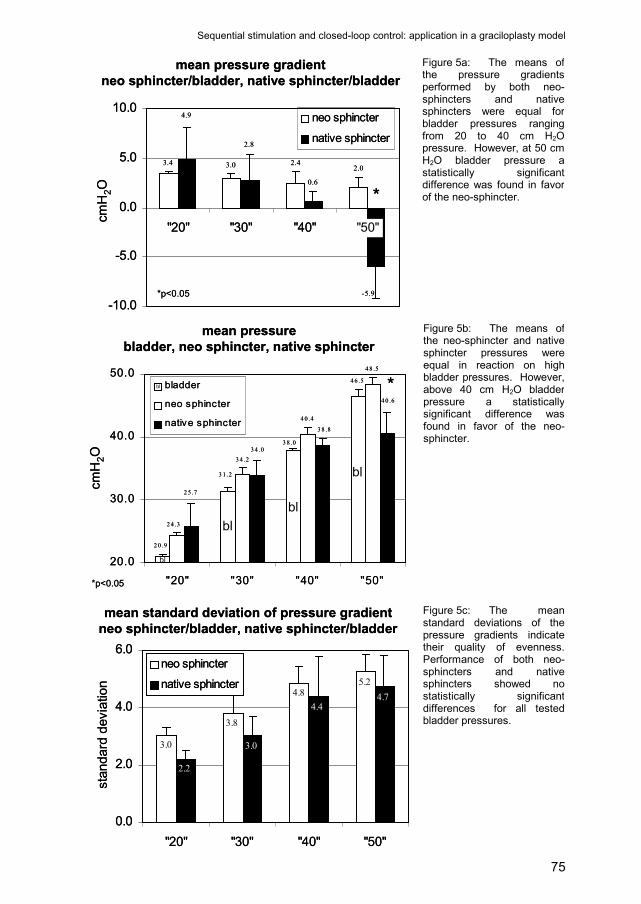
Sequential stimulation and closed-loop control: application in a graciloplasty model
75
Figure 5b: The means ofthe neo-sphincter and nativesphincter pressures wereequal in reaction on highbladder pressures. However,above 40 cm H2O bladderpressure a statisticallysignificant difference wasfound in favor of the neo-sphincter.
mean pressurebladder, neo sphincter, native sphincter
cmH
2O
38 .0
20 .9
31 .2
46 .5
24 .3
34 .2
40 .4
48 .5
34 .0
25 .7
38 .8
40 .6
20.0
30.0
40.0
50.0
"20" "30" "40" "50"
bladder
neo sphincter
native sphincter
*p<0.05
*
mean pressurebladder, neo sphincter, native sphincter
cmH
2O
38 .0
20 .9
31 .2
46 .5
24 .3
34 .2
40 .4
48 .5
34 .0
25 .7
38 .8
40 .6
20.0
30.0
40.0
50.0
"20" "30" "40" "50"
bladder
neo sphincter
native sphincter
*p<0.05
*
Figure 5a: The means of the pressure gradients performed by both neo-sphincters and native sphincters were equal for bladder pressures ranging from 20 to 40 cm H2O pressure. However, at 50 cm H2O bladder pressure a statistically significant difference was found in favor of the neo-sphincter.
3.4 3.0 2.42.0
4.9
2.8
0.6
-5.9
-10.0
-5.0
0.0
5.0
10.0
"20" "30" "40" "50"
neo sphincter
native sphincter
"50"
mean pressure gradientneo sphincter/bladder, native sphincter/bladder
cmH
2O *
*p<0.05
3.4 3.0 2.42.0
4.9
2.8
0.6
-5.9
-10.0
-5.0
0.0
5.0
10.0
"20" "30" "40" "50"
neo sphincter
native sphincter
"50"
mean pressure gradientneo sphincter/bladder, native sphincter/bladder
cmH
2O *
*p<0.05
Figure 5c: The meanstandard deviations of thepressure gradients indicatetheir quality of evenness.Performance of both neo-sphincters and nativesphincters showed nostatistically significantdifferences for all testedbladder pressures.
3.0
3.8
4.85.2
2.2
3.0
4.44.7
0.0
2.0
4.0
6.0
"20" "30" "40" "50"
neo sphincter
native sphincter
mean standard deviation of pressure gradientneo sphincter/bladder, native sphincter/bladder
stan
dard
dev
iatio
n
3.0
3.8
4.85.2
2.2
3.0
4.44.7
0.0
2.0
4.0
6.0
"20" "30" "40" "50"
neo sphincter
native sphincter
mean standard deviation of pressure gradientneo sphincter/bladder, native sphincter/bladder
stan
dard
dev
iatio
n
bl bl
bl
bl
bl

Chapter 5
76
Reaching this point at least one segment of the sequentially stimulated neo-
sphincter needed a higher stimulation amplitude than the maximum of 5 mAmp.
to generate the requested pressure. In the other two cases however, the native
sphincter reflexively relaxed and bladder pressure dropped before neo-sphincter
fatigue occurred. The relative blood-flow increase of the neo-sphincters during
stimulation was more than twofold (mean and SEM: 229% +/-30%). The mean
pressure gradient between bladder and both sphincters during the 8
experiments was 3.1 cm H2O with a SEM of +/-0.2 for the neo-sphincters and
10.1 +/-2.6 cm H2O for the native sphincters.
The mean of the standard deviations, reflecting the accuracy of the mean
pressure gradients, was 2.1 with a SEM of +/-0.2 for the neo-sphincters and 3.7
+/-0.3 for the native sphincters. Both differences were statistically significant
(see figure 7).
1 0 0%
2 2 9%
0
5
10
15
20
native sphincter
neo sphincter
bladder
mean+SEM: 15.9 ± 2.6
mean+SEM48.8 ± 7.2
time in minutes
typical plot of measurement during slow adjustment experiment shtypical plot of measurement during slow adjustment experiment shtypical plot of measurement during slow adjustment experiment shtypical plot of measurement during slow adjustment experiment showingowingowingowingpressures, neopressures, neopressures, neopressures, neo----sphincter blood flow, and stimulation valuessphincter blood flow, and stimulation valuessphincter blood flow, and stimulation valuessphincter blood flow, and stimulation values
relative blood-flow increase(mean+SEM) during stimulation
0123
0
2000
4000 segment 1
segment 2
segment 3
0 25 50
pres
sure
in c
mH
2Oflo
w in
ml
100%
229±30%
2k
4k
0stim
ulat
ion
in µ
Amp
0 25 50
0 25 50
1
2
31
23
12 3
*
*P<0.05
1 0 0%
2 2 9%
0
5
10
15
20
native sphincter
neo sphincter
bladder
mean+SEM: 15.9 ± 2.6
mean+SEM48.8 ± 7.2
time in minutes
typical plot of measurement during slow adjustment experiment shtypical plot of measurement during slow adjustment experiment shtypical plot of measurement during slow adjustment experiment shtypical plot of measurement during slow adjustment experiment showingowingowingowingpressures, neopressures, neopressures, neopressures, neo----sphincter blood flow, and stimulation valuessphincter blood flow, and stimulation valuessphincter blood flow, and stimulation valuessphincter blood flow, and stimulation values
relative blood-flow increase(mean+SEM) during stimulation
0123
0
2000
4000 segment 1
segment 2
segment 3
0 25 50
pres
sure
in c
mH
2Oflo
w in
ml
100%
229±30%
2k
4k
0stim
ulat
ion
in µ
Amp
0 25 50
0 25 50
11
22
3311
2233
1122 33
*
*P<0.05
Figure 6: During the slow-adjustment measurements the bladder was constantly filled andpressure elevated, while the neo- and native sphincters maintained a pressure gradient. Theperfusion of the neo-sphincters increased significantly during stimulation. In this typical plot the meanand standard error values are depicted.
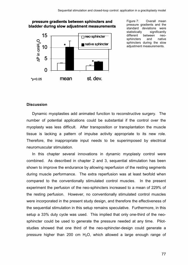
Sequential stimulation and closed-loop control: application in a graciloplasty model
77
Discussion
Dynamic myoplasties add animated function to reconstructive surgery. The
number of potential applications could be substantial if the control over the
myoplasty was less difficult. After transposition or transplantation the muscle
tissue is lacking a pattern of impulse activity appropriate to its new role.
Therefore, the inappropriate input needs to be superimposed by electrical
neuromuscular stimulation.
In this chapter several innovations in dynamic myoplasty control were
combined. As described in chapter 2 and 3, sequential stimulation has been
shown to improve the endurance by allowing reperfusion of the resting segments
during muscle performance. The extra reperfusion was at least twofold when
compared to the conventionally stimulated control muscles. In the present
experiment the perfusion of the neo-sphincters increased to a mean of 229% of
the resting perfusion. However, no conventionally stimulated control muscles
were incorporated in the present study design, and therefore the effectiveness of
the sequential stimulation in this setup remains speculative. Furthermore, in this
setup a 33% duty cycle was used. This implied that only one-third of the neo-
sphincter could be used to generate the pressure needed at any time. Pilot-
studies showed that one third of the neo-sphincter-design could generate a
pressure higher than 200 cm H2O, which allowed a large enough range of
0
5
10
15
mean st. dev.
neo sphincter
native sphincter
pressure gradients between sphincters andbladder during slow adjustment measurements
∆ P in
cm
H2O
*
*p<0.05
*0
5
10
15
mean st. dev.
neo sphincter
native sphincter
pressure gradients between sphincters andbladder during slow adjustment measurements
∆ P in
cm
H2O
*
*p<0.05
*
Figure 7: Overall meanpressure gradients and thestandard deviations werestatistically significantlydifferent between neo-sphincters and nativesphincters during the slowadjustment measurements.
Chapter 5
78
pressures necessary for the experimental designs. However, in other dynamic
myoplasty applications requiring high performance output, division of the muscle
into weaker separate segments, necessary for sequential stimulation, could
make the muscle incapable of reaching maximal performance peaks necessary
to meet the requirements of that specific function. For these applications
sequential stimulation does not suffice.
Closed-loop control made it possible to precisely tune the neo-sphincter
pressure to the required values by constantly repeated corrections of the
stimulation amplitude. The required neo-sphincter pressure values were
calculated by the function-controlling algorithm, which monitored the pressure
gradient between neo-sphincter and bladder.
A major advantage of closed-loop control is its self-regulation under changing
conditions. However, this continuous correction towards a goal value also
creates the possibility of over-correction, thus leading to oscillations. Therefore,
in the present experiment, the same oscillation-damping parameters were
incorporated as described in chapter 4. The stability and quality of the closed-
loop control can be assessed by calculation of the standard deviation of the
produced neo-sphincter pressures. No statistical differences were found
between the calculated standard deviation of the neo-sphincters and the native
sphincters during pressure gradient maintenance in the rapid-adjustment
measurements. However, during the slow adjustment measurements the neo-
sphincters performed better by having statistically lower standard deviations.
These two findings imply that the principle of closed-loop control can meet an
acceptable standard for clinical application. However, limited durability of
biosensors, displacement of- and fibrotic changes around biosensors are just a
few of the current drawbacks, making closed-loop control vulnerable to failure in
chronic applications.
Maintaining a pressure gradient while bladder pressure changed rapidly
showed that the neo-sphincter failed when bladder pressure changed faster than
1 cm H2O.sec.-1 (see also figure 4a). This is probably due to the fact that the
algorithm calculated corrections of the stimulation signals in a linear fashion.

Sequential stimulation and closed-loop control: application in a graciloplasty model
79
Exponential corrections however could provide a more accurate reaction at the
cost of a higher risk of oscillations (see equation 1).
Equation 1
Linear: )(1)1( agnn PPSS −+=+ κ
Exponential: 0)1(0)1(
2
2
)1(<−∧−−≥−∧−+=
−
−
+PaPgeSPaPgeSS
PaPg
PaPg
n
nn κ
κ
Equation 1: S(n+1): next stimulation signal; Sn: actual stimulation signal; κ1 & κ2: correction factors; Pg: goal pressure; Pa: actual pressure
In the experiments the rapid-adjustment measurements mimicked the
situation of sudden abdominal/bladder pressure rises and falls due to posture
changes, lifting objects, etc. The slow-adjustment measurements mimicked
diuresis. The function-controlling algorithm proved to be capable of responding
accurately to these bladder pressure changes by constantly regaining and
maintaining a positive pressure gradient. The value of approximately 49 minutes
of ‘untrained neo-sphincter continence’ is encouraging, but also relative, since
this value depends largely on the rate of bladder pressure rise and thus diuresis
rates. Furthermore, distribution of abdominal and bladder pressure, adding to
the native sphincter pressure as in normal physiology, was not considered.
The use of sensing programmable stimulators adds possibilities to implement
among other things self-learning processes. For instance, in the above-
described setup it often happened that one of the three neo-sphincter segments
fatigued faster than the other two, leading to termination of the measurement.
Therefore, pilot experiments were conducted after termination of the planned
measurements, repeating the slow adjustment-measurements with adapted
algorithms. These algorithms changed the fixed 33% duty cycle per neo-
sphincter segment to a variable duty cycle, depending on the relative fatigue of
each segment, so that a more fatigued segment performed a shorter duty cycle.
Relative individual segmental fatigue was assessed comparing the necessary
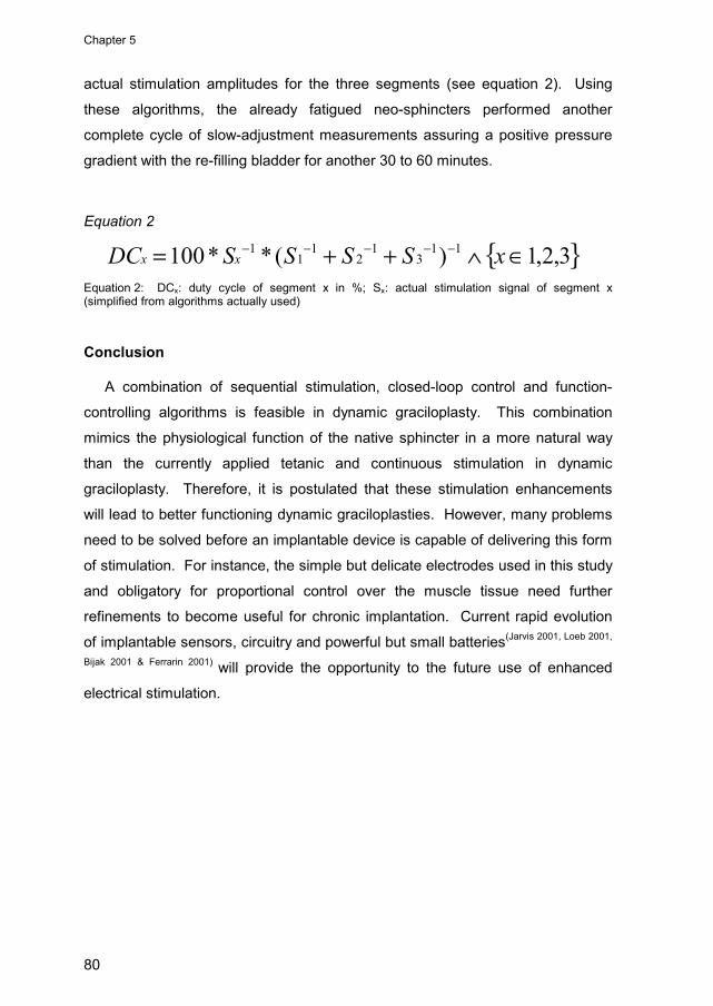
Chapter 5
80
actual stimulation amplitudes for the three segments (see equation 2). Using
these algorithms, the already fatigued neo-sphincters performed another
complete cycle of slow-adjustment measurements assuring a positive pressure
gradient with the re-filling bladder for another 30 to 60 minutes.
Equation 2
{ }3,2,1)(**100 113
12
11
1 ∈∧++= −−−−− xSSSSDC xx
Equation 2: DCx: duty cycle of segment x in %; Sx: actual stimulation signal of segment x (simplified from algorithms actually used)
Conclusion
A combination of sequential stimulation, closed-loop control and function-
controlling algorithms is feasible in dynamic graciloplasty. This combination
mimics the physiological function of the native sphincter in a more natural way
than the currently applied tetanic and continuous stimulation in dynamic
graciloplasty. Therefore, it is postulated that these stimulation enhancements
will lead to better functioning dynamic graciloplasties. However, many problems
need to be solved before an implantable device is capable of delivering this form
of stimulation. For instance, the simple but delicate electrodes used in this study
and obligatory for proportional control over the muscle tissue need further
refinements to become useful for chronic implantation. Current rapid evolution
of implantable sensors, circuitry and powerful but small batteries(Jarvis 2001, Loeb 2001,
Bijak 2001 & Ferrarin 2001) will provide the opportunity to the future use of enhanced
electrical stimulation.

81
Chapter 6
General discussion
Erik Zonnevylle, MD Richard Stremel, PhD Based (in part) upon the article: Stremel R, Zonnevylle E. Re-animation of muscle flaps for improved function in dynamic myoplasty. Microsurgery 21: 281-286, 2001.
“A scientific truth does not triumph by convincing its opponents and making them
see the light, but rather because its opponents eventually die and a new
generation grows up that is familiar with it” -Max Planck-

Chapter 6
82
Introduction
The re-animation of tissue flaps is an important advance in the field of
reconstructive surgery. While tissue flaps are used to repair large tissue defects
and restore form (such as in breast reconstruction), contractile flaps (dynamic
myoplasties) restore function, offering exciting new surgical solutions to clinical
reconstructive problems.
Dynamic myoplasty requires significant surgical skills including microsurgical
neuroraphy and anastomosis of arteries and veins. These techniques are
critical when transplanting tissue flaps from one body location to another. While
free vascularized muscle flap transfer has met with reasonable success (Harii 1976 &
Manktelow 1989), re-innervation of the flap, and thus motor control, is a slow process.
During this re-innervation process, the nonfunctioning transferred muscle
atrophies and the ability of the muscle flap to be animated to produce the
desired performance often results in less than optimal function. But also in the
case of a pedicled graft leaving the nerve intact, the dynamic myoplasty is
lacking a pattern of impulse activity appropriate to its new role. Therefore, it is
often necessary to use external electrical pulse generators eliciting appropriate
muscular contractions and in the case of free flaps preventing muscle atrophy,
during a re-innervation phase.
Most dynamic myoplasty applications currently require that the transferred
muscle flap must perform work that is different from that to which it was
accustomed. Examples include cardiomyoplasty, in which the entire latissimus
dorsi muscle (a muscle whose normal function is adduction and rotation of the
humerus) is wrapped around the myocardium. It is then stimulated by an
implantable pulse generator to contract rhythmically at a fairly high frequency
(approaching every other heart beat) and in synchrony with the heart.(Carpentier 1985,
Chachques 1987 & 1997) Another example is graciloplasty for urinary and fecal
incontinence. The gracilis muscle (a muscle whose normal function is adduction
of the thigh and flexion of the knee) is wrapped around the fecal or urinary outlet
and stimulated to contract tetanically and continuously for periods of 2 to 4
hours.(Williams 1989, Baeten 1991 & Janknegt 1992) These new functional requirements pose
important problems for these muscles; among the most significant and
General discussion
83
compromising of these are rapid muscle fatigue, muscle damage due to over-
stimulation, and ischemic lesions due to constant high pressures.(Williams 1991,
Konsten 1993, Janknegt 1995, Geerdes 1996 & Madoff 1999)
An approach used in dynamic myoplasty to avoid muscle failure due to
fatigue is to train the muscle to enhance fatigue resistance.(Salmons 1976 & 1981 & Koller
1994) Currently used training protocols require an 8-week period of stimulation at
increasing frequency until the muscle is incompletely converted to a fatigue-
resistant fiber type. Skeletal or striated muscles are normally a mixture of
fatigue-resistant, slow-twitch, oxidative (type 1) and fatigue-prone, fast-twitch,
glycolytic (type 2b) muscle fibers. Type 2a fibers have fast twitch, but fatigue
resistant, oxidative qualities and are considered to be an intermediate fiber
type.(Jarvis 1996, Mayne 1996, Pette 1997, Sutherland 1998 & Scott 2001) The innervation and the
function of the muscle determine the predominance of one fiber type over
another, and all fiber types in a given motor unit are the same. However,
striated muscle is plastic in nature, and the training regimen in cardiomyoplasty
and graciloplasty transforms the muscle to predominantly type 1 fibers. The
trade-off for producing fatigue resistance is a slower contracting muscle capable
of generating less power than its original character.(Salmons 1981 & 1994, Pette 1992)
While preliminary outcomes in these clinical applications of dynamic
myoplasty appear to be producing promising results, in general, the results are
variable. The clinical trials of both cardiomyoplasty and graciloplasty are
promising but have met with difficulties.(Williams 1991, Chachques 1997 & Madoff 1999) Many
factors probably contribute to these difficulties. One major reason for the
variable outcomes relates to the way the dynamic myoplasty is controlled using
electrical stimulation. Under normal circumstances, a given anatomical muscle
can respond and adapt to prolonged sustained activity, or brief intense activity.
The response generated by the muscle depends on the types of motor units
(and therefore myofiber types) contained within the muscle and the pattern of
activity in which they are engaged.(Saltin 1983, Scott 2001) Current applications of
dynamic myoplasty use tetanic stimulation and contraction of all myofibers
simultaneously. This tetanic type contraction occurs rarely in normal conditions
and leads to irreversible muscle damage, when prolonged. Furthermore, this
Chapter 6
84
type of on/off control does not allow more intricate applications of dynamic
myoplasty to be developed, in which fine tuned performance control is
mandatory. Therefore, a method of electrically stimulating striated muscle flaps
was developed, mimicking physiological muscle contraction more closely. The
ultimate purpose was the production of effective contraction within dynamic
myoplasties without an undue wait for training, without compromising force
generation, without ischemia during prolonged contractions and with precise
control of the generated performance. To some extent these purposes were met
in this thesis.
Sequential stimulation
The conventional stimulation approach in cardiomyoplasty and graciloplasty
involves stimulation of the nerve where it enters the muscle. This approach
produces neuromuscular stimulation through excitation of the motor nerves.
Excitation of the motor nerve leads to excitation of all the motor units associated
with this motor nerve. A consequence of this conventional stimulation is
simultaneous excitation and contraction of the entire muscle flap. Therefore, a
muscle flap must be divided into separately excitable segments to develop a
stimulation approach that permits segmental recovery from contraction, during
contraction of other segments. A dynamic myoplasty muscle is divided into
multiple individually excitable segments by placing pairs of stimulating
electrodes (anodes and cathodes) in strategic locations. If these segments are
induced to contract in a sequential fashion, one segment rests while adjacent
segments generate the necessary force required from the dynamic myoplasty.
We termed this method of stimulation, sequential segmental neuromuscular
stimulation. In the development of sequential segmental neuromuscular
stimulation, our hypothesis was that this modulus of stimulation would mimic true
physiological contractions of muscle more closely and thus enhances fatigue
resistance of dynamic myoplasties.
General discussion
85
Application of sequential stimulation
Our investigations of sequential stimulation as a method to reduce fatigue
were focused on stimulation of the canine gracilis muscle. In anesthetized dogs,
the gracilis muscles were lifted as uni-pedicled muscle flaps. To mimic human
proportions, the muscles were tailored to a width of 3.5 cm by removing the
posterior aspects of each muscle. The experimentation setup consisted of a
simple fixation of the pelvis while each gracilis muscle was attached to an
isometric force transducer (see figure 1 in chapter 2). Electrode placement was
critical since it was important to establish four segments of the gracilis capable
of generating equal force in each segment (see figure 2 in chapter 2). The
stimulus intensity was adjusted to generate a pre-determined force that was the
same for both sequential and conventional stimulation during any single
comparison.
Since sequential stimulation offers the ability to use variable portions of the
entire gracilis muscle, the amount of time spent in contraction versus relaxation
was considered important to muscle force generation and fatigue resistance.
Thus, a duty cycle was determined for each segment based on the number of
segments contracting simultaneously. For instance, if only one of four segments
were contracting at a time, then the duty cycle was 25% and the resting cycle
was 75% because the muscle was working 25% of the time and resting 75% of
the stimulation period. If all four segments were contracting simultaneously, the
duty cycle was 100% and the contraction was equivalent to that generated by
conventional stimulation.
During these experiments, the time required for the generated force to
decrease by one half (i.e., the half-time to fatigue) was determined, together with
the arterial blood flow. The results were quite striking and unequivocal.
Sequential segmental neuromuscular stimulation significantly enhanced fatigue
resistance in an inverse progressive fashion with respect to the duty cycle.
During the contractions, the muscle blood flow increased more than 100% when
compared to conventional stimulation for duty cycles of 25, 50 and 75 percent.
The success of these experiments suggested that the application of this
stimulus paradigm to the functioning of the graciloplasty neo-sphincter could

Chapter 6
86
provide fatigue resistance due to improved perfusion and avoid prolonged
training, which implies loss of strength and responsiveness. This would permit
earlier functional use of the neo-sphincter and preservation of reactivity, which is
important for the possibility to acutely adjust performance (see section on
closed-loop control). Therefore, the contractile function and perfusion of gracilis
neo-sphincters were studied using both sequential and conventional stimulation.
In anesthetized dogs, both gracilis muscles were converted into neo-sphincters
(see figure 1 and 2 in chapter 3) and electrically stimulated. The stimulation
approach consisted of three-channel sequential stimulation on one side (a duty
cycle of 33%) and conventional stimulation on the other. Both neo-sphincters
were stimulated at an intensity that generated 80 cm H2O pressure. To ensure
that the neo-sphincters could shorten and reduce their lumen, latex tubes
connected to a 40 cm H2O hydrostatic pressure column were also inserted in the
neo-sphincter lumen alongside rigid balloon pressure catheters. The time
necessary for the neo-sphincter to fatigue to one half of the initial pressure was
determined. The results of this neo-sphincter application showed that sequential
stimulation was significantly superior to conventional stimulation. Blood flow in
the sequentially stimulated neo-sphincter actually increased by 86% while
perfusion decreased by 37% in the conventionally stimulated neo-sphincters.
Despite these results, sequential stimulation enhanced the endurance of the
neo-sphincter for only a few more minutes. When used in clinical applications
an untrained sequentially stimulated neo-sphincter would result in short
continence times, which would not be acceptable.
Discussion on sequential stimulation
A universal complication of dynamic myoplasties is muscle fatigue. Muscles
require maintenance of adenosine triphosphate (ATP) within the myofibers to
generate force through the contractile interaction of actin and myosin proteins.
In the absence of adequate oxygen delivery, muscle dependence on aerobic
mechanisms of metabolism for ATP generation will become limited and
dependence on anaerobic metabolic mechanisms will prevail. A consequence
General discussion
87
of this shift to anaerobic metabolism is a reduction in ATP production and an
increase in lactic acid production. While the etiology of muscle fatigue is
complex and can result from many contributing factors, the combination of
decreased ATP availability and decreased intracellular pH from lactic acidosis
certainly contributes to the failure of muscle to produce contractile force.(Fitts 1996)
In dynamic myoplasty, muscle tissue fatigues primarily for two reasons: it is
inadequately perfused and/or it is stimulated to perform in a way that is
incompatible with its basic muscle fiber type. Currently used stimulation devices
produce prolonged tetanic skeletal muscle contractions. As muscle tissue
contracts, the individual myofibers within the muscle place significant pressure
on the blood vessels within the muscle. Due to these high transmural pressures,
a tetanic contraction has the effect of occluding blood flow through the muscle
during the contraction. This reduction of perfusion due to internal muscle
pressure contributes to the failure to deliver adequate oxygen and nutrients to
the contracting muscle, leading to increasing lactic acid and decreasing ATP,
which precipitates fatigue of the muscle and failure to generate force despite
continued stimulation. This can be partially circumvented by transforming the
muscle into a predominantly oxidative type 1 fiber containing muscle, using
chronic stimulation and muscle training. The oxidative pathway replenishes the
ATP, while the type 1 fibers deplete the available resources to a lesser extent.
However, this process costs strength and responsiveness. Availability of
resources on the other hand can be increased somewhat by augmenting
perfusion of the performing muscle tissue as is clearly the case in sequential
stimulation. The lesser performance of the non-isometric sequentially stimulated
myoplasty, reported in chapter 3, when compared to the isometric sequentially
stimulated myoplasty of chapter 2, is at least partly due to the kinking of the
intra-muscular blood-vessels and allowed shortening of the muscle during
stimulation in the non-isometric setup. This induced increased resistance of the
intra-muscular vessels, leading to less effective perfusion, and relative
decreased fatigue resistance in the non-isometric setup, when compared to its
isometric counterpart.
Chapter 6
88
Peckham et al. and Petrofsky described various experimental and clinical
applications of fatigue resistance improving types of stimulus paradigm.(Peckham
1970 &1976, Petrofsky 1979) It has been demonstrated in humans that sequential
alternation of stimulus between agonistic muscle groups enhances fatigue
resistance.(Peckham 1970 & Pournezam 1988) Peckham et al. used a random electrode
placement in cat skeletal muscle to stimulate contraction in three
segments.(Peckham 1976) This random approach was likely to lead to overlap of
stimulus area and precipitate muscle fatigue. Almost all studies utilizing multiple
stimulation channels have also employed a common electrical ground, causing
spread of the stimulation pulse. In the studies presented in this thesis, multiple
differential channels with stimulus isolators were employed. Furthermore,
discrete electrode placement in more distal parts of the muscle permitted
exclusion of recruitment of the main motor nerve branches supplying the muscle.
In this way, it was possible to recruit local intra-muscular nerves, which
produced far less overlap of recruited segments. Petrofsky used a rotary
electrode array to stimulate the peripheral nerve directly to sequentially recruit
motor units and reduce fatigue. Unfortunately, peripheral nerve stimulation is
associated with nerve injury and has not achieved broad clinical application.
More recently, Lau et al. compared the effects of stimulating rabbit triceps at two
to three different sites on fatigue resistance.(Lau 1995) Sequential stimulation
proved to be superior to whole muscle and alternating segment stimulation in
producing fatigue resistance. These observations underscore the results of the
studies of chapter 2 and 3.
Closed-loop control
In the previous paragraphs a relationship is demonstrated between rate of
exertion, duty cycle and rate of perfusion in a dynamic myoplasty. Furthermore,
depletion of ATP, which is counteracted by perfusion and progressed by
exertion, limits, among others, the performance and thus the applicability of
dynamic myoplasties. With sequential stimulation already enhancing perfusion,
it is reasonable to economize exertion to further maximize the possibilities of

General discussion
89
dynamic myoplasties. Therefore a condition is created in which it is possible to
reduce the amount of work the myoplasty performs to the minimal level
necessary to carry out the required physiological function for prolonged periods.
Furthermore, reducing peak exertion to the minimum must have beneficial
effects on surrounding tissues by reducing ischemic lesions, stricture and scar
formation.
Closed-loop control is a powerful cybernetic tool in muscle stimulation(Lemay
1997, Quintern 1997 & Ferrarin 2001) and offers the possibility of continuously controlling the
performance of dynamic myoplasties by cyclically comparing the actual
performance to a target performance. Differences between these two are used
to compute corrections, which are applied in the stimulation signal and evaluated
in the next cycle.
Biosensors feed the closed-loop control tool with the actual performance.
These sensors can offer all kinds of biofeedback. This biofeedback can also
provide input for computing devices running algorithms, which process all sorts
of decisions concerning dynamic myoplasty performance. This ‘artificial
intelligence’ (ranging from simple algorithms to sophisticated self-learning
control systems) offers refinement in complex tasks, opening up possibilities for
more demanding implementations of dynamic myoplasties.
Application of closed-loop control
A simple closed-loop control stimulation algorithm was developed and tested
on sequentially stimulated neo-sphincters designed from canine gracilis muscles
(see chapter 4). Pressure, generated in the lumen of the neo-sphincters, was
used as feedback to regulate the stimulation signal. The typical property of
closed-loop control to oscillate around the target performance was countered
using three oscillation-damping parameters. Effectiveness of these parameters
was determined by calculating the standard deviation of the actual measured
pressures for ranges of parameter values. Each oscillation-damping parameter
showed a significant effect on the standard deviations; the lowest standard
deviations corresponded with the optimal value settings for each parameter.
Chapter 6
90
Additionally, the efficiency of amplitude and frequency modulation techniques
were investigated and compared using a similar procedure. Amplitude
modulation systematically showed lower standard deviations, but frequency
modulation allowed faster bridging of large differences between actual and
target pressures. Closed-loop control enabled the maintenance of a variety of
target pressures within 10% deviation, using optimized oscillation-damping
parameters and amplitude modulation.
Versatile dynamic myoplasties can be designed, when the blood perfusion
enhancement of sequential stimulation and the performance regulation of
closed-loop control are combined with function-controlling algorithms executing
more complex tasks. In chapter 5 a graciloplasty-model (for urinary
incontinence) is described using the sequentially stimulated closed-loop
controlled neo-sphincter of chapter 4. In this model the function-controlling
algorithm was of a low complexity: a pressure gradient between the neo-
sphincter and the bladder was maintained, while the bladder pressure was
rapidly and slowly changed, mimicking respectively change of posture, lifting
heavy objects, etc. and diuresis filling the bladder. Performance of the neo-
sphincter was compared to the native sphincter. The graciloplasty-model proved
to be satisfyingly functional: rapid bladder pressure changes were effectively
compensated with neo-sphincter pressure corrections and without any form of
training the neo-sphincter was capable of maintaining continence during
pressure increase in the bladder due to diuresis for prolonged periods. Overall
the graciloplasty-model functioned as a reasonable alternative for the native
sphincter.
Discussion on closed-loop control
In this thesis, closed-loop control is advocated as an efficient cybernetic tool
to gain proportional control over the performance output of a dynamic myoplasty.
In order to apply proportional control, it is essential to reliably and reproducibly
control small increments and decrements in the performance output of the
muscle. Learning by trial and error we found that the anode and cathode
General discussion
91
electrodes had to be placed distally enough, not to evoke larger common trunks
of the intra-muscularly diverging innervating motor nerve, and proximally enough
to stimulate small branches of this motor nerve before they finished at the motor
end-plates. A 45 degrees angle between the axis of the electrical field, built up
between the anode and cathode on one hand, and the direction of the muscle
fibers and intra-muscular motor nerve branches on the other, allowed for a
workable, pseudo-linear ratio between the amount of recruited motor-units and
signal strength. This important finding made it possible to proportionally control
the output performance with the signal strength in a reliable and reproducible
manner. This proportional control allowed closed-loop control to precisely direct
the performance output of the neo-sphincter.
Closed-loop control is unstable by its nature. This lack of stability is caused
by the constant cyclic repetition of evaluation of the current performance,
followed by correction. The application of corrections has a latency, which is
system specific. If the corrections overtake their own application, caused by
system latency, the closed-loop control starts to oscillate. So, the more cycles
per time unit the evaluation is executed beyond this point, the more unstable the
system becomes. On the other hand however, the performance directed with
this closed-loop control has a tendency to diverge within a certain time and
needs correction, before these diversions become larger than prudent.
Therefore, an ideal frequency of corrections exists, in which corrections are
made fast enough to prevent large diversions in performance, but not too fast to
cause oscillations. The ‘correction frequency’, described in chapter 4, showed
this optimum and proved to have a reproducible value. Furthermore it is
important to realize that the control over performance output is not infinitely
precise. Therefore it is not reasonable to correct for small deviations of
performance output, when the corrected signal will not apply this intended
correction in the first place. Adversely, these corrections of the stimulation
signal will be repeated cycle by cycle until a threshold is passed and the
performance output reacts with an overshoot of the intention. This over-steering
of the closed-loop control will also result in oscillations. Therefore, a certain
deviation must be allowed to prevent this adverse effect. On the other hand, the
Chapter 6
92
threshold for corrections should be as low as the system allows, preventing
gross deviations. In the experiments described in chapter 4, the anticipated
optimized ‘correction threshold’ was found. Concerning the sequential
stimulation, it was also important to consider the latency of application of the
stimulation signal corrections. It is important to consider the segments of the
sequentially stimulated dynamic myoplasty as independent units, which have
their individual steering parameters (see also figure 3 in chapter 4), but with a
common target. An evaluation of the performance output is meaningless, when
the sequential stimulation has just switched to a next segment: in this case, due
to latency, the performance output by the previous segment is still existent. An
evaluation during this latency will lead to inappropriate corrections in the
stimulus signal of the starting segment, which has its own independent setting of
stimulation signal. This causes faults in the stimulation signal and thus
deviations at the start of a segment. Therefore, it is necessary to introduce a
short period in which no evaluations and corrections are executed after
switching to the next segment in sequentially closed-loop controlled stimulation.
However, when the ‘transition time’ is chosen too long, the period of absence of
corrections will lead to unnecessary deviations of the target performance.
Again, a trade-off between system latency and early corrections of the stimulus
signal leads to an optimized value, which was described in chapter 4. It could
be of interest to investigate if the evaluation of the performance during the start
of a next segment can be of use during the next start of the previous segment.
This implies a phase shifting between sequential stimulation and closed-loop
control, determined by the latency of the system.
In the first paragraph of this section the importance of proportional control
over performance is described. Amplitude modulation of the stimulation signal
provided a more precisely proportioned performance output, when compared to
frequency modulation. The modulation of the signal was computed using a
simple linear algorithm in which the correction of the amplitude (and frequency in
the case of frequency modulation) was determined by multiplying the
performance deviation with an empirically fixed correction factor. However, the
use of this linear algorithm to compute the amplitude corrections from the
General discussion
93
performance deviations did not simultaneously allow executing fine-tuning on
one hand and fast bridging of large performance changes on the other, using the
same fixed correction factor. Therefore it could be of interest to use an
exponential design for this algorithm. Another possibility to deal with the
apparent opposite interests of fine-tuning and the capability of quickly bridging
large changes in performance, could be the combination of amplitude and
frequency modulation: the latter proved to be capable of bridging large
performance changes faster, while amplitude modulation can add fine-tuning.
A combination of sequential segmental neuromuscular stimulation, closed-
loop control and function-controlling algorithms provides versatility to dynamic
myoplasties. In chapter 5 a successful application is described in which a neo-
sphincter maintains a pressure gradient with changing bladder pressures.
Based on this model it proved possible to upgrade the function-controlling
algorithm to a more sophisticated version. The upgraded version could learn the
relative state of fatigue of each segment of the triple partitioned neo-sphincter at
that moment, by simply comparing the amplitude changes of each segment.
With this information the duty cycle of each segment was adjusted, taking into
account their individual state of fatigue relative to the other segments. This
improved the endurance of the neo-sphincter, which was otherwise limited by
the fastest fatiguing segment. This underscores that the above-described
combination adds some versatility to the dynamic myoplasty.
Using the present knowledge, but disregarding the lack of availability of
versatile programmable implantable stimulators, the ideal neo-sphincter design
should partition the muscle into two sections. The first more cranially located
section of the neo-sphincter should contain two sequentially stimulated
segments. By training, these two segments should be transformed into units
containing mainly slow twitch fibers. The other more distally located section
should contain three non-transformed fast twitch sequentially stimulated
segments. Both sections should have their own independently functioning
closed-loop control systems. The slow twitch partition should be used to
maintain a mean pressure gradient with the bladder pressure, based upon the
integrated pressure gradient information of probably a minute. The fast twitch
Chapter 6
94
partition however, should be used to safeguard a pressure gradient during acute
moments of bladder pressure raise, which temporarily exceeds the slow twitch
neo-sphincter pressure. Therefore, its functioning should be based upon the
actual pressure gradient between the bladder and the slow twitch partition of the
neo-sphincter. In this concept, the slow twitch partition functions like an
involuntary smooth muscle sphincter, while the fast twitch partition is better
compared to a striated voluntary sphincter.
In fact, replacement of any sphincter in the body (e.g., anal, dynamic
myoplasty for stomal continence etc.) could benefit from similar stimulation
approaches. However, the experiments described in this thesis were
accomplished via a computer/data acquisition system with the size of a fridge,
limiting the experiments to an acute design. The technology for the
miniaturization of this more sophisticated stimulation system exists and is slowly
becoming available.(Jarvis 2001) Moreover, feedback of all sorts of data from
miniature implantable sensors is nowadays feasible with modern electronics.
However, the use of leads/electrodes in chronic designs remains vulnerable due
to the chance of displacement and breakage. A revolutionary design, dealing
with this problem, is the BIONtm system, which disregards all leads, by
introducing radio-controlled stimulation (BION1tm) and sensing (BION2tm)
units.(Loeb 2001) Altogether, the results presented in this thesis justify the effort of
miniaturizing the current computer/data acquisition system, so it can be tested in
chronic experimental designs.
Conclusion
Sequential segmental neuromuscular stimulation improves the (re)perfusion
of dynamic myoplasties during performance of their tasks. This improved
perfusion enhances fatigue resistance in an inverse progressive fashion with
respect to the duty cycle.
These effects are more pronounced in an isometric setup with stretched out
and fixed muscle tissue than in a non-isometric setup, which allows shortening
of the muscle tissue.
General discussion
95
Closed-loop control is a sufficient cybernetic tool to regulate the performance
generated by sequentially stimulated dynamic myoplasties. The use of
optimized damping parameters, can successfully reduce the inherent problem of
oscillations in closed-loop control, whereas amplitude modulation of the
stimulation signal provides better proportional control over the performance of
dynamic myoplasties, then frequency modulation.
A combination of sequential stimulation, closed-loop control and a function-
controlling algorithm offers more versatile dynamic myoplasties, broadening the
abilities of reconstructive surgeons in the repair of functional muscle defects.
All results justify the development of a comparable, but implantable system,
which can be used in chronic studies to further elucidate the benefits of
sophisticated electrical stimulation in dynamic myoplasty.
“The important thing is not to stop questioning” -Albert Einstein-

97
Summary
New electrical stimulation techniques in dynamic myoplasty
98
Summary
It has become common practice in reconstructive surgery to transpose or
transplant a variety of autologous tissues to fill defects at a recipient site. Using
muscle tissue, it becomes possible to dynamically assist or replace an impaired
or lost function. For these procedures the term ‘dynamic myoplasty’ is generally
used. In dynamic myoplasty, control of timing and rate of contraction of the
transposed or transplanted muscle tissue is substantial, but presently not fully
within reach. Currently used electrical stimulation protocols, which stimulate
innervating nerves or the muscle tissue itself, supply gross on-off spasm-like
contractions of the whole dynamic myoplasty muscle. This leads to rapid
fatigue, which can be partly overcome using training protocols converting the
dynamic myoplasty into a more fatigue resistant state at the cost of strength and
responsiveness. Furthermore ischemic lesions, fibrotic changes and abundant
scarring due to constant high tissue pressures are widespread reported in these
procedures.
This thesis reports on efforts to gain more control over dynamic myoplasties,
in order to overcome some of the drawbacks of currently used electrical
stimulation on one hand, and to give rise to more refined applications of dynamic
myoplasties on the other.
Conventional stimulation techniques recruit all or most of the muscle fibers
simultaneously and continuously. Therefore, sequential segmental
neuromuscular stimulation was introduced, in which only segments of the
transferred muscle tissue are stimulated in a sequential fashion, providing
continuous work, but with alternating partitions. This way, the temporary resting
segments are reperfused during functioning of the dynamic myoplasty. The
muscles studied were the canine gracilis and all experiments were acute studies
in anesthetized animals. Comparison of sequential and conventional stimulation
revealed the predicted increase in muscle fatigue resistance and muscle blood
flow. These effects were more pronounced in the isometric setup with stretched
out and fixed gracilis muscles reported in chapter 2, than in a non-isometric
setup with gracilis based neo-sphincters, which were allowed to shorten during
stimulation, as described in chapter 3. The shortening of the muscle tissue and
Summary
99
kinking of the intra-muscular blood vessels within the neo-sphincters could be a
reason for the less distinct increase of perfusion and subsequent fatigue
resistance during non-isometric contractions.
Apart from improving perfusion, reducing the load of the dynamic myoplasty
will improve its endurance as well. Furthermore, it is likely that reducing the load
will decrease ischemia, leading to less fibrotic changes and subsequent scarring
of the involved tissues. To economize the load of the dynamic myoplasty to
exact (and changing) needs, control over the contraction rate of the muscle
tissue needs to be precise. Closed-loop control proved to be a good cybernetic
tool to regulate the pressures generated by gracilis based sequentially
stimulated neo-sphincters in an acute dog study as was elucidated in chapter 4.
The inherent problem in closed-loop control of oscillations was successfully
reduced to less than 10% of the target-pressures by using optimized correction
frequencies, correction thresholds and transition times. Modulation of the
stimulation amplitudes proved to be superior over modulation of the stimulation
frequency in order to tune the performance of the neo-sphincters.
Having increased fatigue resistance and being able to precisely control the
rate of performance, a dynamic myoplasty should be able to perform more
complex tasks. Therefore, an acute dog study was designed in which gracilis
based neo-sphincters were made to maintain a pressure gradient with the
bladder during fast and slowly changing bladder pressures, using a combination
of sequential stimulation, closed-loop control and a function-controlling
algorithm. Results, set out in chapter 5, showed that the neo-sphincter was able
to maintain the desired pressure gradient with the bladder during fast and slowly
changing bladder pressures. Accuracy showed no significant difference when
compared to the native sphincter, which served as control. Addition of a
simplified self-learning component in the algorithm, adapting the duty cycle of
individual segments to their relative state of fatigue, proved feasible and
meaningful in this study design.
Overall this thesis reports improvement in fatigue resistance and control over
a dynamic myoplasty using sequential stimulation, closed-loop control and
function-controlling algorithms in acute studies. A comparable implantable

New electrical stimulation techniques in dynamic myoplasty
100
system, can be used in chronic studies. Availability of this implantable system
should be able to confirm the lesser need for muscle fiber type transformation,
using training regimens, and also reveal other benefits as prevention of ischemic
lesions, fibrotic changes and abundant scar formation. All these improvements
should offer more versatile dynamic myoplasties, broadening the abilities of
reconstructive surgeons in the repair of functional defects.

101
Samenvatting in het Nederlands
Nieuwe elektrische stimulatie technieken in dynamische spierplastieken
102
Samenvatting
In de reconstructieve chirurgie is het steeds meer gebruikelijk om
verschillende eigen weefselsoorten te transponeren of te transplanteren
teneinde wonddefecten te sluiten. Door spierweefsel te gebruiken is het
mogelijk om verzwakte of verloren functies dynamisch te ondersteunen dan wel
te vervangen. Voor deze ingrepen wordt veelal de term ‘dynamische
spierplastiek’ gebruikt. Bij het gebruik van dynamische spierplastieken is
regulatie van het tijdstip en de mate van contractie van het getransponeerde dan
wel getransplanteerde spierweefsel belangrijk, maar op dit moment nog niet
geheel haalbaar. De huidige elektrische stimulatie protocollen, welke de
innerverende zenuwen dan wel het spierweefsel zelf stimuleren, veroorzaken
grove alles-of-niets, spastisch-achtige contracties van de gehele dynamische
spierplastiek, hetgeen leidt tot snelle vermoeidheid van de spierplastiek. Deze
vermoeidheid kan deels worden tegengegaan door het toepassen van
trainingsprotocollen, welke dynamische spierplastieken converteren naar een
minder snel vermoeibare status, echter ten koste van kracht en reactiviteit.
Daarnaast komt bij deze manier van stimuleren ischemische laesies,
fibrotisering en overmatige littekenvorming van de betrokken weefsels voor,
veroorzaakt door de continu hoge druk.
Dit proefschrift beschrijft een manier om meer controle te krijgen over
dynamische spierplastieken; enerzijds door nadelen van de huidig gebruikte
elektrische stimulatie te voorkomen en anderzijds door de mogelijkheid te
creëren voor meer verfijnde toepassingen van dynamische spierplastieken.
Conventionele stimulatie technieken rekruteren alle of de meeste spiervezels
gelijktijdig en continu. Derhalve werd sequentiële segmentale zenuw-spier
stimulatie geïntroduceerd, hetgeen alleen segmenten van het verplaatste
spierweefsel stimuleert op een sequentiële manier, waarbij wel continu
spierarbeid wordt verricht, maar met steeds afwisselende spiergedeeltes. Bij
deze methode worden de tijdelijk rustende segmenten gereperfundeerd terwijl
de dynamische spierplastiek toch continu functioneert. Voor de experimenten
beschreven in dit proefschrift werden de gracilis spieren van honden gebruikt.
Alle experimenten waren acute studies en vonden plaats onder algehele

Samenvatting in het Nederlands
103
narcose. Bij het vergelijken van sequentiële met conventionele stimulatie liet de
sequentiële stimulatie inderdaad de voorspelde afname in spier vermoeidheid en
de toename van perfusie van de spieren zien. Deze effecten waren meer
uitgesproken in de isometrische opstelling met de opgespannen en gefixeerde
gracilis spieren beschreven in hoofdstuk 2, dan in de niet-isometrische opstelling
met van gracilis spieren nieuwgevormde sluitspieren, welke gedurende de
stimulatie mochten verkorten, zoals gerapporteerd in hoofdstuk 3. Het verkorten
van het spierweefsel en het kinken van de bloedvaten in het spierweefsel van de
nieuwgevormde sluitspieren zou een verklaring kunnen zijn voor de minder
uitgesproken toename in perfusie en vermoeidheidsresistentie gedurende non-
isometrische contracties.
Naast het verbeteren van de perfusie, middels sequentiële stimulatie, zal het
verkleinen van de belasting van de dynamische spierplastiek het
uithoudingsvermogen van deze verder verbeteren. Bovendien is het
aannemelijk dat bij een verkleining van de belasting de kans op ischemie
afneemt, hetgeen leidt tot minder fibrotische veranderingen en daaruit volgende
littekenvorming in en rond de betrokken weefsels. Om de belasting van de
dynamische spierplastiek zo economisch mogelijk af te stemmen op de
(veranderende) behoefte, is een precieze regulatie van de mate van contractie
noodzakelijk. Terugkoppeling gestuurde controle bleek een geschikte
besturingsmethode om de druk te reguleren welke werd gegenereerd door van
gracilis spieren gemaakte sluitspieren, zoals beschreven in hoofdstuk 4. Door
gebruik te maken van geoptimaliseerde correctie frequenties, correctie drempels
en overgangspauzes werd het inherente probleem van oscillaties in
terugkoppeling gestuurde controle succesvol gereduceerd tot minder dan 10%
van de doeldruk. Modulatie van de stimulatie amplitudes bewees een
nauwkeuriger methode te zijn dan modulatie van de stimulatie frequenties bij het
reguleren van de gegenereerde drukken in de nieuwgevormde sluitspieren.
Een verbetering van de vermoeidheidsresistentie en de mogelijkheid om de
mate van contractie nauwkeurig te reguleren maken het mogelijk om meer
complexe taken uit te laten voeren door een dynamische spierplastiek.
Derhalve werd een experiment ontwikkeld, waarin van gracilis spieren gemaakte

Nieuwe elektrische stimulatie technieken in dynamische spierplastieken
104
sluitspieren een drukgradiënt met de blaas onderhielden, gedurende snelle en
langzame blaasdruk veranderingen, waarbij gebruik werd gemaakt van een
combinatie van sequentiële stimulatie, terugkoppeling gestuurde controle en
functie-regulerende algoritmen. Resultaten, gerapporteerd in hoofdstuk 5,
toonden dat de nieuwgevormde sluitspieren in staat bleken de gewenste
drukgradiënt met de blaas te onderhouden gedurende zowel snelle als ook
geleidelijke veranderingen van de blaasdruk. De nauwkeurigheid van de
nieuwgevormde sluitspieren was vergelijkbaar met de originele sluitspieren,
welke als controle fungeerden. Toevoeging van een versimpelde zelflerende
component in het besturingsalgoritme, welke de mate van inzet van individuele
segmenten aanpast aan de mate van hun relatieve vermoeidheid, bleek mogelijk
en zinvol in deze experimentele opzet.
Resumerend, dit proefschrift rapporteert verbetering in
vermoeidheidsresistentie en prestatieregulatie in dynamische spierplastieken
door gebruik te maken van sequentiële stimulatie, terugkoppeling gestuurde
controle en functie-regulerende algoritmen in acute experimenten. Een
vergelijkbaar implanteerbaar systeem zou kunnen worden toegepast in
chronische experimenten. Beschikbaarheid van zo’n implanteerbaar systeem
zou de verminderde noodzakelijkheid voor spiervezeltype transformatie middels
training moeten kunnen aantonen, naast andere verbeteringen zoals het
voorkómen van ischemische laesies, fibrotische veranderingen en overmatige
littekenformatie. Al deze verbeteringen zullen de veelzijdigheid van dynamische
plastieken vergroten, teneinde reconstructief chirurgen meer mogelijkheden te
bieden bij het herstellen van functionele defecten.
105
References
Baer GA, Talonen PP, Shneerson JM, Markkula H, Exner G & Wells FC. Phrenic nerve stimulation for central ventilatory failure with bipolar and four-pole electrode systems Pacing Clin. Electrophysiol. 13: 1061-1072, 1990.
Baeten CG, Konsten J, Spaans F, Visser R, Habets AM, Bourgeois IM, Wagenmakers AJ & Soeters PB. Dynamic graciloplasty for treatment of faecal incontinence Lancet 338: 1163-1165, 1991.
Baeten CG, Geerdes BP, Adang EM, Heineman E, Konsten J, Engel GL, Kester AD, Spaans F & Soeters PB. Anal dynamic graciloplasty in the treatment of intractable fecal incontinence N. Engl. J. Med. 332: 1600-1605, 1995.
Baeten CG, Bailey HR, Bakka A, Belliveau P, Berg E, Buie WD, Burnstein MJ, Christiansen J, Coller JA, Galandiuk S, LaFontaine LJ, Lange J, Madoff RD, Matzel KE, Pahlman L, Parc R, Reilly JC, Seccia M, Thorson AG, Vernava AM, III & Wexner S. Safety and efficacy of dynamic graciloplasty for fecal incontinence: report of a prospective, multicenter trial. Dynamic Graciloplasty Therapy Study Group Dis. Colon Rectum 43: 743-751, 2000.
Bardoel JW, Stadelmann WK, Perez-Abadia GA, Galandiuk S, Zonnevylle ED, Maldonado C, Stremel RW, Tobin GR, Kon M & Barker JH. Dynamic rectus abdominis muscle sphincter for stoma continence: an acute functional study in a dog model Plast. Reconstr. Surg. 107: 478-484, 2001.
Bardoel JW, Stadelmann WK, Majzoub RK, Perez-Abadia GA, Quan EE, Francois CG, Maldonado C, Kon M & Barker JH. Fatigue resistance in rectus abdominis stomal sphincters: functional results of two chronic studies Plast. Reconstr. Surg. In press.
Bijak M, Mayr W, Girsch W, Lanmuller H, Unger E, Stohr H, Thoma H & Plenk H, Jr. Functional and biological test of a 20 channel implantable stimulator in sheep in view of functional electrical stimulation walking for spinal cord injured persons Artif. Organs 25: 467-474, 2001.
Buller AJ, Eccles JC & Eccles RM. Differentiation of fast and slow muscles in the cat hind limb. J. Physiol 150: 399-416, 1960.
Carpentier A & Chachques JC. Myocardial substitution with a stimulated skeletal muscle: first successful clinical case Lancet 1: 1267, 1985.
Chachques JC, Grandjean PA, Tommasi JJ, Perier P, Chauvaud S, Bourgeois I & Carpentier A. Dynamic cardiomyoplasty: a new approach to assist chronic myocardial failure Life Support. Syst. 5: 323-327, 1987.
Chachques JC, Grandjean PA & Carpentier A. Latissimus dorsi dynamic cardiomyoplasty Ann. Thorac. Surg. 47: 600-604, 1989.
Chachques JC, Marino JP, Lajos P, Zegdi R, D'Attellis N, Fornes P, Fabiani JN & Carpentier A. Dynamic cardiomyoplasty: clinical follow-up at 12 years Eur. J. Cardiothorac. Surg. 12: 560-567, 1997.
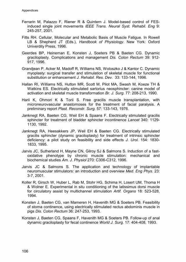
Appendices
106
Ferrarin M, Palazzo F, Riener R & Quintern J. Model-based control of FES-induced single joint movements IEEE Trans. Neural Syst. Rehabil. Eng 9: 245-257, 2001.
Fitts RH. Cellular, Molecular and Metabolic Basis of Muscle Fatigue. In Rowell LB & Shepherd JT (Eds.), Handbook of Physiology. New York: Oxford Univerdity Press, 1996.
Geerdes BP, Heineman E, Konsten J, Soeters PB & Baeten CG. Dynamic graciloplasty. Complications and management Dis. Colon Rectum 39: 912-917, 1996.
Grandjean P, Acker M, Madoff R, Williams NS, Woloszko J & Kantor C. Dynamic myoplasty: surgical transfer and stimulation of skeletal muscle for functional substitution or enhancement J. Rehabil. Res. Dev. 33: 133-144, 1996.
Hallan RI, Williams NS, Hutton MR, Scott M, Pilot MA, Swash M, Koeze TH & Watkins ES. Electrically stimulated sartorius neosphincter: canine model of activation and skeletal muscle transformation Br. J. Surg. 77: 208-213, 1990.
Harii K, Ohmori K & Torii S. Free gracilis muscle transplantation, with microneurovascular anastomoses for the treatment of facial paralysis. A preliminary report Plast. Reconstr. Surg. 57: 133-143, 1976.
Janknegt RA, Baeten CG, Weil EH & Spaans F. Electrically stimulated gracilis sphincter for treatment of bladder sphincter incontinence Lancet 340: 1129-1130, 1992.
Janknegt RA, Heesakkers JP, Weil EH & Baeten CG. Electrically stimulated gracilis sphincter (dynamic graciloplasty) for treatment of intrinsic sphincter deficiency: a pilot study on feasibility and side effects J. Urol. 154: 1830-1833, 1995.
Jarvis JC, Sutherland H, Mayne CN, Gilroy SJ & Salmons S. Induction of a fast-oxidative phenotype by chronic muscle stimulation: mechanical and biochemical studies Am. J. Physiol 270: C306-C312, 1996.
Jarvis JC & Salmons S. The application and technology of implantable neuromuscular stimulators: an introduction and overview Med. Eng Phys. 23: 3-7, 2001.
Koller R, Girsch W, Huber L, Rab M, Stohr HG, Schima H, Losert UM, Thoma H & Wolner E. Experimental in situ conditioning of the latissimus dorsi muscle for circulatory assist by multichannel stimulation Artif. Organs 18: 523-528, 1994.
Konsten J, Baeten CG, van Mameren H, Havenith MG & Soeters PB. Feasibility of stoma continence, using electrically stimulated rectus abdominis muscle in pigs Dis. Colon Rectum 36: 247-253, 1993.
Konsten J, Baeten CG, Spaans F, Havenith MG & Soeters PB. Follow-up of anal dynamic graciloplasty for fecal continence World J. Surg. 17: 404-408, 1993.
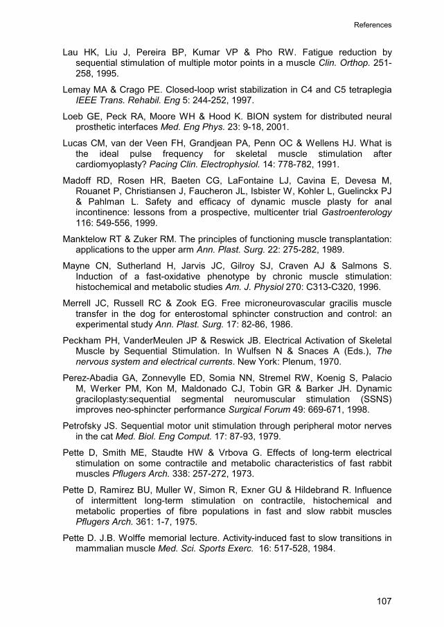
References
107
Lau HK, Liu J, Pereira BP, Kumar VP & Pho RW. Fatigue reduction by sequential stimulation of multiple motor points in a muscle Clin. Orthop. 251-258, 1995.
Lemay MA & Crago PE. Closed-loop wrist stabilization in C4 and C5 tetraplegia IEEE Trans. Rehabil. Eng 5: 244-252, 1997.
Loeb GE, Peck RA, Moore WH & Hood K. BION system for distributed neural prosthetic interfaces Med. Eng Phys. 23: 9-18, 2001.
Lucas CM, van der Veen FH, Grandjean PA, Penn OC & Wellens HJ. What is the ideal pulse frequency for skeletal muscle stimulation after cardiomyoplasty? Pacing Clin. Electrophysiol. 14: 778-782, 1991.
Madoff RD, Rosen HR, Baeten CG, LaFontaine LJ, Cavina E, Devesa M, Rouanet P, Christiansen J, Faucheron JL, Isbister W, Kohler L, Guelinckx PJ & Pahlman L. Safety and efficacy of dynamic muscle plasty for anal incontinence: lessons from a prospective, multicenter trial Gastroenterology 116: 549-556, 1999.
Manktelow RT & Zuker RM. The principles of functioning muscle transplantation: applications to the upper arm Ann. Plast. Surg. 22: 275-282, 1989.
Mayne CN, Sutherland H, Jarvis JC, Gilroy SJ, Craven AJ & Salmons S. Induction of a fast-oxidative phenotype by chronic muscle stimulation: histochemical and metabolic studies Am. J. Physiol 270: C313-C320, 1996.
Merrell JC, Russell RC & Zook EG. Free microneurovascular gracilis muscle transfer in the dog for enterostomal sphincter construction and control: an experimental study Ann. Plast. Surg. 17: 82-86, 1986.
Peckham PH, VanderMeulen JP & Reswick JB. Electrical Activation of Skeletal Muscle by Sequential Stimulation. In Wulfsen N & Snaces A (Eds.), The nervous system and electrical currents. New York: Plenum, 1970.
Perez-Abadia GA, Zonnevylle ED, Somia NN, Stremel RW, Koenig S, Palacio M, Werker PM, Kon M, Maldonado CJ, Tobin GR & Barker JH. Dynamic graciloplasty:sequential segmental neuromuscular stimulation (SSNS) improves neo-sphincter performance Surgical Forum 49: 669-671, 1998.
Petrofsky JS. Sequential motor unit stimulation through peripheral motor nerves in the cat Med. Biol. Eng Comput. 17: 87-93, 1979.
Pette D, Smith ME, Staudte HW & Vrbova G. Effects of long-term electrical stimulation on some contractile and metabolic characteristics of fast rabbit muscles Pflugers Arch. 338: 257-272, 1973.
Pette D, Ramirez BU, Muller W, Simon R, Exner GU & Hildebrand R. Influence of intermittent long-term stimulation on contractile, histochemical and metabolic properties of fibre populations in fast and slow rabbit muscles Pflugers Arch. 361: 1-7, 1975.
Pette D. J.B. Wolffe memorial lecture. Activity-induced fast to slow transitions in mammalian muscle Med. Sci. Sports Exerc. 16: 517-528, 1984.
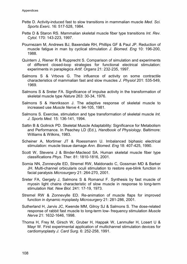
Appendices
108
Pette D. Activity-induced fast to slow transitions in mammalian muscle Med. Sci. Sports Exerc. 16: 517-528, 1984.
Pette D & Staron RS. Mammalian skeletal muscle fiber type transitions Int. Rev. Cytol. 170: 143-223, 1997.
Pournezam M, Andrews BJ, Baxendale RH, Phillips GF & Paul JP. Reduction of muscle fatigue in man by cyclical stimulation J. Biomed. Eng 10: 196-200, 1988.
Quintern J, Riener R & Rupprecht S. Comparison of simulation and experiments of different closed-loop strategies for functional electrical stimulation: experiments in paraplegics Artif. Organs 21: 232-235, 1997.
Salmons S & Vrbova G. The influence of activity on some contractile characteristics of mammalian fast and slow muscles J. Physiol 201: 535-549, 1969.
Salmons S & Sreter FA. Significance of impulse activity in the transformation of skeletal muscle type Nature 263: 30-34, 1976.
Salmons S & Henriksson J. The adaptive response of skeletal muscle to increased use Muscle Nerve 4: 94-105, 1981.
Salmons S. Exercise, stimulation and type transformation of skeletal muscle Int. J. Sports Med. 15: 136-141, 1994.
Saltin B & Gollnick PD. Skeletal Muscle Adaptability: Significance for Metabolism and Performance. In Peachey LD (Ed.), Handbook of Physiology. Baltimore: Williams & Wilkins, 1983.
Scheiner A, Mortimer JT & Roessmann U. Imbalanced biphasic electrical stimulation: muscle tissue damage Ann. Biomed. Eng 18: 407-425, 1990.
Scott W, Stevens J & Binder-Macleod SA. Human skeletal muscle fiber type classifications Phys. Ther. 81: 1810-1816, 2001.
Somia NN, Zonnevylle ED, Stremel RW, Maldonado C, Gossman MD & Barker JH. Multi-channel orbicularis oculi stimulation to restore eye-blink function in facial paralysis Microsurgery 21: 264-270, 2001.
Sreter FA, Gergely J, Salmons S & Romanul F. Synthesis by fast muscle of myosin light chains characteristic of slow muscle in response to long-term stimulation Nat. New Biol. 241: 17-19, 1973.
Stremel RW & Zonnevylle ED. Re-animation of muscle flaps for improved function in dynamic myoplasty Microsurgery 21: 281-286, 2001.
Sutherland H, Jarvis JC, Kwende MM, Gilroy SJ & Salmons S. The dose-related response of rabbit fast muscle to long-term low- frequency stimulation Muscle Nerve 21: 1632-1646, 1998.
Thoma H, Frey M, Girsch W, Gruber H, Happak W, Lanmuller H, Losert U & Mayr W. First experimental application of multichannel stimulation devices for cardiomyoplasty J. Card Surg. 6: 252-258, 1991.
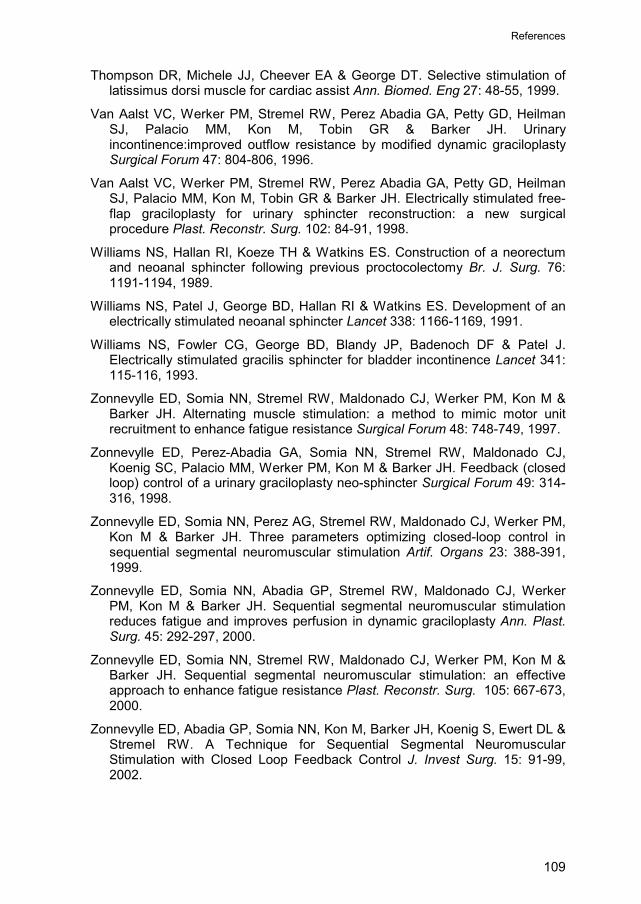
References
109
Thompson DR, Michele JJ, Cheever EA & George DT. Selective stimulation of latissimus dorsi muscle for cardiac assist Ann. Biomed. Eng 27: 48-55, 1999.
Van Aalst VC, Werker PM, Stremel RW, Perez Abadia GA, Petty GD, Heilman SJ, Palacio MM, Kon M, Tobin GR & Barker JH. Urinary incontinence:improved outflow resistance by modified dynamic graciloplasty Surgical Forum 47: 804-806, 1996.
Van Aalst VC, Werker PM, Stremel RW, Perez Abadia GA, Petty GD, Heilman SJ, Palacio MM, Kon M, Tobin GR & Barker JH. Electrically stimulated free-flap graciloplasty for urinary sphincter reconstruction: a new surgical procedure Plast. Reconstr. Surg. 102: 84-91, 1998.
Williams NS, Hallan RI, Koeze TH & Watkins ES. Construction of a neorectum and neoanal sphincter following previous proctocolectomy Br. J. Surg. 76: 1191-1194, 1989.
Williams NS, Patel J, George BD, Hallan RI & Watkins ES. Development of an electrically stimulated neoanal sphincter Lancet 338: 1166-1169, 1991.
Williams NS, Fowler CG, George BD, Blandy JP, Badenoch DF & Patel J. Electrically stimulated gracilis sphincter for bladder incontinence Lancet 341: 115-116, 1993.
Zonnevylle ED, Somia NN, Stremel RW, Maldonado CJ, Werker PM, Kon M & Barker JH. Alternating muscle stimulation: a method to mimic motor unit recruitment to enhance fatigue resistance Surgical Forum 48: 748-749, 1997.
Zonnevylle ED, Perez-Abadia GA, Somia NN, Stremel RW, Maldonado CJ, Koenig SC, Palacio MM, Werker PM, Kon M & Barker JH. Feedback (closed loop) control of a urinary graciloplasty neo-sphincter Surgical Forum 49: 314-316, 1998.
Zonnevylle ED, Somia NN, Perez AG, Stremel RW, Maldonado CJ, Werker PM, Kon M & Barker JH. Three parameters optimizing closed-loop control in sequential segmental neuromuscular stimulation Artif. Organs 23: 388-391, 1999.
Zonnevylle ED, Somia NN, Abadia GP, Stremel RW, Maldonado CJ, Werker PM, Kon M & Barker JH. Sequential segmental neuromuscular stimulation reduces fatigue and improves perfusion in dynamic graciloplasty Ann. Plast. Surg. 45: 292-297, 2000.
Zonnevylle ED, Somia NN, Stremel RW, Maldonado CJ, Werker PM, Kon M & Barker JH. Sequential segmental neuromuscular stimulation: an effective approach to enhance fatigue resistance Plast. Reconstr. Surg. 105: 667-673, 2000.
Zonnevylle ED, Abadia GP, Somia NN, Kon M, Barker JH, Koenig S, Ewert DL & Stremel RW. A Technique for Sequential Segmental Neuromuscular Stimulation with Closed Loop Feedback Control J. Invest Surg. 15: 91-99, 2002.