Embed Size (px)
Citation preview

Equine Ophthalmology
Glenn A. Severin, DVM, MS
This seminar is a review of advances in medication; medication delivery techniques employed inequine ophthalmology; and the diagnosis and treatment of selected ophthalmic diseases. Author’saddress: 1701 S. Whitcomb St., Fort Collins, CO 80526-1932. r 1998 AAEP.
1. Therapeutics Review
This review is intended to describe briefly some ofthe medications, techniques, and devices currentlyemployed in equine ophthalmology. One should notassume that the products and techniques mentionedare the only ones acceptable for treating any givendisease, but they will provide a foundation for thesound management of ocular disease. Some of thedrugs and techniques are new, whereas others arenot.
Since the successful medical management of oph-thalmic disease is directly related to the method ofmedication used and the selection of appropriatedrugs, a brief review of medication techniques isnecessary before drugs can be discussed. The meth-ods of medicating the eye are topical, subconjuncti-val, intraocular, and systemic administration. Thedisease presented will determine the medicationtechnique or combination of techniques that will bemost effective. The efficacy of topical medication isdetermined by the chemical characteristics of thedrug, the health of the cornea and conjunctiva, andthe frequency of application. In the equine, it isfurther limited by the diluting effect of high tearproduction and the difficulty in administering dropsor ointments to a painful eye. The use of subpalpe-bral delivery systems is the most common method ofproviding intensive topical medication. They re-
quire only chemical restraint and local anesthesiafor placement.
2. Topical Medication
Drops are often difficult to administer to horses,especially those with a painful eye. A small syringewith the hub of a 25-gauge needle that has beenbroken off can be used to direct a fine stream ofmedication across the eye. This is especially usefulduring an examination when a topical anesthetic orfluorescein solution is instilled.
Subpalpebral delivery systems are easy to place(Fig. 1) but have disadvantages, which include slip-page of the tubing and infection or irritation of theeyelid.
Double-passage tubing can be easily placed buthas a higher potential of corneal contact, which mayresult in a corneal ulcer (Fig. 2).
Single-passage tubing (Fig. 3) can be made byusing silicone tubing with a small oval of siliconesheeting glued to the end to prevent the tubing frompulling through the eyelid. A commercial siliconetubing for transpalpebral medication is availablefrom Mila Internationala and is sold with trocar,adapter tip, and suture clips. After placement, thetubing can be connected to a bottle for gravity flow ora micrometered delivery system for continuoustherapy.
OPHTHALMOLOGY
NOTES
AAEP PROCEEDINGS 9 Vol. 44 / 1998 105
Reprinted in the IVIS website with the permission of the AAEP Close window to return to IVIS
Proceedings of the Annual Convention of the AAEP 1998

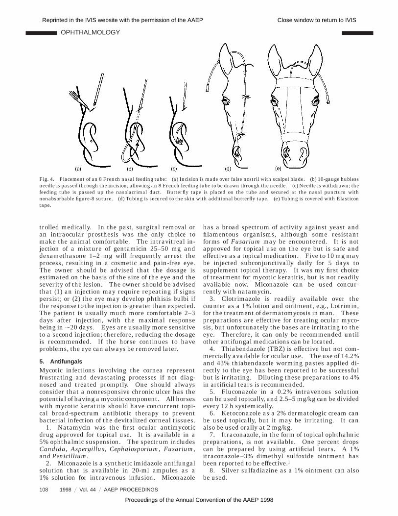
Nasolacrimal systems are more difficult to posi-tion but cause less irritation, allowing them toremain in place for weeks. A commercial nasolacri-mal medication tube, made by Arnold’s VeterinaryProducts Limited, England, is available from Jor-gensen Laboratories.b The tubing is positioned ap-proximately 15 mm up the nasolacrimal duct. An 8French nasal feeding tube can be positioned in asimilar manner (Fig. 4). The backflow of medica-tion along this type of nasolacrimal tube requires alarger volume of medication to treat the ocularsurface adequately.
A nasolacrimal medication tube extending to thepalpebral punctum can be positioned by using a 5French, 36-in. (,91 cm) feeding tube or Stillmanurinary catheter (Fig. 5). With this type of tubing,all of the medication is delivered directly to the eye.
3. Subconjunctival Injection
A subconjunctival injection may have a short-termor prolonged effect, depending on the medicationadministered, and will it result in increased druglevels in the tear film as well as direct ocularpenetration at the injection site. Subconjunctivalinjections provide a means of drug delivery to the
equine eye when frequent topical applications arenot possible or when supplemental therapy is neces-sary. The injections are safe to perform and easilyadministered (Table 1). The injection should begiven under the bulbar conjunctiva to be effective.The injection of drugs into the eyelid or under thetarsal conjunctiva is not effective, because the rapiduptake of such drugs is into the eyelid circulationand away from the globe. Prior to giving a subcon-junctival injection, the veterinarian should be sure ofthe diagnosis and the desired effect of the drug beingadministered. It would be disastrous to inject acorticosteroid in an eye affected with a mycotickeratitis.
4. Intraocular Injection
An anterior chamber (intracameral) injection (Fig. 6)is usually restricted to (1) the use of 1:10,000 epineph-rine in a physiological saline solution to controlanterior uveal hemorrhage or dilate the pupil duringintraocular surgery; or (2) proteases such as plas-minogen activator (tPA) to dissolve fibrin clots.
An intravitreal injection (Fig. 7) is usually re-stricted to cases of suspected bacterial or mycoticendophthalmitis. For most drugs, the appropriate
Fig. 1. Placement of single-passage tubing: (a) fluted tubing, a 12-gauge needle with hub removed, and a needle to cannulate tubing;(b) needle tubing is passed through the upper fornix; (c) medication tube is seated in the superior fornix and anchored to the skin withbutterfly tape; (d) medication tube is covered with tape.
OPHTHALMOLOGY
106 1998 9 Vol. 44 9 AAEP PROCEEDINGS
Reprinted in the IVIS website with the permission of the AAEP Close window to return to IVIS
Proceedings of the Annual Convention of the AAEP 1998

equine dosage has not been established; therefore, Iuse one to two times the maximum human dosagerecommended by the manufacturer. The intravit-real administration of an oculotoxic mixture of genta-micin and dexamethasone should be considered as a
nonsurgical alternative to treat a blind eye withpersistent pain secondary to equine recurrent uve-itis or chronic glaucoma. Unfortunately, a smallpercentage of blind equine recurrent uveitis andglaucoma patients have pain that cannot be con-
Fig. 2. Double-passage tubing: (a) needle is passed through conjunctiva and the upper eyelid at the temporal fornix. Tubing hasbeen placed in the needle before it is withdrawn. (b) Needle is passed through the eyelid, entering the nasal fornix, and tubing isplaced before the needle is withdrawn. (c) Small opening is made in the tubing before it is secured with butterfly tapes and suturing.
Fig. 3. Single-passage subpalpebral delivery systems: (a) Fluted medication tube using polyethylene is shown. 1. Tubing isheated over a flame. 2. Scissors are inserted into the bell tip of the hot tubing. 3. Scissors are opened to form a flutedtip. 4. Edge is trimmed if needed. (b) Silicone tubing is glued to silicone sheeting with silicone adhesive. Allow several days forsilicone adhesive to set up. This has the fewest complications. 1. Silicone tubing and a strip of silicone sheeting with a small holeare shown. 2. Tubing is slipped through the hole in sheeting. 3. Appearance of tubing after adhesive is applied and excess tubing istrimmed. (c) Silicone tubing with trocar, skin suturing clips, and infusion adapter. Continuous infusion units are available but not shown.
OPHTHALMOLOGY
AAEP PROCEEDINGS 9 Vol. 44 / 1998 107
Reprinted in the IVIS website with the permission of the AAEP Close window to return to IVIS
Proceedings of the Annual Convention of the AAEP 1998
trolled medically. In the past, surgical removal oran intraocular prosthesis was the only choice tomake the animal comfortable. The intravitreal in-jection of a mixture of gentamicin 25–50 mg anddexamethasone 1–2 mg will frequently arrest theprocess, resulting in a cosmetic and pain-free eye.The owner should be advised that the dosage isestimated on the basis of the size of the eye and theseverity of the lesion. The owner should be advisedthat (1) an injection may require repeating if signspersist; or (2) the eye may develop phthisis bulbi ifthe response to the injection is greater than expected.The patient is usually much more comfortable 2–3days after injection, with the maximal responsebeing in ,20 days. Eyes are usually more sensitiveto a second injection; therefore, reducing the dosageis recommended. If the horse continues to haveproblems, the eye can always be removed later.
5. Antifungals
Mycotic infections involving the cornea representfrustrating and devastating processes if not diag-nosed and treated promptly. One should alwaysconsider that a nonresponsive chronic ulcer has thepotential of having a mycotic component. All horseswith mycotic keratitis should have concurrent topi-cal broad-spectrum antibiotic therapy to preventbacterial infection of the devitalized corneal tissues.
1. Natamycin was the first ocular antimycoticdrug approved for topical use. It is available in a5% ophthalmic suspension. The spectrum includesCandida, Aspergillus, Cephalosporium, Fusarium,and Penicillium.
2. Miconazole is a synthetic imidazole antifungalsolution that is available in 20-ml ampules as a1% solution for intravenous infusion. Miconazole
has a broad spectrum of activity against yeast andfilamentous organisms, although some resistantforms of Fusarium may be encountered. It is notapproved for topical use on the eye but is safe andeffective as a topical medication. Five to 10 mg maybe injected subconjunctivally daily for 5 days tosupplement topical therapy. It was my first choiceof treatment for mycotic keratitis, but is not readilyavailable now. Miconazole can be used concur-rently with natamycin.
3. Clotrimazole is readily available over thecounter as a 1% lotion and ointment, e.g., Lotrimin,for the treatment of dermatomycosis in man. Thesepreparations are effective for treating ocular myco-sis, but unfortunately the bases are irritating to theeye. Therefore, it can only be recommended untilother antifungal medications can be located.
4. Thiabendazole (TBZ) is effective but not com-mercially available for ocular use. The use of 14.2%and 43% thiabendazole worming pastes applied di-rectly to the eye has been reported to be successfulbut is irritating. Diluting these preparations to 4%in artificial tears is recommended.
5. Fluconazole in a 0.2% intravenous solutioncan be used topically, and 2.5–5 mg/kg can be dividedevery 12 h systemically.
6. Ketoconazole as a 2% dermatologic cream canbe used topically, but it may be irritating. It canalso be used orally at 2 mg/kg.
7. Itraconazole, in the form of topical ophthalmicpreparations, is not available. One percent dropscan be prepared by using artificial tears. A 1%itraconazole–3% dimethyl sulfoxide ointment hasbeen reported to be effective.1
8. Silver sulfadiazine as a 1% ointment can alsobe used.
Fig. 4. Placement of an 8 French nasal feeding tube: (a) Incision is made over false nostril with scalpel blade. (b) 10-gauge hublessneedle is passed through the incision, allowing an 8 French feeding tube to be drawn through the needle. (c) Needle is withdrawn; thefeeding tube is passed up the nasolacrimal duct. Butterfly tape is placed on the tube and secured at the nasal punctum withnonabsorbable figure-8 suture. (d) Tubing is secured to the skin with additional butterfly tape. (e) Tubing is covered with Elasticontape.
OPHTHALMOLOGY
108 1998 9 Vol. 44 9 AAEP PROCEEDINGS
Reprinted in the IVIS website with the permission of the AAEP Close window to return to IVIS
Proceedings of the Annual Convention of the AAEP 1998
6. Anti-Inflammatory Drugs
Anti-inflammatory agents include corticosteroids andantiprostaglandins. Prednisolone acetate 1%, pred-nisolone phosphate 1%, and dexamethasone 0.1%are the most effective topical corticosteroids. Topi-cal antiprostaglandins can be used when ulcerativekeratitis would make corticosteroids contraindicated.Flurbiprofen sodium 0.03%, profenal 1%, and diclo-fenac 0.1% are used in human ophthalmology tocontrol miosis during cataract surgery. Diclofenachas been approved for treating intraocular inflamma-tion. An indomethacin 1–2% suspension can bemade from indomethacin capsules or powder forsolution.
Delayed-absorption corticosteroids are frequentlyused for subconjunctival injections (Table 1). Triam-cinolone 40 mg/ml is excellent. Methyl predniso-
lone is longer acting but has the potential of causinga persistent mineralized plaque at the injection site.This plaque is irritating and may require surgicalremoval.
The long-term systemic administration of highlevels of corticosteroids may be dangerous because ofthe potential to cause secondary laminitis. Becauseof this danger, the antiprostaglandin agents flunixinmeglumine, phenylbutazone, or aspirin are recom-mended in combination with topical and subconjunc-tival corticosteroids. Flunixin meglumine 1 mg/kgq 24 h is very effective in controlling ocular pain fromany cause and is my initial choice of systemicanti-inflammatory agent for acute ocular inflamma-tion. Ketoprofen 10% IV solution, 1 mg/lb q 24 h isalso effective. Phenylbutazone 1–2 g, q 12 or 24 hcan also be used, but in my experience it is not as
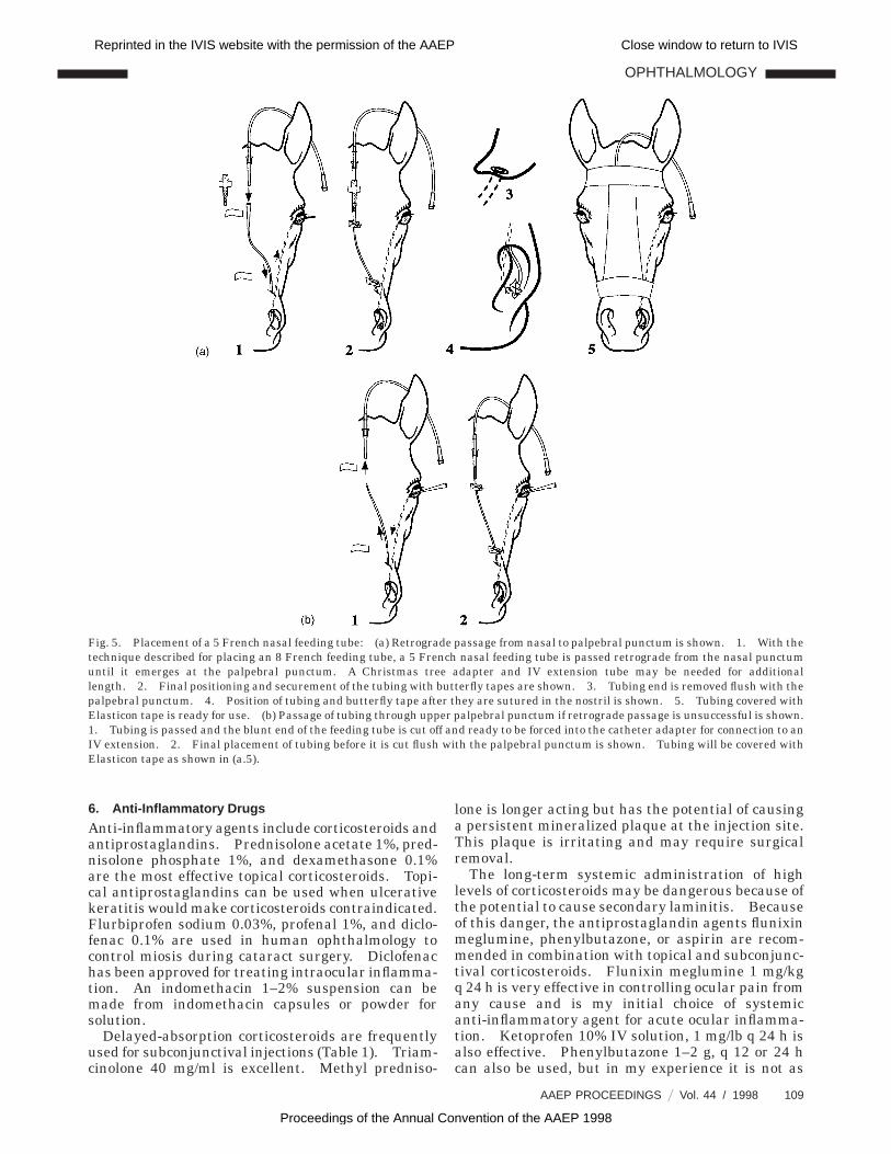
Fig. 5. Placement of a 5 French nasal feeding tube: (a) Retrograde passage from nasal to palpebral punctum is shown. 1. With thetechnique described for placing an 8 French feeding tube, a 5 French nasal feeding tube is passed retrograde from the nasal punctumuntil it emerges at the palpebral punctum. A Christmas tree adapter and IV extension tube may be needed for additionallength. 2. Final positioning and securement of the tubing with butterfly tapes are shown. 3. Tubing end is removed flush with thepalpebral punctum. 4. Position of tubing and butterfly tape after they are sutured in the nostril is shown. 5. Tubing covered withElasticon tape is ready for use. (b) Passage of tubing through upper palpebral punctum if retrograde passage is unsuccessful is shown.1. Tubing is passed and the blunt end of the feeding tube is cut off and ready to be forced into the catheter adapter for connection to anIV extension. 2. Final placement of tubing before it is cut flush with the palpebral punctum is shown. Tubing will be covered withElasticon tape as shown in (a.5).
OPHTHALMOLOGY
AAEP PROCEEDINGS 9 Vol. 44 / 1998 109
Reprinted in the IVIS website with the permission of the AAEP Close window to return to IVIS
Proceedings of the Annual Convention of the AAEP 1998
effective in acute ocular inflammation as flunixinmeglumine. Aspirin is the preferred systemic drugfor chronic disorders. The initial treatment is 25mg/kg q 12 h for 5–30 days, followed by 30 mg/kg q24 h indefinitely. I have not observed any untowardreaction to the aspirin administered at these dosagelevels, and I feel the recurrence rate is higher if thedosage rate is reduced before 2 weeks. In someanimals, treatment can be reduced to every otherday.
7. Glaucoma
Primary equine glaucoma is uncommon. Treat-ment with 2–4% pilocarpine q 6 h has been success-ful, but this frequency of medication is difficult tomaintain for extended periods. A polymerizing gelcontaining 4% pilocarpine (Pilopine HS Gelc) iseffective with twice daily treatment. Humorsol 0.5%is an indirect-acting parasympathomimetic that canbe used twice daily.
Other new drugs are also available. Dipivefrin0.1% is a pro-drug of epinephrine that requiresbiotransformation to epinephrine before therapeuticactivity occurs. Dorzolamide HC1 2% is a topicalcarbonic anhydrase inhibitor that is applied three
times daily. If it is given with other topical medica-tions, allow 10 min between medications. b-adren-ergic blocking agents currently are the mostfrequently prescribed medications for human glau-coma. This has made it difficult to locate some ofthe older medications. Examples of b-blockers arebetaxolol 0.5%, carteolol 1%, levobunolol 0.5%, meti-pranolol 0.3%, and timolol 0.5%. Latanoprost0.005% ophthalmic solution can be given as one dropdaily or twice daily. This is a new prostaglandin F2a
analog that reduces intraocular pressure by increas-ing uveoscleral outflow. This may be helpful inhorses because they have a greater uveoscleral out-flow. If topical medication is not possible, cyclode-structive procedures such as cryotherapy ortransscleral laser therapy may be beneficial.
Secondary (obstructive) glaucoma is generallychronic and a complication to chronic uveitis [seeFig. 11 (b.4) below]. It is usually nonresponsive tomedical treatment. If the eye is still visual, cryo-therapy or transscleral laser therapy should beconsidered. If the eye is blind, enucleation, anintraocular prosthesis, or the intravitreal injec-tion of gentamicin and dexamethasone should beconsidered.
8. Keratoconjunctivitis Sicca
1. The goals in treating keratoconjunctivitis sicca(KCS) are to supplement tear formation, stimulatelacrimal activity, control infection, cleanse the eye,and control corneal changes. Tears can be supple-mented with artificial tears, ointments with or with-out antibiotics, and viscoelastics. Artificial tearshave been improved with the use of artificial mucin,which increases contact time. Viscoelastic agentssuch as sodium hyaluronate and hydroxypropyl meth-ylcellulose 2% are used for intraocular surgery andare also effective as a corneal protectant. Unfortu-nately, they are expensive. A less expensive alterna-tive is sodium hyaluronate preparations for equinejoint disease (e.g., Equron,d Hyalovet,e and Synacidf).
2. Keratoconjunctivitis sicca is not common inthe horse and when seen is usually a transientdisease that can be managed temporarily with tearsubstitution two to six times daily until tears return.I have seen it as a primary inherited disease, but it isusually seen with facial paralysis or guttural pouchinfection. A topical cyclosporine ophthalmic oint-ment 0.2% is the drug of choice if long-term treat-ment is needed. Small-animal practitioners sometimesprepare a 1% solution in corn oil. Some animalsthat do not respond to this drug may respond to a0.5% ointment made by liquefying USP petrolatumin a hot water bath and then adding a 10% cyclospo-rine solution to make a 0.5% concentration. This isstirred and then put into a 6-ml syringe before itsolidifies.
3. A solution can be prepared that includes anti-biotics to control infection, acetylcysteine to dissolvemucus, and pilocarpine to stimulate tear production.
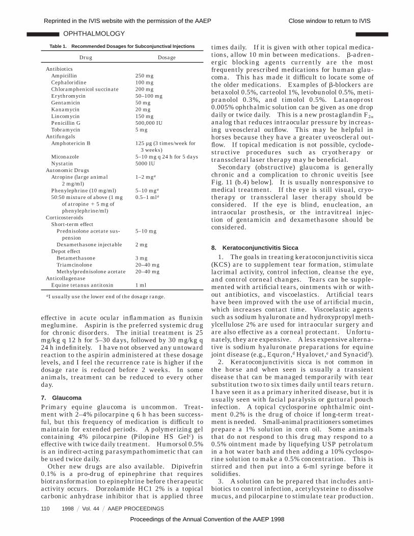
Table 1. Recommended Dosages for Subconjunctival Injections
Drug Dosage
AntibioticsAmpicillin 250 mgCephaloridine 100 mgChloramphenicol succinate 200 mgErythromycin 50–100 mgGentamicin 50 mgKanamycin 20 mgLincomycin 150 mgPenicillin G 500,000 IUTobramycin 5 mg
AntifungalsAmphotericin B 125 µg (3 times/week for
3 weeks)Miconazole 5–10 mg q 24 h for 5 daysNystatin 5000 IU
Autonomic DrugsAtropine (large animal
2 mg/ml)1–2 mga
Phenylephrine (10 mg/ml) 5–10 mga
50:50 mixture of above (1 mgof atropine 1 5 mg ofphenylephrine/ml)
0.5–1 mla
CorticosteroidsShort-term effect
Prednisolone acetate sus-pension
5–10 mg
Dexamethasone injectable 2 mgDepot effect
Betamethasone 3 mgTriamcinolone 20–40 mgMethylprednisolone acetate 20–40 mg
AnticollagenaseEquine tetanus antitoxin 1 ml
aI usually use the lower end of the dosage range.
OPHTHALMOLOGY
110 1998 9 Vol. 44 9 AAEP PROCEEDINGS
Reprinted in the IVIS website with the permission of the AAEP Close window to return to IVIS
Proceedings of the Annual Convention of the AAEP 1998
9. Orbital Prosthesis
Enucleation of the equine eye results in a deep cavitythat, even though covered with skin, is objectionablein appearance. Cosmetic results can be obtainedwhen the orbital cavity is filled with a silicone rubberball implant.g These implants are available in col-ors (I prefer black) and sizes 28, 30, 33, 36, 38, 40, 43,45 and 47 mm in diameter at a cost of approximately$18.00 each. Following surgery, the skin contractsover the implant and results in a smooth contouracross the orbit.
10. Technique
The globe and third eyelid are removed by using atranspalpebral approach (Fig. 8).2 Care is taken toensure the removal of the orbital lacrimal gland andthe gland of the third eyelid, while removing as littleas possible of the extrinsic eye muscles and retrobul-bar fat.
After the eye has been removed, the largest im-plant that will easily fit into the orbit is selected.One side of the implant is trimmed flat until it lieslevel with the orbital rim when facing forward in theorbit. The implant is secured into position by su-tures placed in the periorbital and subconjunctivaltissues. This suture pattern will resemble the lac-ing across a shoe. An optional subcuticular suturemay be placed before closing the skin. Before skinsutures are complete, 0.5–1.0 million units of potas-sium penicillin are injected around the implant.A compression bandage applied over the orbit for24–48 h is recommended. Systemic antibiotics areadministered for 5 days.
Trauma to the orbit at a later date may displace
the prosthesis or create a wound, with subsequentloss of the implant.
11. Intraocular Implant
Chronic uveitis or glaucoma often results in blind-ness. If a blind eye is continually inflamed andpainful, requiring medication, it may be feasible toreplace the contents of the globe with a siliconeimplant (Fig. 9). If the eye is not significantlybuphthalmic, a T incision in the sclera may beneeded to insert the implant (Fig. 10).
The intraocular implant is not a guarantee offreedom from ocular problems. Corneal necrosismay develop or the eye may become infected. Ineither case, enucleation would be required. Blindeyes are more likely to be injured; therefore, theclient should be instructed that any change inthe appearance of the eye or the presence of anocular discharge or pain warrants an immediateexamination.
12. Equine Recurrent Uveitis: Etiology, Signs,and Management
Equine recurrent uveitis (ERU) is an immune-mediated disease affecting the anterior ocular seg-ment (iris, ciliary body, cornea, and lens), theposterior ocular segment (choroid, retina, optic nerve,and vitreous), or both. It is the most common causefor blindness in horses and mules. Synonyms forthe disease include iridocyclitis, moon blindness,and periodic ophthalmia.
The exact etiology of ERU is seldom determinedbecause the initiating uveitis may pass unnoticedbefore the owner is aware ERU exists. Uveitis is
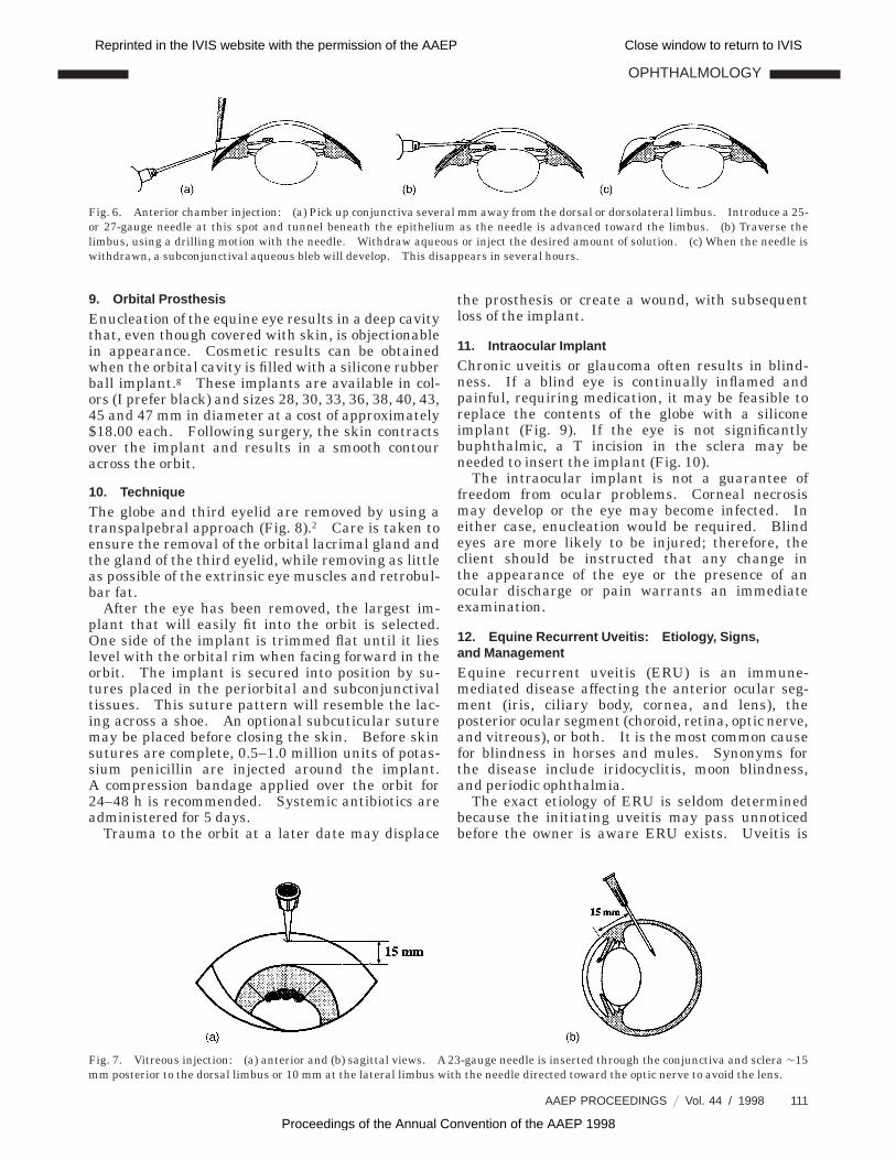
Fig. 6. Anterior chamber injection: (a) Pick up conjunctiva several mm away from the dorsal or dorsolateral limbus. Introduce a 25-or 27-gauge needle at this spot and tunnel beneath the epithelium as the needle is advanced toward the limbus. (b) Traverse thelimbus, using a drilling motion with the needle. Withdraw aqueous or inject the desired amount of solution. (c) When the needle iswithdrawn, a subconjunctival aqueous bleb will develop. This disappears in several hours.
Fig. 7. Vitreous injection: (a) anterior and (b) sagittal views. A 23-gauge needle is inserted through the conjunctiva and sclera ,15mm posterior to the dorsal limbus or 10 mm at the lateral limbus with the needle directed toward the optic nerve to avoid the lens.
OPHTHALMOLOGY
AAEP PROCEEDINGS 9 Vol. 44 / 1998 111
Reprinted in the IVIS website with the permission of the AAEP Close window to return to IVIS
Proceedings of the Annual Convention of the AAEP 1998

the most common intraocular lesion in horses andhas many causes, which include trauma, intraocularsurgery, corneal ulcers, interstitial keratitis, local orsystemic infection, extension of central nervous sys-tem diseases, immune disorders, and severe toxemia.Infectious diseases that have been associated withuveitis are leptospirosis, brucellosis, Staphylococcusequi, Rhodococcus equi, toxoplasmosis, systemicfungi, herpesvirus, and equine viral arteritis. On-chocerca cervicalis has been long proposed as acause, but the high distribution of this parasite andits incidence in many animals with normal eyesmake a causal relationship difficult to establish.It has been demonstrated that when the parasitedies, inflammation can occur. Keratoconjunctivitisor keratouveitis have been observed when largenumbers of microfilaria migrate into the conjunc-tiva, superficial stroma of the cornea, and intraocu-lar tissues. Migrating Setaria and Dirofilariaimmitis may gain access to the anterior chamber,where they are quite active and startling to see.
The initial uveitis may heal uneventfully or lead toa persistent immune-mediated response resulting inERU. This immune response may be a type I,immediate hypersensitivity; type III, arthus im-mune complex hypersensitivity; type IV, cell-medi-ated hypersensitivity involving antigen and sensitized Tlymphocytes; or ocular tissue autoimmunity thatmay result from ocular trauma or be spontaneous.Phacolytic uveitis is associated with the escaping oflens protein from an injured or cataractous lens.
The most frequent observations alerting an owner
to have his or her horse examined include oculardischarge, blepharospasm, change in appearance ofthe eye (this may be a cloudy cornea or a red eye),and, less frequently, signs of disturbed vision.
The examination should begin outdoors in brightlight. The examiner takes a position in front of thepatient, where he or she can determine if the lesionsare unilateral or bilateral. The examiner evaluatesthe eyes and surrounding structures for the presenceof ocular discharge, notes the position of the eye inthe orbit as well as the size of the eye, and deter-mines if there is any periorbital swelling. As thehorse is led indoors to the examination area, itshould be observed for any responses to the environ-ment that would suggest a visual deficit. The exam-ining area should be well lighted and be capable ofbeing darkened so the anterior segment can bere-examined with a transilluminator and ophthalmo-scope. A portable surgical lamp is an excellentadjunct to a lighted room examination. Chemicalrestraint (xylazine, detomidine, or butorphanol) lo-cal nerve blocks, or both may be needed if the horse isuncooperative. If the lesion is unilateral, alwaysexamine the normal eye first.
Clinical signs of anterior segment involvementvary depending on the acuteness of the disease, thetissues involved, and whether the lesion is unilateralor bilateral. Mild involvement may demonstrateonly epiphora, conjunctival catarrh, and slight photo-phobia. In acute uveitis [Fig. 11(a)] the initial signsare photophobia, blepharospasm, and enophthalmoswith marked protrusion of the third eyelid. A ca-
Fig. 8. Transpalpebral enucleation: (a) Eyelids are sutured together and skin is incised ,5–7 mm from the palpebral border.(b) Nasal and temporal attachments of eyelids are severed. Blunt dissection with scissors is continued to the conjunctival attachmentof the limbus. (c) Extrinsic muscle insertions are separated from the sclera with scissors. Care should be taken to remove thetemporal lacrimal gland and the gland of the third eyelid. (d) Carefully elevate the globe enough so that the optic nerve and retractoroculi muscle can be severed with enucleation or curved Mayo scissors. (e) Silicone rubber orbital implant is trimmed for placement.Side view is above; front view is below. (f) Periorbital fascia and tissues are closed over the implant with absorbable sutures.Penicillin G is injected behind the implant (250,000 IU). (g) Subcutaneous tissue is closed and penicillin G is is injected in front of theprosthesis. (h) Skin is closed with a cruciate suture pattern.
OPHTHALMOLOGY
112 1998 9 Vol. 44 9 AAEP PROCEEDINGS
Reprinted in the IVIS website with the permission of the AAEP Close window to return to IVIS
Proceedings of the Annual Convention of the AAEP 1998

tarrhal conjunctivitis will be present; this results inepiphora that may become seropurulent after 24 h.Episcleral injection will be seen. The iris will beswollen, dull in color, and have a miotic pupil (Fig.11). An examination of the aqueous with a slitbeam of light will reveal aqueous flare. The horsewill be head shy and may have signs of diminishedvision.
Tonometry will reveal a very soft eye. If a tonom-eter is not available, digital tonometry can be per-formed by directly touching the cornea or by palpatingthe globe through the eyelids. I prefer to touch thecornea directly. After the eye is topically anesthe-
tized, the examiner can estimate intraocular pres-sure by touching the cornea with his or her finger ora muscle hook. When the cornea of a hypotonic eyeis touched with the examiner’s finger, the cornea willindent before the examiner is aware that pressurehas been put on the cornea. If the intraocularpressure is normal, the examiner is aware that slightfinger pressure must be applied before the corneabegins to flatten. In glaucoma, obvious pressure isneeded to compress the corneal surface. If thelesion is unilateral and both eyes are examined bythis technique, significant differences of intraocularpressure can be detected. When a muscle hook is
Fig. 9. Intraocular prosthesis for a blind glaucomatous eye: (a) Eye is prepared for surgery. Lateral canthotomy and eyelidspeculum provide exposure. Eye is stabilized with fixation or mosquito forceps at 9 and 3 o’clock. (b) Conjunctival incision iscompleted and sclera is exposed. (c) Scleral incision initiated with scalpel results in collapse of the eye. (d) Incision can be completedfrom the 10 to 2 o’clock positions with Mayo scissors. (e) Ciliary body is separated from sclera with a Green spatula.(f) Appearance of the eye after evisceration. Ocular contents are visible to the left of the globe. (g) Carter sphere introducer withsilicone rubber sphere ready to transfer to the globe are shown. (h) Sclera is sutured with a continuous pattern after 3 appositionalsutures are placed. Note the eye is filled with blood. (i) Continuous suture pattern in conjunctiva is shown.
OPHTHALMOLOGY
AAEP PROCEEDINGS 9 Vol. 44 / 1998 113
Reprinted in the IVIS website with the permission of the AAEP Close window to return to IVIS
Proceedings of the Annual Convention of the AAEP 1998

touched to a hypotonic eye, an obvious indentation inthe cornea occurs before corneal resistance can befelt. In the normal eye, resistance will be felt beforethe cornea indents; when glaucoma is present, obvi-ous pressure will be needed to indent the cornea.This technique does not have as much of the inher-ent error that is associated with palpating the globethrough the eyelid.
After the anterior segment has been examined, thepupil should be dilated and the posterior segmentshould be examined by using the ophthalmoscopytechniques described in my previous presentations.The posterior segment changes will be describedlater in this article.
During the next 1–3 days the eye may changerapidly. These changes are progressive eyelid edemaand chemosis. The ocular discharge becomes morepurulent. Corneal edema develops, which may in-terfere with an examination of the deeper structures.A perilimbal corneal flush develops as a result ofcongestion of limbal corneal vascular loops. Kera-titic precipitates appear on the inferior one half ofthe cornea. The nonpigment epithelium of the cili-ary body becomes toxic and the blood–aqueous bar-
rier breaks down, resulting in clotted fibrin or bloodin the anterior chamber. The iris becomes ex-tremely edematous and posterior synechia begin toform. If the pupil is dilated at this time, fibrin,sloughed uveal pigment, and portions of the corporanigra may be seen adhering to the lens. Intraocularpressure continues to be low.
Corneal vascularization begins 3–6 days afteronset and if inflammation persists, may proceedcentrally at a rate of approximately 1 mm/day.Severe corneal endothelial damage may result incorneal edema, and in some patients edema may beso severe that the cornea may show a ripple effectwhen the patient blinks. This may progress to abullae formation that may rupture, resulting incorneal ulceration.
If systemic disease is suspected as a cause forERU, then a complete blood cell count, urinalysis,and blood chemistry are indicated. If infectiousdiseases such as leptospirosis, brucellosis, toxoplas-mosis, or systemic fungi are suspected, then serologi-cal tests should be considered. In some cases, anautoimmune profile may be indicated.
If anterior segment disease, whether mild or se-
Fig. 10. T scleral incision for an equine intraocular implant: (a) incision is started at the 12 o’clock position and extended toward theequator of the globe; (b) the incision is closed with interrupted sutures before the parallel incision is closed.
Fig. 11. Anterior uveitis: (a) Acute. 1. Horse has photophobia and lacrimation. 2. Lacrimation, conjunctival injection, cornealedema, keratic precipitate (arrow), miotic pupil, and swollen iris are shown. 3. Hyphema—note the hemorrhage is stratified withdark red blood near the lower limbus and more recent hemorrhage above. The pupil has been dilated and the lens appears cloudy.4. Corneal vascularization near the limbus indicates several days’ duration, moderate corneal edema, and swollen iris with mioticpupil. (b) Chronic. 1. ERU relapse, showing miotic pupil, hypopyon (arrow), dark iris with superficial vascularization, andepiscleral vascular injection. 2. Quiescent uveitis with corneal vascularization (arrow), mild corneal edema, and dark iris.3. Quiescent ERU with secondary cataract. Note uveal pigment on the lens that was exposed by the dilated pupil. 4. Obstructiveglaucoma. The white lines in the cornea (Haab’s striae) are due to rupture of Descemet’s membrane. The iris is very dark.
(a.1) (a.2) (a.3) (a.4)
(b.1) (b.2) (b.3) (b.4)
OPHTHALMOLOGY
114 1998 9 Vol. 44 9 AAEP PROCEEDINGS
Reprinted in the IVIS website with the permission of the AAEP Close window to return to IVIS
Proceedings of the Annual Convention of the AAEP 1998

vere, were to resolve with treatment at this time, theeye could heal without further deterioration. Butunfortunately in many cases the mechanism for apersisting immune-mediating disease is establishedand ERU results. Clinical signs may subside andgo through quiescent periods followed by mild recur-rent episodes to acute exacerbations. One of themost consistent signs of chronicity is darkening ofthe iris in the diseased eye. It is not unusual tohave a horse presented with a history of no previouseye disease that upon examination is found to have aunilateral dark iris, which indicates that this is ERUand not an initial uveitis episode.
The sequelae [Fig. 11(b)] that may result fromanterior segment ERU are corneal scarring, cornealendothelial degeneration with or without bullouskeratitis, anterior and posterior synechia, compli-cated cataracts, lens luxation, and vitreous opacities.Severely damaged eyes may develop phthisis bulbifrom extensive ciliary body degeneration. Obstruc-tive glaucoma occurs when the filtration angle be-comes obstructed. The signs include buphthalmos,corneal striae (interconnecting parallel lines causedby the rupture of Descemet’s membrane), and a deepanterior chamber.
Symptomatic treatment of anterior ERU withanti-inflammatory therapy and mydriatics should bestarted immediately. Specific treatment is indicatedwhen an etiology has been determined. Anti-inflammatory drugs are the most important part ofERU therapy. They inhibit the immune-mediatedresponse and consequently reduce the uveal conges-tion so that the uveal blood–aqueous barrier canreturn to normal.
My routine protocol for the initial treatment ofacute uveitis consists of the intravenous administra-tion of flunixin meglumine 1 mg/kg, a subconjuncti-val injection of triamcinolone 20 mg or betamethasone3 mg, and a separate subconjunctival injection of 1mg of large-animal atropine. If there is evidence ofsynechia or profound miosis, 5 mg of injectablephenylephrine can be included in the atropine injec-tion. I do not routinely use systemic prednisoloneor dexamethasone because I feel the synergisticeffect of flunixin and subconjunctival and topicalsteroids is as effective as systemic and topical ste-roids. This drug combination eliminates the lamini-tis risk seen with steroid therapy, especially withdexamethasone.
Topical treatment should be started with antibiot-ic–steroid ophthalmic ointments containing predniso-lone (e.g., chloramphenicol and prednisolone) ordexamethasone (e.g., Maxitrolh) four times daily ifpossible. Mydriasis can usually be maintained with1% atropine ophthalmic ointment four times daily.I have never found it necessary to use concentrationshigher than 1% and therefore have never observedatropine-related colic. If topical medications can-not be properly administered, subconjunctival injec-tions may be repeated as needed or a medicationdelivery system can be placed.
Systemic antibacterial agents are indicated whensystemic infections are suspected or as a prophylac-tic measure in acute cases. The animal should beconfined or the eye should be protected with a hood.Warm compresses are beneficial in the presence ofperiocular swelling. When systemic disease is diag-nosed, specific treatment should be initiated and thesymptomatic treatment of uveitis should be contin-ued as long as needed.
Maintenance therapy consists of continuing theflunixin 5–7 days. If the eye is improving, aspirin25 mg/kg PO q 12 h can be substituted. If the eye isnot doing as well as expected, phenylbutazone orsystemic corticosteroids can be given.
As the patient improves, atropine is reduced andcan eventually be given as infrequently as every 4–5days before being discontinued. Atropine will re-main in the iris of horses longer than in the iris ofother species.
Animals with severe corneal edema will benefitfrom treatment four times daily with 5% sodiumchloride ophthalmic ointment. This edema is usu-ally transient and will be self-limiting when thecorneal endothelium returns to normal. Persistentcorneal edema suggests either a continuing endothe-lial immunological response or permanent cornealendothelial degeneration. Topical or subconjuncti-val corticosteroids will be beneficial if an activeimmunological response exists. If corneal endothe-lial degeneration has occurred, corticosteroids willnot help and an unfavorable prognosis must begiven.
Glaucoma is usually chronic when diagnosed. Ifthe eye is still visual, medical or surgical manage-ment is recommended. If the eye is blind, an intra-vitreal injection with 1–2 ml of a 50:50 mixture of 5%gentamicin and 0.2% dexamethasone will controlglaucoma. The owner should be advised that theresponse to this injection is variable. Some eyeswill be extremely sensitive, becoming hypotonic anddeveloping phthisis bulbi. In either event the eyeeventually becomes comfortable and may remaincosmetic. If glaucoma still persists 2 weeks afterthe first injection, a second injection should be given.Blind ERU eyes that are refractory to medical treat-ment and continue to be painful can be treated with1 ml of mixture in the manner recommended forglaucoma.
Posterior ERU is rarely presented during theactive stage unless blindness results. A blind eyemay go undetected by the owner unless the animalinvolved is a performance horse. Some animals willbe presented with a history of sudden blindness, butan examination will reveal that one eye has beenblind for a long time and now the other eye hasrecently become blind. In the active stage of poste-rior ERU, optic neuritis or chorioretinitis or both areseen. In the chronic stage (Fig. 12), optic nerveatrophy, retinal scarring and degeneration, or reti-nal detachment may be present. Mild lesions will
OPHTHALMOLOGY
AAEP PROCEEDINGS 9 Vol. 44 / 1998 115
Reprinted in the IVIS website with the permission of the AAEP Close window to return to IVIS
Proceedings of the Annual Convention of the AAEP 1998

be frequently seen as an incidental finding on ahealth or insurance examination.
Animals with a visual deficiency may be presentedwith a history of personality change, shying, poorperformance, or becoming awkward. The blind sidemay be difficult to examine if blindness is recent andassociated with rapid onset.
The examination should be performed in the samemanner as that previously recommended, i.e., exam-ining the patient outdoors and then indoors in adarkened room. Pupillary reflexes should bechecked before the pupils are dilated with 1% tropi-camide. The modified otoscope is an adequate oph-thalmoscope for a screening examination. Whenabnormalities are noted, a thorough examinationshould be completed with a direct ophthalmoscopeand, if available, an indirect ophthalmoscope, whichwill give a better visualization of the peripheralretina.
Optic neuritis is referred to as papillitis. Theoptic nerve appears red as a result of congested bloodvessels. Small hemorrhages may be seen. Thephysiological cup disappears as papillitis progresses.Circumpapillary edema will make the blood vesselsnear the optic disk appear suspended above thechoroid. Exudate and small blood clots may be seenin the adjacent vitreous.
Optic nerve atrophy will be seen more frequentlythan optic neuritis. The optic disk appears chalkywhite and flattened. Blood vessels will be attenu-ated in early atrophy and absent in advanced atrophy.In rare instances one half of the nerve will beatrophied and the remainder will be normal.
Active circumpapillary chorioretinitis will havecongested blood vessels; the retina may appear hazy.White infiltrates and small hemorrhages are usuallyseen. If large areas are involved, subretinal fluidmay build up, leading to detachment.
As inflammation regresses and the lesion becomesquiescent, retinal degeneration and chorioretinaldepigmentation will develop. Later some irregularareas of repigmentation occur and the lesion maytake on a butterfly appearance. Tapetal lesionstend to be multiple, more linear in appearance, andhave areas of hyperreflectivity and pigment reorgani-zation. Rarely is the retinal degeneration extensiveenough to cause blindness.
Retinal detachment is usually complete, but sec-tional detachments may occur. Acute optic neuritis
and chorioretinitis should be treated with systemicprednisolone or dexamethasone and prophylacticantibiotics until laboratory tests are available.This treatment can be risky if infectious diseases arenot ruled out first, but the treatment must beaggressive if vision is to be saved. If subretinaledema is present, the patient should receive furose-mide 0.5 mg/kg IV q 24 h to assist in the removal offluid and to promote reattachment of the retina.Treatment is not recommended for patients withoptic nerve atrophy, retinal scars, or chronic retinaldetachment.
13. Eyelid and Corneal Lacerations
Lacerations of the eyelids and cornea are commonand, except for minor injuries, require surgical inter-vention for proper healing. The abundant bloodsupply of the eyelid ensures rapid healing of eyelidlacerations, whereas severe corneal injuries healslowly, requiring neovascularization before healingis complete. Common causes for eyelid and corneallacerations are owner-inflicted trauma, and the ani-mal’s striking its head during loading or unloading,catching the eye or eyelids on a sharp object, orfighting with other horses. Owner-inflicted lacera-tions result when the horse is struck with a sharpobject. A blow from a blunt object routinely resultsin contusion to the lids and periorbital soft tissues.If the globe is involved, a corneoscleral rupture mayresult.
A. Eyelid Lacerations
Eyelid lacerations would be expected to bleed pro-fusely for a short period of time and then graduallystop unless the patient continues to injure the area.An eyelid laceration with minimal tissue loss thathas received proper wound closure and after carewill heal without deformity. Immediate treatmentis indicated for wounds with a duration of ,4 h.A delayed repair is indicated if the patient is inshock, has other serious injuries, or if the lacerationhas been present for .12 h. Proper cleaning andbandaging of the wound will reduce swelling andcontrol infection, thereby allowing better surgicalrepair and increasing the possibility of first-inten-tion healing. If the wound is 4–12 h old, the sur-geon should use his or her own judgment as toimmediate or delayed repair. If treatment is to bedelayed, the wound should be gently cleaned with
Fig. 12. Chronic posterior ERU: (a) Lens luxation reveals the detached retina; (b) optic nerve atrophy and retinal folds are shown; (c)optic nerve atrophy and retinal degeneration dorsal to the optic nerve are shown; (d) chorioretinal degeneration in the nontapetal areasurrounding the optic disk is shown.
(a)(b)
(c) (d)
OPHTHALMOLOGY
116 1998 9 Vol. 44 9 AAEP PROCEEDINGS
Reprinted in the IVIS website with the permission of the AAEP Close window to return to IVIS
Proceedings of the Annual Convention of the AAEP 1998

0.2% saline dilution povidone-iodine solution (1/50strength 10% Betadine solutioni).
The eyelids should then be covered with a cottonsquare that has had a liberal application of nitrofura-zone 0.2% ointment and should be bandaged withstretch adhesive tape. The laceration can be re-paired the next day in the same manner as that for afresh wound.
I prefer general anesthesia for optimal woundapposition on all eyelid lacerations, but minor lacera-tions can be repaired by using chemical restraint andinfiltration of a local anesthetic. After the area hasbeen clipped or shaved, the wound, adjacent skin,and the eye are cleansed with a dilute povidone-iodine solution for 2 min and covered for 2 min morewith a sponge saturated with this solution. Stand-ard surgical instruments and sutures are adequateto repair eyelid lacerations, but conjunctival sutur-ing is simplified with an ophthalmic needle holder,conjunctival fixation forceps, strabismus scissors,and ophthalmic suture.
After the wound is draped, it is minimally debrided.If there is severe swelling and the conjunctiva hasretracted, it will be beneficial to place an appositionsuture at the palpebral edge. If this is not the case,suturing the conjunctiva first is preferred. Theconjunctival sutures are continuous and placed sothat the knots are buried in the wound and thereforewill not rub the eye. The appositional skin suture isplaced along the sharp edge of the eyelid, with thesuture going through the meibomian glands. Thesuture ends should be cut short so they will notirritate the cornea. An alternative would be toleave the ends long and place them near the woundbeneath later sutures. The next suture is placed inthe skin ,3 mm from the edge of the eyelid. Thissuture should be deep (nearly to the conjunctiva) andincorporates the meibomial–tarsal plate. This isthe tension suture that holds the wound together.As many sutures as needed are placed to finishclosing the wound. An eyelid sutured in this fash-ion will heal with a smooth palpebral edge (Fig. 13).If there is swelling or eyelid tissue loss that couldresult in postsurgical lagophthalmos, it is recom-mended that the eyelids be sutured together withsplit-thickness horizontal mattress sutures. Band-aging the eye will reduce postoperative swelling andprotect the eyelid during anesthesia recovery. Post-operative drugs should include flunixin meglumineto control pain and inflammation and systemic anti-bacterials. The bandage can be removed in 2–3days. If possible, avoid all forms of ophthalmicmedications and advise the owner not to manipulatethe eyelids. If a corneal injury exists that requirestreatment, a nasolacrimal medication tube would beindicated. A warm compress may be beneficial ifswelling persists. The horizontal mattress suturescan be removed as soon as the eyelids appear normal.The skin sutures in the wound should be left in aminimum of 2 weeks and longer if they do not causeany local reaction (Fig. 14).
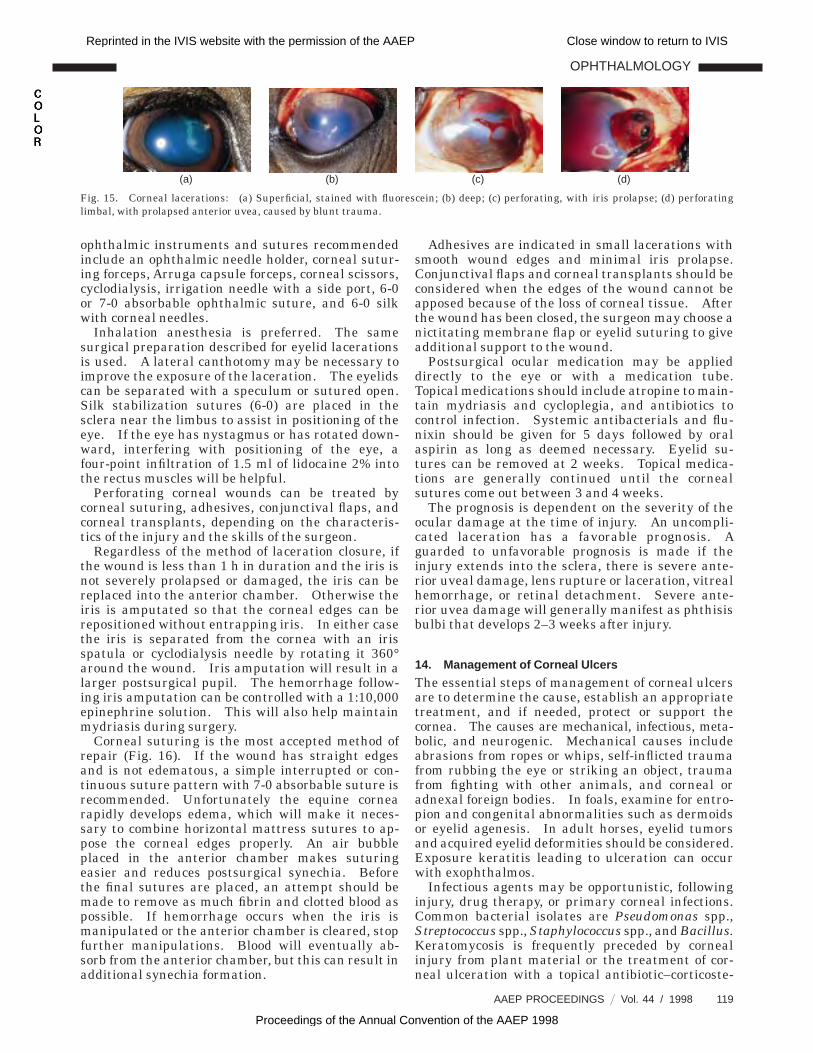
B. Corneal Lacerations
Corneal lacerations may be superficial, deep, orperforating (Fig. 15), with the method of manage-ment dependent on the severity of the injury, thelapsed time since the injury occurred, the amount ofcorneal edema, and the presence or absence ofinfection. A superficial injury will usually heal in24–48 h without treatment unless the source of thelaceration inoculates pathogenic organisms in thestroma. Because of this danger, a topical antibacte-rial ophthalmic preparation should be applied untilthe cornea is fluorescein negative. A deep lacera-tion with minimal corneal damage will often healwith conservative treatment consisting of topicalmedications recommended for ulcer therapy.
If treated conservatively, this patient should beexamined daily. If healing does not proceed asanticipated, the laceration should then be treated inthe same fashion as a wound extending to Desce-met’s membrane.
Lacerations extending to Descemet’s membranerequire surgical attention. Corneal suturing or anadhesive is the treatment of choice. The next bestchoice would be a conjunctival flap. If these are notavailable, indirect corneal support with a third eye-lid flap or a tarsorrhaphy with or without a softcontact is indicated. Indirect corneal support has ahigh risk of perforation and a guarded prognosis.A laceration with severe corneal damage should betreated medically like an ulcer and the cornea shouldbe supported or protected. Lacerations that pro-duce corneal flaps should be sutured if presentedbefore corneal edema develops. After corneal edemais present, the best procedure is to debride the flapand treat the corneal lesion as a deep ulcer.
Small puncture wounds without iris incarceratedin the wound will have local corneal edema, apinpoint pupil, plasmoid aqueous that seals thewound, hypotony, and severe pain. Fluoresceinstaining may be needed to demonstrate the wound.These wounds will not require surgical treatmentbut the potential of intraocular infection will necessi-tate systemic treatment for infectious anterior uve-itis. The eye should be checked every few days.If the wound starts to ulcerate or develop a cornealabscess or stromal necrosis, appropriate treatmentshould be started.
Perforating lacerations with iris prolapse requiresurgery for best results. The examination shouldbe stopped immediately and the patient should beprepared for surgery. Further manipulation at thistime may cause additional damage to the eye. Thepatient should be premedicated with systemic anti-bacterials and flunixin meglumine. The blood–aqueous barrier has been altered, and anyantibacterial drug capable of maintaining a therapeu-tic blood level will also be present in the aqueous intherapeutic levels until the normal blood–aqueousbarrier returns. Do not put topical ointments on aneye with a perforating injury, because of the dangerthat the petroleum base will cause uveitis. The
OPHTHALMOLOGY
AAEP PROCEEDINGS 9 Vol. 44 / 1998 117
Reprinted in the IVIS website with the permission of the AAEP Close window to return to IVIS
Proceedings of the Annual Convention of the AAEP 1998
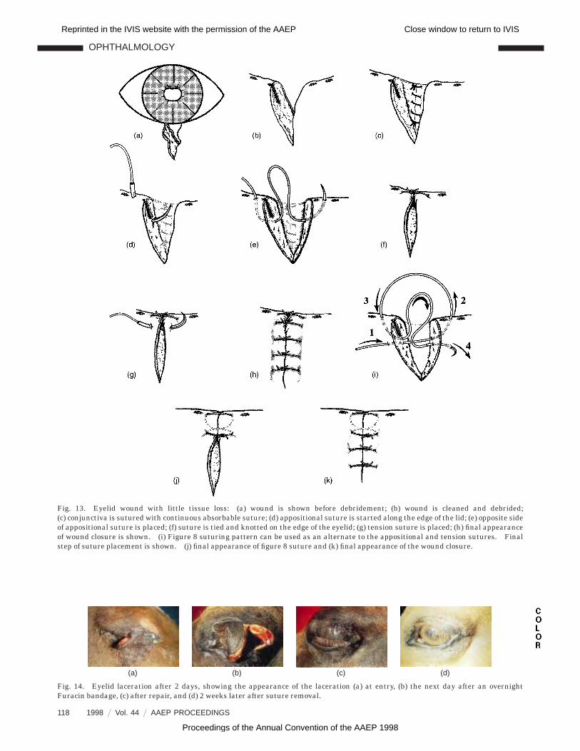
Fig. 13. Eyelid wound with little tissue loss: (a) wound is shown before debridement; (b) wound is cleaned and debrided;(c) conjunctiva is sutured with continuous absorbable suture; (d) appositional suture is started along the edge of the lid; (e) opposite sideof appositional suture is placed; (f) suture is tied and knotted on the edge of the eyelid; (g) tension suture is placed; (h) final appearanceof wound closure is shown. (i) Figure 8 suturing pattern can be used as an alternate to the appositional and tension sutures. Finalstep of suture placement is shown. (j) final appearance of figure 8 suture and (k) final appearance of the wound closure.
Fig. 14. Eyelid laceration after 2 days, showing the appearance of the laceration (a) at entry, (b) the next day after an overnightFuracin bandage, (c) after repair, and (d) 2 weeks later after suture removal.
(a) (b) (c) (d)
OPHTHALMOLOGY
118 1998 9 Vol. 44 9 AAEP PROCEEDINGS
Reprinted in the IVIS website with the permission of the AAEP Close window to return to IVIS
Proceedings of the Annual Convention of the AAEP 1998
ophthalmic instruments and sutures recommendedinclude an ophthalmic needle holder, corneal sutur-ing forceps, Arruga capsule forceps, corneal scissors,cyclodialysis, irrigation needle with a side port, 6-0or 7-0 absorbable ophthalmic suture, and 6-0 silkwith corneal needles.
Inhalation anesthesia is preferred. The samesurgical preparation described for eyelid lacerationsis used. A lateral canthotomy may be necessary toimprove the exposure of the laceration. The eyelidscan be separated with a speculum or sutured open.Silk stabilization sutures (6-0) are placed in thesclera near the limbus to assist in positioning of theeye. If the eye has nystagmus or has rotated down-ward, interfering with positioning of the eye, afour-point infiltration of 1.5 ml of lidocaine 2% intothe rectus muscles will be helpful.
Perforating corneal wounds can be treated bycorneal suturing, adhesives, conjunctival flaps, andcorneal transplants, depending on the characteris-tics of the injury and the skills of the surgeon.
Regardless of the method of laceration closure, ifthe wound is less than 1 h in duration and the iris isnot severely prolapsed or damaged, the iris can bereplaced into the anterior chamber. Otherwise theiris is amputated so that the corneal edges can berepositioned without entrapping iris. In either casethe iris is separated from the cornea with an irisspatula or cyclodialysis needle by rotating it 360°around the wound. Iris amputation will result in alarger postsurgical pupil. The hemorrhage follow-ing iris amputation can be controlled with a 1:10,000epinephrine solution. This will also help maintainmydriasis during surgery.
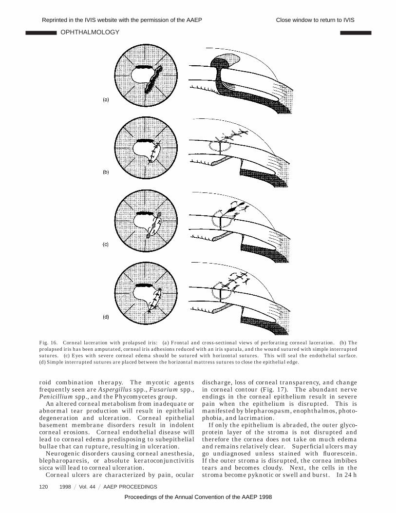
Corneal suturing is the most accepted method ofrepair (Fig. 16). If the wound has straight edgesand is not edematous, a simple interrupted or con-tinuous suture pattern with 7-0 absorbable suture isrecommended. Unfortunately the equine cornearapidly develops edema, which will make it neces-sary to combine horizontal mattress sutures to ap-pose the corneal edges properly. An air bubbleplaced in the anterior chamber makes suturingeasier and reduces postsurgical synechia. Beforethe final sutures are placed, an attempt should bemade to remove as much fibrin and clotted blood aspossible. If hemorrhage occurs when the iris ismanipulated or the anterior chamber is cleared, stopfurther manipulations. Blood will eventually ab-sorb from the anterior chamber, but this can result inadditional synechia formation.
Adhesives are indicated in small lacerations withsmooth wound edges and minimal iris prolapse.Conjunctival flaps and corneal transplants should beconsidered when the edges of the wound cannot beapposed because of the loss of corneal tissue. Afterthe wound has been closed, the surgeon may choose anictitating membrane flap or eyelid suturing to giveadditional support to the wound.
Postsurgical ocular medication may be applieddirectly to the eye or with a medication tube.Topical medications should include atropine to main-tain mydriasis and cycloplegia, and antibiotics tocontrol infection. Systemic antibacterials and flu-nixin should be given for 5 days followed by oralaspirin as long as deemed necessary. Eyelid su-tures can be removed at 2 weeks. Topical medica-tions are generally continued until the cornealsutures come out between 3 and 4 weeks.
The prognosis is dependent on the severity of theocular damage at the time of injury. An uncompli-cated laceration has a favorable prognosis. Aguarded to unfavorable prognosis is made if theinjury extends into the sclera, there is severe ante-rior uveal damage, lens rupture or laceration, vitrealhemorrhage, or retinal detachment. Severe ante-rior uvea damage will generally manifest as phthisisbulbi that develops 2–3 weeks after injury.
14. Management of Corneal Ulcers
The essential steps of management of corneal ulcersare to determine the cause, establish an appropriatetreatment, and if needed, protect or support thecornea. The causes are mechanical, infectious, meta-bolic, and neurogenic. Mechanical causes includeabrasions from ropes or whips, self-inflicted traumafrom rubbing the eye or striking an object, traumafrom fighting with other animals, and corneal oradnexal foreign bodies. In foals, examine for entro-pion and congenital abnormalities such as dermoidsor eyelid agenesis. In adult horses, eyelid tumorsand acquired eyelid deformities should be considered.Exposure keratitis leading to ulceration can occurwith exophthalmos.
Infectious agents may be opportunistic, followinginjury, drug therapy, or primary corneal infections.Common bacterial isolates are Pseudomonas spp.,Streptococcus spp., Staphylococcus spp., and Bacillus.Keratomycosis is frequently preceded by cornealinjury from plant material or the treatment of cor-neal ulceration with a topical antibiotic–corticoste-
Fig. 15. Corneal lacerations: (a) Superficial, stained with fluorescein; (b) deep; (c) perforating, with iris prolapse; (d) perforatinglimbal, with prolapsed anterior uvea, caused by blunt trauma.
(a) (b) (c) (d)
OPHTHALMOLOGY
AAEP PROCEEDINGS 9 Vol. 44 / 1998 119
Reprinted in the IVIS website with the permission of the AAEP Close window to return to IVIS
Proceedings of the Annual Convention of the AAEP 1998
roid combination therapy. The mycotic agentsfrequently seen are Aspergillus spp., Fusarium spp.,Penicillium spp., and the Phycomycetes group.
An altered corneal metabolism from inadequate orabnormal tear production will result in epithelialdegeneration and ulceration. Corneal epithelialbasement membrane disorders result in indolentcorneal erosions. Corneal endothelial disease willlead to corneal edema predisposing to subepithelialbullae that can rupture, resulting in ulceration.
Neurogenic disorders causing corneal anesthesia,blepharoparesis, or absolute keratoconjunctivitissicca will lead to corneal ulceration.
Corneal ulcers are characterized by pain, ocular
discharge, loss of corneal transparency, and changein corneal contour (Fig. 17). The abundant nerveendings in the corneal epithelium result in severepain when the epithelium is disrupted. This ismanifested by blepharospasm, enophthalmos, photo-phobia, and lacrimation.
If only the epithelium is abraded, the outer glyco-protein layer of the stroma is not disrupted andtherefore the cornea does not take on much edemaand remains relatively clear. Superficial ulcers maygo undiagnosed unless stained with fluorescein.If the outer stroma is disrupted, the cornea imbibestears and becomes cloudy. Next, the cells in thestroma become pyknotic or swell and burst. In 24 h
Fig. 16. Corneal laceration with prolapsed iris: (a) Frontal and cross-sectional views of perforating corneal laceration. (b) Theprolapsed iris has been amputated, corneal iris adhesions reduced with an iris spatula, and the wound sutured with simple interruptedsutures. (c) Eyes with severe corneal edema should be sutured with horizontal sutures. This will seal the endothelial surface.(d) Simple interrupted sutures are placed between the horizontal mattress sutures to close the epithelial edge.
OPHTHALMOLOGY
120 1998 9 Vol. 44 9 AAEP PROCEEDINGS
Reprinted in the IVIS website with the permission of the AAEP Close window to return to IVIS
Proceedings of the Annual Convention of the AAEP 1998

polymorphonuclear cells invade the cornea, result-ing in more opacity. In some infections this cellinfiltration may result in a corneal abscess. Ifinfectious organisms such as Pseudomonas spp. thatare capable of producing collagenase or similar pro-teolytic enzymes are present, rapid destruction ofthe collagen framework of the cornea will result inthe melting ulcer. When this occurs, progression ofthe ulcer to Descemet’s membrane will result in aDescemetocele or perforation. Intrinsic collagenasealso produces budding, endothelial cells, leukocytes,and regenerating epithelial cells. It is during thisphase of collagenase activity that corticosteroids arecontraindicated because they increase the activity ofcollagenase by 14 times.
Corneal ulceration with stromal involvement willresult in a secondary anterior uveitis, plasmoidaqueous, and circumcorneal injection. Deep ulcersfrequently stimulate a severe leukotactic responsethat results in hypopyon. If anterior chamber cen-tesis is performed, this exudate will invariably besterile.
As previously mentioned, the first step in success-ful ulcer management is to determine the cause.A thorough examination including culture with sen-sitivity testing and cytology may be necessary.
Severe ocular pain will necessitate chemical restraintand topical anesthesia before a complete examina-tion can be performed. It is not unusual in horseswith a Pseudomonas ulcer to have a sterile samplecollected from the conjunctival sac when a pureculture of Pseudomonas is isolated from the ulcer.Cytology may be collected with a swab or spatula oras an impression smear.
After the cause has been identified, an appropriatetreatment may include systemic and topical medica-tions. Systemic flunixin meglumine 1 mg/kg q 24 hwill control pain and reduce secondary anterioruveitis. After 5–7 days, aspirin 30 mg/kg q 24 h isusually adequate for maintenance until topical medi-cation is discontinued.
An appropriate topical treatment may requiredrugs that are antimicrobial, anticollagenase, painrelieving, and mydriatic. If bacterial infection issuspected, broad-spectrum antibiotics, such as tobra-mycin, gentamicin, or chloramphenicol, or tripleantibiotic mixtures can be used until sensitivityresults are available. Tobramycin is the best ifPseudomonas spp. is suspected. Miconazole 1% par-enteral solution is an excellent presumptive choicefor mycotic ulcers. If not available, Clotrimazole1% dermatological solution or Thiabendazole 14%
Fig. 17. Variations of corneal ulcers: (a) indolent superficial; (b) acute melting, caused by Pseudomonas infection; (c) chronic mycotic,caused by Aspergillus; (d) perforating chronic, with iris prolapse.
(a) (b) (c) (d)
Fig. 18. Peripheral conjunctival flap for a corneal lesion near the limbus: (a) Appearance of a peripheral ulcer is shown.(b) Perilimbal conjunctival incision is made adjacent to the wound. (c) Conjunctiva is undermined with blunt scissors. (d) Suturesare placed in the sclera at the limbus and along the edge of the undermined conjunctiva. (e) Sutures are tied at the limbus. (f) Flap isshown with margins sutured to the cornea.
OPHTHALMOLOGY
AAEP PROCEEDINGS 9 Vol. 44 / 1998 121
Reprinted in the IVIS website with the permission of the AAEP Close window to return to IVIS
Proceedings of the Annual Convention of the AAEP 1998
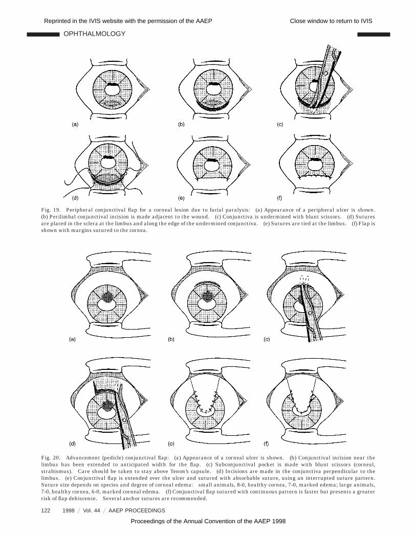
Fig. 19. Peripheral conjunctival flap for a corneal lesion due to facial paralysis: (a) Appearance of a peripheral ulcer is shown.(b) Perilimbal conjunctival incision is made adjacent to the wound. (c) Conjunctiva is undermined with blunt scissors. (d) Suturesare placed in the sclera at the limbus and along the edge of the undermined conjunctiva. (e) Sutures are tied at the limbus. (f) Flap isshown with margins sutured to the cornea.
Fig. 20. Advancement (pedicle) conjunctival flap: (a) Appearance of a corneal ulcer is shown. (b) Conjunctival incision near thelimbus has been extended to anticipated width for the flap. (c) Subconjunctival pocket is made with blunt scissors (corneal,strabismus). Care should be taken to stay above Tenon’s capsule. (d) Incisions are made in the conjunctiva perpendicular to thelimbus. (e) Conjunctival flap is extended over the ulcer and sutured with absorbable suture, using an interrupted suture pattern.Suture size depends on species and degree of corneal edema: small animals, 8-0, healthy cornea, 7-0, marked edema; large animals,7-0, healthy cornea, 6-0, marked corneal edema. (f) Conjunctival flap sutured with continuous pattern is faster but presents a greaterrisk of flap dehiscence. Several anchor sutures are recommended.
OPHTHALMOLOGY
122 1998 9 Vol. 44 9 AAEP PROCEEDINGS
Reprinted in the IVIS website with the permission of the AAEP Close window to return to IVIS
Proceedings of the Annual Convention of the AAEP 1998
paste could be temporarily used. Mycotic keratitisshould be concurrently treated with an antibioticuntil the cornea is negative to fluorescein. Acetylcysteine 5% is an effective anticollagenase agent inulcers with severe collagenase breakdown. Atro-pine 1% is indicated for mydriasis and cycloplegia.In severe ulcers these medications should be givenevery few hours. This is virtually impossible to dounless they are compounded into a single solution.For a simplified administration, a compounded mix-ture referred to as an ulcer solution is sometimesbeneficial. An ulcer solution can be prepared withfresh serum as a diluent for an antibiotic and, ifindicated, atropine. Serum has anticollagenase ac-tivity from a2-macroglobulins, as well as healing
stimulation from endogenous epithelial growth fac-tors. Medication delivery systems are indicated ifthe patient is difficult to treat or if a melting cornearequiring frequent medication is presented.
All patients with severe ulcers are candidates forsubconjunctival injection with antibiotics, mydriatic–cycloplegic agents, and anticollagenase preparationsas supplements to topical medications. The antibi-otics used frequently are chloramphenicol or gentamicin.Tetanus antitoxin is the preferred anticollagenasesource. The antibiotic can be combined with anequal amount of tetanus antitoxin, and then 0.75–1.5 ml of this mixture can be injected. Asubconjunc-tival injection combination of atropine andphenylephrine (Table 1) is a potent mydriatic–
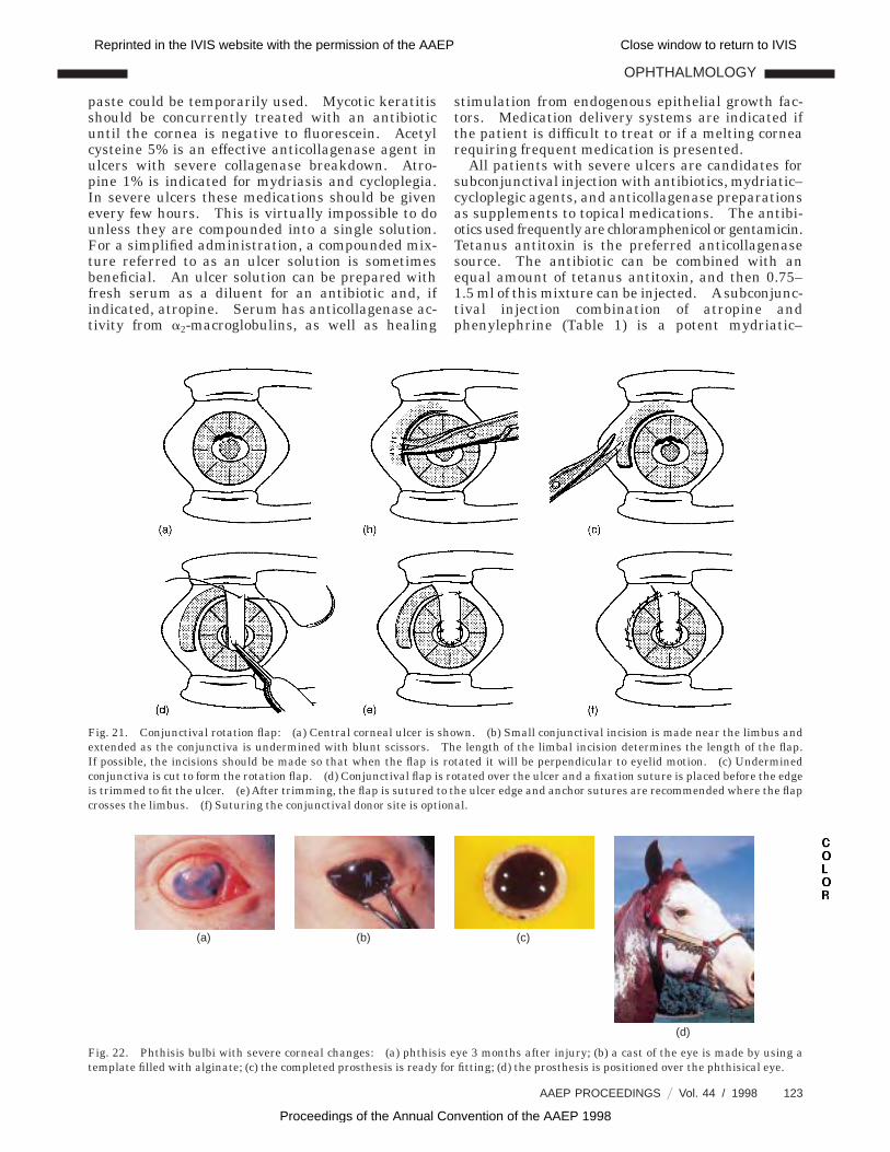
Fig. 21. Conjunctival rotation flap: (a) Central corneal ulcer is shown. (b) Small conjunctival incision is made near the limbus andextended as the conjunctiva is undermined with blunt scissors. The length of the limbal incision determines the length of the flap.If possible, the incisions should be made so that when the flap is rotated it will be perpendicular to eyelid motion. (c) Underminedconjunctiva is cut to form the rotation flap. (d) Conjunctival flap is rotated over the ulcer and a fixation suture is placed before the edgeis trimmed to fit the ulcer. (e) After trimming, the flap is sutured to the ulcer edge and anchor sutures are recommended where the flapcrosses the limbus. (f) Suturing the conjunctival donor site is optional.
Fig. 22. Phthisis bulbi with severe corneal changes: (a) phthisis eye 3 months after injury; (b) a cast of the eye is made by using atemplate filled with alginate; (c) the completed prosthesis is ready for fitting; (d) the prosthesis is positioned over the phthisical eye.
(a) (b) (c)
(d)
OPHTHALMOLOGY
AAEP PROCEEDINGS 9 Vol. 44 / 1998 123
Reprinted in the IVIS website with the permission of the AAEP Close window to return to IVIS
Proceedings of the Annual Convention of the AAEP 1998

cycloplegic combination. Mydriasis will begin in5–10 min and be complete in 1 h. Subconjunctivalinjections can be repeated as needed.
The last phase of ulcer management, if indicated,is corneal support and protection. The cornea canbe protected and supported with eyelid suturing, anictitating membrane flap, a conjunctival flap (Figs.18–21), an extended wear contact lens, corneal adhe-sives, corneal suturing, corneal transplant, and cor-neoscleral transposition. Treatment should becontinued until the ulcer is epithelialized and infec-tion has been controlled. Hypertonic sodium chlo-ride 5% ointment or a 2–5% solution two or threetimes daily promotes epithelial reattachment andshould be used 3–4 weeks after epithelialization.
15. Phthisis Bulbi
Phthisis bulbi is a blind shrunken eye that is due toirreversible ciliary body damage. It is usually notpainful but results in an unsightly eye and, if severe,ocular discharge. Treatment is not needed unlesssecondary problems exist. If there are problems,several treatment options are available. The besttreatment from the patient’s standpoint would bethe removal of the blind eye and positioning of anorbital implant in the orbit before the wound isclosed.
If cosmetic appearance is a factor, the owner maychoose a corneoscleral prosthesis. This prosthesisis placed over the phthisical eye, thereby filling the
anterior orbit and providing support to the eyelids(Fig. 22). The prosthesis is removed periodically forcleaning and then replaced immediately.
Severe phthisis bulbi requires a large prosthesis,which because of its size would be difficult to keep inthe orbit. An alternative procedure would be toenucleate the eye, position a subconjunctival im-plant, and later fit the eye with a corneoscleralprosthesis. This procedure is also indicated foranimals with intraocular tumors.
References and Footnotes
1. Ball M, Rebhun W, Gaarder J, et al. Evaluation of itracona-zole-dimethyl sulfoxide ointment in the treatment of keratomy-cosis in nine horses. J Am Vet Med Assoc 1997; 211:193–203.
2. Figure 8 is modfied from Slatter D. Fundamentals of veteri-nary opthalmology, 2nd ed. Philadelphia: Saunders, 1990.
aMila International, 510 W. 6th St., Covington, KY 41018.bJorgensen Laboratories, 1450 N. Van Buren St., Loveland, CO
80538.cPilopine HS Gel, Alcon Laboratories, Fort Worth, TX 76161.dEquron, Solvay Animal Health, Inc., Mendota Heights, MN
55120-1139.eHyalovet, Ft. Dodge Laboratories, Ft. Dodge, IA 50501.fSynacid, Schering, Kenworth, NJ 07033.gSilicone Implant, Jordan Plastics Research Corp., 17100 W.
12 Mile Rd., Southfield, MI 48076; 313-424-8560.hMaxitrol, Alcon Laboratories, Fort Worth, TX 76161.iBetadine, Purdue Frederick Co., Norwalk, CT 06850-3590.
OPHTHALMOLOGY
124 1998 9 Vol. 44 9 AAEP PROCEEDINGS
Reprinted in the IVIS website with the permission of the AAEP Close window to return to IVIS
Proceedings of the Annual Convention of the AAEP 1998