Embed Size (px)
Citation preview
SOME MORPHOLOGICAL ASPECTS
OF ACTIVE SODIUM TRANSPORT*
The Epithelium of the Frog Skin
CORNELIS L. VOOTTE and HANS H. USSING
From the Institute of Biological Chemistry, the University of Copenhagen, Copenhagen, Denmark.
Dr. Vote's present address is Medizinische UniversitAtsklinik, Basel, Switzerland
ABSTRACT
A method was experimentally tested which allows simultaneous morphological and bioelec-
trical studies of a tissue that performs active sodium transport, i.e., the isolated, surviving
frog skin. In a four cell lucite chamber with four separate electric potential and current cir-
cuits, skin specimens for morphological observation (light and electron microscopy) were
fixed in situ in well-defined functional states. The rate of active sodium transport through the
epithelium of Rana temporaria skin was modified by changing the strength of the electric cur-
rent passed through the specimens. A marked, reversible swelling of the outermost layer of
the stratum granulosum was observed during short circuiting of the skin compared to the
homogeneous appearance of the epithelium under open circuit conditions. Doubling the
ingoing current led to an additional small increase of the swelling or the appearance of islets
of cell necrosis in the same layer. There were signs of a slight shrinkage of the underlying
cell layers. The observations are discussed in the light of previous bioelectrical and morpho-
logical observations.
INTRODUCTION
An enormous amount of information about activetransport of sodium ions across biological mem-branes and its relations to electrical parameterslike potential and short circuit current has accu-mulated in the last two decades. To some extentit has been possible to correlate functional andmorphological observations with encouragingresults (1, 16, 20, 22, 23), but many essentialpoints are still open to discussion. One reason forthis is that in previous investigations morphologicaland functional studies were carried out on differentspecimens. The present paper describes a tech-nique which allows specimens for morphologicalstudies to be fixed under well-defined functional
* Dedicated to Gunnar Bj6rklund, Sigtuna, Sweden.
conditions. The setup was used for investigation othe morphological changes associated with thepassing of ingoing electric currents through iso-lated surviving frog skin. Since such currents,under appropriate conditions, are carried mainlyor (under short circuit conditions) exclusively byactively transported sodium ions (2), the morpho-logical changes may give us information aboutcertain aspects of the active sodium transportmechanism.
Theoretical Considerations
Like many other epithelia, the epithelium of thefrog skin maintains an electric potential differencebetween its inward and outward facing boundaries.
625

() Closed circuit (shorted)
openMembrane
I
vAV AV120 mv Omv
If both sides of an isolated frog skin are in con-tact with Ringer's solution, the inside bathingsolution is positive relative to the outside solu-tion. In the species studied, Rana temporaria, thepotential difference (called the skin potential)may be as high as 170 my but is usually between60 and 120 mv. According to Ussing and Zerahn(2), this potential is created and maintained byactive transport of sodium ions from the outsideto the inside solution. Numerous investigations inthis and other laboratories have substantiated thisview (references 3-6 among others). The electricpotential difference across a given skin is a functionof the efficiency of its "sodium pump" as well as itsleakiness to anions (for details see reference 7).The operation of the active sodium transportmechanism tends to increase the potential differ-ence, whereas the flow of passive ions like chlorideunder the influence of the potential differencetends to lower it. The flow of chloride ions in away represents a shunt to the "sodium battery" sothat a high chloride permeability gives a lowpotential and vice versa. The normally high poten-tials (80-120 mv) are mostly associated with a lowtransport of NaCl, because the low Cl- perme-ability reduces the flow of chloride while the highpotential opposes the inward transport of sodium.If, on the other hand, the electric potential differ-ence is reduced, e.g. by connecting the inside andoutside solutions through an external circuit, moresodium will be transported actively from the out-side to the inside solution (see Fig. 1). The situa-tion is quite simple when the external circuit haszero effective resistance. Under such conditionsthe potential difference is zero, and the passiveions, like chloride, have no reason for movingfaster one way than the other and do not con-
FIGURE 1 Illustration of the theoreticalconsiderations and the methodological setupof the experiments.
tribute to the current. The electric current, there-fore, is carried exclusively by the actively trans-ported sodium ions.
To achieve the condition of total short circuit,one can use the technique developed by Ussingand Zerahn (2).
The principle is to place a battery and a poten-tial divider in series with the skin through suitableelectrodes and to adjust the applied current toexactly the value which makes the potential dropacross the skin equal to zero (measured betweenseparate reversible electrodes). The skin now hasonly to overcome its own internal resistance, andso it is by definition short-circuited; but, as theskin and the external circuit are placed in series,the short circuit current can be read on a micro-ammeter placed in the circuit. Careful measure-ments in instances where the inward and outwardfluxes of sodium were measured with isotopes haveshown that, indeed, the short circuit current isalways equal to the net transport of sodium (2, 8,9); therefore, one can safely use the short circuitcurrent as a measure of the active sodium trans-port.
It has been shown independently by Zerahn(10) and Leaf and Renshaw (11) that the meta-bolic rate of the skin is determined by the amountof sodium transported actively. The oxygen con-sumption attains a basic value when sodium trans-port is stopped, owing to lack of sodium in theoutside medium or a high opposing potential, andincreases linearly with the rate of sodium transport.Metabolically, therefore, the high potential skinis in a "resting state" whereas the short-circuitedskin is working harder. If the current is increasedbeyond the short-circuited state, the oxygen con-sumption continues to increase pari passu with the
626 THE JOURNAL OF CELL BIOLOGY VOLUME 36, 1968
~ open circuit

out
t
out -
tIn
FIGURE 2 Drawing in the left upper corner illustrates the experimental chamber in a sagittal crossection.Attached in- and outlets for the circulating Ringers are shown. The drawing on the right illustrates theseparate electric circuits for PD and SCC measurements.
net sodium transport until the current has reacheda value of somewhat more than double the shortcircuit current; but in this region the sodium trans-port has to be measured with isotopes, if accuratefigures are desired, because the passive ions giverise to a leak current of variable magnitude. Fromthe above considerations, it will be clear that bychanging the strength of an ingoing currentthrough a frog skin one can, within limits, set therate of active transport and the metabolic rate atany desired level.
From the morphologist's point of view theremust be two questions: (a) are the functionalstates which one can define on the basis of opencircuit, short circuit, and double short circuitcurrent associated with characteristic morpho-logical states; (b) is it possible to obtain trust-worthy reproducible pictures of these states?1
1 It may be worth mentioning that the short-circuitedstate should not be considered something very "un-
MATERIALS AND METHODS
The experimental setup, including the electric cir-cuits, was designed on the basis of principles previ-ously described (2). It is composed of four identicalcells, allowing four 1 cm 2 areas of the same skin to bestudied simultaneously. The volume of bathing solu-tion necessary on either side of each skin is 5 ml (seeFig. 2).
Experimental Procedure
All experiments were performed in the monthsSeptember to February on R. temporaria kept in shal-low tap water at 4°C. The skins were obtained in theusual way and placed as gently as possible between
biological." At certain times of the year, especially inthe springtime, one may find skins with a rather highrate of sodium transport and a very low potential(only a few millivolts). These skins have a very lowchloride resistance and can be considered internallyshort-circuited through the chloride shunt.
C. L. VOUTE AND H. H. USSING Morphology of Active Sodium Transport 627

the two half-cells. 5 ml of frog Ringer's solution werepipetted into each half-cell. The bathing solutionswere kept circulating and aerated during the entireexperiment. The composition of the Ringer's usedhas been described elsewhere (12). Special care wastaken to keep pressures as symmetrical as possible inall chambers, as judged by the vertical position of themembranes. The pressure difference across one mem-brane never exceeded 1 cm H2 0. The skins were keptunder open-circuit conditions for 60 90 min until theinitial fluctuations in potential had leveled off.Usually the four areas of one skin agreed well withrespect to the bioelectric parameters potential andshort circuit current. If one of them differed from therest by more than 10 my or 10 amps/cm2 , it was notused.
Each skin was studied histologically with respect tothe following conditions: (a) control, open circuit; (b)short-circuited for 45-50 min; (c) ingoing currenttwice the short circuit current for 20-30 min ("doublecurrent"); (d) recovery control (like c, but fixed 20min after reopening of circuit).
The skins were fixed in situ by the following tech-nique. Circulation of the bathing solutions wasstopped on both sides by clamping the air admissiontubes. One-half of the solution in each half-cell wasremoved and replaced by the same amount of 2%OsO04 dissolved in Ringer's. The final fixative had apH of 7.7 0.1 and an osmolality of 250 milliosmol/liter 5. Circulation was restored symmetrically.The beginning of the fixation could be read as a sharpdrop of the potential, and the potential then declinedless rapidly to reach zero in times varying between 10sec and 2 min. The time it took for the potential tovanish seemed to depend on the intensity of the cir-culation of the fixative. Moreover, skins with lowpotential tended to show a steeper decline of thepotential during fixation than those with high poten-tial. The short-circuited skins were kept short-cir-cuited during fixation, which means that the appliedcurrent had to be reduced gradually to zero as theskin potential vanished. (In a few pilot experimentsnot included in this series, the current was main-tained, throughout fixation, at the level of the shortcircuit current before the beginning of the fixation).The double current skins received a current strengthof twice the short circuit current as measured im-mediately before the beginning of the fixation. Theprogress of the fixation was followed by intermittentpotential readings.
When the skin potentials had dropped to zero, theskins were kept in contact with the circulating fixativefor a period between 30 min and 2 hr (for obviousreasons, the fixed pieces of skin could be removed forfurther work-up only at the end of the whole experi-ment). Some control experiments were done withglutaraldehyde fixative (5% in Ringer's for 30 min
to 2 hr, and 1 hr of washing in Ringer's followed bypostfixation in 1%0 0504 for 15 min). 2 The histologi-cal appearance of the specimens was grossly similarto that obtained with Os05 4 fixation. For the presentpurpose the glutaraldehyde fixation seemed unsatis-factory because it fixes frog skin epithelium ratherslowly. This conclusion is based on the fact that thetime required by the glutaraldehyde fixative to reducethe potential and the short circuit current (SCC) tozero is three to five times the time required by theosmium tetroxide fixative. In a few experimentsglutaraldehyde alone was not able to reduce the bio-electric parameters to.zero.
After the experiment the whole skin was removedfrom the chamber. The four biopsies were cut insmall bits with a razor blade, washed briefly in dis-tilled water, and dehydrated with acetone in in-creasing concentrations. The embedding in Epon812 was performed according to routine procedures,with prolonged infiltration steps. The biopsy piecesin the blocks were all reoriented before cutting. Thiswas done by reembedding them in sealing wax at a90 ° angle to their surface. Sections for light micros-copy were cut, after a last angle correction, on aPorter-Blum MT 1 Ultramicrotome. Their thicknesswas 0.5 for microscopic evaluation and 1 ,u forphotographic purposes. Toluidine blue was used forstaining (13). The measurement of the thickness ofthe different layers of the epithelium and of thethickness of the total skin were performed with a Wildmicroscope, with eyepieces provided with a microin-eter scale (magnification of 100 for measuring totalskin thickness and magnification of 1000 for measur-ing the thicknesses of the different layers of theepithelium).
Of every biopsy piece, three random sections wereused for these measurements. Five measurementswere made on each section. The measurement of theheight of a cell layer was always made through anucleus. The mean values of the measurements aregiven in Table I. Thin sections for electron micros-copy were cut in the same way as those for lightmicroscopy; they were picked up at a cutting thick-ness of 500-800 A and stained with uranyl acetate(14) and lead citrate (15). The light micrographswere taken with a Zeiss microscope with an attachedWild camera. The electron micrographs were ob-tained with a Siemens Elmiskop (type 1A).
RESULTS
For the general morphological description of frogskin, we would like to refer the reader to previously
2 Glutaraldehyde fixative, even when used as a 2%solution in identical Ringer's, has an osmolalitywhich is appreciably above that of Ringer's alone,namely -400 milliosmols/liter.
628 THE JOURNAL OF CELL BIOLOGY VOLUME 36, 1968

TABLE I
Mean Values of Thickness Measurements in Skin
Only the first RCL and the total epithelium are considered
Experi-Condition ments First RCL Total epithelium First RCL excluded
P i % i %o W -± %
Open circuit 20 7.48 - - 64.6 - - 57.1 - -Short circuit 20 9.81 +2.33* +31 68.5 +3.9 +6.0 58.7 +1.6 +2.7Double-current 19 10.24 +2.76: +39 64.2 -0.4 -0.6 54.0 -3.1 -5.5Recovery 6 6.72 -0.76 -!10 57.4 -7.2 -11.1 50.7 -6.4 -12.7
* Statistical evaluation of this value. The numbers in brackets give the statistical evaluation of the fur-ther increase; under double current: mean , 2.33 (0.43; variance s, 11.89 (7.65); variance of the mean52, 0.59 (0.35); standard deviation of the mean s, 0.77 (1.0); degree of freedom, 19 (18); t, 3.15 (0.35;probability P, 0.01-0.001 (0.80-0.70).
published papers (16-21). Electron microscopicfindings will be discussed only if they help toclarify doubtful light microscopic observations orif they offer additional information.
Qualitative description
OPEN CIRCUIT CONTROLS (FIGS. 4 a-6 a,7 a, b): One to three layers of cornified cells arealways found to separate the epithelium from theoutside. These cells appear heavily and uniformlystained on light microscopic observation. Rightnext to an interspace of variable width borderingthe cornified layer, the first layer of the stratumgranulosum will be the one attracting most of ourattention (we shall call it the first reacting celllayer, first RCL). Under open circuit conditions(with a high skin potential) these cells appearuniformly stained and shaped and do not showany appreciable difference compared to the under-lying cells of the stratum granulosum and stratumspinosum. The stratum germinativum may easilybe differentiated by the less dense cytoplasm of itscells, the columnar shape of the cells and theundulating basal surface of the cells which limitsthe epithelium towards the interstitial space.
A network-like system of intercellular space isobserved, the separation of the cells being inter-rupted only by dense-appearing granules (thedesmosomes) found to be especially numerous inthe stratum granulosum. The first cell layer of thestratum granulosum always forms a continuousbarrier, its continuity being preserved by tightlocks closing the intercellular space system towardsthe outside medium. These attachment zones are
found, with very rare exceptions,3 at the outer-most cell angles of the first RCL (Fig. 7 a). Theyare similar to the zonulae occludentes of the corni-fled layer previously described by Farquhar andPalade (18). The intercellular space system is moreor less open throughout the epithelium andreaches the interstitial compartment; it is inter-rupted only by a continuous basement membrane.
SHORT-CIRCUITED SKINS (FIGS. 4 b-6 b,8 a, b): The main morphological change isfound in the cells of the first RCL. These cellsappear swollen. This swelling can be observed as amore or less uniform manifestation of all the cellsbelonging to this layer. A decreased cytoplasmicdensity with a relative increase of the contrast ofthe cytoplasmic structures gives the cells a ratherheterogeneous appearance. In general, however,the cytoplasmic organelles do not present majoralterations. A slight shrinking of the underlyingcells may occasionally be observed (increasednuclear and cytoplasmic density). The stratumgerminativum shows neither shrinking norswelling.
The intercellular space system seems to beopen more uniformly, mainly in the stratumgranulosum and stratum spinosum. No change canbe found in cellular junctions, and the zonulaeoccludentes remain closed (Fig. 8 a). It should be
3 On some rare occasions one finds an intercellularchannel open all the way into the space below thestratum corneum. Since this is a rare observationand since our experimental procedure is not set toevaluate this question, this observation is mentionedonly for completeness.
C. L. VOOTE AND H. H. USSING Morphology of Active Sodium Transport 629

FIGuRE 3 Simplified graphical illustration of bioelectric protocols belonging to the morphological illus-trations shown in this paper.
-

FIGURES 4-6 Light micrographs of frog skin epithelium under different bioelectric conditions (for corre-sponding functional data, see corresponding graphics Fig. 3). Each horizonal row pertains to the sameexperiment (1-3). The letters indicate the experimental condition (a-d): a, open circuit; b, short circuit; c,double current inwards; d, recovery after double current lp sections stained with toluidine blue. X 2000.
Fig. 4. a, Note uniform appearance of all cell layers; b, a slight swelling of the first RCL can be ob-served; c, single necrotic cell in the first RCL; d, the whole epithelium is shrunken. Pyknotic cell in thefirst RCL.
Fig. 5, a, same as Fig. 1 a; b, again, marked swelling of first RCL and shrinking of lower cells are ap-parent; c, same findings as in 5 b but more marked. SC, stratum corneum; SG, stratum granulosum andstratum spinosum; Sg, stratum germinativum.
Fig. 6 a, First RCL seems already slightly swollen (compared to Figs. 4-5 a) (Note lower PI) readingsthan in the two previous experiments); b, Marked increase of swelling of first RCL as compared to Fig. 6 a;c, Extreme swelling of first RCL with pronounced shrinking of underlying epithelium.
631
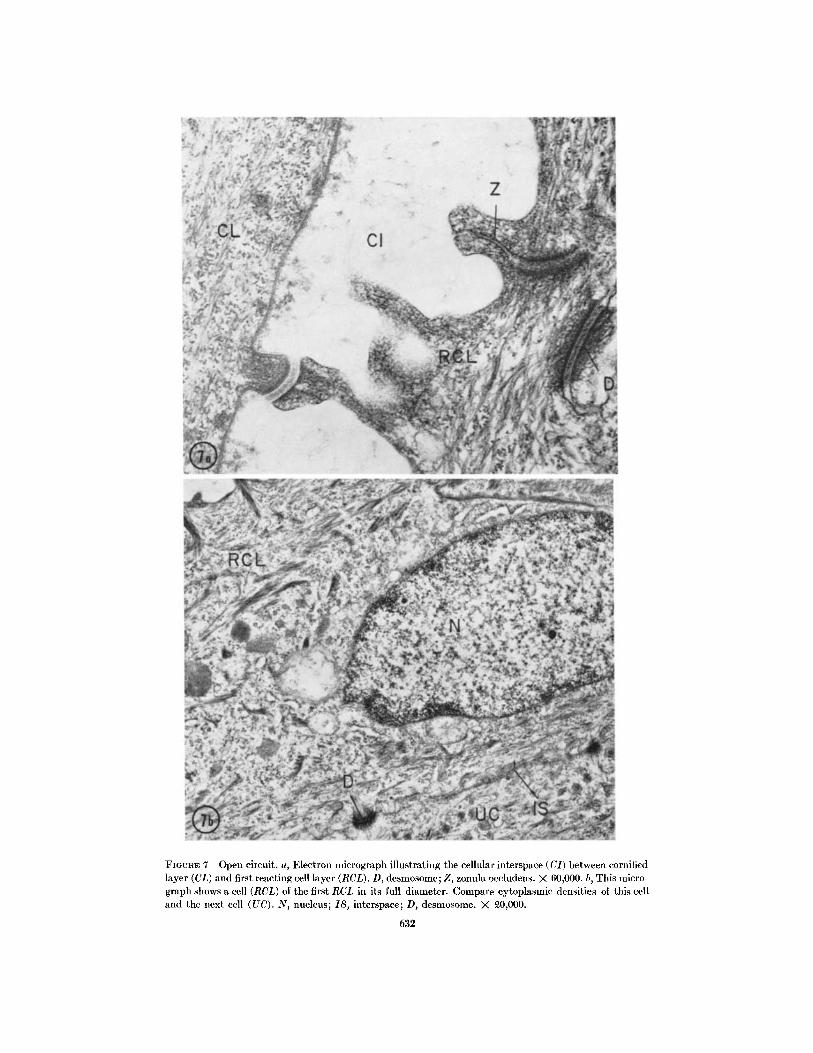
FIGUJRE 7 Open circuit. a, Electron micrograph illustrating the cellular interspace (CI) between cornifiedlayer (CL) and first reacting cell layer (RCL). D, desmosome; Z, zonula occludens. X 60,000. b, This micro-graph shows a cell (RCL) of the first RCL in its full diameter. Compare cytoplasmic densities of this celland the next cell (UC). N, nucleus; IS, interspace; D, desmosome. X 0,000.
632

FIGURE 8 Short circuit. a, As compared to that in Fig. 7 a, the cytoplasmic density of the two cells isnow clearly different. Cellular organelles seem to be unaltered; the zonula occludens region (Z) remainsclosed. Same region is shown at a higher magnification in inset. RCL, first reacting cell; UC, underlyingcell; IS, interspace. Fig. 8, X 15,000; inset, X 40,000. b, Electron micrograph illustrating the cellular junc-tions between swollen cell (S) and underlying cell (UC) with normal cytoplasmic density. Note open inter-space system. X 40,000.
633
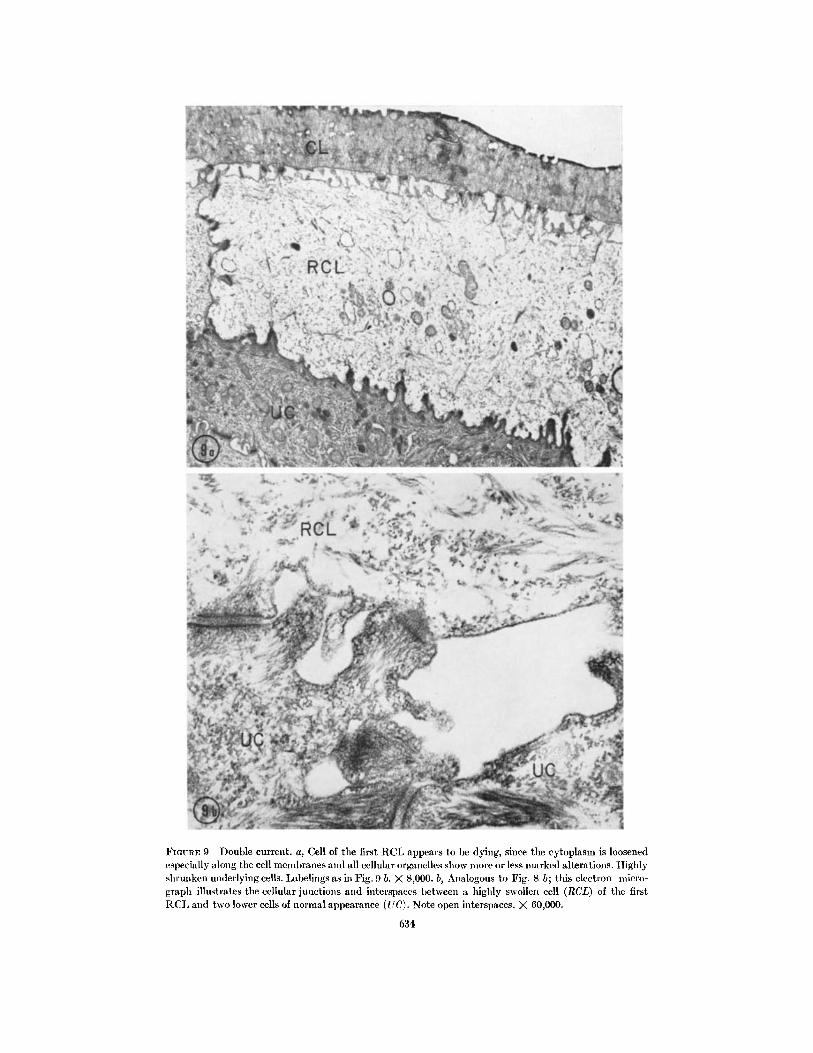
FIGURE 9 Double current. a, Cell of the first RCL appears to be dying, since the cytoplasm is loosenedespecially along the cell membranes and all cellular organelles show more or less marked alterations. Highlyshrunken underlying cells. Labelings as in Fig. 9 b. X 8,000. b, Analogous to Fig. 8 b; this electron micro-graph illustrates the cellular junctions and interspaces between a highly swollen cell (RCL) of the firstRCL and two lower cells of normal appearance (UQ. Note open interspaces. X 60,000.
634
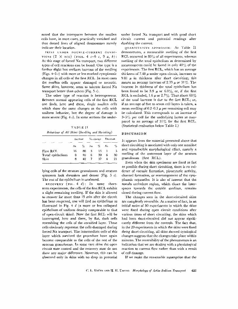
noted that the interspaces between the swollencells have, in most cases, practically vanished andthat dotted lines of aligned desmosomes merelyindicate their location.
SKINS UNDER DOUBLE-CURRENT CONDI-
TIONS (2 X SCC) (FIGS. 4 c-6 c, 9 a, b):At this stage of forced Na transport, two differenttypes of cell reactions can be found. One type is afurther slight but uniform increase of the swelling(Figs. 4-5 c) with more or less marked cytoplasmicchanges in all cells of the first RCL. In most casesthe swollen cells appear damaged or necrotic.Some skins, however, seem to tolerate forced Natransport better than others (Fig. 5 c).
The other type of reaction is heterogeneous.Between normal appearing cells of the first RCLone finds, here and there, single swollen cellswhich show the same changes as the cells withuniform behavior, but the degree of damage ismore severe (Fig. 4 c). In some sections the under-
TABLE II
Behaviour of All Skins (Swelling and Shrinking)
Increase No change Decrease
No. % No. % No. %
First RCL 16 80 3 15 1 5Total epithelium 8 40 6 30 6 30Skin 8 42 7 37 4 21
lying cells of the stratum granulosum and stratumspinosum look shrunken and denser (Fig. 5 c).The rest of the epithelium is unaltered.
RECOVERY (FIG. 4 d): In some short-term experiments, the cells of the first RCL exhibita slight remaining swelling. If the skin is allowedto recover for more than 15 min after the circuithas been reopened, one will find an epithelium asillustrated in Fig. 4 d (a more or less collapsedepithelium of uniform density comparable to thatof open-circuit skins). Now the first RCL will beinterrupted, here and there, by flat, dark cellsresembling the cells of the cornified layer. Thesecells obviously represent the cells damaged duringforced Na transport. The intermediate cells of thislayer which survived the procedure have againbecome comparable to the cells of the rest of thestratum granulosum. In some rare skins the opencircuit state control and the recovery state do notshow any major difference. However, this can beobserved only in skins with no drop in potential
under forced Na transport and with good shortcircuit current and potential readings afterdoubling the current.
QUANTITATIVE APPROACH: As Table II
demonstrates, a measurable swelling of the firstRCL occurred in 80 % of all experiments, whereasswelling of the total epithelium as determined bymeasurements could be found in only 40% of theexperiments. The first RCL, which has an averagethickness of 7.48 p under open circuit, increases to9.81 pu in thickness after short circuiting; thismeans an average increase of 2.33 A or 31 %. Theincrease in thickness of the total epithelium hasbeen found to be 3.9 ju or 6.0%, or, if the firstRCL is excluded, 1.6 pu or 2.7%. Thus about 60%of the total increase is due to the first RCL; or,if an average of five to seven cell layers is taken, amean swelling of 0.2-0.3 , per remaining cell maybe calculated. This corresponds to an increase of3-5 % per cell for the underlying layers as com-pared to an average of 31% for the first RCL.(Statistical evaluation below Table I.)
DISCUSSION
It appears from the material presented above thatshort circuiting is associated with only one manifestand reproducible morphological effect, namely aswelling of the outermost layer of the stratumgranulosum (first RCL).
Even when the skin specimens are fixed as fastas possible during short circuiting, there is no evi-dence of vacuole formation, pinocytotic activity,channel formation, or rearrangement of the cyto-plasmic organelles. It is also of interest that thezonula occludens region, which closes the inter-spaces towards the outside medium, remainsclosed during current flow.
The changes seen in the short-circuited skinsare completely reversible. As a matter of fact, in aninitial series of 50 experiments in which the skinswere fixed during open circuit conditions aftervarious times of short circuiting, the skins whichhad been short-circuited did not appear signifi-cantly different from the controls. The fact that,in the 20 experiments in which the skins were fixedduring short circuiting, all skins showed cytologicalchanges suggests that the changes take place withinminutes. The reversibility of the phenomenon is anindication that we are dealing with a physiologicalreaction to current flow rather than with a resultof cell damage.
If we make the reasonable assumption that the
C. L. VOUTE AND H. HI. USSING Morphology of Active Sodium Transport 635

epithelial cells are in nearly osmotic equilibriumwith the bathing solutions, the swelling must meanthat the number of osmotically active particles inthe first RCL is increased during short circuiting.(This applies whether or not the swelling is due tocell damage.) One possibility is that the increasein metabolic rate, which is associated with the in-creased active transport of sodium during shortcircuiting, leads to an augmentation of the numberof osmotically active organic particles in the cells.However, this would mean that such particlesshould constitute some 209, or more of the totalosmolarity of the cells during short circuiting; thisis much more than one finds in, say, workingmuscle. A hypothesis which perhaps is more likelyis that there is an increase in inorganic electrolytesin the cells of the first RCL during short circuiting.Let us assume, for the sake of argument, that theoutward facing boundary of the cells in question ispermeable to sodium but less so to chloride, andthat the inward facing boundary is more perme-able to chloride and less so to sodium. It can thenbe seen intuitively, and can easily be shown mathe-matically, that there will be an accumulation ofelectrolyte (NaCI) between the membranes whenan ingoing current is passed. In an artificial, twomembrane system, the accumulation would go onas long as the current was running. In the livingcell a steady state might be established if thesodium pump were stimulated by the increase incellular sodium concentration so that the swellingstopped when the entry of Na through the outerbarrier was balanced by pumping through the in-ward facing barrier. The argument can be carriedthrough with essentially the same result for a moresophisticated cell model containing nondiffusiblecolloid anions and potassium ions (24). It is thusclear that a relatively simple cell model might givethe volume response observed during short cir-cuiting, but a detailed evaluation of the merits ofthe hypothesis requires more knowledge about therelative permeabilities of the inward and outwardfacing membranes of the first RCL than we haveat present.
The fact that only one cell layer shows changeswith relation to short circuiting might be taken tomean that this layer alone is performing the activesodium transport. This may be; however, it is quiteconceivable that sodium which has entered thefirst RCL from the outside solution can pass fromone epithelial cell to the other through intercellularconnections of the type described by Kanno and
Loewenstein (22, 23) (as in the case of the cellsof the salivary gland of Drosophila). If so, all epi-thelial cells, even the deeper layers, might partici-pate in the transport. Such an arrangement has, infact, been suggested independently by Farquharand Palade (18 20) and by Ussing and Wind-hager (7). Both groups assume, furthermore, thatthe intercellular space system is relatively tighttoward the outside medium but is open towardsthe inside of the epithelium. This means thatsodium, which is transported actively out of a cell,will end up in the intercellular space system andfrom there pass to the inside solution, irrespectiveof whether it has been transported by a cell in thefirst or in the last cell layer.
If this hypothesis is correct, one may ask whyonly one cell layer swells during short circuiting?The reason may be that all of the sodium ions haveto pass this layer whereas an ever decreasingamount is received by the other layers, the fartherthey are from the surface. Another reason mightbe that the first RCL is approaching the end of itslife and is losing its capacity for osmotic regula-tion. But then one might ask why an aged cellshould respond with an absolutely reversiblephenomenon.
If the strength of the ingoing current is doubled,the swelling of the first reacting cell layer becomesmore pronounced and, in contrast to the resultwith simple short circuiting, there is evidence ofcell damage in many skins. As has already beenmentioned, the oxygen consumption is still closelycorrelated with the sodium transport under suchconditions.4 Therefore, double current must meana very heavy drain on the energy reserves of thecells, and one may speculate that this may lead toexhaustion and premature death of some of thecells. However, there may be other reasons for cellnecrosis, for instance membrane damage owing toexcessive swelling, or direct electrical damage tothe membranes. After recovery from double cur-rent, the cells of the whole epithelium look slightlyshrunken, and many cells of the first RCL havedied. This latter observation is of interest becauseit shows that the cell damage in the double currentexperiments is brought about by this treatment perse and is not an artefact brought about by OsO4fixation during current flow.
This brings us to the general question whetherthe pictures we have obtained of the cellular
4 Andersen, B. Unpublished observations.
636 THE JOURNAL OF CELL BIOLOGY VOLUME 36, 196S

changes during current flow are correct. Among
the factors which determine the volume of living
cells, the osmotic effect of electrolytes plays a
dominating role. With short circuit current, often
more than 1 p/mole of Na passes 1 cm 2 of skin per
hour. This means that an amount of Na corre-
sponding to the total electrolyte content of one cell
layer passes the skin in a few minutes. Therefore,
unless the fixation is very fast, one might anticipate
that the electric current would bring about a re-
distribution of electrolytes and hence of water if,for instance, the sodium pump were inhibited be-fore the whole cell had been fixed.
While such fixation artefacts cannot be ex-
cluded, there is some indication that they are not
important. Thus in one series of short circuit ex-
periments not presented here, the current was
kept at a prefixation level during fixation instead
of being lowered during fixation to keep the poten-
tial at zero. The histological appearance of skins
in that series of experiments was comparable, in all
respects, to that of the material presented here,
the swelling being, perhaps, slightly more pro-nounced. This indicates that the amount of current
passing during fixation is not of decisive im-
portance for the histological appearance of thespecimens.
The morphological part of this work was done in the
Institute of Genetics, University of Copenhagen, andthe Institute of Anatomy, University of Basel.
We would like to express our gratitude to Professorv. Wettstein and Mr. K. Henningsen in Copenhagen
and to Professor R. Schenk and Mr. W. Villiger in
Basel for their kind technical assistance. Our thanks
go as well to Mr. Poul Hansen of the Instiute of Bio-
chemistry for his help in manufacturing the experi-
mental chamber.
Received for publication 10 July 1967, and in revised form3 October 1967.
REFERENCES
1. USSING, H. H. 1965. Transport of electrolytesand water across epithelia. Harvey Lectures,Ser. 59 (1963-1964). 13.
2. USSING, H. H., and K. ZERAHN. 1951. Activetransport of sodium as the source of electriccurrent in the short-circuited isolated frog-skin. Acta Physiol. Scand. 23:110.
3. USSING, H. H. 1949. The active ion transportthrough the isolated frog-skin in the light oftracer studies. Acta Physiol. Scand. 17:1. 1949.
4. KOEFOED-JOHNSEN, V. 1957. The effect of g-
strophanthin (ouabain) on the active trans-
port of sodium through the isolated frog-skin.
Acta Physiol. Scand. Suppl. 42. 145.5. LINDERHOLM, H. 1954. On the behaviour of the
"sodium-pump" in the frog-skin at various
concentrations of Na-ions in the solution onthe solution on the epithelial side. Acta
Physiol. Scand. 31:36.6. TERCAFS, R. R., and E. SCHOFFENIEL. 1961.
Difference de potentiel et courant electriqueau niveau de la peau de grenouille isolee.
Arch. Intern. Physiol. Biochim. 69:459.7. USSING, H. H., and E. E. WINDHAGER. 1964.
Nature of shunt path and active Na-transport
path through frog-skin epithelium. Acta Physiol.
Scand. 61:484.8. KEFoED-JoHNSEN, V., and H. H. USSING. 1958.
The nature of the frog-skin potential. ActaPhysiol. Scand. 42:298.
9. KOEFOED-JOHNSEN, V., H. H. USSING, and K.
ZERAHN. 1952. The origin of the short-circuit
current in the adrenaline stimul ated frog-skinActa Physiol. Scand. 27:38.
10. ZERAHN, K. 1956. Oxygen consumption andactive sodium transport in the isolated andshort-circuited frog-skin. Acta Physiol. Scand.36:300.
11. LEAF, A., and A. RENSHAW. 1957. Ion transport
and respiration of isolated frog-skin. Biochem.J. 65:82.
12. USSING, H. H. 1965. Relationship betweenosmotic reactions and active sodium transportin the frog-skin epithelium. Acta Physiol. Scand.63:141.
13. MUNGER, B. L. 1961. Staining methods appli-cable to sections of osmium-fixed tissue forhigh resolution light microscopy. J. Biophys.Biochem. Cytol. 11:502.
14. BRODY, J. 1959. The keratinization of epidermal
cells of normal guinea pig skin as revealed by
electron microscopy. J. Ultrastruct. Res. 2:482.
15. REYNOLDS, E. S. 1963. The use of lead-citrate at
high pH as an electron-opaque stain in elec-
tron microscopy. J. Cell Biol. 17:208.
16. OTTOSON, D. F., F. SJOSTRAND, S. STENSTR6M,
and G. SWAETICHIN. 1953. Microelectrodestudies on the EMF of the frog-skin related toelectron-microscopy of the dermo-epidermaljunction. Acta Physiol. Scand. Suppl. 106. 611.
17. VOUTE, C. L. 1963. An electron microscopicstudy of the skin of the frog. J. Ultrastruct. Res.
9:497.
C. L. VOOTE AND H. H. USSING Morphology of Active Sodium Transport 637

18. FARQUHAR, M. G., and G. E. PALADE. 1963.
Cell junctions in amphibian skin. J. Cell Biol.19:22A.
19. FARQUHAR, M. G., and G. E. PALADE. 1963.
Junctional complexes in various epithelia. J.
Cell Biol. 17:375.
20. FARQUHAR, M. G., and G. E. PALADE. 1964.
Functional organization of amphibian skin.
Proc. Natl. Acad. Sci. U. S. 51: 569.
21. PARAKKAL, P. F., and A. G. MALTOLTSY. 1964. A
study of the fine structure of the epidermis ofRana pipiens. J. Cell Biol. 20:85.
22. KANNO, Y., and W. R. LOEWENSIEIN. 1964. Low
resistance coupling between gland cells. Some
observations on intercellular contact em-branes and intercellular space. Nature. 201:194.
23. KANNO, Y., and W. . LOEWENSTEIN. 1964.Intercellular diffusion. Science. 143:959.
24. USSING, H. H., and V. KOEFOED-JOHNSEN. 1958.The nature of the frog skin potential. ActaPhysiol. Scand. 42:298.
638 THE JOURNAL OF CELL BIOLOGY VOLUME 306, 1968