Embed Size (px)
Citation preview

1
From the Department of Neurosurgery
Ludwig Maximilians-University of Munich
Campus Grosshadern
Director and Chairman: Prof. Dr. med. Jörg-Christian Tonn
Epilepsy Surgery Around Language Cortex
A study with indepth discussion of cortical stimulation mapping as a gold standard for
detecting language cortex and a comparison of two different cortical mapping techniques to
ensure postoperative language function and seizure control in this group of epilepsy surgery
patients
Dissertation
to Acquire a Medical Doctor Degree
in the Medical Faculty of
Ludwig-Maximilians-University Munich
Offered by
Aksels Ribenis
from
Riga, Latvia
2008

2
With approbation of the Medical Faculty of
University of Munich
Commentator: Prof. Dr.med. Peter A. Winkler_______________________
________________________________________________
Co-Commentator: Prof. Dr. Rolf. R. Engel_____________________________
Prof. Dr. Reiner Frank______________________________
Co-supported by :
Dean: Dr.h.c. M. Reiser, FACR, FRCR
Date of oral exam: 12.03.2009_______________________________________

3
Table of contents
1 Introduction................................................................................................................ 5
1.1 Statistical data on epilepsy and epilepsy surgery …………………………...….. 6
1.2 Criteria for including an epilepsy patient in pre-surgical investigation………… 6
2 Examination of surgery candidates………………………………………………… 7
2.1 Introduction........................................................................................................... 7
2.2 The goal and structure of pre-surgical investigations........................................... 7
2.3 Non-invasive investigations.................................................................................. 8
2.3.1 History and neurological examination............................................................ 8
2.3.2 Ictal and Interictal electroencephalographic recordings................................ 8
2.3.3 Magnetic resonance imaging (MRI)............................................................. 9
2.3.4 Functional magnetic resonance imaging (fMRI).......................................... 10
2.3.5 Positron Emission Tomography (PET)......................................................... 10
2.3.6 Single Photon Emission Computed Tomography (SPECT)........................ 11
2.3.7 Neuropsychological testing.......................................................................... 11
2.4 Invasive investigations....................................................................................... 12
2.4.1 Determination of epileptogenic zone by invasive
electroencephalography and video-EEG-monitoring................................... 13
2.4.2 Determination of functionally significant cortex by cortical stimulation.... 15
2.4.2.1 History of general nerve cell stimulation............................................ 16
2.4.2.2 History of intraoperative cortical stimulation………………………. 19
2.4.2.3 History of extraoperative cortical stimulation………………………. 21
2.4.2.4 First steps of epilepsy surgery close to speech areas……………….. 21
2.4.2.5 The physics of cortical stimulation………………………………… 22
2.4.2.5.1 Current spread and tissue excitability.…………………………. 22
2.4.2.5.2 Current spread…………………………………...……………... 22
2.4.2.5.3 Estimates of excitability and strength – duration functions …… 24
2.4.2.6 Stimulation parameters……………………………………………... 25
2.4.2.6.1 Stimulus intensity (voltage or amperage)…………………….... 26
2.4.2.6.2 Duration of each individual stimulus…………..……………… 27
2.4.2.6.3 Stimulation frequency…………………………………………. 27
2.4.2.6.4 Duration of the stimulus train………………………………….. 27
2.4.2.7 Characteristics of stimulus………………………………………… 27
2.4.2.8 Physiological concerns of cortical stimulation…………………….. 28

4
2.4.2.9 Procedure of extra-operative EEG recording and cortical mapping.. 29
2.4.2.10 Procedure of intra-operative cortical mapping……………………. 34
3 The Operation – Cortical Resection…….………………………………………… 38
4 Hypothesis of the study…………………………………………………………... 38
5 Study questions…………………………………………………………………... 39
6 Goals of the study………………………………………………………………... 39
7 Methods………………………………………………………………………….. 40
7.1 Patients………………………...……………………………………………... 40
7.2 Investigation of language function and cortical language mapping….……..... 41
7.3 Neurological examinations and post-operative seizure outcome………….…. 43
7.4 Statistical analysis……………………………………………………...……. 44
8 Results………………………………………...………………………………..... 44
8.1 Characteristics of patients…………………………………………………… 44
8.2 Non-invasive pre-operative investigations………………………………….. 44
8.3 Invasive language mapping…………………………………………………. 45
8.4 Correspondance of extra- and intra-operative stimulation results………….. 46
8.5 Resective operations………………………………………………………... 48
8.6 Post-operative results as regards language function………………….…….. 49
8.7 Post-surgical outcome as regards seizure control…………………………... 50
8.8 Post-operative complications.......................................................................... 53
8.9 The summary of statistical analysis data........................................................ 53
9 Discussion.......................................................................................................... 54
9.1 Patients and non-invasive investigations........................................................ 54
9.2 Invasive language mapping and post-operative language function................ 56
9.3 Post-operative seizure control......................................................................... 65
10 Drawbacks of the study...................................................................................... 68
11 Final remarks...................................................................................................... 68
12 Conclusions........................................................................................................ 70
13 Possibilities for further improvement in epilepsy surgery around speech areas.72
14 Summary in English…………………………………………………………... 74
15 Summary in German / Zusammenfassung......................................................... 77
16 References.......................................................................................................... 81
17 Curriculum Vitae…………………......…..…………………………………… 97
18 Acknowledgements…………………………………………………………… 99

5
1 INTRODUCTION
Epilepsy surgery is any neurosurgical intervention, whose primary objective is to
relieve medically intractable epilepsy [32]. Its aim is to reduce the number and
intensity of seizures, minimize neurological morbidity and antiepileptic drug toxicity,
and improve the patient’s quality of life [46]. The main challenge of neurosurgery has
always been to preserve maximal physiological, neuronal functions during the
operation. This is even of more concern in epilepsy surgery, which is normally an
elective surgery without any vital indications for resection of the epileptogenic zone.
The epileptogenic zone is a region of the cortex that can generate epileptic seizures.
By definition, it is the “minimal area of cortex that must be resected to produce
seizure freedom” [62].
The concept of the epileptogenic zone is a purely theoretical one. Some are of the
opinion that its extent and location cannot be fully determined until the patient is
actually made seizure free by resective surgery [14]. As the epileptogenic zone is
often located near functionally significant cortical regions, a major concern is to
preserve the higher cortical functions located there. Often it is their preservation that
conflicts with the resection of the entire epileptogenic cortex in order to also achieve
seizure freedom or reduce the seizure frequency after the operation. Those patients
with a potential overlap of pathological alterations and neurophysiological function
pose the frequently observed dilemma of a necessary tradeoff between seizure relief
and permanent neuropsychological deficits [49], [51], [105].
One of the most significant higher cortical functions, of great importance for
neurosurgeons and their patients, has always been and remains speech. According to
Ojemann and colleagues, the location of language zones varies from one individual to
the next. They are found in a wide area of the left lateral cortex, extending beyond the
traditional anatomical limits of the Broca and Wernicke areas [84], [86], [88], [89],
[120]. Thus, the location of the epileptogenic zone should more frequently be
suspected to lie around the speech cortex. Extensive investigations must often be
performed to clarify this.
Therefore epilepsy surgery close to the speech cortex has become a special,
independent, and even more problematic subgroup of epilepsy surgery as regards the
6
successful surgical treatment of medically refractory seizure activity. To better
explain the actual investigation and treatment options for this epilepsy surgery
subgroup, we compiled an overview of our 10 years of experience with such patients
in Munich University Hospital, Grosshadern. This overview comprises descriptions of
current examinations used in our work, especially focusing on the invasive language
mapping method, the current gold standard for language localization. It also includes
our data on 22 left hemisphere epilepsy surgery patients whose epileptogenic zone
was located around the speech cortex and who underwent operations in our clinic
between 1997 and 2007. We also assess two different tactics of language mapping in
those patients and appraise our indications for their use.
1.1 Statistical data on epilepsy and epilepsy surgery
Almost one percent of the world’s populace suffer from epilepsy. There are
approximately 50 million epilepsy patients in the whole world [54]. In Germany there
are 6 to 7 epilepsy patients per 1 000 inhabitants, thus about 500 000 in the entire
country. Every year 30 to 50 persons out of 100000 in Germany are diagnosed to have
epilepsy, making all together about 30 000 new epilepsy cases each year [54].
According to data of the National Society for Epilepsy (United Kingdom), up to 70%
of persons with epilepsy achieve full seizure freedom through medication [73].
Sixty percent of all epilepsies are of focal onset. In about 30% of focal epilepsy
cases the seizures continue in spite of adequate antiepileptic (AED) medication or
patient develops intolerable side effects [21]. If half of these patients were evaluated
for epilepsy surgery and half of those evaluated would eventually benefit from
epilepsy surgery, this means that about 4.5% of all patients with epilepsy (0.03% of
the total population) could profit from epilepsy surgery [31]. There is currently a
considerable backlog of 5,000 people waiting for surgery and between 300 to 500 new
cases each year in the United Kingdom [73].
1.2 Criteria for including an epilepsy patient in pre-surgical investigation
On the basis of the above-mentioned definition of epilepsy surgery, a potential
epilepsy surgery patient must have medically intractable epilepsy.

7
Although the definition of medical intractability differs among the various epilepsy
centers, it mainly refers to patients whose seizures have continued despite adequate
monotherapy in trials of at least two antiepileptic drugs (AEDs) with or without one
trial of combination of two drugs [25], [121]. Medical intractability can also indicate
that control of seizures is achieved, but the necessary medication is accompanied by
intolerable side effects. Another criterion of medical intractability is that seizures
are of sufficient severity and/or frequency to interfere with the patient’s quality of life
[33]. The impact of epileptic seizures on a patient’s quality of life is assessed during
several visits to an epileptologist.
2 Examination of surgery candidates
2.1 Introduction
If the above-mentioned criteria for medical intractability are met, the patient is
included in our epilepsy surgery program and specific investigations are initiated to
find the epileptogenic zone.
Per definition epilepsy surgery does not include normal surgical treatment of
intracranial lesions, where the primary goal is to diagnose and possibly remove the
pathological target, often a progressing tumor. In these patients, epileptic seizures are
only one symptom of the lesion and are treated as part of the procedure [46].
However, a few tumor patients in whom the primary goal of operation was still to
decrease an intolerable seizure frequency were also included in our epilepsy surgery
program.
2.2 The goal and structure of pre-surgical investigations
The goal of pre-surgical evaluation is to precisely define the location and extent of
the epileptogenic zone together with nearby functional zones using both non-invasive
and invasive investigation methods.
Pre-operative investigations are of great significance in surgery of the dominant
hemisphere. Their ability to precisely localize the functionally significant (dominant)
8
cortex and to specify its relation to the epileptogenic cortex (epileptogenic zone)
determines the objectives and results of surgery as regards seizure control and post-
surgical neuropsychological/ neurological morbidity.
Like many other epilepsy surgery centers, we begin with relatively less expensive
and simple, non-invasive methods and progress to invasive investigations only if non-
invasive investigations do not provide enough information to define an epileptogenic
zone and determine its relation to functionally significant cortex. We then
proceed with resective surgery.
2.3 Non-invasive investigations
Non-invasive or extra-cranial investigations are relatively safe methods that provide
sufficient information in the majority of medically intractable epilepsy cases
regarding the localization of the epileptogenic zone. They often allow us to proceed
with resective surgery, without requiring more invasive (intracranial) examinations.
The following list gives short descriptions of non-invasive investigational methods
used in the pre-surgical evaluation.
2.3.1 History and neurological examination
A detailed history of epileptic attacks and a neurological examination are essential
to differentiate between epileptic and non-epileptic attacks. Both are also important
for understanding the seizure semiology, which can indicate the possible seizure
origin [65].
2.3.2 Ictal and interictal electroencephalographic recordings
The electroencephalogram (EEG) is a graphic recording of the brain’s electrical
activity. By registering epileptogenic potentials in some of the head-surface
electrodes, we can narrow down the possible localization of an epileptogenic zone.
Hans Berger (1873 – 1941) first described an EEG in 1929. The following ten years
witnessed revolutionary changes in the diagnosis of epilepsy, mainly due to the
9
implementation of EEG in clinical practice [11]. The first purely EEG-directed
temporal lobe resection was performed in 1942 (Boston, USA) by Percival Bailey
(1892 – 1973) and Frederick Gibbs (1903 – 1992) [111].
Although surface EEG recordings are less sensitive than invasive studies, their role
has continued to evolve with the advent of high resolution volumetric magnetic
resonance imaging (MRI) and other imaging techniques. They provide the best
overview and therefore the most efficient way of defining the approximate
localization of the epileptogenic zone [75], [107].
As already mentioned, the main limitation of extra-cranial EEG is its decreased
sensitivity to cortical generators [40], [108]. Surface recordings also have significant
difficulty “seeing” seizure onsets occurring in cortical regions located relatively deep
with respect to the scalp (interhemispheric, mesial temporal, etc.). This lack of
sensitivity implies that surface recordings only detect EEG seizures after they have
spread to involve extensive areas of cortex. EEG also has a spatial limitation - it can
only record electrical activity of the brain in an area of approximately 6 cm2 [62].
For proper investigation both ictal and interictal EEG have to be recorded.
Interictal EEG gives evidence of the region of cortex that generates epileptiform
discharges in the EEG (some authors also call this zone the epileptogenic focus [70]).
Many patients have, however, multiple, bilateral, fronto-temporal, or poorly localizing
interictal irritative abnormalities. The definition of interictal epileptiform discharge,
which is highly subjective and varies among electroencephalographers, poses a major
limitation of the method. Thus, ictal electroclinical documentation of seizures is
considered the gold standard in non-invasive electroencephalography [46].
In about 80% of adult patients with temporal lobe epilepsy, extracranial ictal EEG
video- monitoring, in combination with MRI, sufficiently localizes the seizure origin
to permit a decision about surgery [34]. If the patient has a mesial temporal lobe
epilepsy, then the percentage rises to 90% [46], [107].
2.3.3 Magnetic resonance imaging (MRI)
MRI is a sensitive and specific method for detecting various abnormalities of the
brain structures. As mentioned earlier, if a structural lesion is found and its location is

10
consistent with clinical and EEG data on the epileptogenic zone, the removal of the
lesion may be sufficient to control seizures [16]. Most epilepsy surgery centers use
high-resolution MRI images on 1,5-Tesla systems with standardized protocols that
consider seizure semiology and EEG findings to detect lesions [115]. The potential
usefulness of 3-Tesla high-field MRI is currently being investigated [19]. When
augmented by special techniques, image algorithms, and increasing experience,
the sensitivity of MRI is now close to 98% [20], [80], [115].
In those patients in whom scalp EEG recordings provide insufficient information to
proceed with resective surgery, an MRI may be helpful to make a hypothesis about
the optimal site for intracranial electrode implantation [79].
2.3.4 Functional magnetic resonance imaging (fMRI)
Functional MRI (fMRI) can detect regional hemodynamic increases in response to
simple, complex, or imagined finger movements, visual stimuli, and a variety of
auditory stimuli, as well as language tasks. It can also provide preoperative
localization information on the essential functional cortex [22]. Thus, fMRI is also
one of the methods available for cortical or functional mapping (attributing a location
to some particular functionally significant site in the cortex). It also continues to be
studied as a non-invasive alternative to the Wada test for language lateralization [98].
The most important difference between the Wada test and fMRI is that fMRI is an
activating test while the Wada test is a deactivating test; fMRI allows examination of
patients without any time limitations and repeatedly, if necessary [46]. One drawback
of fMRI for epilepsy surgery is the fact that it detects involved language cortex
instead of essential language cortex [106]. Consequently, the cortical language areas
visualized in fMRI are broader than those defined with direct cortical stimulation.
This makes the resection of nearby epileptogenic cortex problematical, if intra-
operative orientation is based only on this investigation.
2.3.5 Positron Emission Tomography (PET)
PET provides images of local blood flow, metabolism, and brain transmitter systems
in vivo, using short-lived radioisotopes as markers. An epileptic focus appears
interictally as low glucose metabolism. It is mainly used to diagnose extra-temporal

11
focal epilepsy, especially in children with equivocal findings [46]. [18F] FDG-PET can
visualize hypometabolic area that correlates with the focus in 80% of patients with
focal temporal lobe epilepsy (TLE) [52]. Here surgery can achieve good results also
in patients without MR-documented lesions. Indeed a distinct, surgically remediable
syndrome of “MRI-negative, PET-positive TLE” has been proposed [15]. The
underlying pathophysiological mechanisms are still unclear [19].
2.3.6 Single Photon Emission Computed Tomography (SPECT)
SPECT is based on radioactive isotopes that emit gamma radiation with a much
longer half-time than isotopes used in PET scanning. SPECT can be used to measure
ictal cerebral blood flow in the focal epileptogenic zone and identify regions of acute
ictal hyperperfusion within the temporal lobe. These regions are a surrogate of
the epileptic zone, whose excision correlates with satisfactory seizure control.
However, the spatial resolution of SPECT alone is considered insufficient, especially
when considering limited resections [45], [108].
2.3.7 Neuropsychological testing
Neuropsychological testing can provide information about the patient’s preoperative
cognitive functions (it tests intelligence, attention, visual and verbal memory,
language, higher verbal and visual reasoning). This is helpful for counselling on the
possible risks of cognitive deficits after surgery and for planning post-surgical
rehabilitation. Epilepsy surgery must always be weighed against the attendant risks of
cognitive deficits.
An IQ below 70 in adults is considered a poor prognostic factor for resective epilepsy
surgery, since it usually indicates diffuse brain damage often associated with a wide-
spread epileptogenic zone [75].
One part of a neuropsychological evaluation is the Wada test, used for lateralization
of speech and memory. This test is actually an invasive investigation: a barbiturate
(125 – 175 mg sodium amobarbital) is injected by means of a catheter placed in the
carotid arteries. The purpose of the investigation is to suppress the ipsilateral
functional capacity for a few minutes, enabling the testing of speech and memory in

12
one hemisphere at a time [117]. Assessment of memory function in the Wada test is
based on the hypothesis that pharmacologic inactivation of a single temporal lobe will
not create global amnesia if the awake temporal lobe is healthy [71]. Assessment of
language function is based on the hypothesis that pharmacologic inactivation of a
dominant temporal lobe will create global aphasia. The indications for the Wada test
differ from one center to the next; at some centers it is used systematically, however,
at others very rarely [116].
This test may not be needed if only mesial temporal lobe resection (amygdalo-
hippocampectomy) is done, since these operations require no language mapping and
the test may not reliably lateralize the hemisphere that supports memory. However,
further investigations are needed to determine the role of the Wada test in pre-surgical
investigations for epilepsy patients [91]. Today the test has been replaced in many
cases by non-invasive fMRI. It is applied mainly in selected patients to determine
language dominance, particularly in hemispherectomy and callosotomy candidates
and in patients with epileptic foci close to or overlapping with putative language areas
[29], [35], [56].
2.4 Invasive investigations
If non-invasive investigational techniques cannot provide a sufficient amount of
information to proceed with surgery, the collected information is too heterogeneous,
or the suspected epileptogenic zone is located very close to functionally significant
cortex, invasive investigation methods must be considered [40].
Today invasive investigations are required in temporal lobe epilepsy, i.e., in about
20% of all cases. This value differs among the different epilepsy surgery centers.
Immonen and colleagues reported that about 45% of all their temporal epilepsy
patients underwent invasive investigations [46]. The need for invasive investigations
is more frequent in extra-temporal epilepsy than in temporal lobe epilepsy.
In contrast to non-invasive methods, invasive methods carry an increased risk of
patient morbidity. They are limited by the size of the investigational region and the
time required. One must first propose a strong hypothesis about the seizure origin
zone before turning to invasive investigations. The strength of the hypothesis is based
on the results of the non-invasive evaluation, which is a key to successful use of
13
invasive techniques. The clearer the question is for testing, the greater is the chance of
success with the invasive evaluation [9].
The questions to be answered by invasive methods include determination of the
epileptogenic zone, the functionally significant cortex, the brain lesions, and their
interactions.
2.4.1 Determination of the epileptogenic zone by invasive
electroencephalography and video-EEG monitoring
To perform an invasive electroencephalography, subdural electrodes are placed during
a neurosurgical operation (craniotomy) on the brain surface, under the dura mater.
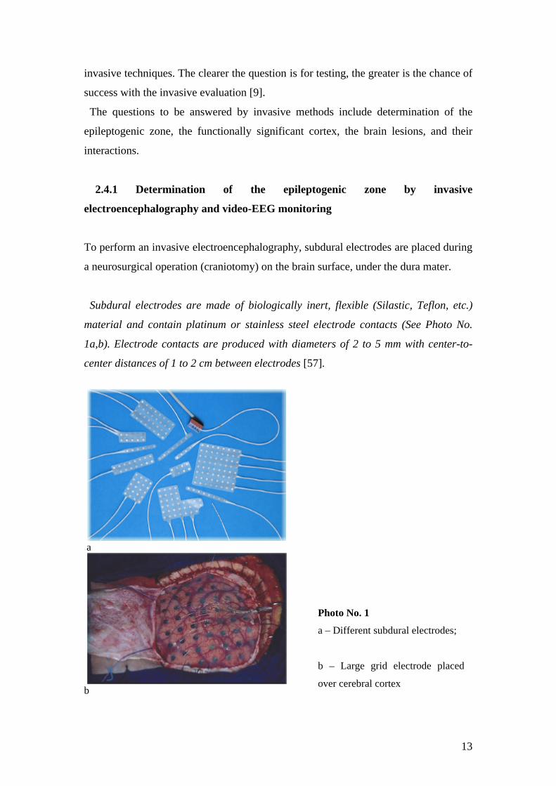
Subdural electrodes are made of biologically inert, flexible (Silastic, Teflon, etc.)
material and contain platinum or stainless steel electrode contacts (See Photo No.
1a,b). Electrode contacts are produced with diameters of 2 to 5 mm with center-to-
center distances of 1 to 2 cm between electrodes [57].
a
b
Photo No. 1
a – Different subdural electrodes;
b – Large grid electrode placed
over cerebral cortex

14
There are two types of subdural electrodes – strip and grid electrodes. They differ in
the number of encompassed electrode lines. Strip electrodes contain only one line of
electrodes, from 5 to maximally 16 cm long. Grid electrodes contain up to 10 lines of
electrodes of different lengths, thus allowing coverage of broader cortical areas. The
decision to use one or the other is based on the pre-operative hypothesis (for example,
the width of the cortical area to be explored).
Accordingly, there are also differences in terms of the extent of surgery needed for
electrode placement. Strip electrodes can be placed through a simple burr hole,
whereas grid electrodes require a vaster craniotomy.
Depending on the extent of surgery, there are also limitations inherent in the
unilateral or bilateral placement of subdural electrodes. Strip electrodes can be
placed intracranially within a smaller area of surgery, thus carrying less risk for the
patient. Therefore these electrodes can be placed bilaterally, if needed. Subdural grid
electrode placement, in contrast, requires a broader craniotomy, carries more risks
for the patient, and can be performed only unilaterally. Therefore, grid electrode
placement necessitates an even stronger hypothesis of the epileptic zone location.
The main indications for invasive video-EEG monitoring can be divided into three
overlapping groups: to define (1) the extent and distribution of the epileptogenic zone,
(2) the epileptogenic zone versus structural lesion, if present, and (3) epileptogenic
zone versus eloquent cortex [40].
The main limitation for precisely defining the epileptic zone with invasive electrodes
is the fact that they can only cover a very limited portion of the brain [62].
The following are more detailed examples of instances that may require invasive intracranial
EEG monitoring:
• Seizures are lateralized but not localized (e.g., a left-sided, widespread frontal-
temporal onset);
• Seizures are localized but not lateralized (e.g., ictal EEG patterns that appear
maximally over both temporal lobes);
• Seizures are neither localized nor lateralized (e.g., stereotyped complex partial
seizures with diffuse ictal changes or initial changes obscured by artifacts);

15
• Seizure localization disagrees with other data (e.g., EEG ictal scalp data different
with neuroimaging [MRI, PET, SPECT] or neuropsychological data);
• The relation of seizure onset to functional tissue must be determined (e.g., seizures
with early involvement of language or motor function);
• The relation of seizure onset to lesion must be determined (e.g., dual pathology or
multiple intracranial lesions);
• Seizures are clinically suspected, but video-EEG is inadequate to define them (e.g.,
simple partial seizures with no detectable scalp EEG ictal discharge or suspected
epileptic seizures with unusual semiology that suggests psychogenic seizures
[pseudo-pseudo seizures]) [19], [40], [104], [122].
2.4.2 Determination of functionally significant cortex by cortical stimulation
If after completion of EEG registration, the results indicate a possibly resectable
epilepsy focus in the cortical region covered and we suppose functionally significant
zones to be located in close proximity, we can proceed to direct electrical stimulation
of the cortex in order to state the correct localization of the latter areas [66].
Direct intraoperative electrical stimulation is a safe, precise, and reliable method for
detecting functional cortical areas and white matter pathways [83], [85], [102]. It has
been the gold standard for mapping brain function in preparation for surgical resection
since the 1930s [83], [92]. This is mainly due to fact that false negative results
are intrinsically impossible. Indeed, each eloquent structure, whatever its actual role
in brain function, will be in essence electrically disturbed by direct electrical
stimulation, which thus induces an obligatory functional consequence [28], [68].
However, it is of utmost importance to use certain physical parameters (see below) in
cortical stimulation, since the slightest technical approximation can result in false
negatives [59], [111].
In cortical stimulation a small electrical current is passed through individual
electrodes, and any symptoms of interference with the cortical function are closely
observed [66], [82]. Stimulation is either by electrodes placed in subdural or
intracerebral space (extra-operative stimulation), or during the operation (intra-
operative stimulation). The cortical stimulation process is supposed to define

16
functionally significant cortical regions that should be preserved in epilepsy surgery.
On the basis of the results of cortical stimulation we can draw a map of cortical
representations of different functionally more and less significant areas. This is called
“cortical mapping”. According to such a cortical map of representations of
functionally significant cortex and earlier estimated epileptogenic zone, we can plan
the actual epilepsy surgery – if it is possible at all (to what extent), without damaging
significant cortical areas.
Since cortical stimulation mapping (either extra-operative or intra-operative) plays
an essential role in epilepsy surgery around language areas, we give here a short
history of cortical stimulation mapping, and also describe the physics on which it is
based.
2.4.2.1 History of general nerve cell stimulation
There is little agreement between the data and opinions appearing in the literature, as
to who first discovered nerve cell excitability and who first actually performed brain
stimulation. However, according to reliable data, the first scientists to discover nerve
cell excitability were Luigi Galvani (See photo No. 2) and Alessandro Volta (See
photo No. 3) in the 18th century [44].
Photo No. 2 Luigi Galvani Photo No. 3 Alessandro Volta
Galvani showed that the muscle could be made to contract if a zinc electrode
attached to the muscle and a copper electrode attached to the nerve were brought in

17
contact with each other. Galvani incorrectly concluded that the contractions were the
result of "animal electricity" released from storage in the muscle, only to return via
the closed zinc and copper path through the nerve. In 1793, one year after Galvani's
initial publication on "animal electricity", the Italian physicist Alessandro Volta
proposed that the electrical stimulus responsible for the contraction was due to
dissimilar electrical properties at the metal-tissue saline interfaces. It was not until
1800 that Volta conclusively proved that the stimulus was of electrical origin: the
voltage difference due to the unbalanced half-cell potentials of the zinc-saline and
copper-saline interfaces excited the neuromuscular preparation. The early work of
Galvani and Volta provided physiologists with a basic understanding of the
mechanisms of neural and muscular excitation. While the mechanistic details would
be filled in nearly 150 years later, it was clear that neural and muscular signals could
be generated and transported by electrical means [44].
Data on the first brain stimulation mention an Italian scientist Felice Fontana (See
photo No. 4), who worked in the beginning of the 19th century and was influenced by
Galvani and Volta.
Photo No. 4
Soon thereafter several groups of scientists started experiments on animal brain
stimulation. One of the first scientists to describe electrical stimulation of an animal’s
Using a series of voltaic cells, Fontana carried
out the first known human brain stimulation
experiments on cadavers, invoking facial spasms
in the recently deceased by applying the voltaic
cell to specific brain regions. When public
concern over his experiments led to a law
forbidding such work, Fontana responded by
continuing his work on living volunteers [44].

18
brain were Gustav T. Fritsch and Eduard Hitzig (See photo No. 5) in the year 1870.
Their work was entitled “Über die elektrische Erregbarkeit des Grosshirns”.
Photo No. 5 Fritsch and Hitzig
One of the clearest and most detailed early account of human brain stimulation was
published in 1874 by the American physician Roberts Bartholow (See photo No. 6),
who stimulated the cortex of the 30-year-old patient Mary Rafferty.
Photo No. 6 Roberts Bartholow
The predecessors of Fritsch and Hitzig
did not resolve the critical question of
whether the cerebral cortex could be
electrically excited. Their demonstration
that it was electrically excitable is
considered one of their major
contributions. Perhaps the greatest
importance of their research, however,
was its contribution to the theory that
functions are localized in the brain [113].
She was said to be of good health until
an ulcer appeared on her scalp a little
more than a year before she was
admitted to the hospital. Mary's ulcer
was attributed to the "friction of a piece
of whalebone in her wig and the skull is
eroded and has disappeared over a
space of two inches in diameter, where
the pulsations of the brain are plainly
seen". Bartholow reported on a series of
six observations, during which needle
electrodes caused a mechanical
stimulation. Stimulation was performed
in varying depths and current strengths.
The results varied from no response to
19
distinct muscular contractions, very evident pain, great distress, and finally – to loss
of consciousness and violent convulsions. Later the publication of his observations
resulted in Bartholow's being forced to leave Cincinnati [113].
2.4.2.2 History of intraoperative cortical stimulation
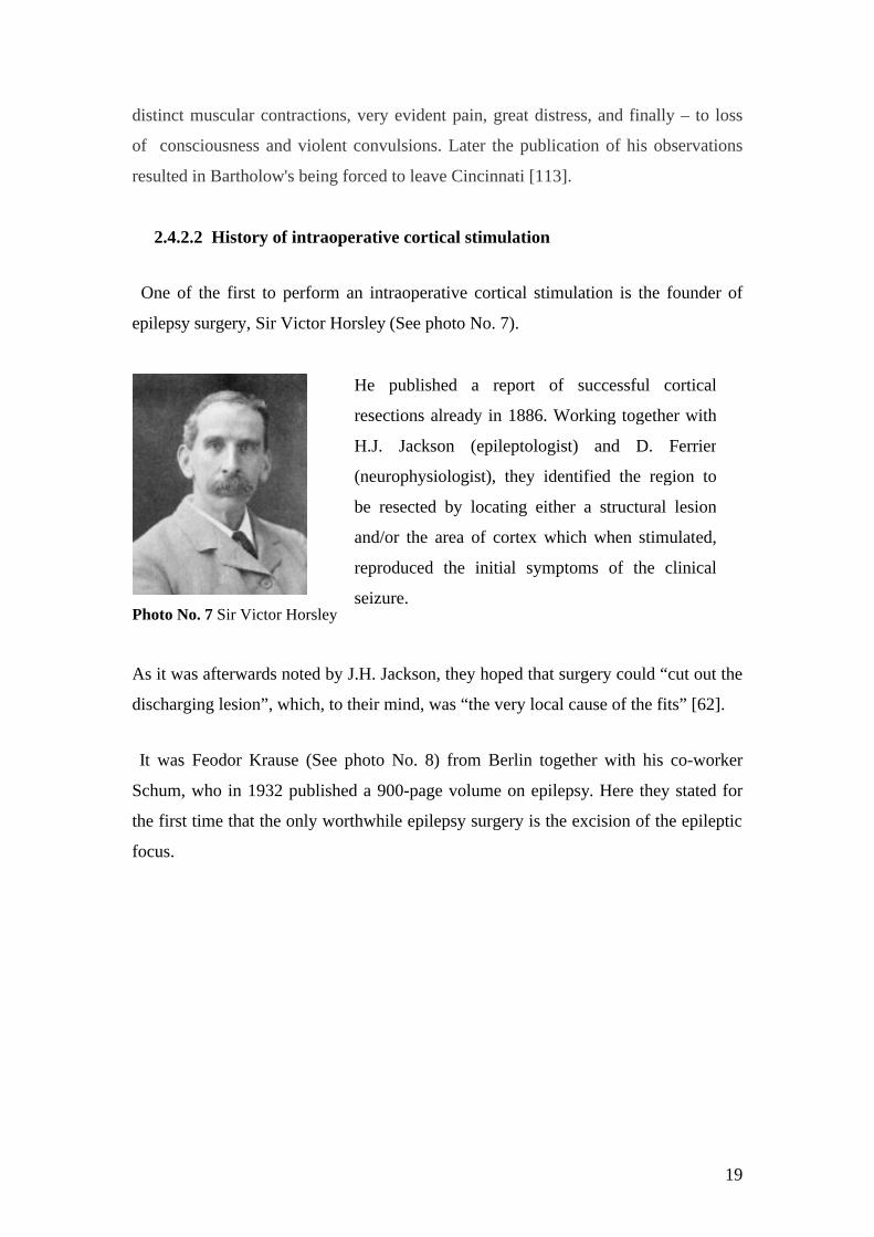
One of the first to perform an intraoperative cortical stimulation is the founder of
epilepsy surgery, Sir Victor Horsley (See photo No. 7).
Photo No. 7 Sir Victor Horsley
As it was afterwards noted by J.H. Jackson, they hoped that surgery could “cut out the
discharging lesion”, which, to their mind, was “the very local cause of the fits” [62].
It was Feodor Krause (See photo No. 8) from Berlin together with his co-worker
Schum, who in 1932 published a 900-page volume on epilepsy. Here they stated for
the first time that the only worthwhile epilepsy surgery is the excision of the epileptic
focus.
He published a report of successful cortical
resections already in 1886. Working together with
H.J. Jackson (epileptologist) and D. Ferrier
(neurophysiologist), they identified the region to
be resected by locating either a structural lesion
and/or the area of cortex which when stimulated,
reproduced the initial symptoms of the clinical
seizure.

20
Photo No. 8 Feodor Krause
Thus, together with Sir Horsley, Krause seems to have been the first one to
systematically stimulate the human motor cortex during epilepsy surgery. In his work
Krause included a detailed functional map of the motor strip, which was based on
stimulation results from 142 operations. He also advocated monopolar faradic
stimulation and described the method in detail, because he felt it induced less severe
seizures than galvanic stimulation, which was more favored by O. Foerster (See photo
No. 9), another very prominent personality in the history of epilepsy surgery.
Photo No. 9 Otfrid Foerster Photo No. 10 Wilder Penfield
The earliest stimulation Krause performed took
place on 16 November 1893. The patient was a
15-year-old girl, who suffered from Jacksonian
seizures and Jacksonian status starting at age 3. It
was due to a postencephalitic cyst following
meningitis at the age of 2. After removal of the
cyst, the patient remained seizure free for the rest
of her life and also markedly improved in her
mental performance.

21
It was Otfrid Foerster, together with Wilder Penfield (See photo No. 10), who in 1930
produced a less detailed, but much more extensive cortical map than that of Krause
[125]. He also had a much keener and more detailed interest in the semiology of
seizures and its localizing significance. This provided important information for
epilepsy surgery in the time before the development of EEG and the
electrocorticogram [126]. While Foerster initially used cortical mapping to identify
motor and sensory cortex, Penfield and colleagues subsequently applied the technique
to identify language cortex, with the goal of sparing these functional areas from
resection.
2.4.2.3 History of extraoperative cortical stimulation
The first brain electrode implantation took place in the early 1940s, followed in
1946 by the introduction of the first stereotactic instrument for use in humans by
Spiegel and Wycis [41]. Large subdural grids were introduced and systematically
produced beginning in the 1980s. They have had a major impact on identifying
patients who are eligible for surgery [1], [66].
2.4.2.4 First steps of epilepsy surgery close to speech areas
In the early years of focal epilepsy surgery, patients with seizures that arose from the
left hemisphere were refused surgical treatment, unless it was certain that the lesion
was located in the anterior of the frontal lobe or in the posterior of the occipital lobe.
Any other area in the left hemisphere was considered “forbidden territory” for fear of
producing postoperative aphasia [93]. The clinical use of cortical stimulation mapping
for language began with Wilder Penfield and colleagues in the 1940s. Due to
Penfield’s innovative technique of cortical language mapping, surgical treatment
became a viable treatment option for numerous patients who had not been helped by
pharmacological treatment of epilepsy. Thus, the implementation of cortical
stimulation was the starting point for epilepsy surgery close to speech areas.
22
2.4.2.5 The physics of cortical stimulation
As mentioned before, the use of certain physical parameters in direct cortical
stimulation is of utmost importance, because the slightest technical approximation can
result in false negatives. As noted by Taylor and co-workers, if the intensity of
stimulation is too low, if the duration is too short, or if a stimulation is performed
during a transient post-epileptic refractory phase, an erroneous “negative mapping”
may result [111].
2.4.2.5.1 Current spread and tissue excitability
There are two very important physical properties that play an important role in
electrical stimulation of brain tissue. These are current spread and tissue excitability.
Both of these issues have been investigated by several methods (single-cell recording,
behavioral methods, and neuroimaging) [112].
2.4.2.5.2 Current spread
It is commonly accepted that the initial segment and the nodes of Ranvier are the sites
at which a neuron can be directly activated by electrical microstimulation [36], [76],
[77], [99]. These zones contain the highest concentrations of sodium chanels, thus
making them the most excitable segments of a neuron [18], [76], [77].
The amount of current injected through a microelectrode to directly activate a
neuron (cell body or axon) is proportional to the square of the distance between the
neuron and the electrode tip.
This is expressed as:
I – the current level (µA)
I = Kr2 r – distance (mm)
K – excitability constant (µA/mm2)
This relationship is derived from studies of cortical and corticospinal neurons of rats,
cats, and primates [4], [69], [78].

23
The effective current spread from an electrode tip can be expressed as the square
root of the current divided by the square root of the excitability constant (I/K) 1/2. This
relationship is illustrated in Fig. 1.
Current spread and excitability properties of pyramidal tract neurons determined using single-cell recordings within motor
cortex of the cat [122].
0
0,2
0,4
0,6
0,8
1
1,2
0 200 400 600 800 1000 1200 1400
Current (µA)
Dist
ance
(mm
)
Fig. 1: Radial distance (in millimeters) of a direct activation of pyramidal tract neurons
using the equation radial distance = (K/I)1/2. The curve represents the amount of current
required for the antidromic elicitation of an action potential 50% of the time using a single
cathodal pulse of 0.2 ms duration. The average K value was 1,292 µA/mm2 for12-cell studies.
Fig.1 shows that the higher the current used, the larger is the current spread.
Another important factor influencing current spread is the conduction velocity of
axonal elements. The conduction velocities of myelinated pyramidal tract neurons
range from 3 to 80 m/s, with the largest of these neurons exhibiting the highest
velocities [13], [23], [67]. The conduction velocities of small unmyelinated cortical
fibers are <1 m/s [78]. Thus, the excitability constant (the constant reflecting the
excitability of a neural element 1 mm away from the electrode tip) derived with a 0.2
ms pulse can be as low as 300 µA/mm2 for the largest myelinated cortical neurons and
as high as 27,000 µA/mm2 for the smallest unmyelinated cortical neurons [78], [109].

24
This explains why large myelinated cortical neurons are easier to excite than small
unmyelinated cortical neurons.
The current spread characteristics have always been a subject of debate – to what
extent current spreads through directly activated neurons subcortically and to what
extent through transynaptic or lateral connections. The most precise responses are
achieved through direct cortical – subcortical activation, but at the moment of
stimulation there is also an indirect current spread laterally, which can involve more
distant cortical areas and give some false-positive responses. To assess the functional
localizing value of cortical stimulation, we have to know the extent of the direct and
indirect neuronal activations.
Several factors let us assume that current spreads mainly in a direct (cortical -
subcortical) way. First, there is the scientifically based fact that lateral connections
within the cortex are often unmyelinated and therefore much less excitable [78],
[110]. Second, microstimulation activates the most excitable elements in the cortex,
that is, by and large the fibers of the pyramidal cells, which project subcortically
rather than laterally [112], [78], [13], [23]. Third, microstimulation of the neocortex
evokes precise responses because directly activated neurons make more significant
contribution to the evoked response. This is due to fact that these neurons are more
synchronously activated in contrast to neurons further away from the electrode tip
which are activated transynaptically in the cortex [114].
Using a modern diagnostic tool, like functional MRI, scientists recently recorded
higher current lateral spread, which is contradictory to data published earlier. An
obvious reason for these differences is the appreciably larger currents and longer train
durations used in the fMRI study [114].
2.4.2.5.3 Estimates of excitability and strength – duration functions
To deduce the excitability of stimulated neurons, current can be traded-off against
pulse duration to elicit some response [3], [4], [5]. Normalized strength –
duration functions for pyramidal tract neurons are illustrated in Fig.2.

25
Fig. 2: Normalized strength – duration functions of pyramidal tract neurons [4], [109].
As the pulse duration is increased, the amount of current needed to evoke an action
potential 50% of the time diminishes to an asymptotic level; this level is called the
rheobase current.
The excitability or chronaxie of a stimulated element is expressed as the pulse
duration at twice the rheobase current. The shorter the chronaxie, the more excitable
is a directly stimulated neural element (shorter pulse duration is necessary for their
activation). Chronaxie depends on the characteristics of the tissue being stimulated,
specifically on its impedance. The few studies in this area have produced resistance
values of 250 Ohms for gray matter, 500 Ohms for white matter, and 65 Ohms for
cerebrospinal fluid [72]. Axons have shorter chronaxies than their cell bodies (axons:
0.03 – 7 ms; cell bodies: 7 – 31 ms [76]), and large, myelinated axons have shorter
chronaxies than small, nonmyelinated axons (large: 0.03 – 7 ms; small: >1.0 ms
[60],[97], [119]). Moreover, impedances can be modified in patients in an awake or
anesthetized state. Also any pathological process, whether lesional (tumor) or non-
lesional (epilepsy, post-ictal status), can interfere directly with the tissue’s excitability
[48]. Research on current spread and excitability investigations is still continuing.
2.4.2.6 Stimulation parameters
Cortical stimulation produces clinical effects only when very special stimulation
parameters are used. The four following essential factors must be considered [63]:
• stimulus intensity;
• duration of each individual stimulus;
• stimulation frequency;
• duration of the stimulus train.

26
2.4.2.6.1 Stimulus intensity (voltage or amperage)
Ideally the stimulation intensity should be strong enough to produce significant
depolarization (or hyperpolarization) of all the neurons underlying the stimulating
electrode but without affecting surrounding brain tissue or producing brain damage. A
stimulation of 15 mA seems to accomplish this. There are various reasons why the
“ideal” stimulation intensity of 15 mA can frequently not be used. The main reason is
that afterdischarges and painful or unpleasant sensations are produced by
electrical stimulation [66].
Afterdischarge per definition is “the portion of the response to stimulation in a nerve
which persists after the stimulus has ceased and consists of rhythmic, high-voltage,
high-frequency spikes, sharp waves, or spike-wave complexes which occur at the
region stimulated and are distantly different from background activity” [17].
Afterdischarges can be triggered only in certain circumstances, for example, if
electrical stimulation is at sufficient intensity, has a repetitive rate, and is of certain
duration. Initially they tend to be limited to the stimulating electrode, but they often
spread to adjacent electrodes, activating extensive cortical areas. The symptomatology
elicited when afterdischarges are triggered is not only an expression of the area
directly stimulated electrically but also of the whole region activated by the
afterdischarges. Therefore, in such cases we cannot be sure if the response at the
electrode site, where the afterdischarges are elicited, is due to the stimulation or if it is
produced by the afterdischarge. Consequently only those symptoms and signs elicited
by stimuli that do not produce afterdischarges are counted.
In some cortical sites even quite low intensities (for example, 2 mA) produce
striking positive effects, such as muscle twitches. Clinical trials warn that too high a
stimulus intensity could cause tissue damage due to excessive heat, produced
especially by hydrolysis; or “leaking” of the intracellular current, which goes from the
anode to the cathode through the cytoplasm, posing a risk of lesion to the
mitochondriae and the endoplasmic reticulum; or even alter the homeostasis if
neurons are activated in a manner that is too repetitive and synchronous [127].
Usually the initial stimulus intensity is very low. It is gradually increased until a
positive response, afterdischarges, or the maximum intensity is reached.

27
2.4.2.6.2 Duration of each individual stimulus
The duration of each individual stimulus in cortical stimulation varies from 0.1 to 0.3
ms [106]. Usually it is 0.2 ms.
2.4.2.6.3 Stimulation frequency
Single stimuli produce functional effects only at very high intensity. Repetitive
stimulation, most probably due to temporal facilitation, produces functional
alterations at a much lower intensity [100]. The ideal stimulus frequency (stimulus
frequency producing clinical effects at the lowest effective stimulus intensity) is
approximately 15 to 50 Hz.
2.4.2.6.4 Duration of the stimulus train
Repetitive electrical stimulation and relatively low stimulus intensities frequently
trigger clinical symptoms after a variable delay of 1 to 3 seconds. The temporal
summation of stimuli of the human cortex is an essential factor in the generation of
clinical symptoms. It is necessary to note that with longer stimulation durations, the
effect of the stimulation on both positive or negative symptoms not infrequently tends
to diminish after 5 to 10 sec of stimulation (due to alternative pathways [58] or
cortical adaptation) [66]. Usually the cortex is stimulated either until there is a
positive effect or the maximal timing (15 seconds) is reached [63].
2.4.2.7 Characteristics of a stimulus
Normally a biphasic stimulus is used for cortical stimulation. It is not as effective as
a monophasic (sinusoidal) stimulus, but it is safer for the brain, since the second
stimulus phase inverses the effects of the first.
If sinusoidal impulses were used for stimulation, they would increase the threshold
needed to be reached in order to generate the impulse (because the neural structures
are kept in a state of infraliminar depolarization). This phenomenon is known as
“accommodation”. “Accomodation” carries the risk of inducing a cerebral lesion due

28
to the accumulation of negative charge at the level of the cathode or the production of
metal ions at the level of the anode. Therefore, rectangular (biphasic) impulses
are recommended [2], [66], [68].
2.4.2.8 Physiological concerns of cortical stimulation
Electrical stimulation of the human cortex is the best experimental model of the
effect of activation of the cortex by an epileptiform discharge [64].
Contrary to mapping of the rolandic cortex, language cortex mapping depends on the
electrical blockade of cortical function rather than on eliciting function [83].
Electrical stimulation generates membrane excitability (membrane potential (MP) of
the neuron at rest varies between -60 mV and -100 mV) via an initial phase of passive
modification of local MP at the level of the cathode (the negative electrode). Before
this happens, the inner side of the membrane becomes progressively less negative than
the outer side (the membrane becomes inversely hyperpolarized with regard to the
anode). The intensity of this phenomenon depends on the parameters of the
stimulations and of the characteristics of the membrane (as mentioned before, the
membrane can be more easily stimulated at the level of the initial segment of the
axon, at the level of fibers that are myelinized and of larger diameter) [50], [55], [61],
[96]. If the MP reaches the laminar depolarization threshold, a second phase occurs
that begins with the opening of voltage-dependent ionic channels, which allow entry
of Na+ ions, and which therefore invert the MP between +20 mV and +30 mV. A
secondary output of K+ ions, associated with an inhibition of the entering flux of Na+
ions, brings the MP back to its resting state. Once generated, this rapid sequence of
MP fluctuation – the action potential – is still the same, no matter what the stimulation
parameters are (law of “all or nothing”) [68].
The effect of stimulation is more or less strictly limited to the area of brain beneath
the two electrodes being stimulated. The current flow only reaches sufficiently high
current density to stimulate the brain at the two poles (electrodes) and their immediate
vicinity. (These considerations apply, however, only when no afterdischarges are
triggered by the stimulus.) [95]
It is important to point out that cortical stimulation, even in the primary
afferent/efferent cortical areas, has a highly non-physiological effect. This explains
why most effects of stimulation in cortical areas are non-physiological (paresthesias,
29
unusual motor movements, etc.). Also in associative cortical areas, the massive
synchronized activation or deactivation of neurons by the electrical stimulus is
extremely un-physiological [95]. The full details of the physiological basis of nerve-
cell activation by electrical stimulation, however, remain unclear.
2.4.2.9 Procedure of extra-operative EEG recording and cortical mapping
Grid electrodes in our clinic are mainly used for both - extra-operative EEG
recording and cortical stimulation, whereas strip electrodes are mainly used for EEG
recording alone - often in situations, when seizure lateralization is necessary and
electrodes must be implanted bilaterally.
Consecutive, extra-operative cortical mapping is indicated in cases when localization
of the detected epileptogenic zone is close to or overlaps with eloquent areas [19],
[40], [104].
The type, number, and position of the electrodes are determined by the location of the
suspected epileptogenic zone in each patient, according to data gathered from all non-
invasive investigations (pre-investigational hypothesis). After implantation of
subdural electrodes by means of surgery and possibly after monitoring in the intensive
care unit (depending on the extent of surgery), the patient is brought for further
observation, recording of EEG, and cortical stimulation to the epilepsy intensive
station. Meanwhile a CT scan has also been made to locate the subdural electrodes.
This scan is merged with pre-operative MRI images to yield a three-dimensional
picture of the precise electrode locations over the cerebral sulci [124] (See Picture No.
1).
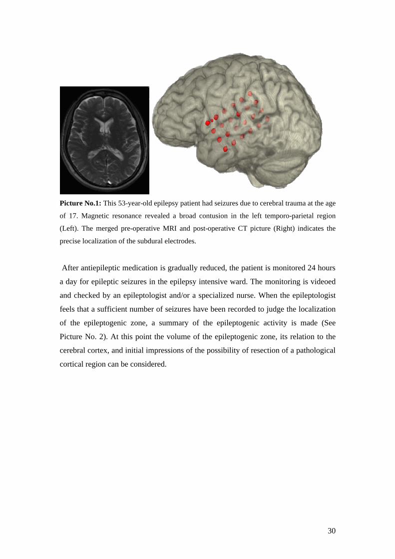
30
Picture No.1: This 53-year-old epilepsy patient had seizures due to cerebral trauma at the age
of 17. Magnetic resonance revealed a broad contusion in the left temporo-parietal region
(Left). The merged pre-operative MRI and post-operative CT picture (Right) indicates the
precise localization of the subdural electrodes.
After antiepileptic medication is gradually reduced, the patient is monitored 24 hours
a day for epileptic seizures in the epilepsy intensive ward. The monitoring is videoed
and checked by an epileptologist and/or a specialized nurse. When the epileptologist
feels that a sufficient number of seizures have been recorded to judge the localization
of the epileptogenic zone, a summary of the epileptogenic activity is made (See
Picture No. 2). At this point the volume of the epileptogenic zone, its relation to the
cerebral cortex, and initial impressions of the possibility of resection of a pathological
cortical region can be considered.

31
The next stage is the localization of functionally significant cortex - cortical
stimulation/ mapping. The physical parameters of stimulation are shown in Tab.1.
Physical parameters Unit
Stimulus intensity 1 – 15 mA
Duration of each individual stimulus 0.2 ms
Stimulation frequency 50 Hz
Duration of the stimulus train 5 – 15 sec
Tab. 1: Physical parameters used in extra-operative cortical mapping.
Summary of Seizure Origin
1
7
12
2
3
4
5
6
18
13
19 20
21
22
23
24
135
2
1
1
1
Picture No. 2: Summary of seizure origins (overall 17 seizures were recorded in this case) in the same 53-year-old epilepsy patient after invasive EEG registration by subdural electrode.
32
At the beginning, each pair of electrodes on the grid are stimulated and the reaction
is observed. In this way the “reference electrode”, where no function has been
triggered, is found. Later all the other electrodes are stimulated with reference to this
one electrode.
Initially the cortical stimulation begins at a minimal current strength and duration
(for example 1 mA for 5 sec.) and continues until some response, afterdischarges, or
maximal current strength – 15 mA is reached. During the stimulation the patient has
to perform certain tasks, depending on the stimulated zone (expected) and the
observed response. The main tasks include motor activities (moving arms and
fingers); also neuropsychological tests (naming several objects presented, counting
numbers or months of the year; reading aloud from a book or journal, sorting different
objects by their colour, shape, etc.). If any changes in these actions are observed or the
patient reports any uncustomary feelings, more detailed tasks to clarify this response
are required. Symptoms during stimulation may include positive motor phenomena
(tonic or clonic contraction of muscle groups), negative motor phenomena (inhibition
of voluntary movements of the tongue, fingers, or toes), somatosensory phenomena
(tingling, tightness, or numbness of a part of the body), or language impairment
(speech hesitation or arrest, anomia, or repetitive difficulties) [9]. Sites where
stimulation produces consistent speech arrest or anomia (anomia - impaired recall of
words with no impairment of comprehension or the capacity to repeat the words) are
considered essential to language function [106].
A significant response is considered to be any response during stimulation which is
observed or which is noted by the patient during at least three consecutive
stimulations at the same cortical site.
The duration of invasive monitoring greatly depends on the seizure frequency, the
success of any planned stimulation, and patient compliance [41].
By combining acquired stimulation results with the previous localization of the
epileptogenic zone on the 3-dimensional cortical picture, we obtain a reflection of the
relation between the epileptogenic zone and functionally significant cortex in the
investigated cortical region. On the basis of these data, we make the final decision
about resective surgery and estimate the resection borders (See Picture No. 3).

33
Summary of extra-operative language mapping and suggested resection
1
7
12
2 3
4 5
6
18
13
19 20
21
22 23
24
Speech Motor function of Face and Tongue Negativ mot. Face & Tongue
Suggested resection
Three speech points on the border of resection
Picture No. 3: Summary of extra-operative investigation by subdural electrodes in the same 53-year-old epilepsy patient. Extra-operative language mapping revealed 3 language points located on the border of epileptogenic zone. An intra-operative language mapping was performed to validate the border of maximal cortical resection.
34
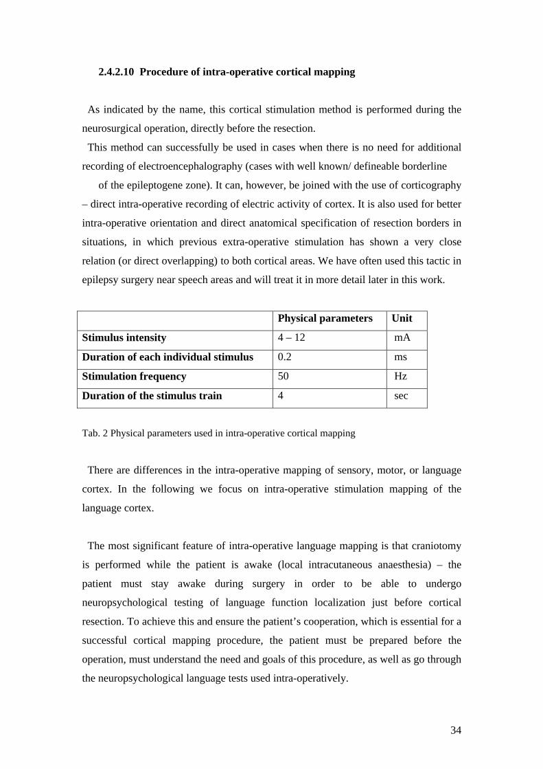
2.4.2.10 Procedure of intra-operative cortical mapping
As indicated by the name, this cortical stimulation method is performed during the
neurosurgical operation, directly before the resection.
This method can successfully be used in cases when there is no need for additional
recording of electroencephalography (cases with well known/ defineable borderline
of the epileptogene zone). It can, however, be joined with the use of corticography
– direct intra-operative recording of electric activity of cortex. It is also used for better
intra-operative orientation and direct anatomical specification of resection borders in
situations, in which previous extra-operative stimulation has shown a very close
relation (or direct overlapping) to both cortical areas. We have often used this tactic in
epilepsy surgery near speech areas and will treat it in more detail later in this work.
Physical parameters Unit
Stimulus intensity 4 – 12 mA
Duration of each individual stimulus 0.2 ms
Stimulation frequency 50 Hz
Duration of the stimulus train 4 sec
Tab. 2 Physical parameters used in intra-operative cortical mapping
There are differences in the intra-operative mapping of sensory, motor, or language
cortex. In the following we focus on intra-operative stimulation mapping of the
language cortex.
The most significant feature of intra-operative language mapping is that craniotomy
is performed while the patient is awake (local intracutaneous anaesthesia) – the
patient must stay awake during surgery in order to be able to undergo
neuropsychological testing of language function localization just before cortical
resection. To achieve this and ensure the patient’s cooperation, which is essential for a
successful cortical mapping procedure, the patient must be prepared before the
operation, must understand the need and goals of this procedure, as well as go through
the neuropsychological language tests used intra-operatively.
35
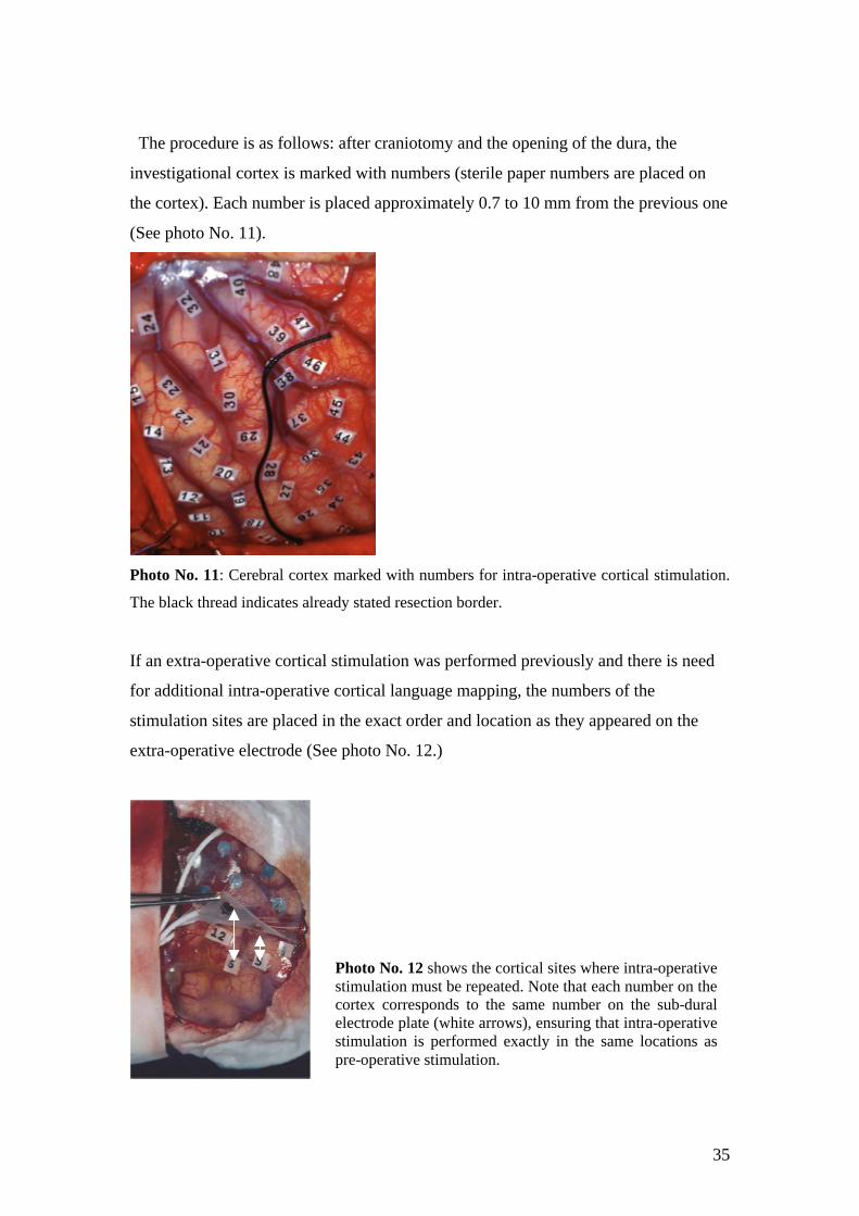
The procedure is as follows: after craniotomy and the opening of the dura, the
investigational cortex is marked with numbers (sterile paper numbers are placed on
the cortex). Each number is placed approximately 0.7 to 10 mm from the previous one
(See photo No. 11).
Photo No. 11: Cerebral cortex marked with numbers for intra-operative cortical stimulation.
The black thread indicates already stated resection border.
If an extra-operative cortical stimulation was performed previously and there is need
for additional intra-operative cortical language mapping, the numbers of the
stimulation sites are placed in the exact order and location as they appeared on the
extra-operative electrode (See photo No. 12.)
Photo No. 12 shows the cortical sites where intra-operative stimulation must be repeated. Note that each number on the cortex corresponds to the same number on the sub-dural electrode plate (white arrows), ensuring that intra-operative stimulation is performed exactly in the same locations as pre-operative stimulation.

36
Then a direct cortical stimulation is performed with bipolar stimulation tweezers at
each of these points. Simultaneously, the patient is asked to name different objects
(visual naming test, indicating visual naming sites) presented on the computer screen
in front of him. The patient has to say a full sentence, for example “This is a dog”;
“This is a house”. In order to maximize the validity of the stimulation results, the
patient has undergone identical visual naming tests pre-operatively. Later intra-
operatively only those visual stimuli are used, for which there was no pre-operative
failure in naming.
Parallel to the stimulation, the patient’s verbal response is observed by the
neurophysiologist. Similarily as in extra-operative language mapping, sites where
stimulation produces consistent speech arrest or anomia are considered essential to
language function [106].
Stimulation is at first done sequentially at all points and then is repeated twice,
increasing the current strength each time, since some points give positive response at
only higher current strengths. The upper limit of current strength is 15 mA. For
patients in whom speech has already been extra-operatively mapped and additional
intra-operative language mapping is now indicated, the latter is normally performed
only in the region where there is a close relation between the epileptogenic zone and
language sites or where overlapping of both areas has been seen. Thus, the exact
borders of the language cortex can also be directly determined intra-operatively. In
cases where no speech was found extra-operatively and repeated intra-operative
stimulation is indicated to approve this, stimulation normally includes a broader area
of the cortex (all the cortex accessible in craniotomy) as in extra-operative language
mapping. This way of mapping language is also used in cases, in which only intra-
operative language mapping is performed. Only those cortical sites, where language
disturbances are found in all three consecutive stimulations, are considered essential
for language and are preserved during resection.
All essential language sites are registered, and the final summary of results provides
a direct anatomical image of the cortical representation of language sites, as well as
the resection border. The resection is then performed, while keeping a distance of 10
mm from the essential cortical locations.

37
Phase I (non-invasive)
Purpose:
• diagnosis non-epileptic vs. epileptic spells
• localisation of the epileptogeniczone
Phase II (invasive)
Purpose:
• localisation and extent
of the epileptogenic zone
• History
• Neurological examination
• EEG-video monitoring
• Neuropsychological examination
• MRI, SPECT, PET
epileptic ? non- epileptic ?
• convergence of results ?
• resectable focus ?
yesno
• subdural electrodes
Phase III (surgery)
• Vagal stimulation
• Corpus Callosotomy
Resection of theepileptogeniczone
epileptogeniczone overlappingwith or adjacent to eloquent cortex
resectableepileptogeniczone
• epileptogeniczone not localized
• multifocal
cortical stimulation
• new hipothesisabout seizure origin
• no resective surgery
Pre-surgical evaluation of epilepsies
results still unclear
Drawing Nr 1: Algorithm of epilepsy surgery
38
3 The Operation – Cortical Resection
After detailed and in-depth pre-surgical investigation, a decision is made as to
whether it is possible to resect an epileptogenic zone and to what extent. Once a
cortical resection operation is considered justified, the operation is performed.
Operations around the language cortex typically include a resection of neocortex
in which the epileptogenic zone is found. A resection margin of 1 cm away from the
essential language site is currently considered satisfactory for functionally safe
(regarding language) surgery [12].
To better illustrate the implementation of the above-mentioned measures in epilepsy
surgery around speech areas and to present its complexity and results, as well as to
analyze the best tactic of language mapping in this surgery group, we have
summarized our 10 years of experience with this subgroup of epilepsy patients.
4 Hypothesis of the study:
Since the surgical tactics in this epilepsy surgery subgroup are mostly shaped by the
data gathered in language mapping, the accuracy of invasive language mapping is of
utmost importance. We have used two different tactics for language mapping, our
decision based on the significance of conflict between epileptogenic and language
areas. Habitually the language was mapped by extra-operative method alone. In those
cases, where very close relationships (less than 10 mm) between epileptogenic zone
and speech cortex or overlapping of both areas was seen in extra-operative mapping,
additional intra-operative language mapping was used.
We hypothesize that additional intra-operative language mapping is beneficial for a
better postoperative language and seizure frequency outcome in cases in which a close
relation between epileptogenic and language areas had been detected in previous
extra-operative language mapping.

39
5 Apart from confirming our hypothesis, we also sought answers to several other
questions:
1. What is the common investigational characteristic of epilepsy surgery patients
whose epileptogenic zone is close to neocortical language areas?
2. How are cortical mapping techniques typically used in this group of epilepsy
surgery patients?
3. What results as regards post-operative language outcome are seen in the whole
group of patients with epileptogenic zone around speech cortex and what
results are seen in both language mapping subgroups (extra-operative and
combined extra- plus intra-operative cortical mapping)?
4. What results as regards seizure outcome are seen in the whole group and two
different language mapping subgroups? Does the combined language mapping
technique influence post-operative results as regards seizure control?
6 Therefore the following goals of the study were stated:
1. To summarize 10 years of experience in epilepsy surgery around speech areas
in the Neurosurgery Clinic of Munich University Hospital, Grosshadern;
2. To analyze the use of pre-surgical investigation methods in this group of
patients;
3. To compare the use of two invasive language mapping techniques in two
different groups of patients (extra-operative versus combined extra-intra
operative);
4. To compare the results of both language mapping methods per se;
5. To analyze the post-operative results as regards language function in the whole
group of patients and compare them in both invasive mapping groups;
6. To appraise our indications for using extra-operative or combined extra- and
intra-operative language mapping tactics (these indications are stated in the
following section “Investigation of language function and cortical language
mapping”);
7. To analyze the post-operative results as regards seizure control in this group of
patients and both subgroups of cortical stimulation;

40
8. To estimate the percentage of situations in which the epileptogenic zone could
not be fully resected due to overlapping or close relationships with language
cortex;
9. To discuss our results and possible measures for their improvement.
7 METHODS
7.1 Patients
Between September 1997 and June 2007, a total of 22 medically refractory epilepsy
patients whose epileptogenic zone was close to the speech areas underwent
operations. In all cases the primary reason for neurosurgical treatment was medically
refractory epilepsy that significantly influenced the patient’s quality of life. However,
in one case a low-grade astrocytoma had been diagnosed pre-operatively, in another
case a low-grade astrocytoma had been diagnosed post-operatively, and in one other
case operation for a dysembryoplastic neuroepithelial tumor (DNET) had been
repeated. Four patients underwent repeated operations for epilepsy.
All patients, except one, were examined with both non-invasive and invasive
methods described earlier. In one case only non-invasive investigations were used. In
this case speech mapping had been done by functional magnetic resonance imaging
(f-MRI) for an insular cavernoma, diagnosed as the cause of the epileptic seizures. It
was well confined and safely (with regard to language function) accessible by
neuronavigation, when combined with f-MRI data. There was thus no need for
additional invasive investigations.
We included in our study only those cases in which
• positive speech points were found during direct language mapping;
• these speech points were located close to the epileptogenic zone (in the
majority of cases detected by direct subdural EEG recording).
Five patients had an epileptogenic zone located close to frequently described
language sites (posterior portion of Gy Frontalis superior, Gy angularis), but we did

41
not manage to find any positive speech point by direct cortical stimulation here. In
two of these cases only extra-operative cortical stimulation was used, and in three
cases a combination of extra- and intra-operative stimulation was used. Due to the
negative language mapping results (apparently no speech sites were located close to
the epileptogenic zone), these patients were not included in our study. None of these
patients had a post-operative language deficit.
Three patients who had needed invasive investigations, which proved unsuccessful,
were also excluded from the study. In one case the reason was a personal wish of the
patient to have the invasive electrodes removed after 12 days of invasive monitoring
when no seizures were registered. In another case subdural electrodes could not be
placed due to severe adhesions between the dura mater and the cerebral cortex. In the
third case the patient had a subdural hematoma following placement of the subdural
electrodes as a result of sudden drug-induced coagulation disorders. For reasons
of patient safety it was decided to remove the electrodes and not perform resective
surgery.
Three patients who underwent left hemisphere neocortical epilepsy surgery and in
whom language was localized on the right hemisphere (detected by the Wada test)
were also excluded from our study.
7.2 Investigation of language function and cortical language mapping
Language testing before and after the operation was performed by a
neuropsychologist, a neurosurgeon, and a neurologist. The neuropsychologist used the
Token test (part of Aachen Aphasia Test) to evaluate language. The neurologist and
neurosurgeon assessed language through everyday observations. This pre-operative
language assessment was done several days to weeks before the surgery.
Post-operative assessment of language was done during the hospitalization period
after surgery and in the following visits to the neurosurgeon (the same surgeon who
examined patient before and performed the operation) or the neurologist (first visit
normally 4 to 6 months after surgery or earlier if needed, next visit after every 4 to 6
months on average or earlier if needed). The patient was sent for repeated
neuropsychological evaluation (Token test) post-operatively if any kind of language
disturbance was detected by the neurologist or neurosurgeon or was reported by the
42
patient. If a patient still had disturbed language function 6 months after the last
resective surgery, it was classified as a permanent deficit.
The following language deficits were classified: anomia – patient cannot name
objects, but is able to repeat sentences and speaks fluently; expressive
aphasia – patient’s expression in speech or writing is impaired; receptive aphasia –
patient’s speech is fluent, but meaningless, the ability to understand spoken or written
words is also impaired.
All cases patients had left-sided language dominance.
The localization of cortical language areas was done invasively, except in one patient
(well-demarcated insular cavernoma, mapped by f-MRI).
In one case (pre-operatively known low-grade astrocytoma) only intra-operative
language mapping was used. Here non-invasive EEG investigations credibly indicated
tumor as an epileptogenic zone, and no further invasive EEG investigation was
necessary. The remaining 20 patients with an epileptogenic zone close to the cortical
speech areas can be divided into two groups. In one group only extra-operative
language mapping was used, in the other group a combination of extra- and intra-
operative cortical mapping was used.
Our indications for the use of either only the extra-operative or the combined extra-
and intra-operative cortical language mapping method are as follows:
1. Only extra-operative language mapping (Ex-M) was used in situations in
which language mapping (measured in 3-dimensional cortical maps) indicated
a distance of at least 10 mm between cortical language points and the
epileptogenic zone. In such situations this distance was assumed to be safe to
perform a resection with a diminished possibility of resection-caused damage
to language areas and subsequent permanent post-operative language
deterioration.
(It was also used for one patient, in whom part of the speech cortex overlapped
with the epileptogenic zone. Since the overlapping part of the language cortex
was identified as the basal temporal language cortex, it was considered as safe
for resection and no intra-operative mapping was performed. The case is more
profoundly presented later in the discussion chapter.)
43
2. Combined extra- and intra-operative language mapping (Co-M) was used in
situations, when language cortex found extra-operatively overlapped with the
epileptogenic zone, or the distance between these cortical regions was less
than 10 mm. Here there was an increased risk for resection-caused damage to
the language areas. Additional intra-operative language mapping was applied
in order to achieve the most precise resections, while at the same time
preserving a safe amount of language cortex. The next step was the precise,
maximal resection of the epileptogenic zone, keeping a distance of 10 mm
from the language cortex. Additional intra-operative cortical stimulation was
also used in a few cases, in which the results of extra-operative language
mapping appeared to contradict the data in the literature or our previous
experience. Thus, the repeated intra-operative stimulation was used here partly
to confirm the extra-operative language mapping data and partly as an
additional investigation to obtain more in-depth information.
In one case in which additional intra-operative language mapping was indicated, this
could not be done due to the youth (10 years) and psychological problems of the
patient.
The procedures of intra- or extra-operative stimulation, physical parameters, and
materials used are described in the corresponding above sections.
7.3 Neurological examinations and post-operative seizure outcome
Pre-operative neurological examination was done by both the neurosurgeon and the
neurologist; the pre-surgical seizure frequency was documented by the neurologist.
Post-operative follow-up was done by the neurosurgeon, neurologist, or both on a
regular basis (every 4 to 6 months or more frequently if needed). The post-operative
seizure frequency data were summarized, starting from 2 years after last resective
surgery, and were assessed using the Engel post-surgical seizure outcome scale [30] in
four classes: Class Ia – seizure free, Ib – only auras; Class II – rare seizures (not more
than 2 per year); Class III – worthwhile improvement (reduction of seizures by 85%
or more); Class IVa – significant reduction, IVb – unchanged seizure frequency.

44
In most cases (19; 86.3%) data were gathered retrospectively from in-patient
documents and out-patient letters. Some (3) prospective patients were assessed post-
operatively only as regards language function, since the post-operative control period
occurred after at least 6 months, but less than 2 years at the endpoint of this study
(12/2007).
7.4 Statistical analysis
The statistical analysis was done using Windows Excel program and Fischer’s Exact
Test. The “p” value was considered significant, if p < 0.05.
8 RESULTS
8.1 Characteristics of patients
The study included 11 men (50%) and 11 women (50%) with a mean age of 31.9
years (range 10 to 53 years). The mean duration of epilepsy was 16.3 years (range 6
months to 38 years). All 22 patients had medically refractory epilepsy and underwent
neurosurgical operation for resection of an epileptogenic cortex. In all of these
patients the language cortex was located near the epileptogenic cortex or directly
overlapped with it.
Four epilepsy patients (18.1%) underwent repeated operations for epilepsy. One of
them had undergone an operation for frontal arterio-venous malformation (AVM) and
needed additional frontal resection. Another had first undergone a neurosurgical
operation for left frontal cerebral abscess, and another operation later on for epilepsy
but had needed an additional frontal resection for seizure freedom. Two others had
had a previous resection of the temporo-mesial structures and in one case additional
temporal neocortical resection was necessary; the other required an additional frontal
neocortical resection.
8.2 Non-invasive pre-operative investigations
All the patients were examined neurologically by both the neurosurgeon and the
neurologist. Testing for speech function showed that one patient (4.5%) had an

45
insignificant, preoperative light dysphasia and one patient (4.5 %) light expressive
aphasia. Eight patients (36.3%) had short post-ictal aphasia, seen as a lateralizing sign
for language function.
All patients were examined by magnetic resonance imaging with the following
results: unspecified lesion (including changes after previous resective operation) – 8
(36.4 %), post-contusional cortical changes – 3 (13.7%), no visible pathology – 3
(13.7%), cortical dysplasia – 4 (18.2%), low-grade tumor 2 (9.0%), cavernoma – 1
(4.5%).
Non-invasive EEG-video recording was also used for all 22 patients.
Positron emission tomography (PET) was needed in 19 cases (86%). It was not used
in situations, when we had a strong pre-operative hypothesis of the epileptogenic
cortex location after non-invasive EEG-video investigation. In one case the patient
had insular cavernoma, another patient had had previous AVM resection, and one
patient was suspected to have an astrocytoma.
Single proton emission computer tomography (SPECT) was used for 10 patients
(45.4 %) when the previous search results for the epileptogenic zone were still not
persuasive.
As described before, a functional magnetic resonance imaging (fMRI) was used for
language mapping in one case, when insular cavernoma was the reason for the
epileptogenic seizures.
The Wada test for language lateralization and memory assessment was used for 8
patients (36.3%). The use of this invasive test has greatly declined in the last 7 years
due to its invasive nature and relatively high complication risk, as well as the
possibility of now using fMRI for language lateralization. We used the Wada test for
patients, in whom there was a strong possibility of speech dislocation due to long
persisting lesion and brain plasticity.
8.3 Invasive language mapping
Invasive investigations to locate language cortex were performed in 21 out of 22
patients (95.4%) (in one case language was mapped non-invasively by functional
magnetic resonance). (See drawing No. 2)

46
In one patient (4.5%) only intra-operative stimulation was done (a case of pre-
operatively known low-grade astrocytoma). In eight cases (36.4%) only extra-
operative language mapping was used. Both stimulation methods were combined for
12 patients (54.5%).
If we consider the applicability of both language mapping methods separately, the
following results are seen: extra-operative stimulation was used in 20 cases; intra-
operative stimulation in 13 cases. In all 20 extra-operatively examined patients,
positive speech points were found in 18 cases (90%). In all intra-operatively examined
patients positive speech points were found in 12 cases (92.3%). The statistical value
of differences in language finding by both methods was p=1.0 (insignificant).
8.4 Correspondence of extra- and intra-operative stimulation results
(in the Co-M group):
The results of both mapping methods corresponded in 9 cases (75%) out of 12. (If
we also add to this number those 3 patients, who had both language mappings and no
speech was found in either mapping (not included in our study), the results
corresponded in 12 cases (80%) out of 15.)
Three cases out of 12 had discordant results: in 2 cases (16.7%), no language cortex
was found extra-operatively. However, intra-operative language mapping showed
positive language points. In one case (8.3 %) (See Picture No. 3), some positive
18 12 1
Group in which language was mapped extra-operatively (Ex-M)
Group in which language was mapped extra- and intra-operatively (Co-M)
Language mapped intra-operatively
Language mapped non-invasively by f-MRI Drawing No. 2 The frequency of different language mapping methods used in our work.

47
language points found extra-operatively were stimulated also intra-operatively, but no
language function could be confirmed in these locations.
SUMMARY OF RESULTS: Variable Value Men 11 (50 %) Women 11 (500 %) Mean age 30.3 y - Age range 10 – 53 Duration of Epilepsy 16.3 y - Range 0.5 – 40 y Significant language deficite deficits pre-operatively - Light expressive aphasie aphasia 1 (4.5 %) Magnetic resonance 22 (100%) - Unspecified lesion 8 (36.4 %) (including changes after previous resective operation)
- Post-contusional changes 3 (13.7 %) - No visible pathology 3 (13.7 %) - Cortical Dysplasie Dysplasia 4 (18,2 %) - Low grade tumor 2 (9.0 %) - Cavernoma 1 (4.5 %) - Hippocampus sclerosis 1 (4.5 %) Non-invasive 24 h video – EEG 22 (100 %) PET 19 (86 %) SPECT 10 (45.4 %) f MRI 1 (4.5 %) WADA Wada test 8 (36.3 %) Invasive EEG rec./ language mapping 21 (95.4 %) - Only i/op mapping 1 (4.5 %)
SUMMARY OF RESULTS: Variable Value Resective operation 22 (100 %) - Frontal 8 (36.4 %) - Fronto-Parietal 2 (9.2 %) - Fronto-Temporal 1 (4.,5 %) - Temporal 8 (36.4 %) - Temporo-Occipital 1 (4.5 %) - Temporo-Occipito-Parietal 1 (4.5 %) - Parietal 1 (4.5 %) Pathology (n=20) - Sclerosis/ Gliosis 10 (50 %) - Dysplasie Dysplasia (Cortical, Glioneural) 7 (35 %) - TumourTumor (Astroc WHO II; DNET) 2 (10 %) - Cavernous angioma 1 (5 %) Post-operative complications 3 (13.5 %) - Meningitis 1 (4.5 %) - Subdural hematoma 1 (4.5 %) - Epidural hematoma 1 (4.5 %) Post Post-operative language deficite deficits 10 (45.4 %) - new permanent deficite deficits 1 (4.5 %) Post-surgical outcome regarding seizure control * (n-18) - Engel I 9 (50 %) - Engel II 0 - Engel III 2 (11.1 %) - Engel IV 7 (38.9 %)

48
8.5 Resective operations
All 22 patients underwent a resective operation. It included the following cerebral
lobes: frontal 8 (36.4%), fronto-parietal 2 (9.2%), fronto-temporal 1 (4.5%), temporal
8 (36.4%); temporo-occipital 1 (4.7%); temporo-occipito-parietal 1 (4.5%), parietal 1
(4.5%).
In 6 (54.5%) out of 11 temporal lobe resections, mesial temporal structures were also
removed. This was done in cases, in which invasive EEG investigations showed some
mesial epileptogenic activity.
Four patients (18.2%) underwent repeated operations for epilepsy.
In four cases (18.2%) of the total group, after repeated intra-operative stimulation of
language sites (all cases in Co-M group) there was clear overlapping of language
cortex with the epileptogenic zone. In these cases our inability to resect the
whole epileptogenic cortex had been clear before the actual resection.
In one young epilepsy patient, an additional intra-operative language mapping was
also indicated (language sites were very close to the epileptogenic zone). An operation
could not be performed due to the patient’s youth and psychological instability.
Therefore we had also assumed pre-operatively that we would not be able to resect a
full epileptogenic zone while preserving the language cortex intact. In another case,
no full resection of the epileptogenic zone (low-grade astrocytoma) was possible due
to the patient’s complaints of short eyesight disturbances and our observation of
horizontal nystagmus, while removing the rest of posterio-mesial temporal tumor in
awake brain surgery. Thus, in six cases (27.2 %) out of all, we knew that we had not
resected the complete epileptogenic zone at the endpoint of resective surgery. In all
the other cases we assumed that we had resected the compete epileptogenic zone.
Pathologically following diagnosis were made: Sclerosis/ Gliosis (50 %); Cortical /
Glioneural Dysplasia (35 %), Tumor (Astrocytoma WHO grade II, DNET) (10 %),
Cavernous angioma (5 %).

49
8.6 Post-operative results as regards language function
Overall, postoperative speech deterioration was noted in 10 cases (45.4 %) out of 22
(anomia – 5, expressive aphasia – 3, receptive aphasia – 1, combination of expressive
aphasia and anomia – 1 case). In all of these cases speech disturbances regressed soon
after surgery; however, only in 8 out of 10 patients did speech return to its pre-
operative performance either already during the hospitalization period or by the time
of the first check-up (4 to 6 months after surgery). In the remaining two cases a
complete regression of speech disturbances was not seen even after 6 months of
observation; these speech deficits were considered permanent. In one of these two
cases, language disturbance (light expressive aphasia) was noted already before the
resective operation; therefore, new permanent language deficit occurred in one case
(4.5%) of the whole group.
If we compare the post-operative language deterioration between the two groups
(Ex-M and Co-M), the results are as follows. Post-operative speech deterioration was
seen in 6 (75%) out of 8 patients in the Ex-M group (See Fig. No. 3) and in 4 (33.3%)
out of 12 patients in the Co-M group (See Fig No. 4). The statistical significance of
difference in post-operative language deterioration in both groups was p = 0.169
(insignificant).
A comparison of new, permanent language deterioration in both groups revealed one
case (12.5%) in the Ex-M group. One patient of the Co-M group had had permanent
language deterioration already pre-operatively. The statistical significance of
difference between the two groups as regards new, permanent post-operative language
deficit was p=0.4 (insignificant).

50
Postoperative outcome regarding speech function in the Ex-M group (n-8)
1
5
2
0%
10%
20%
30%
40%
50%
60%
70%
80%
90%
100%
1
No speech deterioration
Transient speechdeterioration
Permanent speechdeterioration
Fig. No. 3
Postoperative outcome regarding speech function in the Co-M group (n-12)
1
3
8
0%
10%
20%
30%
40%
50%
60%
70%
80%
90%
100%
1
No speech deterioration
Transient speechdeterioration
Permanent speechdeterioration
Fig. No. 4
8.7 Post-surgical outcome as regards seizure control
Post-surgical seizure outcome of 2 or more years after last resective epilepsy surgery
(mean follow-up period 46.6 months, range 24 to 96 months) was determined for 18
(81.8%) out of 22 patients. These were all only retrospectively analysed cases. Four
patients were not included in this summary of post-operative results. In one case the
seizure frequency had not declined since resective surgery, and a vagal nerve

51
stimulator had been implanted 1 year later. The remaining three patients hade not had
a sufficient follow-up period post-operatively by the endpoint of this study (12/2007).
The Engel classification data for 18 patients who underwent operations for epilepsy
close to speech areas are as follows (See Fig No. 5):
Engel I – 9 patients (50%) (Ia – 8 cases, Ib – 1 case); Engel III – 2 patients (11.1 %),
Engel IV – 7 patients (38.9 %) (IVa – 3 cases, IV b – 4 cases).
In six cases (27.2 %) we were not able to resect the complete epileptogenic zone and
stated it already during the resective surgery. Half of these cases were in the IVb
group, two cases in the IVa group (all together 5 cases). In one case the follow-up
period was too short (8 months) by the endpoint of our study (12/2007), this patient
was not included in the evaluation of seizure outcome.
1
Engel IEngel IIEngel IIIEngel IV
7
2
0
9
012345678
9
Postoperative outcome regarding seizure control at least 2 years after last resective operation (n-18)
Fig. No. 5
If we compare the post-operative outcome for seizure control in both groups of
different language mapping methods (extra-operative and combined language
mapping group), the results are as follows. In the Co-M group, data were compiled for
nine patients. Engel class I outcome was seen in three cases (33.3 %), Engel class II –

52
none, Engel class III – one case (11.1 %), Engel class IV – five cases (55.6 %). (See
Figure No. 6)
In the Ex-M group, data were compiled on seven patients. Engel class I outcome was
seen in four cases (57.1%), Engel class II – none, Engel class III – 1 (14.2%); Engel
class IV – two (28.7%). (See Figure No. 7)
The statistical significance of differences between Ex-M and Co-M groups, as regards
Engel class I outcome, was p=0.63 (insignificant).
5
1
03
0
1
2
3
4
5
Number of patients
Engel IEngel II
Engel III
Engel IV
Engel Score in Co-M group
Fig. No. 6: Post-operative outcome for seizure control (Engel Score) in combined language
mapping group.
Engel IEngel II
Engel IIIEngel
IV
2
1
0
4
0
1
2
3
4
Number of patients
Engel Score in Ex-M group
Fig. No. 7: Post-operative outcome regarding seizure control (Engel Score) for extra-
operative language mapping group.

53
8.8 Post-operative complications
The following post-operative complications were diagnosed: meningitis (4.5%),
subdural hematoma (4.5%), epidural hematoma (4.5%). All complications occurred in
cases in which large craniotomies with insertion of subdural electrodes had been
performed. All patients recovered well after the operations, and no permanent
neurological deficits were apparent.
8.9 The summary of statistical analysis data
The significance of differences between extra-operative (Ex-M) and combined (Co-
M) language mapping groups as regards postoperative language and seizure outcome:
Comparing difference as regards p value Difference estimated as
Immediate post-operative language
deterioration
0.169 insignificant
New, persistent language deterioration 0.4 insignificant
Engel class I outcome 0.63 insignificant
The significance of differences between extra-operative and intra-operative language
mapping technique as regards the identification of language sites:
Comparing difference as regards p value Difference estimated as
Identification of language sites 1.0 insignificant
54
9 DISCUSSION
9.1 Patients and non-invasive investigations
Our study demonstrates that epilepsy surgery around speech areas is a very complex
subgroup of epilepsy surgery. This complexity is reflected in the diversity of patient
characteristics and investigation results (significant age range, varying duration of
epileptic seizures, localization of epileptogenic zone, localization of speech cortex,
various pathological data, etc.). A significant variability in patient characteristics, in
their turn, makes difficult any decisions on the site and amount of resection as well as
prediction of post-surgical seizure control outcome.
The preservation of intact language areas and postoperative language function has
always been of primary importance in our work, even superior to the full resection of
the epileptogenic zone.
The suspicion that language cortex is located close to the epileptogenic zone must be
considered a serious, difficult condition for surgery, in which both good post-
operative seizure control and the simultaneous preservation of language function are
expected. Such proximity almost always necessitates vast pre-surgical investigations,
often including the most recent and expensive investigational methods available in
neurosurgery. This, of course, restricts the investigational process of epilepsy surgery
to only a few neurosurgical clinics.
One of the first serious factors that necessitates more detailed investigations is the
difficulty of determining the precise borders of lesions in magnetic resonance
imaging. Although it was possible to identify a certain lesion in 86% of our cases, in
most of these situations the lesion was rather diffuse, and we had to resort to
additional investigation methods to specify the exact borders of the epileptogenic
zone.
Non-invasive EEG also provided insufficient information about the topography of
the seizure focus. This investigation is considered too insensitive to be used for
determining exact borders of the epileptogenic zone [40], [108]. It also has a spatial
limitation - it can only record brain electrical activity in an area of about 6 cm2 [62].

55
In 80% of adult temporal lobe epilepsy cases (in mesial temporal lobe epilepsy, the
frequency rises to 90%), magnetic resonance and ictal non-invasive EEG recording
alone provide sufficient information for localizing the seizure origin and making a
decision about surgery. In contrast, epilepsy surgery around speech areas normally
requires the implementation of several additional investigational techniques to achieve
the same goal [33], [46]. Only in one case (4.5%) were we able to proceed with
surgery on the basis of MRI and EEG alone: the patient had a well-confined insular
cavernoma. In this case we were certain that the pathology corresponded to the
epileptogenic zone and the danger of damaging functionally significant language
areas was stated here as low. In the remaining 21 patients (95.5%) several additional
non-invasive (PET) and (SPECT) as well as invasive investigations were necessary to
more precisely delimit the epileptogenic zone. The danger of damage to the
functionally significant language cortex during the resective surgery was variable in
these cases.
An invasive Wada test was used in situations, in which non-typical speech
lateralization was suspected. Due to its invasive nature, we have increasingly
restricted the use of this investigation in our work over the last 7 years only to cases in
which the presence of epileptogenic lesion and epileptic seizures had been seen or had
been expected for a considerable time (especially since childhood). This strategy is
based on findings that early onset of left hemisphere seizure foci is associated with
altered language lateralization and increased incidence of right hemisphere dominance
[10], [47]. However, in our epilepsy surgery practice only three (2,9%) out of all 103
left hemisphere epilepsy surgery patients had a right-sided language representation. In
only one of these (epidermoid tumor, epileptic seizures since the age of 16, lasting for
15 years) could a reorganization of language cortex be validly suspected due to the
early pathology and long duration of the seizures. The second case involved a young
patient with left-sided hippocampal sclerosis. Seizures had first occurred at the age of
15 and lasted for 3 years. In this case it was hard to say if 3 years of epileptic seizure
history were sufficient for language function dislocation. We also do not have
information about the duration of the “silent period” – time interval between first
pathological changes in the hippocampus and their clinical manifestation. Therefore it
is not clear if the patient had had these pathological changes already for a longer time
and if this factor played a significant role in the displacement of language. The third
56
patient was 56 years old, had a left temporal lesion, histologically diagnosed as
dysplasia, and a 10-year history of epilepsy. These three patients, of course, were not
included in our study; their cases simply illustrate the complexity of deciding whether
to give the Wada test for language lateralization. With such few data we cannot
conclude that early development of an epileptic lesion and/or seizures is directly
connected with language dislocation to the opposite hemisphere. Duchowny and
colleagues also confirmed this, reporting that the language cortex tended to remain in
the left hemisphere, in proximity to, or even overlapping developmental lesions (e.g.,
dysplasia) and the epileptogenic cortex in patients with early seizure onset (age <5
years). Only very large early lesions acquired before age five and which
destroyed language cortex were associated with right hemisphere language [27].
In summary, we cannot infallibly prove that a long history of seizures and
epileptogenic lesion is a definite indication for the use of the Wada test in this group
of patients. The use of this test largely depends on the experience made with it in each
individual neurosurgery clinic.
A good alternative to the Wada test for language lateralization is functional
magnetic resonance imaging (f-MRI) [24]. In our study, however, it was not used for
language lateralization, but instead for non-invasive language mapping in one patient
with insular cavernoma as an epileptogenic lesion. Although Roux and co-workers
found that f-MRI cannot be used for making surgical decisions in the absence of
direct (invasive) brain mapping [102], we performed a resective operation without
direct cortical language mapping in this one case due to the fact that good intra-
operative orientation and preservation of the speech cortex were possible by
combining of neuronavigation and f-MRI data. No post-operative speech deficits
were seen in this case.
9.2 Invasive language mapping and post-operative language function
For the remaining 21 patients (95.5%) in our group, language was mapped
invasively by direct cortical stimulation. In one case epileptic seizures were caused by
a low-grade tumor. On the basis of previous non-invasive investigations, we were
quite sure that the epileptogenic zone corresponded with the tumor, and we saw no

57
indications for inserting subdural electrodes to record additional invasive EEG data.
Nevertheless, language mapping was needed and it was done intra-operatively during
awake brain surgery.
For the remaining 20 patients (91%), no well-confined cortical lesions (in 13.7% no
lesions at all) were seen, nor did other successive non-invasive investigations assure
us of precise epileptogenic zone boundaries. We did not consider it safe to proceed
directly with resective surgery with only intra-operative language mapping; therefore,
subdural electrodes were used to better specify the epileptogenic zone as well as for
successive language mapping.
Poor EEG localization of interictal spikes, seizure onset, the presence of a broad,
often ill-defined epileptogenic area, as well as the extension of the proposed area of
resection into brain areas of high functionality have also been mentioned as
significant problems in several other studies [90], [94], [123]. However, these studies
involved only extra-temporal epilepsy cases. In contrast, our study included also
temporal neocortical epilepsy cases that required additional invasive pre-surgical
investigations.
Thus, the majority of epilepsy surgery patients with an epileptogenic zone located
close to speech areas required apart from the usual resective operation, a complex
invasive investigation, including craniotomy, insertion of subdural electrodes, and 8 to
12 days of continued monitoring in the epilepsy intensive care unit.
The use of extra-operative or combined extra-intra-operative language mapping
techniques was in almost all cases based on the measure of overlap between
epileptogenic and language zones. The few exceptions will be discussed later.
Our assumption that epileptogenic areas, located at least 10-mm away from language
areas, are at a rather safe distance from each other and thus the maximal amount of
epileptogenic zone can be resected without significant fear of post-operative language
disturbances was based on data published by Haglund and colleagues. They reported
that a resection margin of >1 cm from the language area results in significantly less
permanent language deficits [37]. According to Silbergeld, any injury to essential
language areas will lead to permanent difficulties [106]. Thus, the strategy of leaving
58
a 10-mm resection margin from the language areas is also followed in centers with
considerable experience in neurosurgery around speech areas [12].
In the first group of patients (only extra-operative language mapping group (Ex-M))
we also included one patient in whom the basal temporal language area directly
overlapped with the epileptogenic zone (Picture No. 4). There was no need for
additional intra-operative specification of language cortex borders in this case. We
based our decision on data firstly published by Krauss GL et al,, who reported that
resection of basal temporal language areas does not cause permanent decrease in
naming in most cases and therefore might be an acceptable risk, when the goal is
treatment of severe partial epilepsy [53]. This particular patient directly developed
post-operative deterioration of the language function, but these changes were of a
regressive nature and at the time of first post-operative visit to the neurosurgeon (4
months after surgery) no more speech disturbances were observed.
59
Picture No 4 A 37-year-old patient with epileptic seizures due to cerebral contusion defect (trauma 10 years previously). Speech was detected at three temporo-occipito-basalpoints, two of which were located in the area of the planned resection. Due to the basal localization of these points and insignificant functional value, it was decided to perform resection without repeated intra-operative language mapping. Initially language disturbances were observed post-operatively, but speech returned to its pre-operative state 4 months after surgery.
Suggested resection
Three speech points, two posterior of themconsidered as basal speech points
It was necessary in the second group of patients (combined cortical mapping group
(Co-M)) to repeat intra-operative language mapping. We assumed that there was a
significant trade-off between maximal resection of the epileptogenic zone and
preservation of the language cortex (preservation of a margin of at least 10 mm from
the language cortex) due to the close relation between the two cortical areas. In our
opinion, intra-operative orientation and resection of the epileptogenic cortex could not
be performed here only on the basis of extra-operative invasive investigation data
(summarized in report with 3-dimensional image as seen in Picture No.3).
60
Interestingly, the initial post-operative data showed more frequent decreases of
immediate post-operative language performance (in 75% of the cases) in the Ex-M
group, although pre-operatively a rather in-significant trade-off between epileptogenic
and language cortex had been supposed. In contrast, the immediate post-operative
language performance in the Co-M group showed a decrease in only 33.3% of the
cases. In the majority of these cases, however, language deterioration was of a
regressive nature. Permanent language deterioration was seen in only one patient
(12.5%) in the Ex-M group and in one patient (8.3%) in the Co-M group (in this one
case deterioration was seen before the resective operation).
Even if these differences are considered statistically insignificant (due to the often
similar performance of both language mapping techniques and the small number of
study patients), they illustrate quite well the impact of larger resections on post-
operative language function in cases in which the epileptogenic zone is located close
to speech areas. In the Ex-M group, where a less significant trade-off between both
cortical areas was supposed, resection could be performed in a somewhat freer
manner and include broader cortical areas. This slightly freer manner and larger
resection could account for more frequent temporary post-operative language
deterioration.
An explanation for more frequent temporary speech deterioration and for one case of
new, permanent language deterioration in the Ex-M group could be that the resection
caused damage to the language association (supplementary) cortex (in one case
permanent deficit – damage to the essential language cortex). If at least some of the
supplementary language cortex had been missed in extra-operative language mapping,
we could have also included some part of it or part of the subcortical language tracts
in the resection. This may indicate the need for better detection of different speech
zones (essential and supplementary).
An additional factor is the difficult intra-operative anatomical orientation, based on
3-dimensional extra-operative language mapping images, not on actual anatomical
representation of language sites that were more precisely detected by intra-operative
mapping. Thus, it may be that a safe distance of at least 10 mm from language sites
was not maintained at all sites.
Among some in the literature expressed views, Hamberger and colleagues suggest
that a resection of auditory naming (AN) sites, undetected during mapping based on

61
the sole use of visual naming tests (in our work also only visual naming tests were
used for intra-operative language mapping), possibly contributes to such decline of
post-operative language function [38]. They also mention the possibility that
stimulation produces more limited localized response, whereas resection results in
more extensive damage to neuronal/ cognitive processes underlying word retrieval.
Another reason for transitory speech disturbances could be a post-surgical edema at
the resection site.
The reasons for transient speech deterioration in the Co-M group could include the
presence of post-operative edema or damage to the language association cortex,
auditory naming sites (also here exclusive use of visual naming tests during
neuropsychological investigation) or subcortical language tracts. The reason for the
fewer temporary post-operative language deficits in this group is most probably
somewhat more cautious surgery in combination with better intra-operative
orientation by intra-operative language mapping. However, we were not able to
ascertain the location of associative language cortex in these patients, and we did not
perform a subcortical language mapping or auditory naming tests. Thus, this remains
only a presumption of the cause of post-operative transitory language deterioration.
Bello and colleagues report that even if cortical structures are preserved, permanent
morbidity may depend on surgical damage to the subcortical pathways. They
have advocated an additional use of subcortical intra-operative language mapping [7].
Regarding resection caused damage to the supplementary language cortex (also
called “sites of partial naming errors” by Ojemann [89]) was reported in one small
study of 10 patients. The authors noted that removal of these sites is not associated
with persistent language decline, whereas encroachment on essential sites (where
function disturbance was found in 100% by cortical mapping) is related to
postoperative (at 3 months) decline of language function [89]. In their study two
patients out of ten had their supplementary language sites removed but
postoperatively did not have any permanent language disturbance. However, further
and larger scale studies are needed to clarify this question.
Another observation has been made on the post-operative consequences after
resection of the supplementary motor area. Although its stimulation can induce motor
and even language problems, it is described as possible to remove this area with only
a transient “supplementary motor area syndrome” followed by a complete recovery

62
[28]. These experiences of only transient disturbances, combined with observations
after resection of basal speech sites, recommends considering the resection of
suspected associative language cortex in situations when it hinders full resection of
the epileptogenic zone. The only problem is the precise localization of the associative
language cortex.
Our current knowledge and investigative methods are insufficient for clearly
distinguishing between essential and associative language areas as well as for defining
the actual neurophysiological defect after resection of the latter. The complex
organization and significant individual variability of the language cortex still remain a
topic for many further studies [83], [103].
On the basis of our current knowledge and these study data, both extra- and combined
extra-intra operative language mapping techniques are good and reliable for use
in epilepsy surgery close to speech areas. There are no statistically significant
differences between post-operative language outcomes in either group; however, a
combination of both methods is associated with less frequent temporary postopertive
language deterioration and should be considered at least in cases in which there is a
very close relation between the epileptogenic and the language cortex. Large-scale
studies are needed to better evaluate each language mapping tactic used as well as to
determine the organization of the language cortex, the role of associative language
cortex in language function, and the best diagnostic measures for defining associative
and essential language cortex.
A comparison of the results of both stimulation methods in the Co-M group reveals
the congruence of language mapping data in 9 (75%) out of 12 cases (if we also add
the 3 excluded cases in which both cortical mapping methods consistently showed no
presence of speech cortex close to the epileptogenic zone, both mapping methods
correspond in 12 (80%) out of 15 cases). This proves that they are highly reliable
methods for language mapping.
In three cases the two language mapping methods yielded incongruent results. In two
of these cases no speech could be found extra-operatively, but only intra-operatively.
The subdural electrodes were placed over the classic Wernicke (Gy Angularis) and
Broca areas (posterior portion of Gy Frontalis Inferior). By additional intra-operative

63
language mapping we wanted to verify here the negative extra-operative language
mapping results. In one case, stimulation indicated language sites in the same
location, where subdural electrodes had previously been located. It is not clear why
the two mapping techniques yielded different results in this case. There might have
been poor contact between the subdural electrodes and the cerebral cortex, although
no problems were noted during direct EEG recording at these sites. It might have been
due to the very small size of the speech site, localized close to (in between) the
subdural electrodes, but not overlapping them. Earlier observations indicated that
unique and reliable responses occur at sites within only a few millimeters of each
other [87], [88]. However, this is an extraordinary case and should not be seen as a
typical difficulty of subdural mapping.
In the other discrepant case, a broader cortical area was stimulated intra-operatively,
and language sites were found close to the area covered by subdural electrodes.
Perhaps the choice of subdural electrode size was inappropriate, and the distribution
of the language areas was atypical due to the early onset of epilepsy [26]. (This
patient had had seizures for 38 years, ever since the age of 14.) However, we cannot
clearly attribute the dislocation of speech areas in this patient to the long duration of
epileptic seizures. Also Ojemann G. and co-workers could not prove that dislocation
of language areas was due to abnormal early development in their study of 117 left
language-dominant frontal or frontotemporoparietal craniotomies [83]. However,
a study published by Bell and colleagues reported reorganization of the language
function within the left hemisphere as a result of an early precipitating injury and/or
early onset left temporal lobe epilepsy [6]. In their study, the mean onset age for
early-onset temporal lobe epilepsy group was 4.2 years, which is significantly earlier
than in our patients.
These results prove the importance of the investigational hypothesis for placement of
subdural electrodes and its complexity, as well as the need for a critical weighing of
the investigational data.
In the third case of incongruent language mapping results, three extra-operatively
found language sites overlapped with the epileptogenic zone (See Picture. No. 3).
Repeated intra-operative stimulation at these three points (See Photo No. 13) found
no language function, apparently indicating their associative nature. In view of

64
the intra-operatively assumed associative function of these points and severe epileptic
seizures of this patient, we chose to resect the complete epileptogenic zone, including
all three extra-operatively found language sites. This particular patient had light
expressive aphasia preoperatively. Immediately after the resective operation the
aphasic disturbances increased, but they returned to their pre-operative level at the end
of the first month after surgery. This experience seems to support the earlier presumed
non-significant role of particular (in this case, presumably associative) speech areas in
language function. This also exemplifies a possible strategy in situations in which the
suspected associative language cortex overlaps with the epileptogenic zone, arguing in
favor of the resection of associative language areas without fear of permanent
language deterioration postoperatively.
Photo No. 13 Intra-operative stimulation of 3 overlapping language points
The use of two different language mapping techniques in this study allowed us to
additionally evaluate the general performance of both methods individually. The two
methods yielded quite similar results: extra-operative language mapping identified
positive language points in 90% of cases, intra-operative language mapping, in 92.3%.
Also statistically the difference between the two language mapping methods was
insignificant. However, these data do not agree with other published data comparing
the stimulation methods. This might be due to the fact that we did not measure the
number of positive responses in each language mapping method, but only whether
However, as mentioned earlier, the best
method for defining an associative cortex
remains unclear. We cannot prove here that
intra-operative language mapping is the best
means for differentiating between essential and
associative language cortex. This, as well as
the consequences of associative language area
resection, must be investigated in a specific
large-scale study.

65
speech was found or not. Noachtar reported that more positive responses were
detected by extra-operative stimulation in a case in which both stimulation methods
were used to localize the motor cortex [74].
Analysis of the post-operative results regarding language function in the whole
group of patients with epileptogenic cortex close to speech areas revealed that
permanent language deficits were found in only two cases (9.0%), whereas the deficit
was new in only one case (4.5%). Comparison of these data with that of the
post-operative language function in a recently published study of 250 glioma patients
who underwent resective operations close to the speech cortex showed there was a
permanent language deterioration 6 months after surgery in four cases (1.6%) [103].
The increased percentage of permanent speech deteriorations in our study might be
due to the small number of cases.
9.3 Post-operative seizure control
Analysis of the seizure outcome at least 2 years post-operatively (18 patients)
revealed almost only diametrically opposite results – the best possible outcome (Engel
I) in nine cases (50%) versus the worse possible outcome (Engel class IV) in seven
cases (38.9%). For easier further evaluation the results of Engel class III and IV (9
patients) were combined in one group. This group can be called the unfavorable
seizure frequency outcome group.
In the majority of cases in this unfavorable outcome group (five cases, all Engel IV),
no full resection of the epileptogenic zone was possible. This was known already
during the surgery. In four of these patients, full resection of the epileptogenic zone
was not possible due to its close relation to the language cortex. In the fifth case a full
resection of the epileptogenic zone (low-grade astrocytoma) was not possible due to
intraoperative complications - patient’s complaints (during awake brain surgery) of
eyesight disturbances and intra-operatively observed transient horizontal nystagmus.
In the remaining 13 cases (out of 18 cases with 2 years postoperative follow-up
period), we initially supposed that the complete epileptogenic zone had been resected.
During the follow-up period, four more cases had repeated seizures; apparently

66
no complete resection of the epileptogenic zone had been achieved by the resective
surgery. In two of these cases the seizure frequency was reduced by about 85%,
compared with the pre-operative results (Engel class III); two other cases
corresponded to Engel class IV outcome.
Analyzing retrospectively the reasons for poor seizure control in these four patients,
we reached the following conclusions:
• In two cases a significant trade-off between complete resection of the
epileptogenic cortex and preservation of the language cortex was seen pre-
operatively. In both cases an additional intra-operative language mapping
was used with subsequent careful resection around language areas. This more
restricted mode of surgery could be the cause for unsatisfactory seizure
outcome.
• In all 4 cases there were difficulties with determination of the epileptogene
zone. In two cases rather diffuse lesions (cortical dysplasia and cortical
contusion in combination with hippocampus sclerosis) were identified in the
MRI. In two other cases no lesions at all could be found in the MRI (non-
lesional cases). Also despite vast invasive investigations, no complete
detection and/or resection of the epileptogene zone was achieved.
Here we can state the main reasons for unfavorable seizure frequency outcome in the
whole group:
• conflict between full resection of the epileptogenic zone and simultaneous
preservation of safe distance to language cortex together with
• difficult localization of the epileptogenic zone.
The latter reason is specific for general neocortical epilepsy surgery, and has been
described in several studies of extra-temporal epilepsy surgery [90], [94], [123].
Analysis of the post-operative seizure freedom in both language mapping groups,
assuming equal results in the definition of the epileptogenic zone, revealed better
results in the Ex-M group, where the trade-off between the epileptogenic zone and the
language cortex was suspected to be less significant preoperatively. Thus, more of the
epileptogenic cortex could be resected in a freer manner and more completely. And

67
exactly this increased extent of surgery might account for the more complete resection
of the epileptogenic zone and better post-operative seizure outcome.
Although the difference in seizure outcome in both groups was found to be
statistically insignificant, the less positive seizure control in the Co-M group seems
indicative of the complexity of epilepsy surgery close to speech areas. It once more
highlights the significance of the trade-off between full resection of the epileptogenic
zone and preservation of the language cortex in this epilepsy surgery group. It was not
that the combined mapping technique was the reason for worse post-operative seizure
outcome, but rather the above-mentioned trade-off and our inclination to less
aggressive surgery around speech cortex, fearing to cause significant post-operative
neuropsychological deficits.
However, despite the complex pathology of these patients - often unequivocal
noninvasive findings, including wide-spread neocortical lesions or non-lesional cases,
the complicated definition of the epileptogenic zone and language cortex, their close
interactions or even overlapping as well as cases in which complete resection of the
epileptogenic focus was not possible, 50% of the cases had complete seizure freedom
and satisfactory results.
Described post-operative complications indicate the high risks of invasive
investigation. All of these complications occurred in patients in whom an extensive
investigation with subdural electrodes was performed. This once more stresses the
importance of strong indications for invasive extra-operative language mapping.
68
10 Drawbacks of the study
One of the main drawbacks of this study is the small number of cases. This is
directly connected with the relatively small number of epilepsy surgery patients in
neurosurgery and the even smaller number of patients with the epileptogenic zone
close to the language cortex. The second drawback of this study is the mainly
retrospective mode of analysis.
11 Final remarks
This work illustrates the complicated nature of epilepsy surgery close to speech
areas. Apart from the complicated localization of the epileptogenic zone, which is not
unusual for neocortical epilepsy cases, its close location to or overlapping with the
language cortex makes a complete resection of the epileptogenic zone and subsequent
full post-operative seizure control difficult or sometimes even impossible. The success
of surgical treatment can be promoted by accurate and extensive pre-surgical
investigation. Its quality greatly depends on the experience of the individual epilepsy
surgery team.
These few study data do not permit us to statistically, prima facie, prove that
additional intra-operative language mapping is beneficial for better postoperative
language and seizure frequency outcome in cases in which previous extra-operative
language mapping detected the close relation of epileptogenic and language areas.
Thus, we also cannot statistically confirm our initial hypothesis. Nonetheless, better
postoperative language function is associated with the additional use of such methods.
The combined method is especially advisable in cases in which a full resection of the
epileptogenic zone would significantly endanger the preservation of language areas.
Better postoperative seizure outcome is seen in cases in which there is a less
significant tradeoff between the two cortical areas and thus larger resections are
possible.
The use of invasive methods for language mapping is well-founded due to still many
uncertainties towards the performance of different non-invasive language mapping
techniques and despite the many unknowns about the organization and function of

69
language. In some individual cases of severe epilepsy, resection of the basal temporal
or associative language areas may be considered for better seizure control without fear
of permanent post-operative language deterioration. Nevertheless, which methods are
best for distinguishing between essential and associative language cortex remain
unclear.
Large-scale studies are needed to study the organization of the language cortex, to
define the supplementary and essential language areas, to determine their role in
language function, as well as to recommend the best methods/ tactics for language
mapping and resective surgery close to speech areas.

70
12 CONCLUSIONS
1. Epileptogenic cortex located close to or overlapping with speech areas is a
very complex pathology. The complexity is based on the diversity of patient
characteristics, localization of the epileptogenic zone and language sites.
2. Parallel to the common burdensome factor for full resection of the
epileptogenic zone in neocortical epilepsy surgery – its adequate localization,
a significant additional, specific factor in this sub-group is seen. This is a close
localization or overlapping of language cortex with the epileptogenic zone.
3. Therefore in the majority of cases in our clinic, pre-surgical investigations in
this subgroup of epilepsy surgery patients include a wide range of specific and
high-cost procedures.
4. The use of the Wada test for language lateralization in this group of patients
depends largely on the experience of each epilepsy surgery center. Successful
language lateralization can also be done with non-invasive functional magnetic
resonance.
5. In the majority of cases a vast neocortical extension of the epileptogenic zone
necessitates insertion of subdural electrodes; the close and individually
different location of language areas requires successive language mapping.
6. The main factor that influences the extensiveness of resective surgery is the
need to avoid any new permanent neuropsychological morbidity, especially in
connection with language function.
7. The overall post-operative language outcome is satisfactory and thus justifies
the use of invasive language mapping as currently the best language
localization method.
8. The patient groups that underwent extra-operative and extra-intra operative
language mapping showed no statistically significant difference post-
operatively as regards language function. This can be attributed to the small
number of study cases and the quite similar performance of both language
mapping techniques.
9. Both invasive language mapping tactics can be successfully used in epilepsy
surgery close to speech areas. However, the use of combined extra-intra
operative language mapping is associated with better post-operative language
outcome. Thus, combined language mapping is strongly indicated at least in

71
cases in which the tradeoff between epileptogenic and language areas is
significant. It can be seen as significant, if the distance between both cortical
areas is less than 10 mm.
10. Post-operative seizure control in the whole group of epilepsy surgery close to
speech areas can be considered satisfactory, in view of the complex pathology
of these patients. Complete seizure freedom is seen in 50% of these cases.
11. Better post-operative seizure outcome is seen in those cases, in which there is
less significant tradeoff between the two cortical areas and thus more
aggressive resections of the epileptogenic zone are possible.
12. The choice of language mapping technique did not statistically influence the
level of postoperative seizure control.
13. The main reasons for unfavourable seizure outcome in this epilepsy surgery
subgroup are inability to perform complete resection of the epileptogenic zone
due to its direct overlapping or close relationships with the language cortex
and/or difficult location of the epileptogenic zone.
14. Better post-operative seizure freedom might be possible in situations in which
the epileptogenic cortex overlaps with the supplementary language cortex. In
individual cases of severe epilepsy a resection of the supplementary areas
might be reasonably considered. However, the best methods for precisely
distinguishing between essential and supplementary/associative language
cortex are unclear and remain a subject for further study. The precise impact
of the resection of associative language sites must also be investigated in
large-scale studies.
15. The impact of the disperse location of the epileptogenic zone in neocortical
epilepsy can be minimized by patient examination in experienced epilepsy
surgery centers and the use of the most advanced investigational techniques.
16. Larger scale studies are needed for more profound evaluation of both
language mapping tactics in epilepsy surgery.

72
13 Possibilities for further improvement in epilepsy surgery around speech
areas
Subcortical intra-operative language tract mapping should be seen as a useful
addition to the usual cortical stimulation for epilepsy surgery close to speech areas,
when the epileptogenic lesion involves both cortical and subcortical tissues. Bello and
colleagues advocate its use in their study of patients with gliomas that involved
language pathways [7]. Even when preserving cortical structures, permanent
morbidity may depend on the surgical damage to the subcortical pathways. Bello and
co-workers describe the use of the same current threshold for subcortical stimulation,
as used in stimulation of the cortex. Subcortical stimulation was alternated with
surgical removal and used when the resection came close to the subcortical structures
located near the cortical language sites, all around the surgical cavity, and at its
boundaries. They recommend subcortical stimulation as a reliable tool for guiding
surgical resection and, at the same time, for predicting the likelihood of postoperative
language deficits in cases of tumors involving speech areas.
Henry and colleagues advocate the combination of diffusion tensor MRI fiber
tracking with intraoperative mapping for better detection of the subcortical
pathways in surgery close to eloquent regions [43].
In another study, Bello and co-workers recommend the use of cortical and
subcortical language mapping for all the languages in which a patient is fluent [8].
Bilingual patients are known to have the same, but also different, cortical areas for the
various languages they speak; these are located in both the temporo-parietal and the
frontal areas [83], [101],[102], [118], but moreso in the temporo-parietal area. Due to
the various representations of different languages in the cerebral cortex, Roux and
colleagues also recommend the use of different language tasks for better language
mapping in their study of language organization in bilingual patients [103]. We do
not have any data on different languages spoken by our epilepsy surgery patients. All
were evaluated only in one language (German), however, all did not have German as
their first language. Therefore, we should consider the use of different language
mapping tasks for mapping different languages in the future. The importance of the
language tasks chosen for language mapping accuracy has also been noted by
Ojemann and co-workers [81].

73
Hermann and colleagues reported that the exclusive use of only visual naming tasks
in language mapping and the consecutive sparing of only visual naming (VN) sites
from resection does not appear to consistently protect left temporal lobe epilepsy
patients from post-operative naming decline [42]. They suggest the additional use of
auditory-based naming tasks (test includes questions like “What a king wears on his
head”) instead of using only VN tests that detect VN sites [38], [39]. They found that
patients who had auditory naming (AN) sites removed tended to exhibit worse
objective naming postoperatively, whereas patients who did not have AN sites
included in their resection tended to perform as before the operation or in some cases,
even improved in naming tasks. Despite the sparing of VN sites in all patients of this
study, those who had AN sites removed were worse not only in AN tasks, but in VN
tasks as well.
Therefore we consider the implementation of an auditory naming test in
language mapping as a reasonable addition to achieve better post-operative language
function; however, it has not yet been definitely determined whether sparing AN sites
influences seizure outcome.
For better intra-operative localization of language cortex borders, Silbergeld
recommends having the patient continue naming objects during that part of the
resection that is close to the identified language site (within 2 cm of the identified
language area) [106]. The resection can then be halted if naming errors occur.

74
14 Summary in English Background. Both epilepsy surgery and surgery close to functionally significant
cortical areas have challenged neurosurgeons in the last two decades.
With this work we wanted to illustrate the current status of epilepsy surgery close to
language cortex in our clinic (Neurosurgery Clinic, University of Munich) and to
evaluate our tactic of repeated intra-operative language mapping after initial extra-
operative language mapping in cases, where language areas lie very close to or
overlaps with the epileptogenic zone.
First part of this work describes the process of decision making in epilepsy surgery –
patient admission criteria, gradual investigational process from non-invasive to
invasive.
The main emphasis is put to the analysis of invasive language mapping (extra- and
intra-operative) as this is the current gold standard of language localization in
neurosurgery. Here the historical development of language mapping, together with its
physical and physiological concerns is discussed.
The next part of this work is devoted to the analysis of two different invasive
language mapping tactics – extra-operative versus combined extra- and intra-operative
mapping.
Methods. Group of retrospective (19) and prospective (3) patients, operated in our
clinic in time period from 1997 to 2007, was gathered. Among these 22 patients were
11 male and 11 women with a mean age of 31,9 years and mean epilepsy duration of
16,3 years. Only those patients, by whom either by extra-, intra-operative or both
stimulation methods a language cortex close to or overlapping with epileptogenic
zone was found, were included in our study.
The patients were divided in 2 groups, basing on the language mapping tactic, used
during the investigation. Only extra-operative language mapping was used in cases,
where rather safe distance (more than 10 mm) between language sites and
epileptogenic zone was seen (Ex-M group). The necessity for additional intra-
operative language mapping was seen in cases, where rather small (less than 10 mm)
distance between language sites and epileptogene cortex or overlapping of both zones
was seen (Co-M group).
75
Results. Only extra-operative language mapping was used for 8 patients and the
combination of both language mapping techniques was used in 12 cases. In 1 case
language was mapped by functional magnetic resonance and in 1 case – only intra-
operatively.
All patients underwent resective operations.
Immediate post-operative language deterioration was seen only in 10 (45,4 %) cases
(6 (75%) cases in Ex-M sub-group and 4 (33,3%) in Co-M sub-group) out of the
whole group. In 2 cases (1 in each group) the language deterioration was permanent
(detectable also 6 months after surgery). The patient in the Co-M sub-group had
permanent language deterioration already pre-operatively. Thus the only new
permanent post-operative language deterioration was seen in 1 case of Ex-M sub-
group, where rather safe distance between language and epileptogenic zone was
thought pre-operatively.
Regarding seizure outcome, patients were evaluated for at least 2 years (mean follow
up 46,6 months). The results were gathered from 18 patients (only retrospective
patients) and were as follows: Engel I – 9 cases (50%), Engel II – none, Engel III – 2
(11,1%) cases, Engel IV – 7 (38,9%) cases.
In 9 unfavourable seizure outcome cases (combination of Engel class III and IV
cases) apparently no full resection of the epileptogene zone was achieved. In 5 cases
this was known already intra-operatively, in the remaining 4 cases it was noted during
the follow up period. In 8 of these cases the reason for incomplete resection of the
epileptogene zone was its close relationship or overlapping with speech cortex and/or
difficult localization of the epileptogenic zone. In 1 case complete resection could not
be done due to intra-operative complications.
In the Co-M sub-group (n=9) the results were following: Engel I – 3 (33,3%) cases,
Engel II – none, Engel III – 1 (11,1,%) case and Engel IV – 5 (55,6%) cases. In the
Ex-M sub-group (n=7), the results were following: Engel I – 4 (57,1%), Engel II-
none, Engel III – 1 (14,2%), Engel IV – 2 (28,7%) cases.
No statistically significant differences were observed between both groups regarding
immediate post-operative language deterioration, new persistent language
deterioration and Engel class I outcome.

76
Conclusions. Apart from casual neocortical epilepsy surgery, neocortical surgery
close to speech areas identifies the need for language mapping in order to state safe
resection borders. The long term post-operative results regarding language outcome in
our study are satisfying and justify the use of invasive language mapping as the best
language localization method.
As no statistically significant differences regarding language outcome are seen in
comparison of both groups, we can conclude that both invasive language mapping
tactics can be successfully used in epilepsy surgery. However, the use of combined
extra-intra operative language mapping is associated with better post-operative
language outcome. Here we can appraise our indications for combined language
mapping to be considerable at least for cases where significant tradeoff (distance of
less than 10 mm) between epileptogenic and language areas is seen.
Also seizure outcome is found not to be significantly influenced by use of one or
another language mapping technique. Better post-operative results are seen in cases,
where less significant conflict between both cortical areas is seen and thus somewhat
more aggressive resections of the epileptogenic zone are possible. The post-operative
results regarding seizure control in the whole group of epilepsy surgery close to
speech areas can be seen as satisfactory, taking into account the complex pathology of
these patients. A complete seizure freedom is seen in 50% of cases. The main reasons
for unfavourable seizure outcome were significant conflict between full resection of
the epileptogene zone and preservation of safe distance from speech cortex together
with difficult localization of the epileptogene zone.
Finally, we can conclude that epilepsy surgery close to speech cortex is a very
complex treatment method. The complexity is based on the diversity of patient
characteristics, localization of the epileptogenic zone and language sites. However,
with the use of vast investigational techniques and gathered experience, it is possible
to achieve good post-surgical results.
We would also like to advocate a need for similar study with larger number of
patients. This could provide more significant analysis of both language mapping
tactics in epilepsy surgery close to speech areas.
77
15 Summary in German / Zusammenfassung
Einleitung: Neurochirurgie in der Nähe von funktionell bedeutsamen Cortexarealen
im Allgemeinen und Epilepsiechirurgie im besonderem stellen in den letzten zwei
Jahrzehnten eine grosse Harausforderung dar. Epileptogene Foci und zu entfernendes
Hiranareal bei der Focusresektion befinden sich manchmal in enger Nähe von oder
überlappen mit sprachtragenden Cortexarealen.
Mit der vorliegenden Arbeit sollte der aktuelle Stand der Epilepsiechirurgie in der
Nähe von Spracharealen an der Neurochirurgischen Klinik der Ludwig Maximilians-
Universität München dargestellt und der Einsatz eines zusätzlichen intraoperativen
Sprachmonitorings nach initialem extraoperativen Sprachmapping in besonders
gelagerten Fällen überprüft werden. Es geht dabei primär um Patienten, bei denen sich
eine enge Nachbarschaft zwischen Sprachareal und epileptogener Zone bzw. eine
Überlappung beider Regionen zeigt.
Die Arbeit beschreibt im ersten Teil den Prozess der Entscheidungsfindung in der
Epilepsiechirurgie, die Kriterien der Aufnahme der Patienten in des
Epilepsiechirurgie-Protokoll und das stufenweise Vorgehen und Anwendung der nicht
invasiven und invasiven diagnostischen und therapeutischen Techniken.
Es folgt dann eine Beschreibung der verschiedenen invasiven Methoden der
Sprachlokalisation. Bei der extraoperativen Sprachlokalisation oder Sprachmapping
erfolgt die Zuordnung der sprachrelaventen Areale über die Stimulation von in den
Subduralraum implantierten Gitterelektroden, bei der intraoperativen
Cortexstimulation wird am ebenfalls wachen Patienten beim sogenannten
Sprachmonitoring eine Benennungsschleife durch Stimulation unterbrochen und
daraus eine individuelle Landkarte für die Sprachfunktion erstellt. Beide Methoden
sind derzeit als der Goldstandard in der Lokalisierung von Spracharealen anzusehen.
Die extraoperative Stimulation nach subduraler Gitterelektrodenimplanatation fand
für die prächirurgische Diagnostik der Patienten auf der Video-EEG-Monitoring-
Station der Neurologischen Klinik statt, während die intraoperative Stimulation
bei der anschliessenden epilepsiechirurgischen Fokusresektion im Operationssaal
durchgeführt wurde.

78
Methoden: 22 Patienten mit epileptogenen Foci in unmittelbarer Nähe zu
sprachrelevanten Regionen wurden in der Zeit von 1997 bis 2007 operiert und in diese
Studie aufgenommen. Dabei wurden die Daten von 19 Patienten retrospektiv und von
3 Patienten prospektiv ausgewertet. Die Gruppe bestand aus 11 männlichen und 11
weiblichen Patienten. Beide Gruppen hatten ein mittleres Alter von 31,9 Jahre
und eine mittlere Epilepsiedauer von 16,3 Jahren.
Bei diesen Patienten wurde entweder mit der extra- oder intraoperativen bzw. einer
Kombination beider Stimulationsmethoden die Entfernung des Sprachkortex von der
epileptogenen Zone oder die Überlappung des Sprachkortex mit derselben bestimmt.
Auf Grund der im Einzelfall angewandten Methode des Sprachmappings wurden die
Patienten in 2 Gruppen unterteilt:
In der Gruppe, in der sich eine sichere Distanz (mehr als 10 mm) zwischen den
Spracharealen und der epileptogene Zone ergab, wurde ausschließlich das
extraoperative Mapping verwendet (Ex-M Gruppe).
In den Fällen, in denen das extraoperative Sprachmapping eine geringe Distanz
zwischen den Spracharealen und dem epileptogenem Kortex zeigte (weniger als 10
mm), wurde zusätzlich ein intraoperatives Mapping für notwendig erachtet (Co-M
Gruppe).
Ergebnisse: Alle 22 in die Studie eingeschlossenen Patienten haben die
Resektionsoperation gut toleriert. Die Mortalität war 0%.
8 Patienten wurden einem alleinigem extraoperativen Mapping unterzogen, während
die Kombination aus extra- und intra-operativem Sprachmonitoring bei 12 Patienten
eingesetzt wurde. In einem Fall haben wir die Sprache mittels funktioneller
Kernspintomographie untersucht, bei einem weiteren Patienten kam ausschließlich
das intraoperative Sprachmonitoring zur Anwendung.
Eine sofort nach der Operation aufgetretene Sprachstörung wurde bei 10 (45,4 %)
von 22 Patienten beobachtet (6 (75%) der Ex-M Gruppe, 4 (33,3 %) der Co-M
Gruppe). Diese Störung war in den meisten Fällen vorübergehend. Nur in 2 Fällen
(jeweils ein Patient aus jeder Gruppe) war die Sprachstörung permanent, d.h. auch 6
Monate nach der operativen Resektion noch vorhanden. Dabei war die Sprachstörung
bei einem der Patienten aus der Co-M Gruppe bereits vor der Operation nachweisbar.
Somit wurde nur in einem Fall, bei dem präoperativ eher eine gefahrlose Distanz

79
zwischen Sprachregion und epileptogener Zone vermutet worden war, eine
neuaufgetretene permanente Sprachstörung beobachtet.
Bezüglich des postoperativen epileptischen Anfallsleidens haben wir die Patienten
über mindestens 2 Jahre verfolgt. Die mittlere Beobachtungszeit betrug 46,6 Monate.
Bei 18 Patienten - die Auswertung betraf nur die retrospektive Gruppe - ergab sich
folgendes: Engel I – 9 (50%), Engel II – keine, Engel III – 2 (11,1%), Engel IV – 7
Fälle (38,9%). Bei 9 Patienten mit einem ungünstigen postoperativen Verlauf des
Anfallsleidens (Klasse Engel III und IV) wurde demnach keine vollständige
Resektion der epileptogenen Zone erreicht. Bei 5 der Patienten wurde dieses bereits
intraoperativ erkannt, bei den übrigen 4 Fällen wurde dieses erst während der
postoperativen Periode offensichtlich. Bei 8 Fällen lag die Ursache für eine
inkomplette Resektion der epileptogenen Zone in ihrer engen Beziehung zum
Sprachkortex und/oder in der ungünstigen Lokalisation der neokortikalen
epileptogenen Zone. In einem Fall einer zusätzlichen temporomesialen Resektion
musste diese aufgrund des Auftretens von störendem Nystagmus und Doppelbildern
in Hirnstammnähe vorzeitig beendet werden. In keinem Fall tragen intraoperative
Komplikationen auf.
In der Co-M Gruppe (n=9) wurden folgende Resultate erzielt: Engel I – 3 (33,3%),
Engel II – keine, Engel III – 1 (11,1%) und Engel IV – 5 Fälle (55,6%).
In der Ex-M Gruppe (n=7) erzielten wir folgende Ergebnisse: Engel I – 4 (57,1%),
Engel II – keine, Engel III – 1 (14,3%) und Engel IV – 2 Fälle (28,6%).
Wir haben keinen statistisch relevanten Unterschied zwischen den Gruppen
Ex-M und Co-M in Bezug auf die Häufigkeit einer transienten bzw. permanenten
postoperativen Sprachstörung und in Bezug auf das Operationsergebnis, repräsentiert
durch die Zuordnung zu Engel Klasse I gesehen. Dieses Fehlen deutet daraufhin, das
unsere beiden Methoden des Sprachmappings eine ähnliche Wertigkeit haben, es aber
einer größeren Patientenzahl bedarf, um eine statistische Signifikanz zu zeigen.
Diskussion: Zusammenfassend stellten wir fest, daß Epilepsiechirurgie in der Nähe
von Spracharealen eine sehr komplexe Behandlungsmethode darstellt. Die
Komplexität beruht auf der Inhomogenität der Patientencharakteristika, der
Lokalisation der epileptogenen Zone und ihrer Beziehung zu den Spracharealen.
Für die Mehrzahl der Patienten mit einem epileptogenen Fokus in der Nähe des
Sprachkortex ist die Einlage einer subduralen Gitterelektrode und ein anschließendes
80
Sprachmapping erforderlich. Deshalb ist diese Behandlung nur in speziell dafür
eingerichteten neurochirurgischen Zentren möglich.
Alle postoperativen Resultate bezüglich der Sprachstörung sind sehr befriedigend
und bestätigen den Einsatz eines invasiven Sprachmapping als beste Methode zur
Lokalisation der sprachrelevanten Areale. Beide invasiven Methoden des
Sprachmappings können in der Epilepsiechirurgie für Focusresektionen in der Nähe
von Spracharealen erfolgreich angewandt werden. Allerdings ist die kombinierte
Anwendung des extra- und intraoperativen Sprachmappings mit einem
befriedigenderen Ergebnis in Bezug auf das postoperative Sprachvermögen
verbunden. Hier konnten wir zeigen, daß unsere Indikation für ein kombiniertes
Sprachmapping vor allem für jene Patienten entscheidend sein kann, bei denen durch
die Nähe der prospektiven Resektionszone zum Sprachkortex Komplikationen zu
erwarten sind.
Auch die Ergebnisse im Hinblick auf das postoperative Ergebnis bezogen auf
die Anfallsfrequenz sind erfreulich, vor allem wenn man die komplexe Pathologie
dieser Patienten in Betracht zieht. Eine komplette Anfallsfreiheit wurde bei 50% der
Fälle erreicht.
Neben den allgemeinen Risiken, die bereits die komplette Resektion der
epileptogenen Zone einer neokortikalen Epilepsie mit sich bringt liegt bei der in der
vorliegenden Studie beschriebenen Patientengruppe in der Lokalisation der Foci in
unmittelbarer Nähe zur Sprachregion ein zusätzlicher bedeutsamer Risikofaktor. Er
beruht auf der engen lokalisatorischen Nachbarschaft zwischen Sprachareal und dem
geplanten Resektionsareal bzw. in extremis auf der Überlappung von beiden. Bessere
postoperative Ergebnisse in Bezug auf das Anfallsgeschehen sind dort zu erwarten,
wo ein grösserer Abstand zwischen beiden Kortexarealen vorliegt und somit eine
ausgedehnte Resektion möglich ist.
Die enge Nachbarschaft und in extremis Überlappung von sprachrelevanten
Corterxarealen einerseits und epileptogener Zone sowie prospektivem
Resektionesareal andererseits bleibt ein kritischer und erschwerender Faktor in Bezug
auf eine positive Beeinflussung des Anfallsleidens für diese Subpopulation von
Patienten in der Epilepsiechirurgie.

81
16 References
1 Adelson PD, Black PM, Madsen JR, et al. (1995) Use of subdural grids
and strip electrodes to identify a seizure focus in children,
Pediatr Neurosurg 22:174 – 180.
2 Agnew WF, McCreery DB (1987) Considerations for safety in the use
of extracranial stimulation for motor evoked potentials, Neurosurgery,
20:143 – 147
3 Armstrong DM, Harvey RJ, Schild RF (1973) Cerebello-cerebellar
responses mediated via climbing fibres, Exp Brain Res 18:19 – 39
4 Asanuma H, Arnold A, Zarzecki P (1976) Further study on the
execution of pyramidal tract cells by intracortical microstimulation,
Exp Brain Res 26:443 – 461
5 Bartlett JR, DeYoe EA, Doty RW, Lee BB, Lewine JD, Negrao N,
Overman WH Jr (2005) Psychophysics of electrical stimulation of striate
cortex in macaques, J Neurophysiol 94:3430 – 3442
6 Bell B, Hermann B, Seidenberg M, Davies K, Cariski D, Rosenbek J,
Woodard A, Rutecki P, Bishop M (2002) Ipsilateral reorganization of
language in early-onset left temporal lobe epilepsy,
Epilepsy & Behavior 3, 158 - 164
7 Bello L, Gallucci M, Fava M, Carrabba G, Giussani C, Acerbi F,
Baratta P, Songa V, Conte V, Branca V, Stochetti N, Papagano C,
Gaini SM (2007) Intraoperative subcortical language tract mapping
guides surgical removal of gliomas involving speech areas,
Neurosurgery 60:6 – 82

82
8 Bello L, Acerbi F, Giussani C, Baratta P, Taccone P, Songa V, Fava M,
Stochetti N, Papagano C, Gaini SM (2006) Intraoperative language
localisation in multilingual patients with gliomas,
Neurosurgery 59:115 – 125
9 Benbadis S, Wyllie E, Bingman WE (2001) Intracranial
Electroencephalography and Localisation Studies, The treatment of
Epilepsy, 3rdedition, edited by E. Wyllie, Lippincott Williams & Wilkins.
10 Benton AL, Hamsher KDS, Varney NO., Spreen O (1983) Contributions
to neuropsychological assessment. New York: Oxford University Press
11 Berger H (1929) Über das Elektrenkephalogramm des Menschen, Arch
Psychiat Nervenkr 87:527 – 570
12 Berger MS (2008) Introduction, Language localisation in the dominant
hemisphere, J Neurosurg 108:410
13 Calvin WH, Sypert GW (1976) Fast and slow pyramidal tract neurons:
an intracellular analysis of their contrasting repetitive firing properties in
the cat, J Neurophysiol 39:420 – 434
14 Carreno M, Lüders HO (2001) General principles of presurgical
evaluation, Epilepsy surgery 20:185 – 199
15 Carne RP, O’Brien TJ, Kilpatrick CJ, MacGregor LR, Hicks RJ,
Murphy MA, Bodwen SC, Kaye AH, Cook MJ (2004) MRI-negative
PET-positive temporal lobe epilepsy: a distinct surgically remediable
syndrome. Brain; 127:2276 – 2285
16 Cascino GD, Kelly PJ, Sharbrough RW, Hulihan JF, Hirschon KA,
Trenerry MR (1992) Long – term follow – up of stereotactic

83
lesionectomy in partial epilepsy: Predictive factors and
electrencephalographic results, Epilepsia 33:639 – 644
17 Cataltepe O, Comair YG (2000) Intrasurgical Cortical Electrical
Stimulation, Epileptic Seizures Pathophysiology and Clinical Semiology,
Edited by Lüders HO, Noachtar S, 10:172 – 186
18 Catterall WA (1981) Localisation of sodium chanells in cultured
neuronal cells, J Neurosci 1:777 - 783
19 Clusmann H, Kral T, Schramm J (2006) Present Practice and Perspective
of Evaluation and Surgery for Temporal Lobe Epilepsy, Zentralblatt für
Neurochirurgie; 67:165 – 182
20 Clusmann H, Schramm J, Kral T, Helmstaedter C, Ostertun B,
Fimmers R, Haun D, Elger CE (2002) Prognostic factors and outcome
after different types of resection for temporal lobe epilepsy.
J Neurosurg 97:1131 – 1141
21 Cockerell OC, Sander JW, Shorvon SD (1995) Remission of epilepsy.
The NGPS. National General Practice Study of Epilepsy.
Lancet 346:1228
22 Cross SA, Smith JL (1982) Crossed-quadrant homonymous
hemianopsia. The “checkboard” field defect,
J Clin Neuroophtalmol 2:149 – 158
23 Deschenes M, Labelle A, Landry P (1979) Morphological characteristics
of slow and fast pyramidal cells in the cat, Brain Res 178:251 – 274
24 Desmond JE, Sum JM, Wagner AD, Demb JB, Shear PK, Glover GH,
Gabrieli JDE, Morell MJ (1995) Functional MRI measurement of
language lateralisation in Wada-tested patients, Brain, 118: 1411 – 1419

84
25 Devinsky O, Pacia S (1993) Epilepsy surgery, Neurol Clin, 11:951 – 971
26 Devinsky O, Perrine K, Llinas R, Luciano DJ, Dogali M (1993) Anterior
temporal language areas in patients with early onset of temporal lobe
epilepsy, Ann Neurol, 34:727 – 732
27 Duchowny M, Jayakar P, Harvey AS, Resnick T, Alvarez L, Dean P, et al
(1996) Language cortex representation: Effects of developmental versus
acquired pathology, Annals of Neurology, 40, 31 - 38
28 Duffau H, Gatignol P, Mandonnet E, Peruzzi P, Tzourio-Mazoyer N,
Capelle L (2005) New insights into the anatomo-functional connectivity
of the semantic system: a study using cortico-subcortical
electrostimulations, Brain Apr; 128 (Pt 4): 797 – 810
29 Elger CE, Grunwald T, Lehnertz K, Kutas M, Helmstaedter C,
Brockhaus A, Van Roost D, Heinze HJ (1997) Human temporal lobe
potentials in verbal learning and memory processes, Neuropsychologia
35:657 – 667
30 Engel J Jr (1993) Outcome with respect to epileptic seizures. In: Surgical
treatment of the epilepsies. Raven Press, New York: 609 – 621
31 Engel J Jr (1993) Update on surgical treatment of the epilepsies.
Summary of the Second International Palm Desert Conference on Surgical
Treatment of the Epilepsies (1992). Neurology 43:1612 – 1617
32 European Federation of Neurological Societies Task Force (2000)
33 Foldvary N (2001) Recognition of Potential Surgical Candidates and
Video-Electroencephalography Evaluation, The Treatment of Epilepsy,
3rd edition, edited by Wyllie E, Lippincott Williams & Wilkins,
73: 1019 – 1030

85
34 Foldvary N, Klem G, Hammel J, Bingman W, Najm I, Lüders H (2001)
The localizing value of ictal EEG in focal epilepsy.
Neurology 57:2022 – 2028
35 Grunwaldt T, Elger CE, Lehnertz K, Van Roost D, Heinze HJ (1995)
Alterations of intrahippocampal cognitive potentials in temporal lobe
epilepsy, Electroencephalogr Clin Neurophysiol, 95:53 – 62
36 Gustaffson B, Jankowska E (1976) Direct and indirect activation of
nerve cells by electrical pulses applied extracellularly,
J Physiol 258:33 – 61
37 Haglund MM, Berger MS, Shamseldin M, Lettich E, Ojemann GA
(1994) Cortical localisation of temporal lobe language sites in patients
with gliomas, Neurosurgery, 34(4): 567 – 76
38 Hamberger MJ, Seidel WT, Mckhann II GM, Perrine K, Goodman RR
(2005) Brain stimulation reveals critical auditory naming cortex,
Brain; 128:2742 – 2749
39 Hamberger MJ, Tamny TR (1999) Auditory naming and temporal lobe
epilepsy, Epilepsy Research, 35:229 – 243
40 Hamer HM, Morris HH (2001) Indications for invasive video-
electroencephalographic monitoring. In: LüdersHO, Comair YG (eds)
Epilepsy surgery. Lippincott Williams & Wilkins,
Philadelphia, pp 559 – 566
41 Harkness WFJ, McEvoy A (2005) Methods of epilepsy surgery,
http://www.e-epilepsy.org.uk/pages/articles/text.cfm?id=63
86
42 Hermann BP, Perrine K, Chelune GJ, Barr W, Loring DW, Strauss E,
Trennery MR, Westerveld M (1999) Visual confrontation naming
following left anterior temporal lobectomy: a comparison of surgical
approaches, Neuropsychology; 13:3 – 9
43 HeNo.y RG, Berman JI, Nagarjan SS, Mukherjee P, Berger MS (2004)
Subcortical pathways serving cortical language sites: initial experience
with diffusion tensor imaging fiber tracking combined with
intraoperative language mapping, Neuroimage, Feb; 21(2):616 – 622
44 History of Brain Stimulation
http://www.bidmc.harvard.edu/display.asp?node_id=4638
45 Ho SS, Berkovic SF, Berlangieri SU, Newton MR, Egan GF,
Tochon-Danguy HJ, McKay WJ (1995) Comparison of ictal SPECT and
interictal PET in the presurgical evaluation of temporal lobe epilepsy.
Ann Neurol; 37:738 – 745
46 Immonen A, Jutila L, Kälviäinen R, Mervaala E, Partanen K, Partanen J,
Vanninen R, Ylinen A, Alafuzoff I, Paljärvi L, Hurskainen H, Rinne J,
Puranen M, Vapalahti M, (2004) Preoperative Clinical Evaluation,
Outline of Surgical Technique and Outcome in Temporal Lobe Epilepsy.
Advances and Technical Standards in Neurosurgery, 29: 88 – 132
47 Jastak S, Wilkinson GS (1984) Wide range achievement test-revised.
Wilmington, DE: Jastak Associates Inc
48 Jayakar P (1993) Physiological principles of electrical stimulation, In:
Devinsky O, Beric A and Dogali M, eds. Electrical and Magnetic
Stimulation of the Brain and Spinal cord, Raven press, Ltd., New York
87
49 Jones-Gotman M, Hardanek MC, Kubu CS (2000) Neuropsychological
assessment for temporal lobe surgery, Can J Neurol Sci;
27 (Suppl1):39 – 43
50 Jayakar P (1993) Physiological principles of electrical stimulation,
In: Devinsky O, Beric A and Dogali M, eds. Electrical and Magnetic
Stimulation of the Brain and Spinal cord, Raven press, Ltd., New York
51 Kim H, Yi S, Son El, Kim J (2004) Lateralisation of epileptic foci by
neuropsychological testing in mesial temporal lobe epilepsy,
Neuropsychology; 18:141 – 151
52 Knowlton RC, Laxer KD, Klein G, Sawrie S, Ende G, hawkins RA,
Aasar OS, Soohoo K, Wong S, Barbaro N (2001) In vivo hippocampal
glucose metabolism in mesial temporal lobe epilepsy,
Neurology 57:1184 – 1190
53 Krauss GL, Fisher R, Plate C, Hart J, Uematsu S, Gordon B, Lesser RP
(1996) Cognitive effects of resecting basal temporal language areas,
Epilepsia, 37(5): 476 - 483
54 Krämer G, Schweizerische Epilepsie-Zentrum Zürich (2007)
http://www.swissepi.ch/web/swe.nsf/swehomepage?OpenPage
55 Krnjevic K, Randic M, Strnoghan DW (1966) An inhibitory process in
the cerebral cortex, J Physiol (lond) 184:16 – 48
56 Kurthen M, Linke DB, Elger CE, Schramm J (1992) Linguistic
perseveration in dominant-side intracarotid amobarbital tests, Cortex,
28:209 – 219

88
57 Lesser RP, Gordon B, Fisher R, Hart J, Uemats S (1991) Subdural Grid
Electrodes in Surgery of Epilepsy, Epilepsy Surgery, Raven Press,
New York, 46:399 – 408
58 Lesser RP, Lüders H, Morris HH, Dinner DS, Kleim G, Hahn J,
Harrison M (1986) Electrical stimulation of Wernicke’s area interferes
with comprehension, Neurology, 36:658 – 663
59 Leuthardt EC, Miller K, Anderson NO., Schalk G, Dowling J, Miller J,
Moran DW, Ojemann JG (2007) Electrocorticographic frequency
alteration mapping: a clinical technique for mapping the motor cortex,
Neurosurgery 60 [ONS Suppl 2]:ONS-260-ONS-271
60 Li CL, Bak A (1976) Excitability characteristics of the A- and C- fibres
in peripheral nerve, Exp Neurol 50: 67 – 79
61 Li CL, Chou SN (1962) Cortical intracellular synaptic potentials and
direct cortical stimulation, J Cell Comp Physiol, 60:1 – 66
62 Lüders HO, Najm I, Nair D, Widdess-Walsh P, Bingman W (2006) The
epileptogenic zone: general principles, Epileptic Disord,
8 (Suppl.2):51 – 59
63 Lüders HO (2000) Symptomatogenic Areas and Electrical Cortical Stimulation, Epileptic Seizures Pathophysiology and Clinical Semiology, Edited by Lüders HO, Noachtar S, 6:131 – 140
64 Lüders H, Noachtar S (1995) Atlas und Video epileptischer Anfälle und
Syndrome, Wehr, Germany

89
65 Lüders HO, Awad I (1991) Conceptual Considerations, Epilepsy Surgery,
New York, Raven Press, 7:51 – 62
66 Lüders H, Lesser RP, Dinner D, Morris HH, Hahn JF, Friedman L,
Skipper G, Wyllie E, Friedman D (1987) Commentary: Chronic
Intracranial Recording and Stimulation with Subdural Electrodes, Surgical
Treatment of the Epilepsies, edited by Engel J, Jr,
Raven Press, 297 – 321 New York
67 Machperson J, Wiesendanger M, Marangoz C, Miles TS (1982)
Corticospinal neurons of the supplementary motor area of monkeys:
a single unit study, Exp Brain Res, 48:81 – 88
68 Mandonnet E, Winkler PA, Duffau H (2007) Electrical stimulation : an
input gate to brain functional networks – publication in progress
69 Marcus S, Zarzecki P, Asanuma H (1979) An estimate of effective
current spread of stimulation current (Appendix),
Exp Brain Res 34:68 – 72
70 Morris HH, Lüders H (1985) Electrodes, Long-term monitoring in
epilepsy, Amsterdam, Elsevier Science Publishers Biomedical Division,
3 – 25
71 Milner B (1997) Amobarbital memory testing: some personal reflections,
Brain Cogn 33:14 – 17
72 Nathan SS, Lesser RP, Gordon B, Thakor NV (1993) Electrical
stimulation of the human cerebral cortex. Theoretical approach.
In: Devinsky O, Beric A and Dogali M, eds. Electrical and Magnetic
Stimulation of the Brain and Spinal cord, Raven press, Ltd., New York

90
73 The National Society for Epilepsy (United Kingdom) (2007)
www.epilepsynse.org.uk
74 Noachtar S (2003): Subdural electrodes in focal cortical dysplasia,
Epileptic Disord; 5 (Suppl 2):S91 – S94
75 Noachtar S, Winkler P, Lüders HO (2003) Surgical Therapy of Epilepsy,
Neurological Disorders: Course and Treatment, Second Edition,
Elsevier Science
76 Nowak LG, Bullier J (1998) Axons, but not cell bodies , are activated by
electrical stimulation in cortical gray matter. I. Evidence from chronaxie
measurements, Exp Brain Res 118: 477 – 488
77 Nowak LG, Bullier J (1998) Axons, but not cell bodies , are activated by
electrical stimulation in cortical gray matter. I. Evidence from selective
inactivation of cell bodies and axon initial segments,
Exp Brain Res 118: 489 – 500
78 Nowak LG, Bullier J (1996) Spread of stimulating current in the cortical
grey matter of rat visual cortex studied on a new in vitro slice
preparation, J Neurosci Methods 67:237 – 247
79 von Oertzen J, Urbach H, Jungbluth S, Kurthen M, Reuber M,
Fernandez G, Elger CE (2002) Standard magnetic resonance imaging is
inadequate for patients with refractory focal epilepsy,
J Neurol Neurosurg Psychiatry; 73: 643 – 647

91
80 von Oertzen J, Urbach H, Blumcke I, Reuber M, Traber F, Peveling T,
Menzel C, Elger CE (2002) Time – efficient T2 relaxometry of the entire
hippocampus is feasible in temporal lobe epilepsy.
Neurology; 58:257 – 264
81 Ojemann JG, Ojemann GA, Lettich E (1997) Cortical stimulation
mapping of language cortex by using a verb generation task: effects of
learning and comparison to mapping based on object naming;
J Neurosurg 97:33 – 38
82 Ojemann GA, Sutherling WW, Lesser RP, et al. (1993) Cortical
stimulation, Surgical treatment of the Epilepsies, 2nd Ed, edited by
Engel J Jr, New York, Raven Press, 399 – 414
83 Ojemann GA, Ojemann JG, Lettich E et al. (1989): Cortical language
localisation in left dominant hemisphere: an electrical stimulation
mapping investigation in 117 patients, Jneurosurg 71:316 – 326
84 Ojemann GA, Dodrill CB (1985) Verbal memory after left temporal
lobectomy for epilepsy: mechanism and intraoperative prediction,
J Neurosurg; 62:101 – 107
85 Ojemann GA (1983) Brain organisation for language from the perspective
of electrical stimulation mapping. Behav Brain Sci; 6:189 – 206
86 Ojemann GA (1982) Interrelationships in the localisation of language,
memory, and motor mechanisms in human cortex and thalamus.
In: Thompson RA, Green JR, eds. New perspectives in cerebral
localisation. New York: Raven Press, 1982:157 – 175
87 Ojemann GA (1982) Intrahemispheric localisation of language and
visuospatial function: Evidence from stimulation mapping during
92
craniotomies fro epilepsy. Advances in Epileptology: XIIIth Epilepsy
International Symposium 373 – 378
88 Ojemann GA, Whitaker HA (1978) Language localisation and variability.
Brain Lang; 6:239 – 260
89 Ojemann GA (1979) Individual variability in cortical localisation of
language, J Neurosurg; 50: 164 – 169
90 Olivier A, Awad I (1993) Extratemporal resections. In: Engel J, ed.
Surgical treatment of epilepsies, New York: Raven Press, 489 – 500
91 Ozdemir A, Black PM (2005) Mapping of Human Visual Cortex,
Neurosurg Q, Vol 15, Numb 2:65 – 71
92 Penfield W (1937) The cerebral cortex and consciousness, in The Harvey
Lectures, Baltimore, Williams & Wilkins Co., pp 35 – 69
93 Penfield W, Roberts L (1959) Evidence from cortical mapping. In L.R.W.
Penfield (Ed.), Speech and brain mechanisms (pp. 119 – 137), Princeton,
NJ: Princeton University Press.
94 Quesney LF (1992) Extra-temporal epilepsy: clinical presentation,
preoperative EEG localisation and outcome,
Acta Neurol Scand Suppl; 140:81 – 94
95 Racine RJ (1972) Modification of seizure activity by electrical
stimulation. I. Afterdischarge treshold, Electroencephalogr Clin
Neurophisiol 32: 269 – 279
96 Ranck JB (1981) Extracellular stimulation, Petterson MM, Kesner RP,
eds. Electrical stimulation research methods, New York:
Academic press, 2 – 36

93
97 Ranck JB Jr (1975) Which elements are excited in electrical stimulation
of mammalian central nervous system: a review, Brain Res 98:417 – 440
98 Rausch R (2002) Epilepsy surgery within the temporal lobe and its short-
term and long-term effects on memory, Curr Opin Neurol 15:185 – 189
99 Rattay F (1999) The basic mechanism for the electrical stimulation of the
nervous system, Neuroscience 89:335 – 346
100 Rosenthal J, Walter HJ, Amassion VE (1967) An analysis of the
activation of motor cortical necrosis by surface stimulation,
J Neurophysiol, 30:844 – 858
101 Roux FE, Treumolet M (2002) Organisation of language areas in
bilingual patients: a cortical stimulation study,
J Neurosurg 97(4):867 – 864
102 Roux FE, Boulanouar K, Lottiere JA, Nejdoubi M, LeSage JP, Berry I
(2003) Language functional magnetic resonance imaging in preoperative
assessment of language areas: Correlation with direct cortical
stimulation. Neurosurgery; 52:1335 – 1347
103 Sanai N, Mirzadeh Z, Berger MS (2008) Functional outcome after
language mapping for glioma resection, N Eng J Med, 358: 18 – 27
104 Schramm J, Kral T, Grunwald T, Blümcke I (2001) Surgical treatment
for neocortical temporal lobe epilepsy: clinical and surgical aspects and
seizure outcome, J Neurosurg 94:33 – 42

94
105 Seidenberg M, Hermann B, Wyler AR, Davies K, Dohan Jr FC,
Leveroni C (1998) Neuropsychological outcome following anterior
temporal lobectomy in patients with and without the syndrome of
mesial temopral lobe epilepsy, Neuropsychology; 12:303 – 316
106 Silbergeld DL (2001), Cortical mapping, Epilepsy Surgery, edited by
Lüders HO, Comair YG, Second edition, Lippincott Williams &
Wilkins, 633 – 635
107 Smith SJM (2005) EEG in the diagnosis, classification, and
management of patients with epilepsy,
J Neurol Neurosurg Psychiatry; 76 (Suppl 2): 112 – 117
108 Spencer SS (1994) The relative contributions of MRI, SPECT, and PET
imaging in epilepsy. Epilepsia; 35:72 – 89
109 Stoney SD Jr, Thompson WD, Asanuma H (1968) Excitation of
pyramidal tract cells by intracortical microstimulation: effective extent
of stimulation current, J Neurophysiol 31:659 – 669
110 Swadlow HA, (1985) Physiological properties of individual cerebral
axons studied in vivo for as long as one year.
J Neurophysiol 54: 1346 – 1362
111 Taylor MD, Bernstein M (1999) Awake craniotomy with brain
mapping as a routine surgical approach to treating patients with
supratentorial intraaxial tumors: a prospective trial of 200 cases,
J Neurosurg; 90:35 – 41
112 Tehovnik EJ, Tolias AS, Sultan F, Slocum WM, Lagothetis NK (2006)
Direct and indirect activation of cortical neurons by electrical
microstimulation, J Neurophysiol 96:512 – 521
95
113 Thomas RK, Donaldson Young C (1993)
Journal of General Psychology
http://findarticles.com/p/articles/mi_m2405/is_n1_v120/ai_14174315
114 Tolias AS, Sultan F, Augath M, Oeltermann A, Tehovnik EJ,
Schiller PH, Logothetis NK (2005) Mapping cortical activity elicited by
electrical microstimulation using fMRI in the macaque,
Neuron 48: 901 – 911
115 Urbach H, Hattingen J, von Oertzen J, Luyken C, Clusmann H, Kral T,
Kurthen M, Schramm J, Blumcke I, Schild HH, (2004) MR imaging in
the presurgical workup of patients with drug-resistant epilepsy,
Am J Neuroradiol 25:919 – 926
116 Valton L, Mascott CR (2004) What is the current role of a Wada test in
the pre-surgical work-up of pharmacologically intractable epilepsy in
adults? Rev Neurol (Paris);160 Spec No 1:5S164-9. Review. French
117 Wada J, Rasmussen T (1960) Intracarotid injection of sodium Amytal for
the lateralization of cerebral speech dominance: Experimental and
clinical observations, J Neurosurg 17:226 – 282
118 Walker JA, Quinones-Hinojosa A, Berger MS (2004) Intraoperative
speech mapping in 17 bilingual patients undergoing resection of a mass
lesion, Neurosurgery 54:113 – 118
119 West DC, Wolstencroft JH (1983) Strength – duration characteristics of
myelinated and non-myelinated bulbospinal axons in the cat spinal cord,
J Physiol 337:37 – 50
120 Whitaker H, Ojemann GA (1977) Graded localisation of naming from
electrical stimulation mapping of left cerebral cortex,

96
Nature; 270: 50 – 51
121 Wilensky A (1993) A history of focal epilepsy and criteria for medical
intractability, Neurosurg Clin North Am, 4:193 – 198
122 Wilkus RJ, Thompson PM, Vossler DG (1990) Bizarre ictal
automatisms: frontal lobe epileptic or psychogenic seizures?
J Epilepsy ; 3: 207-13
123 Williamson PD, Spencer SS (1986) Clinical and EEG features of
complex partial seizures of extratemporal origin, Epilepsia;
27 (suppl 2):S46-63
124 Winkler PA, Vollmar C, Krishnan KG, Pfluger T, Brückmann H,
Noachtar S (2000) Usefulness of 3-D reconstructed images of the human
cerebral cortex for localization of subdural electrodes in epilepsy surgery
Epilepsy Res; 41: 169-178
125 Winkler PA, Piotrowska N (2007) Otfrid Foerster: (1873 -1941) – the
great neurologist and neurosurgeon from Wroclaw (Breslau) in his time
- his traces in todays Wroclaw. Journal Neurosurg, 107: 451-456
126 Wolf P (1991) The History of Surgical Treatment of Epilepsy in Europe.
Epilepsy Surgery, Raven Press, New York, 2:9 – 16
127 Yeomans JS (1990) Principles of brain stimulation, New York, Oxford University Press

97
17 CURRICULUM VITAE Private data Name: Aksels Ribenis Birth date, place 01.11.1979, Riga, Latvia Nationality: Latvian Parents: Inguna Ribena (mother, political figure) Andris Ribenis (father, architect) Twin sister: Asnate Ribena (journalist) Education 2006 – 2008 Doctoral Student, Guest-doctor in Neurosurgery Clinic, University of Munich 2004 – 2006 Neurosurgery resident in Paul Stradins Clinical University Hospital, Neurosurgery Clinic 1998 – 2004 Riga Stradin’s University, Faculty of Medicine Final degree – medical doctor 1986 – 1998 Riga Secondary school Nr.49 Additional professional education 2004 – 2008 Several National and International Conferences in Neurosurgery and Epileptology January 2004 Clinical elective period, The National Hospital for Neurology and Neurosurgery, Queen Square, London University College September 2003 Course in microsurgery, Department of Neurosurgery, University Hospital Zurich July 2003 Clinical elective period, Neurosurgery Department of Kuopio University (Finland) October 2002 Lectures on neuronathomy and aneurysm surgery, Prof. M. Yasargil, Tartu, Estonia

98
Professional memberships Full member of Neurosurgeon’s society of Latvia Full member of Latvian Doctors Association
Full member of Latvian Junior Doctors Association
Social / voluntary activities
2004 – 2006 Vicepresident, Latvian Junior Doctors Association 2003 – 2004 Assistant to neurosurgeon, Department of Neurosurgery Riga 7th Clinical hospital
2001 – 2003 President of IFMSA Latvia (Latvian Medical Student Association)
2000 – 2004 Member of board, Students Scientific Committee of Riga Stradin’s University
1999 – 2001 Creator and leader of medical students international exchange program within IFMSA Latvia November 1999 Co-Founder of IFMSA Latvia Awards
2002 Youth Action Net award (International Youth Foundation, USA) for
“Making a positive changes in society”
1999 – 2001 Awards for scientific works in Riga Stradin’s University
1997 Special award from Prime minister and minister of Education of Republic of Latvia for good success in state and international competitions;
1996 - 1998 Several State Awards in Chemistry, Biology, Human Anatomy competitions Hobbies Social activities, Mountain climbing, 20 Century literature, Boating Language skills Latvian (native), English, German, Russian (all fluently).
99
18 Acknowledgements
My acknowledgements goes firstly to Prof. Dr. Peter A. Winkler, who was very
enthusiastic to take me in his epilepsy surgery team and guide my work on this
dissertation, as well as to ensure that my stay in Munich University Clinic is as
enjoyable, as possible.
I would also like to thank Prof. Dr. Soheyl Noachtar together with the whole team
of Epilepsy Intensive Station for their kind assistance in the gathering of patient data
for this study.
I would also like to thank following foundations and persons, who supported me
during these 2 years of studies and work. Without their help this work also would not
be possible. Foundations are listed in the order of period of support.
Dr. Aina Galeja Foundation and Dr. Aina Galeja
Dr. Veide Foundation and Dr. Kaspars Tuters
Hella Langer Foundation and Mrs. Hella Langer
Deutscher Akademischer Austausch Dienst (DAAD) and Mrs. Katharina Ertle,
Mrs. Tatjana Rauch.