Embed Size (px)
Citation preview

EPI
LE
PSY
: A
man
ual
for
Med
ical
and
Clin
ical
Off
icer
s in
Afr
ica
International Bureau for Epilepsy
World HealthOrganization
International League Against Epilepsy
A manual for Medicaland Clinical Officersin Africa
E P I L E P S Y
WHO/MSD/MBD/02.02
Epy•AfricaCov 25/11/02 15:38 Page 1

Epilepsy
A manual forMedical and Clinical Officers
In Africa
P.A. Dekker, M.D.†
Revised edition
World Health OrganizationGeneva
2002

Revised edition 2002
SecretariatILAE/IBE/WHO Global Campaign Against Epilepsy
Achterweg 5, 2103 SWHeemstede, the Netherlands
First edition © P.A. Dekker, 1990Second edition © P.A. Dekker, 1994
Reprint © Epicadec 1998
© World Health Organization 2002
All rights reserved. Publications of the World Health Organization can be obtained from Marketing and Dissemination,World Health Organization, 20 Avenue Appia, 1211 Geneva 27, Switzerland (tel: +41 22 791 2476; fax: +41 22 791 4857;email: [email protected]). Requests for permission to reproduce or translate WHO publications – whether for sale or fornoncommercial distribution – should be addressed to Publications, at the above address (fax: +41 22 791 4806; email:[email protected]).
The designations employed and the presentation of the material in this publication do not imply the expression of anyopinion whatsoever on the part of the World Health Organization concerning the legal status of any country, territory, city orarea or of its authorities, or concerning the delimitation of its frontiers or boundaries. Dotted lines on maps representapproximate border lines for which there may not yet be full agreement.
The mention of specific companies or of certain manufacturers’ products does not imply that they are endorsed orrecommended by the World Health Organization in preference to others of a similar nature that are not mentioned. Errorsand omissions excepted, the names of proprietary products are distinguished by initial capital letters.
The World Health Organization does not warrant that the information contained in this publication is complete and correctand shall not be liable for any damages incurred as a result of its use.
The named author and editors alone are responsible for the views expressed in this publication.
Text layout: Harry Meinardi and Annemarie HartingDesign: Tushita Graphic Vision, Tushita Bosonet & Carine MottazCover photo: © WHO. H. Anenden
Printed in France

iii
CONTENTS
vvi
1
1
344477
111515151517171819282931323239394142434546474750
Contents
Preface and acknowledgements to the revised edition 2002Introduction and acknowledgements to the first edition
ABBREVIATIONS
GLOSSARY
CHAPTERS
1. Definitions2. Epidemiology
PrevalenceIncidence
3. CausesCauses of a seizureCauses of epilepsy
4. Contributory factorsGenetic factorsEffects of brain maturationOther precipitating factors
5. SeizuresComponents of a seizureCharacteristics of a seizureInternational classification of epileptic seizuresWhich seizures do we see in daily practiceStatus epilepticus
6. Classification of epilepsiesLocalization-related epilepsiesGeneralized epilepsies
7. History, examination and investigationsMedical historySocial historyExaminationInvestigationsPsychological evaluation
8. Disorders to be distinguished from epilepsySyncope (fainting)Psychogenic non-epileptic seizuresOther disorders

iv
CONTENTS
9. Conditions co-existing with epilepsyCerebral palsyMental retardationPsychiatric problemsBehaviour disordersLearning disordersDementia
10. TreatmentManagement during a seizureDrug therapyMonitoring serum levels of AEDsDietSurgeryManagement of status epilepticusPromoting compliance
11. Febrile convulsionsManagement of febrile convulsions
12. Prognosis13. Prevention14. Social aspects
Fire hazards15. Special epilepsy clinics
First visitThe “Epilepsy Aide”Registration cards and record keepingMedicines and costs
APPENDICES
Appendix A: Review of the epilepsies and their treatmentaccording to age linked groups
Appendix B: Antiepileptic Drugs (AEDs)PhenobarbitonePhenytoinCarbamazepineValproic AcidEthosuximideClonazepamDiazepam
Appendix C: Head circumference graphsAppendix D: Record cards
BIBLIOGRAPHY
INDEX
USEFUL ADDRESSES
5353535555565657575865656566686970727476777979798082
85
9494969899
101102103106108
113
115
122

v
PREFACE AND ACKNOWLEDGEMENTS
Preface and acknowledgements tothe revised edition 2002
The initiative of the International League Against Epilepsy (ILAE),the International Bureau for Epilepsy (IBE) and the World HealthOrganisation (WHO) to start a Global Campaign Against Epilepsykindled the search for a textbook, in order to provide medical andclinical officers working in rural areas in Anglophone Africa withsufficient and sound background information.
The choice fell on a manual, written by the late Nelly Dekker, aDutch expatriate doctor who had worked in Ethiopia and Kenya formany years. She was a co-founder of the Kenyan Association for theWelfare of Epileptics, an initiative of Caroline Pickering, mother of achild with epilepsy. After Dr Dekker’s untimely death in 1995 herhusband released the copyright of the book to allow for non-profitdistribution in the developing world.
In order to update the book and adapt it for use in all of AnglophoneAfrica the Secretariat of the Global Campaign Against Epilepsyappointed an editorial committee with expertise in Africa, Americaand Europe. Members of the committee were Bryan Kies (SouthAfrica), Cecilia Bartels (Kenya), Gretchen Birbeck (USA/Zambia), HarryMeinardi (Netherlands), Jens Mielke (Zimbabwe), and Zenebe GedlieDamtie (Ethiopia). As the original was in English the editorial committeewas Anglophone, however, a similar book will be made available inFrench and in Portuguese.
This committee was charged to refrain from rewriting the book orproduce a completely new text, yet to ensure its accordance withpresent day expert knowledge and opinions. They gratefullyacknowledge helpful suggestions and comments from the secretariat ofthe Global Campaign Against Epilepsy (Pete Engel, Hanneke de Boerand Leonid Prilipko), and the regional advisor for mental health WHO/AFRO Custodia Mandlhate.
The project would have been much more costly if the editors had notbeen assisted by the staff of the printer-division of PASWERK, a shelteredworkshop also employing people with epilepsy, CRUQUIUS, TheNetherlands. In particular the expert help of Ms. Jacky van Ruitenassured that hurdles and pitfalls in the conversion and adaptation ofthe original text to its present printable form were overcome.
The final responsibility for its contents remains with the editors.They realise that some of their choices are open to criticism but theyare confident that an evaluation in five years time whether the bookhas served its purpose will be positive.

vi
FIRST EDITION INTRODUCTION AND ACKNOWLEDGEMENTS
Introduction andacknowledgementsto the first edition
The Kenya Association for the Welfare of Epileptics (KAWE) wasestablished in 1982. Through its activities, which include clinics andpublic education in the form of film and printed materials, people havebeen made aware that epilepsy is a medical condition and that it can,therefore, be treated medically and be controlled. As a result, thenumber of patients with epilepsy seeking treatment is increasingrapidly.
Epilepsy is a major public health problem in Kenya.For this reason a manual has been prepared to help those people
(medical officers, clinical officers and nurses) who are responsible forthe primary health care of these patients and who may be working inthe rural areas.
As early treatment of convulsions and of epilepsy is very important,it is essential to start correct treatment immediately.
But in order to be able to start this treatment, the doctor or clinicalofficer needs to know the causes of the seizures and epilepsy, whattype of seizure and epilepsy the patient has, and which drug should beused.
It is emphasized that not all seizures are a form of epilepsy andtherefore it is necessary to know the different causes of seizures inorder to be able to distinguish those not due to epilepsy and to treatthem adequately. Knowing the cause of epilepsy will also help patientsovercome their superstitions about the disease.
The different types of seizures and their general classification of theepilepsies are given in Chapters 7 and 8 [Chapters 5 and 6 in the 2002edition], while a more detailed discussion of the epilepsies and theepileptic syndromes is in Appendix A.
In this manual the patterns of the EEG findings in differentepilepsies are not illustrated or discussed as an EEG can only be donein Nairobi.
As the drugs used in treating patients with epilepsy may be dispensedfor years, it is necessary to know a few facts about these medicines.Although the side-effects and interactions with other drugs are notalways understood or predictable, these actions must be known byeveryone dealing with these drugs. General information about theseactions is given, while the individual anticonvulsants are discussed indetail in Appendix B. However, the therapeutic blood levels of thevarious medicines are not mentioned as they are not routinelydetermined in Kenya.

vii
FIRST EDITION INTRODUCTION AND ACKNOWLEDGEMENTS
Thanks are due to Professor H. Meinardi (Instituut voor Epilepsiebestrijding, Heemstede), for his continuous advice and comments, andmy colleagues L.T. Oei and J.C. Doelman (EpilepsiecentrumKempenhaeghe, Heeze) in the Netherlands, for their help and forproviding the necessary references.
I also thank the following colleagues in Kenya who read themanuscript and made valuable comments: Professor R. Ruberti, P.Muiva, P. Muthiga, C. Forbes, G. Rietkerk, D. Franssens, H. Wouters,M. Gajjar (psychologist), W. Nieuwstraten, and M.C.J. Bosman.
Special thanks are due to Dr J. Moore-Webster who was indefatigablein helping to formulate the contents in as concise a manner as possibleand to Drs Miyanji and Stanfield who helped make the informationreadable and arranged in a logical manner.
Mrs A. Melvin and Mrs. C. Pickering are thanked for patient typingand retyping, Mrs C. Agola for editing and Mr G.C. Backhurst fortypesetting.
Finally, we are grateful to Christelijke Vereniging voor de Verplegingvan Lijders aan Epilepsie, Heemstede, The Netherlands, for assistancein printing this manual.
Nelly Dekker, Nairobi, 1990.

viii

1
Abbreviations
AED Antiepileptic drugAIDS Acquired immune deficiency syndromeEEG Electro-encephalogramGTCS Generalized tonic-clonic seizureHHE Hemiconvulsions-hemiplegia-epilepsyHIV Human immunodeficiency virusILAE International League Against EpilepsyKAWE The Kenya Association for the Welfare of EpilepticsREM Rapid eye movementSSPE Subacute sclerosing pan-encephalitis
Glossary
Absence absent; not being there; name given to seizures that only causea brief lapse of consciousness
Anoxia lack of oxygen reaching the tissues of the bodyAnticonvulsant agent preventing or arresting a convulsionAntiepileptic drug a term often preferred instead of anticonvulsantAphasia loss or impairment of the power to use words due to a lesion in
the brainAsphyxia lack of oxygen or excess of carbon dioxide in the body, usually
caused by interruption of breathingAstatic not able to standAtaxia loss or impairment of muscular coordinationAtonic without (muscle) tone (used interchangeably with astatic)Auditory relating to the sense of hearingAutomatism more or less coordinated movements independent of conscious
controlAutonomic relating to that part of the vertebrate nervous system that
regulates involuntary action (e.g., intestines, heart, glands)Calcification the process by which tissue becomes hardened by deposits
of calcium, or the calcified structureChoreo-athetosis constantly occurring slow, involuntary abnormal
movementsClonic alternatingly flexing and stretching of limbsCryptogenic symptomatic, but whose cause is hidden or occultDéjà vu an illusion in which a new situation is incorrectly perceived as
being a repetition of a previous situationDementia organic deterioration of mental or intellectual faculties
GLOSSARY

2
Diplegia paralysis of corresponding parts on both sides of the bodyDyskinetic disordered movementsDystonic disordered muscle toneDysphasia words are distorted or used incomprehensiblyFocus an area in the brain that is the starting point of a partial (as
opposed to generalized) seizureFortification spectra a flashing array of black and white zigzags seen as
a warning sign of migraineGustatory relating to the sense of tasteHalf-life time (i.e., in serum) time it takes the body to eliminate half of
an administered dose of drug.Hemianopsia blindness in one half of the visual fieldHemiplegia paralysis of one side of the bodyIctus refers to seizure or strokeIdiopathic arising spontaneously (often used interchangeably with genetic)Idiosyncrasy individual hypersensitivity, for example, to a drug or foodLesion an abnormal change in the structure of an organ, or part of an
organ, due to injury or disease, especially an abnormality that iscircumscribed and well defined
Location a particular spot, place, site or positionOlfactory related to the sense of smellOpisthotonus a form of spasm in which the body curves backwardsPartial part of, or relating to, a part rather than the whole; not general
or totalPyknolepsy composite word formed by pyknos (Greek for numerous) and
epilepsy. A term used to describe frequent daily absence seizureslike in Childhood Absence Epilepsy
Sensory related to sensation or the sensesSomato-sensory related to sensory activity having its origin elsewhere
than in the special sense organs (e.g., eyes or ears) and conveyinginformation about the state of the body proper and its immediateenvironment
Symptomatic having the characteristics of a particular disease, adisturbance of function due to a disease and not to a geneticcause.
Tetraplegic paralysis of all four limbsVertiginous causing, or tendency to cause, dizzinessVisual related to the sense of sight
GLOSSARY

3
1Definitions
EPILEPSYThe word “epilepsy” comes from the Greek and means to be taken, seizedor attacked.
Epilepsy is a condition characterized by repeated seizures due to adisorder of the brain cells. It is a life-long tendency, though the seizuresmay start at any time during life and occur sporadically or frequently.Some of the epilepsies are confined to particular age groups. Some sufferfrom it their whole lives and others only for a few years (averageapproximately 13 years).
Epilepsy may develop after a particular identifiable event (e.g., asphyxia,head injury, meningitis), in which case it is called symptomatic epilepsy,or it may develop without any identifiable cause, and then it is calledidiopathic epilepsy.
Sometimes the term “secondary epilepsy” was used for symptomatic epilepsyand “primary epilepsy” for idiopathic epilepsy. But this is confusing andshould not be done any more. In this manual the terms primary andsecondary are only used in relation to seizures and not in relation to epilepsy.A secondary generalized seizure is a seizure which starts in one place and thenbecomes generalized, while a primary generalized seizure is one generalizedfrom its onset.
Further discussion of these terms is in chapters 5 and 6.
SEIZUREA seizure is a result of excessive nerve-cell discharges in the brain. It isseen as a sudden abnormal function of the body, often with loss ofconsciousness, an excess of muscular activity, or sometimes a loss of it, oran abnormal sensation.
The excessive nerve-cell discharges or excitation may remain in asmall area of the brain (a localized lesion or focus) giving rise to partial(focal) seizures, or start immediately in the whole brain or spread fromthe small area (focus) to the whole brain and spinal cord giving rise togeneralized seizures.
Not only may these discharges vary in site, but also in severity andextent, therefore a wide variation of clinical forms is seen.
A seizure is also referred to as a convulsion, fit, or attack. However,the words “convulsion” or “fit” are usually used to refer to seizures withtonic-clonic muscle movements.
1 — DEFINITIONS

4
2 — EPIDEMIOLOGY
2Epidemiology
PREVALENCEPrevalence is the ratio of those with a certain disease to the entire population.
Epilepsy is a chronic disease with a high prevalence rate. Thereforeevery medical practitioner will see patients with epilepsy and be asked totreat them. Epilepsy is more common in the developing countries than inthe developed ones. Racial differences have not been observed butenvironmental and social differences seem to be important.
Significant variation in epilepsy prevalence has been noted in relativelylocal geographic regions despite similar methodologies and case ascertainmentsuggesting that local circumstances may strongly influence epilepsyepidemiology.
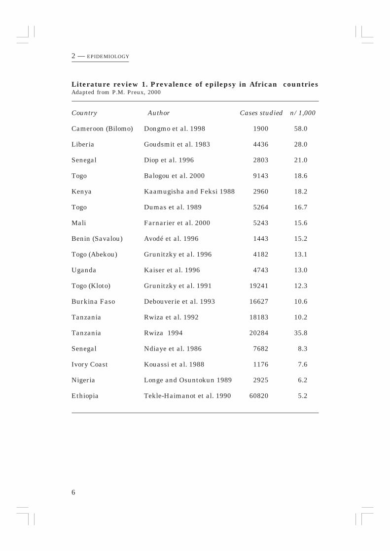
Pierre Marie Preux (2000) reported the prevalence of epilepsy in Africancountries and found a variation of 5.2/1,000 to 58/1,000. In his review (Literaturereview 1, page 6) one paper from Tanzania was reported presenting a prevalenceof 10.2/1,000. Two years later Rwiza published a second paper the outcomeof which is added to the table of Preux reporting a prevalence of 35.8/1,000from an area near the one reported in his first survey.
INCIDENCEIncidence is the rate at which new cases of a disease occur within a givenperiod (e.g., a year) in a given population. In the case of epilepsy, theannual incidence is usually calculated per 100,000 population.
In all known surveys the annual incidence rate is highest in theyoungest age groups, decreases during childhood, diminishes amongadults, and rises again in old age (see fig. 1).
About incidence Hauser (1997) writes: The only data on the incidence of allepileptic syndromes come from Bordeaux, France. The incidence of idiopathiclocalization-related epilepsy was 1.7/100,000 (7% of all cases). An additional13.6/100,000 (56%) had symptomatic localization-related epilepsy. Thus, ifthe same criteria are used as in most other contemporary incidence studies,about 60% of cases can be classified as partial seizures. Each of the followingsyndromes accounted for about 1% of new cases: juvenile myoclonic epilepsy,awakening grand mal, and West syndrome. About 2% had pyknolepsy(childhood absence epilepsy). These proportions are similar to those providedby the Rochester, Minnesota, studies. Crude incidence for all epilepsy (about24.5/100,000) was about half that reported in other recent studies inindustrialized countries. A few reports of incidence of specific epilepticsyndromes in other total-population studies provide data consistent with theabove figures.

5
2 — EPIDEMIOLOGY
Figure 1. Incidence, prevalence and cumulative incidence rates for epilepsyin Rochester, Minnesota
(adapted from Hauser et al. 1993)
Incidence of epilepsy
Prevalence of active epilepsy
Cumulative incidence of epilepsy
Age (yr)
Inci
den
ce p
er 1
0000
0
Pre
vale
nce
/cu
mu
lati
ve in
cide
nce
per
cen
tage
_________ Incidence of epilepsy
-------------- Prevalence of active epilepsy
................... Cumulative incidence of epilepsy
Inci
denc
e pe
r 10
0.00
0
Prev
alen
ce /
cum
ulat
ive
inci
denc
e pe
rcen
tage
Age
6
2 — EPIDEMIOLOGY
Country Author Cases studied n/1,000
Cameroon (Bilomo) Dongmo et al. 1998 1900 58.0
Liberia Goudsmit et al. 1983 4436 28.0
Senegal Diop et al. 1996 2803 21.0
Togo Balogou et al. 2000 9143 18.6
Kenya Kaamugisha and Feksi 1988 2960 18.2
Togo Dumas et al. 1989 5264 16.7
Mali Farnarier et al. 2000 5243 15.6
Benin (Savalou) Avodé et al. 1996 1443 15.2
Togo (Abekou) Grunitzky et al. 1996 4182 13.1
Uganda Kaiser et al. 1996 4743 13.0
Togo (Kloto) Grunitzky et al. 1991 19241 12.3
Burkina Faso Debouverie et al. 1993 16627 10.6
Tanzania Rwiza et al. 1992 18183 10.2
Tanzania Rwiza 1994 20284 35.8
Senegal Ndiaye et al. 1986 7682 8.3
Ivory Coast Kouassi et al. 1988 1176 7.6
Nigeria Longe and Osuntokun 1989 2925 6.2
Ethiopia Tekle-Haimanot et al. 1990 60820 5.2
Literature review 1. Prevalence of epilepsy in African countriesAdapted from P.M. Preux, 2000

7
3 — CAUSES
3Causes
CAUSES OF A SEIZUREAny person may develop a seizure in certain circumstances.
A seizure is a symptom of an abnormal condition. It is imperative toestablish which condition is the cause. The various causes of seizuresare listed in table 1. Some are common, some are very rare. If noinvestigations are carried out it is not possible to find out what iswrong. The better our investigation methods are, the more likely wecan find a cause. If, for instance, in a newborn baby with a seizure noblood is examined, no one will ever know if the seizure was due to ahypoglycaemia, hypocalcaemia, or something else. X-rays, CT scans,and even more modern investigation methods can show structurallesions, while chemical and serological investigations will showmetabolic and parasitic abnormalities. In rural areas where none ofthese investigations can be done, the cause will often remain obscure.But it is extremely important that every Health Centre, even in themost rural of areas, has at least a small laboratory, so that simple testscan be done. Then common disorders can be recognized as the cause ofthe seizure and appropriate treatment given—instead of, or beforeanticonvulsants are used—thus preventing further harm. Many ofthe metabolic disturbances, bacterial and parasitic infections areeasily treated when recognized. The relationship of fever and seizuresis discussed on page 69. Table 2 presents causes that may be direct orfever mediated. Three examples, often seen in paediatric practice, aregiven to illustrate these points:
– A child with vomiting and diarrhoea started to convulse and is broughtto the clinic. There is dehydration and therefore an electrolyte imbalanceis likely. Treatment consists of fluids and salts.
– A mother cannot wake her child in the morning and brings him/her tothe clinic. The child is unconscious and is convulsing. First determinethe blood glucose level. If it is low, treat with glucose solution and donot give anticonvulsants.
– A child with high fever and convulsions is brought to the clinic. If noother disease is obvious, a blood smear for malaria parasites and alumbar puncture must be done to diagnose possible meningitis. Abacterial meningitis should be treated as early as possible with adequateintravenous antibiotics to prevent later neurological sequelae. As ittakes some days before the antibiotics are effective, anticonvulsantshave to be given to control the seizures.

8
3 — CAUSES
Table 1. Causes of a seizure
MetabolicHypoglycaemia Pyridoxine deficiency/dependencyHypocalcaemia UraemiaElectrolyte imbalance Phenylketonuria**Hypomagnesaemia PorphyriaHyperbilirubinaemia (kernicterus)
InfectionsINTRACRANIAL EXTRACRANIAL— meningitis* — febrile illnesses (febrile— encephalitis* convulsions)*— AIDS* — pertussis— Neurosyphylis — pertussis immunization— cerebral malaria* — tetanus— rabies— toxoplasmosis— cysticercosis— encephalopathy (SSPE)
TraumaBirth trauma* Cold injury in newbornsHead injury in later life* Hypothermia
AnoxiaBirth asphyxia* Conditions later in life
ToxicAlcohol and withdrawal from alcohol*Carbon monoxide poisoningDrugs (high dose i.v. penicillin, strychnine, etc.)Lead poisoningOrgano-phosphorus insecticide poisoning
Space-occupying lesionsHaemorrhage Tuberculoma*Abscess CysticercosisTumour Toxoplasmosis
Circulatory disturbancesCerebro-vascular accident (stroke) Sickle-cell crisis*Vascular anomalies
Cerebral oedemaHypertensive encephalopathy Eclampsia
CongenitalMalformations of the brain (hydrocephalus, microcephaly, etc.)Tuberous sclerosis (Bourneville disease)**Neurofibromatosis (von Recklinghausen disease)**Encephalo-trigeminal facial angiomatosis (Sturge-Weber’s syndrome)**
Degenerative diseasesNiemann-Pick disease** Dementias*Cerebromacular degeneration**
Epilepsy
*Most common causes; **Rare topics treated in more detail on page 9

9
3 — CAUSES
Notes to Table 1
PhenylketonuriaThis is a rare disease transmitted by a recessive gene. There is inability to form tyrosinefrom phenylalanine, resulting in the formation of excessive phenylketone bodies which areexcreted in the urine. Infants appear to be normal at birth, but when the plasmaphenylalanine levels rise, progressive brain damage begins and reaches a limit at two tothree years of age unless a diet low in phenylalanine is started in early life. If no diet isinstituted, mental retardation, skin changes (excessive oiliness, scaliness, excematouslesions), musty odour of the urine and of the body occur.Tuberous sclerosis (T.S., Bourneville disease)An ectodermal dysplasia inherited as a dominant trait, although almost 50% occur for thefirst time (i.e., as a result of a sporadic mutation). Mental retardation varying from mildto severe occurs in 60–70% of people with T.S., and in about 90% there is epilepsy. Theseizures usually develop during the first year of life in the form of infantile spasms. Laterin life they may be myoclonic-astatic seizures.
Behaviour disorders such as hyperactivity and destructiveness are common.There are lesions in the skin (fibroadenomas of the sebaceous glands over the nose and
cheeks, leathery patches, hypopigmented patches, subungual fibromas). Small tumoursmay occur in the kidneys, liver, spleen, lungs or heart. Nodules and cystic lesions mayoccur in the eyes, bone or brain. The cortical and subependymal nodules frequently calcifyand may then be seen on the skull X-ray or CT scan.Neurofibromatosis (von Recklinghausen disease)Inherited as a non-sex-linked dominant. Freckling in the axilla is pathognomonic, andsharply outlined café au lait areas occur on the skin. Neurofibromas are found cutaneouslyand subcutaneously along cranial and spinal nerves, osteitis fibroma cystica in the bonesand nodules in the central nervous system.Encephalo-trigeminal facial angiomatosis (Sturge-Weber’s syndrome)Patients are often mentally retarded, and all develop epilepsy with lateralized seizures.There is a large telangiectasis (port-wine stain) in the trigeminal area involving skin,scalp, skull and meninges. There are typical opacities on the skull X-ray. There may bea haemangioma in the choroid causing glaucoma.
Degenerative diseasesThe features of these diseases are progressive loss of previously acquired intellectual,motor and sensory functions. Early manifestations of degeneration of cerebral grey mattershow as dementia and seizures, whereas degeneration of cerebral white matter shows asspasticity, hypotonia or ataxia. But eventually in both forms the entire nervous systemtends to be affected and the person becomes totally helpless with loss of intellectual andvoluntary motor functions.
Cerebromacular degenerationTransmission is on an autosomal recessive basis. These degenerations are ganglioside-storage diseases.• Infantile form (Tay-Sachs’ disease)
Onset is at 2–6 months of age. The infant becomes apathetic and loses interest in hissurroundings. There is progressive blindness (macular degeneration with macularcherry-red or black spots in the fundi), spasticity, seizures, wasting, dementia, anddeath usually occurs before the third birthday.
• Late infantile form (Bielschowsky syndrome)Onset is between the ages of 1 and 3 with seizures, ataxia, dementia, spasticity andblindness. Death occurs within three to five years.
• Juvenile form (Spielmeyer-Vogt or Batten disease)Onset between 3 and 7 years with the same symptoms but a much slower course thanabove.
Niemann-Pick diseaseThis is a heredo-familial disease with a disturbance of the lipid metabolism. Soon afterbirth physical and mental retardation starts. There is general emaciation, but theabdomen is distended due to the enlarged liver and spleen. In the fundus a cherry-redspot may be seen, as in Tay-Sachs’ disease. There is progressive deterioration, anddeath usually occurs before the third year of life.DementiasOf the dementias in later life Alzheimer’s disease is probably the most frequent cause.

10
3 — CAUSES
Table 2. Fever and convulsions
Intracranial infections– meningitis/encephalitis– cerebral malaria
Extracranial infectionsFebrile Convulsions (FC) are often associated with– otitis media– upper respiratory infections– lower respiratory infections– shigellosis– urinary tract infections– measles, roseola infantum– malaria
When the disorder that caused seizures is cured the brain may have beenpermanently altered so as to give rise to spontaneous seizures. This personthen has epilepsy.

11
CAUSES OF EPILEPSYEpilepsy is a condition with recurrent seizures. These may be idiopathicor symptomatic. Epilepsies can start at any age. A rough outline of therelationship between cause and age of onset is presented in fig. 2.
If an acute disturbance, a metabolic like hypocalcaemia, or an infectionsuch as meningitis, or a poisoning or any of the other causes mentionedin table 1 are recognized and treated adequately, epilepsy will not follow.
If the acute disorder was too severe, or not treated correctly, convulsionsmight have become prolonged and continuous, resulting in anoxia of thebrain with subsequent brain damage followed by epilepsy.
In some infections, e.g., tuberculosis, toxoplasmosis, cysticercosis, thedisease may leave calcified areas in the brain.
Some diseases—tuberous sclerosis, Sturge-Weber’s syndrome—presentwith calcifications in the brain.
A haemorrhage, abscess or tumour may present with repeated seizuresbut when the blood, pus or tumour has been successfully removedsurgically, no epilepsy needs to follow.
Any head injury, including birth trauma, may result in permanentchanges of brain tissue, i.e., scar tissue.
Any area with abnormal brain tissue (calcifications, scars, or vascularabnormalities) may act as a focus from where abnormal activity of theneurons takes place causing “symptomatic epilepsy”.
The causes mentioned in table 1 might lead to epilepsy. Some of them arevery common in Africa (cerebral malaria, birth trauma, infectious diseasesand accidents later in life), while others are very rare.
Neurocysticercosis, a parasitic infection associated with pig-rearing andpoor hygiene is very common in Latin America and India. Only people comingor returning from a country with prevalent neurocysticercosis might sufferfrom it.
Oncocerciasis has recently been suspected of causing epilepsy as well. Ithas to be kept in mind if a patient has come from an endemic area.
Some diseases are so rare that the average doctor will not know aboutthem and will not, therefore, recognize them if they are present. Suchdiseases can only be diagnosed by specialists. A number of these diseasesare mentioned on page 9 so that if any are suspected, the patient shouldbe referred to a specialist.
3 — CAUSES

12
3 — CAUSES
Figure 2. Approximate frequency of different causes of epilepsy, developingat different years
* Genetic brain disorders with epilepsy
**Idiopathic = genetic epilepsy without other disorders = epilepsy sui generis
Idiopathic**/cryptogenicVascular
Tumor
TraumaBirth trauma
Age (years)
Rel
ativ
e fr
eque
ncy
10 20 60504030
Genetic*

13
3 — CAUSES
EPILEPSY AFTER HEAD INJURYThe onset of these seizures depends on the age at the time of the accident,e.g., birth trauma will give seizures in the first year of life.
Not everyone who has had a head injury will develop seizures. Seizuresare more common when the injury has been penetrating, when there wasa depressed skull fracture, an intracranial haematoma, or if in the acutestate there was post-traumatic amnesia of more than 24 hours’ duration.Fifty percent of post-traumatic seizures develop in the first year followingthe accident, and another 20% will develop by the end of the second year.The use of prophylactic anticonvulsants after a serious head injury shouldbe considered.
EPILEPSY CAUSED BY A BRAIN TUMOURA tumour, benign, malignant or a metastasis, may occur in any agegroup, but is more common in the older age group.
Epilepsy starting after the age of 20 years should always raisesuspicion for a tumour, and full investigations, preferably with CT scanshould be done.
EPILEPSY FOLLOWING CEREBRO-VASCULAR DISEASEIn people over 50 years old, cerebro-vascular disease is a common causeof epilepsy. Seizures may follow a cerebro-vascular accident (stroke), ormay develop during the course of subclinical cerebro-vascular disease.
In all these groups (injury, tumour, cerebro-vascular accident) the type ofseizure depends on the localization of the injury.
Preferred treatment is with phenytoin or carbamazepine.When there are secondary generalized seizures, phenobarbitone may
be used as an alternative.

14
It is worth bearing in mind that a cause for the underlying seizurescan be found in only 30–40% of epilepsy patients. In the rural areas,where people live far from health care facilities, the percentage ofsymptomatic epilepsy is probably higher than in the urban areas. And theprevalence rate of epilepsy in developing countries is likely higher thanin developed countries due to higher rates of perinatal injuries, moreperinatal and childhood infections, and less timely treatment than isavailable in industrialized countries.
Prevalence of epilepsy among mentally handicapped is high, between 20–35%. Conversely, concomitant mental handicap is present in about 10–15% of people with epilepsy.In Tanzania, Matuja (1990) found that 48% of persons with epilepsytreated in Muhimbili Medical Centre had organic brain disease andpsychological disturbances.
In 60–70% of patients (at least in developed countries) where no cause forthe epilepsy can be found, part of these persons suffer from “idiopathicepilepsy” (idiopathic = spontaneous, self-generated, genetic).In those cases where secondary damage is the probable cause, althoughsuch damage cannot be proven, the epilepsy is classified as “cryptogenic”.
3 — CAUSES

15
4 — CONTRIBUTORY FACTORS
4Contributory factors
GENETIC FACTORSIn many, if not all, epilepsies there is a genetic factor which influencesthe threshold for seizures (fig. 3). Even in symptomatic epilepsy thisfactor plays a role, e.g., many people have had a head injury but onlysome develop epileptic seizures afterwards (see page 93).
If one parent has idiopathic epilepsy the risk of a child developingepilepsy is 4–6%, compared to a risk of 0.3-0.5% in the general population(Europe). If both parents have idiopathic epilepsy, the risk rises to 12–20%.
In parents with symptomatic epilepsy, there is still a slight increase inthe risk—up to 2% in European studies.
EFFECTS OF BRAIN MATURATIONThe resistance to seizures also depends on the maturation of the brain.The resistance in the first year of life (except during the newborn period)is very high, and therefore only a severe injury such as severe braindamage since birth, meningitis or tuberous sclerosis, will produce seizures.
Between the ages of one and four the resistance to seizures is very low.A simple febrile disease may precipitate seizures. After the age of four theresistance is again high, and seizures are mainly seen in already-brain-damaged children. This resistance diminishes again from about the seventhyear when the idiopathic epilepsies tend to appear. The resistance is at itslowest around the time of the prepubertal growth spurt (Brown, 1982).
OTHER PRECIPITATING FACTORSApart from the condition and maturation of the brain and the geneticthreshold, other factors may trigger a seizure. These factors may bedifferent for each individual patient. Some patients learn which factorsare important for them, and so they can modify their behaviour oractivities to try to avoid seizures (see also page 75).
The most common factors are mentioned in table 3.

16
4 — CONTRIBUTORY FACTORS
grey: seizures, white: no seizures
* Charles: moderate tendency, no brain damage, no seizures** Esther: strong tendency, mild brain damage, has seizures*** Mary: mild tendency, severe brain damage, has seizures**** William: very slight tendency, severe brain damage, no seizures
Table 3. Common precipitating factors
– Flashing lights (resulting in reflex epilepsy)– Hyperventilation– Lower alertness, sleep itself and lack of enough sleep– Emotion– Physical stress– Special smells, sounds or sensations of touch– Alcohol– Hormonal changes, e.g., during menses– High fever– Overhydration
Some of these triggers are used to provoke epileptic activity for an EEGrecording (see page 44).
normal mild moderate severe very severe
very strong
strong
mild
slight
Esther **
Mary ***
William ****
BRAIN DAMAGE ➩
moderate Charles *
Figure 3. Relation between brain damage and tendency to develop seizures
TENDENCY ➩

17
5 — SEIZURES
Table 4. Components of a seizure
– Prodromal phase– Aura– Seizure (ictus)– Post-ictal phase
5Seizures
Four components of a seizure can be distinguished (table 4). Not allseizure types will have all these stages. The presence or absence and thenature of them are important for diagnosing the seizure type.
Prodromal phaseThis phase begins a few hours or even days before the actual seizure andshould not be confused with the aura. Prodromal symptoms are: headache,irritability, insomnia, bad temper, depression or increased activity.
AuraAn aura precedes the seizure by seconds or a few minutes. It is thebeginning of the seizure and signals the focal onset of the seizure. Thesymptoms depend on the location of this focus. The feelings of the auraare often vague and indescribable, leading to extreme fear. Strangeepigastric sensations, dreamlike experiences, unpleasant smells, etc. mayoccur. The patient remembers the aura very well, and although he/shewill not always be able to recount it, he/she can affirm the presence of it,as it happens before consciousness is lost.
Seizure (ictus)The characteristics important to know for their classification are mentionedin table 5. In most seizures there is a loss of consciousness, and thepatient is therefore not able to give any information about the actualictus. For this we are dependent on witnesses who have seen the actualseizure. The patient has no memory of the seizure.

18
5 — SEIZURES
Table 5. Characteristics of a seizure
– The type of seizure (see classificationof seizures table 6)
– The duration of the seizure– The frequency of the seizures– The time of day or night that the
seizure occurs, and its relation tosleep
– The presence of an aura– The presence of a post-ictal phase– The age of onset
Post-ictal phaseThis phase may be absent, brief or may last several hours, and sometimeseven days. There is usually a deep sleep and waking up with headache,tiredness, irritability, vomiting, confusion, muscular aches or ataxia.Transient paralysis of a part of the body, known as Todd’s paresis mayoccur for a few hours or days. Altered speech or aphasia may occur whenthe dominant hemisphere of the brain has been involved. Altered behaviourand emotional outbursts may occur, and if these are interfered with,violent behaviour is likely.
19
5 — SEIZURES
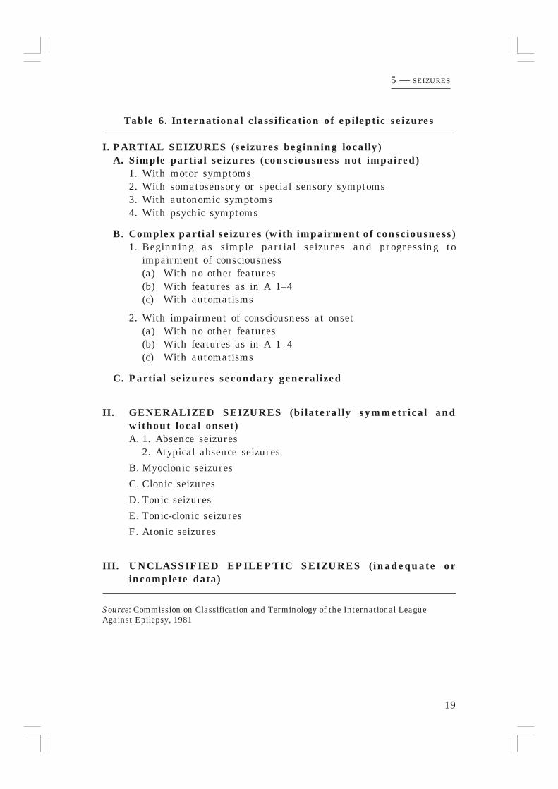
Table 6. International classification of epileptic seizures
I. PARTIAL SEIZURES (seizures beginning locally)A. Simple partial seizures (consciousness not impaired)
1. With motor symptoms2. With somatosensory or special sensory symptoms3. With autonomic symptoms4. With psychic symptoms
B. Complex partial seizures (with impairment of consciousness)1. Beginning as simple partial seizures and progressing to
impairment of consciousness(a) With no other features(b) With features as in A 1–4(c) With automatisms
2. With impairment of consciousness at onset(a) With no other features(b) With features as in A 1–4(c) With automatisms
C. Partial seizures secondary generalized
II. GENERALIZED SEIZURES (bilaterally symmetrical andwithout local onset)A. 1. Absence seizures
2. Atypical absence seizures
B. Myoclonic seizures
C. Clonic seizures
D. Tonic seizures
E. Tonic-clonic seizures
F. Atonic seizures
III. UNCLASSIFIED EPILEPTIC SEIZURES (inadequate orincomplete data)
Source: Commission on Classification and Terminology of the International LeagueAgainst Epilepsy, 1981

20
CLASSIFICATION OF EPILEPTIC SEIZURESTable 6 is the International classification of epileptic seizures proposed bythe Commission on Classification and Terminology of the InternationalLeague against Epilepsy (ILAE) and approved in September 1981. Thisclassification is based on the clinical expression of the seizure and theelectroencephalographic picture during and between the seizures. Themain division in this classification is into partial seizures and generalizedseizures.
In the partial seizures the abnormal electrical discharges start in alocalized area of the brain. The symptoms/signs are dependent on whichpart of the brain is affected. These discharges may remain localized, orthey may spread to other parts of the brain and then the seizures becomegeneralized (secondary generalized seizures).
In generalized seizures, on the other hand, the seizure is generalizedfrom the onset (i.e., primary generalized seizures), starting in bothhemispheres of the brain simultaneously.
As we often do not have an EEG to help in making this division, weare completely dependent on the clinical expression: the medical history,and the ability of the observer to describe the seizure. The patient himselfhas no memory of the seizure, except in simple partial seizures and onlyof the aura of other seizures. If he is not too young, he can inform usabout the presence and the nature of an aura.
A definite aura is an indication that the seizure is of focal (partial) onset.
During his lifetime, a patient does not necessarily have only one typeof seizure. The type may change over the years, depending on the age andmaturation of the brain. Moreover, one patient may have a combinationof different seizure types.
PARTIAL SEIZURES (fig.4)
The partial seizures are first divided into two groups, those where theconsciousness is maintained, and where there is an impairment of theconsciousness. Both these groups may develop into generalized seizures,then forming a third group.
Simple partial seizuresThe patient does not lose consciousness, and therefore is able to tell whathappened, but the experience may be so strange that he may not be ableto express himself properly. What happens is dependent on the locationof the affected area.
5 — SEIZURES

21
5 — SEIZURES
Figure 4. Partial seizure: discharge remains localized
22
In motor seizures, the focus is in the primary motor cortex. There aretwitchings, starting in a distal part of the extremity, or in the face. Thetwitching may remain there, or spread up the whole extremity and evenbecome completely generalized. The spreading is called a Jacksonianmarch (named after Huglings Jackson 1835 - 1911).
The sensory seizures have their focus in the post central gyrus (primarysensory cortex). There might be feelings of tingling, pins and needles, coldor heat, or numbness of a limb. Sometimes there may be strange feelingswith visual signs, or hearing or smelling sensations.
The autonomic seizures are associated with foci in the temporal lobe.There maybe: a sensation rising from the epigastrium to the throat,palpitations, sweating or flushing.
The psychic symptoms may consist of changes in mood, memory, orthought (thinking). There may be distorted perceptions (time, space, orperson) or problems with language. Structured hallucinations could occur(music, scenes).
These simple partial seizures are usually only recognized as epilepticseizures when they develop into generalized seizures.
Complex partial seizuresHere the patient has impaired consciousness, there is no complete loss ofconsciousness, he is slightly aware of what is going on, but he cannotrespond to anything, neither can he change his behaviour during anattack. There is an aura, a strange feeling in the stomach rising up to thethroat and head, or a sensation of light, smell, sound or taste. The seizuremay occur with changes in perception, e.g., of time (time seems to passtoo slowly or too fast), of light or sound or space. The surroundings maysuddenly seem completely strange and different in scale (things seemlarger or smaller than usual), or there is déjà vu (a sensation of thingshaving happened before). These feelings can cause the patient a greatdeal of anxiety.
Sometimes the seizure occurs with hallucinations or with psychomotorsymptoms such as automatisms, automatic movements, e.g., pulling at theclothes, chewing, lip smacking, or repeated aimless movements. Theseautomatisms may become very complex, the patient is able to performdifficult tasks, or travel somewhere, but later not remember having donesuch a thing. He suddenly comes to again and finds himself in a completelydifferent place. During such an automatism the patient may becomeaggressive and violent when restrained. There is a slow recovery after acomplex partial seizure, with a period of confusion. After the attack thereis complete amnesia of it. These seizures were previously called ‘psychomotorseizures’, and as the localization of the abnormal discharge is often in thetemporal lobe, the epilepsy is often called ‘temporal lobe epilepsy’ (the focusmight occur in the frontal lobe too).
5 — SEIZURES

23
5 — SEIZURES
Partial seizures secondary generalized (fig. 5)Both the simple partial seizures and the complex partial seizures maybecome generalized tonic-clonic seizures. The beginning is as describedabove, but they end alike the primary generalized tonic-clonic seizures asdescribed below.
GENERALIZED SEIZURES (fig. 6)
The primary generalized seizures are characterized by a complete loss ofconsciousness and the absence of an aura. They come on suddenly andunexpectedly, and if the patients fall, they may injure themselves.
The generalized seizures consist of six different seizure types, of which theprimary generalized tonic-clonic seizure (GTCS) is the most common. Althoughless common, all seizure types can be seen in special epilepsy clinics.
Absence seizuresThese are short periods of loss of consciousness lasting only a few seconds(not more than half a minute). They are of sudden onset, there are usuallyno, or only minimal motor manifestations. There is a blank stare, briefupward rotation of the eyes, an interruption of ongoing activity. The childis unresponsive when spoken to. It is suddenly over, and the child continueswhat he was doing before the seizure came. The child has no memory ofthese seizures. They should not be confused with brief complex partialseizures (table 7).Typical absences occur in school-aged children, during childhood because ofChildhood Absence Epilepsy, and in adolescence because of Juvenile AbsenceEpilepsy (see classification of epilepsies). They occur many times a day.During such an absence seizure the child does not hear what the teacher issaying, and as they occur so often the child cannot follow the lessons anymore. Unless the teacher is aware of this condition, he will scold the childfor daydreaming and inattentiveness.
Most parents are unaware of these small seizures, and even when theyobserve them, do not think them important and will not mention them tothe doctor. Unless these children also suffer from generalized tonic-clonicseizures they are not brought to a clinic, and especially not to an epilepsyclinic, as people are unaware that these absences are epileptic seizures.
Absences are easily provoked by overbreathing (hyperventilation). Theyhave a typical EEG pattern and therefore are easily recognized on an EEG.
A child with absence seizures may, in addition, have other types ofseizures, such as primary GTCS, or myoclonic seizures (see Appendix Apage 85 and following where the special syndromes are discussed). Previously,these seizures were called petit mal seizures, or pyknolepsy (because theyoccurred so frequently). The term “petit mal” (little illness) should no longerbe used as it is very unspecific.

24
5 — SEIZURES
Figure 6. Generalized seizure: epileptic dischargeaffects both hemispheres
Figure 5. Secondary generalized seizure: epileptic discharge initiallylocalized and then spreads to trigger a generalized seizure

25
5 — SEIZURES
Myoclonic seizuresThese seizures consist of sudden, brief, shock-like muscle contractions,either occurring in one limb, or more widespread and bilateral. They maybe single jerks, or jerks repeated over longer periods. They are often seenin combination with other seizure types occurring in special epilepticsyndromes as discussed in Appendix A.
Clonic seizuresThese seizures are generalized seizures, where the tonic component is notpresent, only repetitive clonic jerks (clonic jerks are repetitive rhythmicflexing and streching of limbs). When the frequency diminishes theamplitude of the jerks do not.
Tonic seizuresTonic seizures are sudden sustained muscle contractions, fixing the limbsin some strained position. There is immediate loss of consciousness. Oftenthere is a deviation of the eyes and head towards one side, sometimesrotation of the whole body. They are seen mainly in paediatric practice.
Tonic-clonic seizures (GTCS)The patient loses consciousness, falls down, sometimes with a scream,and develops a generalized stiffness (the tonic phase). Breathing stops, asall the muscles of the trunk are in spasm, and the patient becomescyanotic, the head is retracted, the arms flexed and the legs extended.After a while, this tonic phase is followed by the clonic phase, when themuscles alternately contract and relax, resulting in clonic movements.With this jerking the patient might bite his tongue, pass urine, orsometimes stool. The clonic phase may last several minutes. When all thejerking stops and the patient regains consciousness, he may feel verytired with a headache and confusion. He has no memory of whathappened, and may find himself on the floor in a strange position. Oftenhe falls into a deep sleep.
These seizures are not as frequent as absence seizures. Their frequencymay vary from one a day to one a month or once a year, or even onceevery few years.
Generalized tonic-clonic seizures can occur in generalized epilepsies andin partial epilepsies. To distinguish the two they are called primary orsecondary generalized (table 8). Primary GTCS occur without any warning,i.e., they are not preceded by an aura or other partial seizure. SecondaryGTCS occur in partial epilepsies and are always preceded by an aura or otherpartial seizure, however, the generalization may be so rapid that thepreceding seizures are not noticed.
This type of seizure is not seen in the newborn period or infancy.

26
5 — SEIZURES
Table 7. Differences between complex partial seizures andgeneralized absence seizures
complex partial generalizedseizures absence seizures
Age any age childhood or earlyadult
Aetiology symptomatic or idiopathicidiopathic
Duration of attacks several minutes short, usually <30 s
Other clinicalmanifestations there may be marked slight tone changes
tone changes or or motormotor phenomena, phenomenaincluding automatism
Post-ictal gradual recovery quick recoveryoften with confusion
Frequency not as frequent numerous, oftenclustered
EEG focal temporal 3-Hz spikedisturbance and wave

27
5 — SEIZURES
Table 8. Points in favour of secondary and of primary GTCS
secondary GTCS primary GTCS
Aura present absent
Time of occurrence often during sleep often daytime, orjust after fallingasleep, or just whenwaking up
Aetiology symptomatic idiopathic
Age of onset early in life between 5 and< 5 yr or 20–25 yrlate in life> 20 or 25 yror after known trauma
Other handicaps might be present usually absent
In family less often more often

28
Atonic seizures (astatic seizures)There is a sudden loss of muscle tone causing the head or a limb to drop,and often the patient falls suddenly to the floor. They are therefore alsocalled “drop attacks”. There is loss of consciousness, a sudden onset andno post-ictal phase. The patient stands up and continues what he was doing.The seizure is very short, only seconds, but may occur several times a day.The patients often present with scars or fresh wounds on chin, cheek orforehead, or the back of the head. A protective helmet is recommended forthese patients. Sometimes these patients may have, in addition to atonicseizures, absence or myoclonic seizures (pages 87-88).
Infantile spasmsBefore 1981, infantile spasms were seen as one of the seizure types. In thepresent classification they are classified under the generalized syndromes(table 10, page 34) and discussed in Appendix A (page 87). They are flexorspasms of the head, bending of the knees and flexion with abduction ofthe arms. They occur in the first year of life, and are very difficult totreat. ACTH or prednisolone is the drug of choice.
UNCLASSIFIED EPILEPTIC SEIZURESThis category includes all seizures which cannot be classified because ofinadequate or incomplete data, or seizures that defy classification in thecategories as presently defined.
WHICH SEIZURES DO WE SEE IN DAILY PRACTICE?The majority of our patients (70–80%) present with generalized tonic-clonic seizures, GTCS for short. This is a new name for what werepreviously called “grand mal” (French for the big illness) seizures.
But GTCS is a combination of two seizure types, the secondary GTCS(group I, C) and the primary GTCS (group II, E)— see table 6. Both theseseizure types are seen equally commonly: 35–40% will have secondaryGTCS, and the same percentage will have primary GTCS. The differentiationis important, as the drug of choice is different in each group. As most of ourpatients have to be treated without the help of an EEG recording, we relyon the medical history for this differentiation. In table 8 some points arementioned in favour of one or the other.
The four main antiepileptic drugs (AEDs) can be used for all GTCS.But the drug of choice for the secondary GTCS is phenytoin orcarbamazepine, and for the primary GTCS phenobarbitone or valproate.
The minority of our patients present with the other seizure types as theyare often not recognized by the patient, their family or even the primaryhealth workers to be caused by epilepsy. Or they are considered to cause toolittle problems to take the trouble of a visit to a clinic.
5 — SEIZURES
29
5 — SEIZURES
Most of the patients who do not present with GTCS present withcomplex partial seizures (I, B); approximately 15%. They are a strangemixture of feelings and signs, and were previously called psychomotorseizures or temporal lobe epilepsy because the origin is often in thetemporal lobe. These complex partial seizures should not be calledcomplex absences, but should be differentiated from the generalizedabsences. In table 7 the points in favour of one or the other arementioned. The drug of choice for complex partial seizures is carbamazepineor phenytoin.
From 1–4% of our patients will present with generalized absences (II, A).valproate is the drug of choice. When they have only generalized absences,they can be treated with ethosuximide, but when they have other types aswell, then valproate is the drug of choice. Phenobarbital alone can take careof primary GTCS but is not effective against absences.
Simple partial seizures might occur in a small percentage of ourpatients, and the drug of choice is then phenytoin or carbamazepine,while the rest of our patients might have myoclonic or astatic seizures, forwhich the drug of choice is valproate.
Definite figures of the occurrence of these seizures in Africa cannot begiven, as the diagnosis is made on a clinical impression, and not verifiedby an EEG recording.
STATUS EPILEPTICUSA status epilepticus occurs whenever a seizure persists for at least 30minutes, or is repeated so frequently that recovery between attacks doesnot occur.
A status is a medical emergency and the patient should betransferred to a clinic where, with i.v. injections the status could bestopped as quickly as possible (table 22, page 66). It is a dangerouscondition which may result in brain damage (cerebral necrosis) withsevere morbidity or death. A status may be the patient’s first epilepticevent, or may be precipitated by suddenly discontinuing anticonvulsanttherapy.
An initial status epilepticus in an adult may be due to a brain tumour andrequires full investigation.
A status is again classified according to the different seizure types, butin two groups as presented in table 9.

30
5 — SEIZURES
Table 9. Classification of status epilepticus
I Convulsive statusFocal or partial– Partial motor status (epilepsia partialis continua, Kojewnikow’s
syndrome)Continuous motor seizures with retained consciousness. Occursmainly in adults with structural brain damage due to trauma orvascular disease, or in children with severe brain disease
Generalized– Tonic-clonic (previously grand mal) status– Myoclonic status– Febrile status epilepticus
Prolonged febrile convulsions (see page 69)
II Non-convulsive or stupor status– Complex partial status, or psychomotor status
Prolonged period of automatic behaviour– Absence status
Repeated absence seizures may cause prolonged periods of confusedbehaviour in children. Is occasionally seen in adults. This may bedetected on an EEG, and controlled by diazepam

31
6 — CLASSIFICATION OF EPILEPSIES AND EPILEPTIC SYNDROMES
6Classification of epilepsies
In addition to the international classification of epileptic seizures, thereis an International Classification of Epilepsies and Epileptic Syndromes,of which the latest revised classification is given in table 10 (page 34),according to the Commission on Classification and Terminology of theInternational League Against Epilepsy (1989).
An epileptic syndrome is an epileptic disorder characterized by acluster of signs and symptoms customarily occurring together. Theseinclude such items as seizure type, aetiology, precipitating factors, age ofonset, severity, chronicity, diurnal and circadian cycling, and sometimesprognosis.
Two main groups are again distinguished, depending on the characterof the presenting seizure type:
PARTIAL or LOCALIZATION-RELATED, and
GENERALIZED.
In this new classification, each is then divided into three groups as theterm cryptogenic has now been introduced. The three groups are:
– Idiopathic epilepsiesThere is no underlying cause other than a possible hereditarypredisposition. Idiopathic epilepsies are defined by age-related onset,clinical and electroencephalographic characteristics, and a presumedgenetic aetiology of the epilepsy. This does not include other genetic braindisorders associated with epilepsy such as those grouped under congenitaland degenerative diseases in table 1.
– Symptomatic epilepsiesThey are considered to be the consequence of a known or suspecteddisorder of the central nervous system.
– Cryptogenic epilepsiesThe term refers to a disorder whose cause is hidden or occult. Theseare presumed to be symptomatic, but there is no clear evidence of anaetiological factor. The cryptogenic epilepsies are also age-related, butoften do not have well defined electro-clinical characteristics.

32
SYNDROMES where it is impossible to determine whether the seizuresare focal (localized) or generalized, is the third main group.
SPECIAL SYNDROMES in which epileptic seizures are the main component,and of these febrile convulsions (page 69) are the most common andimportant are collected in a fourth main group.
This classification is important as it often tells us about the prognosis,and indicates the appropriate treatment (table 12, page 37) it alsofacilitates discussion with the patient or other medical experts, e.g., bymail or telephone.
1. LOCALIZATION-RELATED EPILEPSIESMost of the localization-related (partial) epilepsies are symptomatic. Withintensive investigation methods, a lesion or focus can be found. Thepattern of the seizures and other characteristics are dependent on the siteof the lesion. The focus might be in the frontal, temporal, parietal oroccipital lobes of the brain or in the motor cortex (table 10).
These focal symptomatic seizures respond better to treatment than thegeneralized symptomatic group, but not as well as the idiopathic epilepsies.
The appropriate drugs in this group are phenytoin or carbamazepine.With these anticonvulsants, the seizures are less severe and less frequent,but will sometimes still occur. If there is insufficient improvement,phenobarbitone or valproate may be tried. Fatigue, sleep deprivation,alcohol and emotional stress may trigger these seizures. Lifestyleinstructions are important.
The idiopathic localization-related epilepsies are not common, buthave a good prognosis, and respond well to treatment (phenytoin orcarbamazepine). They are discussed in more detail in Appendix A.
2. GENERALIZED EPILEPSIESMost of the generalized epilepsies are idiopathic. There are no neurologicaldeficits, psychological abnormalities or mental handicaps. Epilepsy in thefamily is more common than in the other groups. The prognosis is good.The response to treatment is usually good. The seizure types found areabsences (treatment: valproate or ethosuximide), myoclonus (treatment:valproate and benzodiazepines, but difficult to treat if it occurs in infancyor early childhood), and generalized tonic-clonic (treatment:phenobarbitone, valproate or carbamazepine).
Some of the generalized epilepsies are symptomatic (in Europeapproximately 10% of all epilepsies). They result from diffuse braindamage following birth asphyxia or occur in diseases with inborn errors
6 — CLASSIFICATION OF EPILEPSIES AND EPILEPTIC SYNDROMES

33
of metabolism. These children often have multiple handicaps and havespasticity and/or mental retardation in addition to their epilepsy.Treatment is very difficult and phenobarbitone, phenytoin, carbamazepine,valproate and benzodiazepines may be tried. The seizure types seen aretonic-clonic, tonic, atonic, atypical absences and infantile spasms. In thelatter case ACTH or prednisolone are often used.
Age of onsetIn addition to the seizure type and the aetiology, the age of onset, i.e., theage at which the first seizure occurs, is a helpful tool in classifying thedifferent epilepsies and syndromes.
All the symptomatic epilepsies occur after (sometimes years after) theevent resulting in the epilepsy and therefore depend upon the age (birthor later) at which the event took place.
The idiopathic epilepsies, however, might commence at any time, butage-related groups are now well recognized. As the age of onset is adefinite fact, table 10 is discussed not as it is presented, but according tothe age of onset of the different epilepsies in Appendix A.
Time of occurrence of seizureAnother important factor is the time of day or night that the seizureoccurs and its relation to the sleep-waking cycle.
I. – Partial seizures of the idiopathic type (benign focal epilepsy) occurmainly during sleep.
– Complex partial seizures are uncommon during sleep.
– Partial seizures secondary generalized are much more common atnight than primary generalized seizures.
II. – Absences occur mainly during the day-time.
– Seizures seen just after awakening are often associated withmyoclonic seizures.
– Primary generalized tonic-clonic seizures are more commonly seenduring the daytime, but if they do occur at night it is soon afterfalling asleep, or in the period of early morning sleep.
The treatment for these seizures is indicated in table 12. However,phenobarbitone increases the amount of deep sleep, and is therefore bestavoided in epilepsy with predominantly nocturnal seizures. Phenytoinand carbamazepine, which have less hypnotic effect, are indicated forsleep provoked epilepsy.
6 — CLASSIFICATION OF EPILEPSIES AND EPILEPTIC SYNDROMES

34
6 — CLASSIFICATION OF EPILEPSIES AND EPILEPTIC SYNDROMES
Table 10. International classification of epilepsies and epilepticsyndromes and related seizure disorders
1. Localization-related (local, focal, partial) epilepsies and syndromes1.1 Idiopathic (with age-related onset)
Benign childhood epilepsy with centro-temporal spikesChildhood epilepsy with occipital paroxysmsPrimary reading epilepsy
1.2 SymptomaticChronic progressive epilepsia partialis continuaSyndromes characterized by seizures with specific modes of
precipitationTemporal lobe epilepsies *Frontal lobe epilepsies *Parietal lobe epilepsies *Occipital lobe epilepsies *
* see table 11 page 36
1.3 Cryptogenic
2. Generalized epilepsies and syndromes2.1 Idiopathic (with age-related onset)
Benign neonatal familial convulsionsBenign neonatal convulsionsBenign myoclonic epilepsy in infancyChildhood absence epilepsyJuvenile absence epilepsyJuvenile myoclonic epilepsy (impulsive petit mal, Janz
syndrome)Epilepsy with grand mal seizures (GTCS) on awakeningOther generalized idiopathic epilepsiesEpilepsies with seizures precipitated by specific modes of
activation
2.2 Cryptogenic or symptomaticWest syndrome (infantile spasms)Lennox-Gastaut syndromeEpilepsy with myoclonic-astatic seizuresEpilepsy with myoclonic absences
2.3 Symptomatic2.3.1 Non-specific aetiology
Early myoclonic encephalopathyEarly infantile epileptic encephalopathy with
suppression burstsOther symptomatic generalized epilepsies
2.3.2 Specific syndromesEpileptic seizures complicating other disease states
continued on p. 35

35
6 — CLASSIFICATION OF EPILEPSIES AND EPILEPTIC SYNDROMES
Table 10, continued
3. Epilepsies and syndromes undetermined whether local or generalized3.1 With both generalized and focal seizures
Neonatal seizuresSevere myoclonic epilepsy in infancyEpilepsy with continuous spike waves during slow wave sleepAcquired epileptic aphasia (Landau-Kleffner syndrome)Other undetermined epilepsies
3.2 Without unequivocal generalized or focal features
4. Special syndromes4.1 Situation-related seizures
Febrile convulsionsIsolated seizures or isolated status epilepticusSeizures occurring only with acute metabolic or toxic events
From the Commission on Classification and Terminology of the International League AgainstEpilepsy, 1989
36
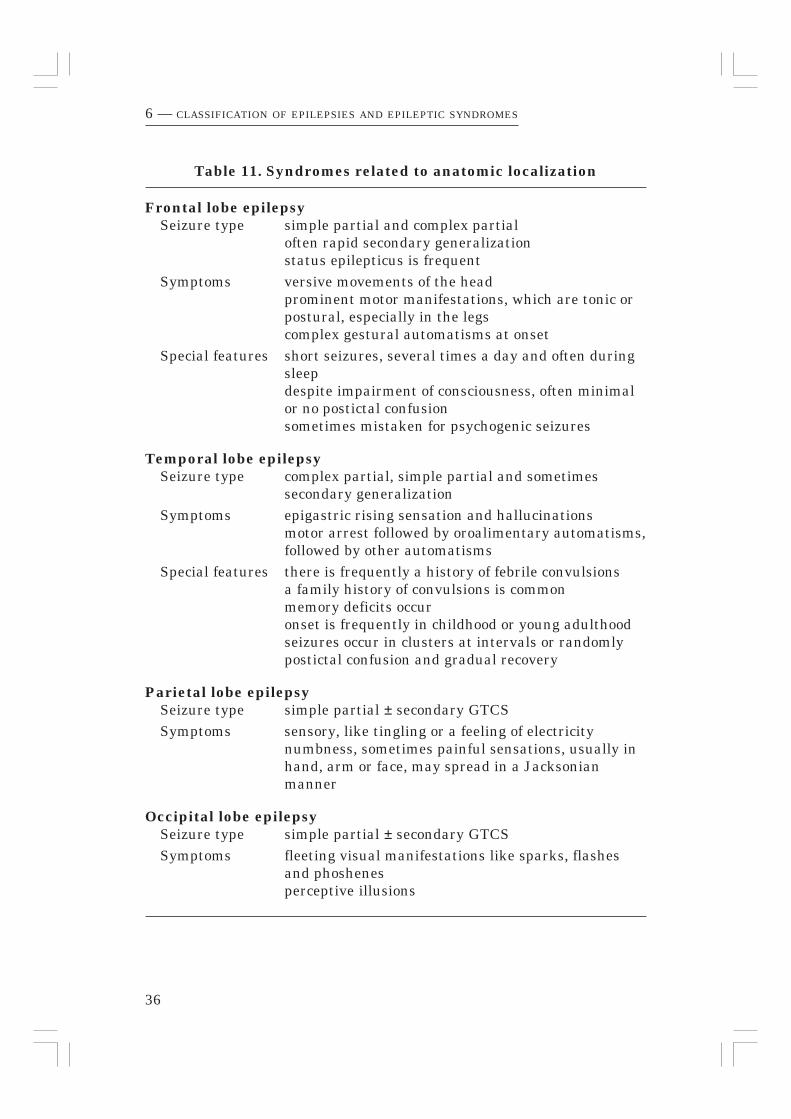
Table 11. Syndromes related to anatomic localization
Frontal lobe epilepsySeizure type simple partial and complex partial
often rapid secondary generalizationstatus epilepticus is frequent
Symptoms versive movements of the headprominent motor manifestations, which are tonic orpostural, especially in the legscomplex gestural automatisms at onset
Special features short seizures, several times a day and often duringsleepdespite impairment of consciousness, often minimalor no postictal confusionsometimes mistaken for psychogenic seizures
Temporal lobe epilepsySeizure type complex partial, simple partial and sometimes
secondary generalizationSymptoms epigastric rising sensation and hallucinations
motor arrest followed by oroalimentary automatisms,followed by other automatisms
Special features there is frequently a history of febrile convulsionsa family history of convulsions is commonmemory deficits occuronset is frequently in childhood or young adulthoodseizures occur in clusters at intervals or randomlypostictal confusion and gradual recovery
Parietal lobe epilepsySeizure type simple partial ± secondary GTCSSymptoms sensory, like tingling or a feeling of electricity
numbness, sometimes painful sensations, usually inhand, arm or face, may spread in a Jacksonianmanner
Occipital lobe epilepsySeizure type simple partial ± secondary GTCSSymptoms fleeting visual manifestations like sparks, flashes
and phoshenesperceptive illusions
6 — CLASSIFICATION OF EPILEPSIES AND EPILEPTIC SYNDROMES

37
Table 12. The relationship between epilepsy, prognosis andtreatment
Epilepsy Prognosis Treatment
1. Localization-related1.1 Idiopathic good phenytoin,
carbamazepine
1.2 Symptomatic depends on phenytoin,the lesion carbamazepine
(phenobarbitone)
2. Generalized2.1 Idiopathic good
Absence ethosuximide,valproate
Myoclonus valproate,benzodiazepines
Generalized tonic-clonic phenobarbitone,valproate,carbamazepine
2.2 Cryptogenic/symptomatic poorInfantile spasms ACTH, oral steroids,
valproate,benzodiazepines
Lennox-Gastaut prednisolone,valproate,benzodiazepines
2.3 Symptomatic poor phenobarbitone,phenytoin,carbamazepine
3. Epilepsies and syndromesundetermined as to whetherthey are focal orgeneralized see Appendix A
4. Special syndromes4.1 Situation-related seizures good valproate
4.2 Febrile convulsions good benzodiazepines
6 — CLASSIFICATION OF EPILEPSIES AND EPILEPTIC SYNDROMES

38
CONCLUSIONTo classify an epilepsy (and therefore to know which anticonvulsant is themost appropriate and to attempt to make a possible prognosis) thefollowing factors are important:
– Type of seizure
– Aetiology
– Age of onset of seizures
– Time of occurrence of seizure.
If the medical history and the physical examination do not enabledetermination of seizure type and aetiology, treat with phenobarbitonefor daytime seizures and phenytoin for nocturnal seizures. If the seizuresoccur both in the daytime and at night treat with phenobarbitone. If noimprovement occurs, change over to phenytoin.
In patients with true absences, however, neither of these drugs iseffective and phenytoin and carbamazepine may even aggravate thecondition.
6 — CLASSIFICATION OF EPILEPSIES AND EPILEPTIC SYNDROMES

39
7History, examination andinvestigations
MEDICAL HISTORYA carefully taken medical history is the most important need of all in apatient with repeated seizures. As the patient cannot recollect theseizures an accompanying person has to help in answering questionsabout the seizures.
First we let the patient and his companion tell their story, then wehave to ask pertinent questions about the present seizures and previousmedical history.
SeizuresOnset
– At what age was the first seizure? (Explain what is covered by theterm “seizure”, mimic absences, complex partial seizures, etc.)
– Was it in association with a particular event, accident or illness?
– Was there fever with the first seizure?
– Is there always fever with the seizures?
Pre-ictal phase
– Does the patient know of any precipitating factors such as hunger(pointing to hypoglycaemia), lack of sleep, emotion, alcohol, flashinglights, etc?
– Are there any prodromal symptoms?
– Is there an aura? What does it consist of?
Ictal phase (description of seizure itself)
– Does the patient get flushed or become pale? Does he see greyscintillations (pointing to a faint)? Does he scream?
– Where and how does it start, turning face to one side, or in one hand?
– Does he jerk? If so, with both arms and both legs, or only one side?
– Is the patient unconscious? Does he fall down?
– Does he fumble with his clothes, smack his lips, mumble or make anyother noises?
Duration
– How long does the seizure last?
7 — DIAGNOSTIC PROCEDURES

40
Post-ictal phase
– What is the patient’s behaviour like after the seizure—sleepy, aggressive,continues what he was doing?
– Is there any focal sign (e.g., Todd’s paralysis)?
Time
– Is it always in the day time, always when he is asleep, or onawakening?
Frequency
– How often does it happen?
Family history– Is there any similar disease in any of the siblings, the parents, or the
parents’ families?
Perinatal historyHow was the pregnancy:
– Any diseases or complaints?
– Were medicines or alcohol used, was the mother a chain smoker?
– Was it of normal duration?
How was the delivery:
– Normal, vacuum, forceps, or Caesarean section?
– Was it of normal duration, prolonged, or precipitate?
– Did the baby cry immediately after birth, or had he to be resuscitated?
– Did the baby have a low birth weight?
– Did he look yellow?
– Did the baby suck well?
– Was there any disease after birth?
– Did he get the usual vaccinations?
Development– Were the milestones normal?
– Does or did he go to school and how is/was he performing there?
– If of school-age and not attending school, why not?
– If over school age does he have a job?
– What is his behaviour like?
– Does he sleep well?
7 — DIAGNOSTIC PROCEDURES

41
What has been done up till now about the seizures?– Has he been admitted to a hospital for his seizures or for any other disease
or accident? (Or would he have been admitted if a hospital had beenaccessible?)
– What kind of treatment has he had in the past?
– Is he on treatment now?
– Which medicines, what dosage?
– If his drugs were changed what was the reason—allergy orineffectiveness?
– Is he treated by a traditional healer? If so what treatment has beenprescribed?
SOCIAL HISTORY– Where does the patient live? With parents? With sibs? With wife/husband
or child?
– Is it far from the clinic? How long does it take to reach the clinic?
How does he travel—by foot, etc.
– What are the parents’ occupations? If he is married, who earns a living?
– What is the educational level of the parents/the household member?
– Are the parents together?
– Who is looking after the patient,in case of need?
– How many siblings are there?/How many children are there?
– Are they all well/normal? If not, what is wrong?
– How is the patient doing at school/in his job? Is he coping?
– Are there any others in the class, the school, the workplace, or theneighbourhood with the same problem?
– Are there any problems with his teachers, superiors or colleagues,neighbourhood, (extended) family because of the epilepsy?
ANY PROBLEMS NOT YET MENTIONED
– Ask the patient or the parent to tell what problem they would like to talkabout that has not yet been dealt with.
7 — DIAGNOSTIC PROCEDURES

42
EXAMINATIONPhysical examinationThis starts with:
Observation
– From the moment the patient enters the consulting room noticewhether he walks normally, or is there weakness or spasticity on oneor both sides?
– Does he act appropriately to the new surroundings or is he too quiet,or too active?
– Is he able to communicate?
– Is the speech normal for his age?
Measurements
– Weight
– Height
– Head circumference (a graph of normal values is given in Appendix C).
General physical examination
Routine examination of all the systems. Note especially:
– Scars, bruises, change in pigmentation, adenoma sebaceum,haemangioma
– Asymmetry, congenital anomalies
– In acutely ill children note:
fever, bulging fontanels, neck stiffness, rash, level of consciousness
Neurological examination
– Eyes—size and reaction of pupils, visual fields, nystagmus, fundoscopyif possible
– Other cranial nerves
– Muscle power and tone—hyper- or hypotonic?
– Reflexes
– Is there any difference between right and left?
– Are there any signs of drug toxicity, e.g., ataxia, drowsiness, sleepiness,nystagmus
– Finger-nose test
– Dysdiadochokinesis (slowing or overshooting when alternating pronationand supination of the hands)
7 — DIAGNOSTIC PROCEDURES

43
INVESTIGATIONSLaboratory investigationsIf available, it is good to have baseline information on the generalcondition of the patient:– Haemoglobin, WBC count and differentiation, blood platelets, ESR,
blood film, urinalysis, and stool examination
– Specific tests are sometimes indicated to find the basic cause of theconvulsions (blood glucose, electrolytes, parasites, etc.)
– HIV/syphylis serology in suspected cases
– Liver-function tests are indicated in the first months after starting anantiepileptic drug
– Lumbar puncture is indicated when there is an acute illness withfever, convulsions and signs of meningitis (neck stiffness, in infantsbulging fontanels) or, in the absence of these signs, in a very ill patientwith no other obvious disease
– Exceptionally it may be necessary to measure the level of AED inserum (see page 65)
Skull X-ray– Signs of intracranial calcifications may be detected. Calcifications may
follow tuberculosis (tuberculoma), tuberous sclerosis, intrauterinecytomegalovirus (CMV) disease, Sturge-Weber syndrome, toxoplasmosisor cysticercosis.
– Signs of raised intracranial pressure might be seen (tumour).
Electroencephalography (EEG)This is a painless non-invasive and safe procedure whereby the electricalactivity of the brain is registered, amplified and recorded by a number ofelectrodes placed in a specific manner on the head. (The head does nothave to be shaved.)
– There are nearly always abnormal discharges to see on the EEGduring a seizure, and often in between seizures (inter-ictal).
– An EEG is often helpful in differentiating between partial andgeneralized seizures.
– The EEG has a characteristic pattern in a number of the epilepticsyndromes (e.g., absences).
7 — DIAGNOSTIC PROCEDURES

44
– Epileptic abnormalities can sometimes be provoked by hyperventilation,photic stimulation, sleep and deprivation of one night’s sleep. Duringan EEG recording, hyperventilation and photic stimulation are alwaysused. When sleep is required, it can be induced, for instance withchloral hydrate. On indication, the EEG is recorded after a night’ssleep deprivation.
However, this examination can be carried out only in a few top-referralhospitals on selected cases only, and treatment is usually given withoutthe help of the additional information given by an EEG.
Computer-assisted tomography (CT) scanComputer-assisted tomography (computerized axial tomography) is a safeand non-invasive procedure. Serial X-ray pictures of horizontal sections ofthe skull and brain are taken resulting in an accurate anatomical picture.A low radiation dose is used. This technique is able to demonstratetumours, haemorrhages, subdural haematomas, vascular anomalies andother structural abnormalities. Sometimes a contrasting medium is givenintravenously, and the two pictures, with and without medium, can becompared to further facilitate a diagnosis. CT scanning is a very helpfulbut very costly examination and can only be carried out in top-referralhospitals.
Magnetic resonance imaging (MRI)
Magnetic resonance imaging is based on the absorption and emission ofenergy in the radio frequency range of the electromagnetic spectrum. Thehuman body is primary fat and water. Fat and water have many hydrogenatoms. The human body is comprised of approximately 63% hydrogen atoms.Hydrogen nuclei have a Nuclear Magnetic Resonance (NMR) signal. Forthese reasons MRI primary images the NMR signal from the hydrogennuclei. MRI started out as a tomographic imaging technique, i.e., it producedan image of the NMR signal in a thin slice through the human body. MRI hasadvanced beyond a tomographic imaging technique to a volume imagingtechnique.
(From Joseph P. Hornak, Ph.D. at http://www.cis.rit.edu/htbooks/mri/inside.htm)
MRI has enormously advanced knowledge of the central nervous system.However, it is still a very expensive technique that is only sparsely availablein economically evolving countries.
7 — DIAGNOSTIC PROCEDURES

45
PSYCHOLOGICAL EVALUATIONPsychological evaluation by a psychologist is desirable, but opportunitiesfor this are limited in Africa.
Assessment of the intelligence, potential, or particular problems of thepatient is of value, for instance in placement at an appropriate school andto provide counselling for the patient, family and teachers to enable him/her to live as normal a life as possible.
Information may be obtained by carefully watching the child and byquestioning the mother who can compare this child to her other children(milestones, speech, daily activities such as eating, dressing, toilet trainingand behaviour).
A simple grading of the intelligence can be done as described on pages53-54.
Institutional care may be necessary, not only because of the degree ofmental retardation, but also because of the need to control the epilepticseizures and the degree of other related physical handicaps (e.g., spasticity).
For a review of this chapter see table 13.
7 — DIAGNOSTIC PROCEDURES
Table 13. What has to be asked and done
Medical historySeizuresFamily historyPerinatal historyDevelopmentPrevious seizure management
Social history
ExaminationObservationMeasurementsGeneral physical examinationNeurological examination
InvestigationLaboratorySkull X-rayElectroencephalography (EEG)Computer-assisted tomography (CT) scan(Magnetic Resonance Imagining )
Psychological evaluation

46
8 — DISORDERS TO BE DISTINGUISHED FROM EPILEPSY
8Disorders to be distinguishedfrom epilepsy
Before treatment with anticonvulsants is started, the diagnosis of epilepsymust be as certain as possible. When treatment with anticonvulsants hasstarted, and no improvement occurs at all, then the diagnosis of epilepsyshould be reconsidered.
The main conditions, which are often mistaken for epileptic seizures, arefainting attacks and psychogenic pseudoepileptic seizures (pseudo = lookalike). Also physiological jerks when falling asleep (hypnagogic or hypnic,jerks) are sometimes mistaken for a brief seizure.
A detailed medical history, and a clear witnessed account of theattacks are a must for differentiating these conditions (table 14).
Table 14. Conditions often confused with epilepsy
– Reflex syncope
– Cardiac syncope
– Postural syncope
– Psychogenic seizures
– Breath-holding spells
– Nightmares, night terrors, sleepwalking
– Narcolepsy
– Sleep apnoea
– Transient ischaemic attacks
– Senile falls
– Dizziness
– Migraine
– Trigeminal neuralgia
– Tetanus
– Hypoglycaemia
– Jitteriness in the newborn
– Temper tantrum
– Hypnagogic (hypnic) jerks
47
8 — DISORDERS TO BE DISTINGUISHED FROM EPILEPSY
SYNCOPE (FAINTING)Syncope is much more common than epilepsy, and the main differencesfrom epileptic seizures are shown in table 15. There is loss of consciousnessdue to a sudden decrease in the cerebral blood flow. Syncope may bedivided further into three groups:
Reflex syncope (vasovagal syncope)This is the most common type and includes simple fainting. Precipitatingfactors are anxiety, hunger or any unpleasant experience causing vagalnerve stimulation and reduced venous return. Standing at parades in warmweather (when venous pooling occurs in the legs) is a common cause offainting. The onset is often gradual, with a feeling of faintness, nausea ordizziness. Blurring or blacking out of vision is often present before thepatient slumps to the floor. Loss of consciousness is short, and often thereare some uncoordinated clonic jerks (causing the confusion with epilepticseizures). There is pallor and sweating and the pulse is slow. Recovery isquick without confusion. Treatment is to increase the blood flow to the headby putting the head between the knees, or to lie down.
Cardiac syncopeThis is caused by heart disease, disturbance of the heart’s rhythm orreduced cardiac output. The attacks can occur in any situation; inchildren it is often seen after exertion. A prolonged ECG may be necessaryto make the right diagnosis.
Postural syncopeThis occurs within seconds or minutes of assuming an upright position inpatients whose postural reflexes are impaired (elderly people, diabeticpatients, or due to medication or alcohol).
PSYCHOGENIC NON-EPILEPTIC SEIZURES(PSYCHOGENIC PSEUDO-EPILEPTIC SEIZURES, FUNCTIONAL SEIZURES, HYSTERIA)
These seizures simulate generalized tonic-clonic seizures and may occur inpatients with epilepsy or in patients who do not have epilepsy themselves.These pseudo-seizures may occur in several pupils in the same class orschool after having witnessed someone with a real epileptic seizure. It is anattention-seeking device in patients with emotional disturbances who cannotcope with the expectations of their surroundings. The seizures are variable,do not follow a typical pattern, and usually occur when there is an audienceand are often prolonged. In the classical hysterical seizure, the hips arelifted from the floor. (However, pelvic thrusting may also occur in frontallobe seizures.) There is rarely incontinence. There is no improvement withanticonvulsants. In table 16 the main differences are shown.

48
8 — DISORDERS TO BE DISTINGUISHED FROM EPILEPSY
Table 15. Differentiation between epileptic seizures and faintingattacks (reflex syncope)
Epileptic seizure Syncope
Precipitant unusual usual (emotion, pain)Circumstances any usually upright
position; crowded,hot surroundingsemotional stressfulsituations
Onset sudden or aura gradualMotor phenomena tonic or flaccid
tonic-clonic uncoordinated clonicclonic movements with jerks of low amplitudecharacteristic amplitudeand frequency
Skin colour pale or flushed paleRespiration stertorious, foaming shallow, slowIncontinence common rareTongue-biting common rareVomiting unusual oftenInjury common rarePost-ictal drowsy, confusion usally rapid recovery
sleep without confusionDuration minutes seconds

49
8 — DISORDERS TO BE DISTINGUISHED FROM EPILEPSY
Table 16. Differentiation between epileptic seizures andpsychogenic seizures
Epileptic seizure Psychogenic seizure
Precipitant unusual usual (emotion)
Circumstancesin sleep common rarewhen alone common less common
Prodroma rare common
Onset sudden or aura gradual
Cry at onset common unusual
Vocalization during automatism only groans and moansor cries usuallythroughout fit
Motor phenomena stereotyped, usually variable, usuallyboth tonic and clonic; tonic or clonic;clonic movements slow clonic movementsas seizure continues and vary in amplitudeincrease in amplitude and frequency;
pelvic thrusting(also in frontal lobeepilepsy!)
Injury common rare
Incontinence common rare
Tongue-biting common rare
Consciousness lost in GTCS, usually not lost; mayreduced in CPS not respond
Resistance to passivelimb movement oreye opening unusual common
Duration seconds or minutes many minutes
Terminationof attack usually rapid; gradual, often with
confusion, drowsiness emotional display;or sleep common confusion,
drowsinessor sleep unusual
Recall ofseizure events very rare possible or elicitable
by hypnotic recall

50
OTHER DISORDERSBesides these pseudo-seizures, there are also other attacks, such as
swoons and tantrums, which might be confused with simple partial orcomplex partial seizures. Jerks (hypnagogic startles; hypnic jerks) whenfalling asleep are not rare, but usually are not mistaken for epilepsy.
In a swoon, there is a sinking to the floor, with closed eyes, followedby lying inert on the floor with peculiar eyelid flickering. Swoons are away of avoiding difficult situations, unpleasant thoughts or memories.
In a tantrum, the subject throws himself to the floor screaming,thrashing about and kicking. Often the subject bites himself or even bitesonlookers. Tantrums frequently occur in children and immature, brain-damaged or underprivileged people as an expression of anger andfrustration.
Two other forms of pseudo seizures are panic attacks and episodicconfusion. A panic attack is characterized by shortness of breath,smothering sensations, choking and chest pains while episodic confusioncan occur after the use of such drugs as barbiturates, organic solvents(glue sniffing), and adulterated alcohol.
Less common disorders to be differentiated are covered below.
Breath-holding spellsThese occur in infants and children between one and five years old. Theyare caused by anger or pain in children who cannot restrain theiremotions. There is a sudden arrest of the respiration followed by cyanosis,unconsciousness, and sometimes twitching lasting a few seconds. Theattack stops spontaneously and never results in brain damage.
Nightmares, night terrors and sleepwalkingThese are often familial and occur in childhood, mostly between the agesof 4 and 14. Nightmares occur during REM sleep. Night terror (pavornocturnus) and sleep walking occur during deep sleep.
NarcolepsyThese are attacks of sleeping many times a day under the strangestcircumstances. It is often familial and occurs mostly between the ages of15 and 30. The condition usually becomes chronic, but spontaneousimprovement sometimes occurs. Stimulants like ephedrine ormethylphenidate or antidepressants like clomipramine may be tried.
8 — DISORDERS TO BE DISTINGUISHED FROM EPILEPSY

51
Sleep apnoeaSleep apnoea is a disturbance of the respiration during sleep, withdiscontinuation of the respiration for about half a minute or longer upto a hundred times a night. The patient is not aware of these spells.
Transient ischaemic attack (TIA)These attacks occur in an older age group and are caused by a transientreduction in blood supply to parts of the brain, usually secondary toarteriosclerosis of the cerebral vessels and vessels in the neck. Disturbancesof consciousness may be associated with focal paralysis, disturbance ofspeech or loss of vision which occurs for minutes or hours. More prolongedsymptoms may indicate a more serious cerebrovascular accident (stroke).
Senile fallsIn elderly people sudden falls may be triggered by turning of the head orlooking upwards. In this case reduced cerebral blood flow is caused bykinking of a calcified blood vessel.
DizzinessReal vertigo is an attack associated with a spinning sensation, loss ofposture, nausea and vomiting caused by diseases of the inner ear.
In Menière’s disease, i.e., deafness and tinnitus in one ear, attacks ofvertigo may last from half an hour to several hours.In acute labyrinthitis severe vertigo may last for several days.
Vague sensations of dizziness with loss of balance is a commoncomplaint in many people.
MigraineMigraine is a paroxysmal headache, often unilateral, but sometimesgeneralized. The headache may be preceded by an “aura” which iscommonly a visual disturbance, e.g., flashing lights, fortification spectraor hemianopsia, but there may be numbness and tingling in the face orlimbs or even a hemiparesis. These symptoms last for 15–30 minutes andare invariably followed by severe headache. The aura in an epilepticattack lasts only a few seconds. Very rarely, an acute migraine attackmay result in a permanent neurological disturbance, for example epilepsyor a defect of vision. Migraine attacks may precipitate seizures.
Trigeminal neuralgiaPeriodic attacks of severe pain on one side of the face, of unknown cause,but most effectively treated by carbamazepine.
8 — DISORDERS TO BE DISTINGUISHED FROM EPILEPSY

52
TetanusA wound infection with Clostridium tetani. After a variable incubationperiod (5–30 days) the first clinical manifestations are intermittent clonicconvulsions. Episodic muscle spasms, especially of jaw and neck, withdifficulty in opening the mouth (lockjaw) occur. There is a typical facialexpression, “risus sardonicus”, board-like rigidity of the body, andpronounced opisthotonus with legs and feet extended, stiff arms andclenched fists. These spasms are precipitated by the slightest stimulus(touching the patient, noise and light). As the disease progresses theintervals between the spasms are less obvious. Fever is common. Withouttreatment the disease is usually fatal within a week.
In addition to the conditions mentioned in this chapter, all the conditionsgiven in table 1 have to be considered in the differential diagnosis of epilepsy.The most common of these conditions are hypoglycaemia, meningitis, cerebralmalaria, electrolyte imbalance and poisoning. If these conditions areappropriately treated and the patient recovers, prolonged protection withantiepileptic drugs (anticonvulsants) like in epilepsy will not be necessaryuntil there is proof that the disorder, e.g., meningitis or cerebral malaria hasbrought epilepsy in its wake.
8 — DISORDERS TO BE DISTINGUISHED FROM EPILEPSY

53
9Conditions co-existing withepilepsy
Sometimes the insult to the brain is so severe (e.g., in birth trauma, birthasphyxia, etc.) that in addition to the epilepsy there may be otherimpairments, e.g., cerebral palsy and/or mental retardation. Of thepatients attending KAWE clinics, 32% have, besides their epilepsy,another handicap, while 12% of all patients have multiple handicaps.
CEREBRAL PALSYIn Europe the incidence of cerebral palsy is 2 per 1,000 live births. Thecondition is classified into the following syndromes:
– Spastic (hemiplegic, tetraplegic and diplegic)– Ataxic– Dyskinetic (choreo-athetotic and dystonic)
Epilepsy is most common in the spastic and rare in the ataxic anddyskinetic syndromes.
MENTAL RETARDATIONIn Europe about 3% of all children have a certain degree of mentalretardation, while 0.3% of all children have it in a severe degree. Thecondition is graded from borderline, mild, moderate, severe to profound,depending on the results obtained by testing the IQ (intelligence quotient).As the tests for measuring IQ are not yet standardized in every country,a simple grading can be done according to the following definitions:
– Mild mental retardation, the educable groupThese children can master basic academic skills up to Standard IV toVI with extra help, and are able to master simple vocational skills andadjust fairly well to social demands (social accomplishment level of a7–9 year-old child).
– Moderate mental retardation, the trainable groupMay acquire very basic academic skills (5–7 year-old level). May betaught self-help-care skills and could work in a sheltered workshopunder supervision.
9 — CONDITIONS CO-EXISTING WITH EPILEPSY

54
– Severe mental retardationRequire custodial and sometimes nursing care. Can be taught mostbasic self-help-care skills such as eating, drinking and toilet training(3–5 year-old level).
– Profound mental retardationThese children cannot be tested (neither can they be trained) andrequire complete care (3 year-old level or less).
Epilepsy is common in mentally handicapped children, the proportionrising as the degree of retardation increases (from 6 to 30%; Corbett, 1985and from 13.7% to 44.4% in Kibwezi, Kenya, see Baldwin 1990).
The occurrence of epilepsy varies in the different syndromes associatedwith mental retardation. It is very common in children with tuberoussclerosis, common in those with spastic tetra- and hemiplegia, and not socommon in children with Down’s syndrome.
Although epilepsy is common in mentally handicapped children, mentalretardation is not as common in patients with epilepsy. In Europe it issaid that one out of three retarded children cared for institutions hasepilepsy, while one out of ten epilepsy patients is mentally handicapped.
In Africa there is, however, a great difference between the findings ina rural clinic and an urban clinic. For example in Kenya in the ruralclinic 32.5% were retarded, in an urban clinic this was 21.4% (table 17).Also, the physically handicapped were more common in the rural clinicthan in the urban one—20.3% and 11.4% respectively.
The overall distribution of people with epilepsy, with or withoutmental retardation and or physical handicap for the KAWE clinics ispresented in table 18.
Most probably, in the rural areas, where people live far from healthfacilities, especially hospitals, more children develop severe brain damagefollowing birth trauma, birth asphyxia, or childhood infections resulting inmore multiple handicapped children with severe forms of epilepsy. However,another explanation may be that assistance of health authorities is more likelyto be looked for when there are multiple handicaps or severe epilepsy.
9 — CONDITIONS CO-EXISTING WITH EPILEPSY
Table 17. Difference between data from one rural clinic, n = 442,and one urban clinic, n = 597, as percentages
Epilepsy + Epilepsy +Physical handicap Mental retardation
(± Mental retardation) (± Physical handicap)
Rural 20.3% 32.5%Urban 11.4% 21.4%

55
Table 18. Data of 1493 patients with epilepsy attending threeKAWE clinics (90% of the patients were below the age of 20 yr)
Epilepsy + Epilepsy + Epilepsy +Physical handicap Physical handicap + Mental retardationwithout Mental Mental retardation withoutretardation Physical handicap
54 180 246(3.5%) (12%) (16.5%)
Epilepsy + Epilepsy +Physical Mentalhandicap retardation
234 426
PSYCHIATRIC PROBLEMSEpilepsy is not a mental disease, although a small number of patientswill develop psychiatric problems.
This is more likely when there is organic brain damage, an early ageof onset, a chronic form of epilepsy, special location (e.g., temporal lobeepilepsy), or a difficult adjustment to social surroundings.
Psychotic breakdown does not usually occur before puberty. Referral toa psychiatrist is indicated in all cases where the patient or his relativescannot cope with the emotional and/or psychiatric morbidity. When thepsychiatric problems need medical treatment, it must be rememberedthat chlorpromazine (Largactil) and haloperidol (Serenace) lower thethreshold for seizures. If possible, benzodiazepines or other less seizure-inducing antipsychotics should be used.
BEHAVIOUR DISORDERSBrain lesions (from minimal brain damage to more severe damage) canalso result in behaviour problems such as hyperactivity, irritability, lackof concentration and aggression, especially in left-sided temporal lobe (ifdominant) dysfunction. Right temporal lobe dysfunction tends to makethe patient introverted. Occasionally, especially in children, the behaviourproblem is a side-effect of the medication (phenobarbitone or clonazepam).
9 — CONDITIONS CO-EXISTING WITH EPILEPSY

56
Table 19. Causes for learning disabilities
– Presence of actual seizures
– Presence of subclinical epileptic activity
– Structural brain abnormality
Lesions in the left temporal lobe (if dominant) carry a greater riskof speech and language disorders.
Lesions in the right temporal lobe (if non-dominant) carry a greaterrisk of problems with mathematics.
– Use of anticonvulsant drugs
– Unfavourable environmental factors at school and at home causingemotional problems, which might be the most important factors causingacademic and social under-achievement.
LEARNING DISORDERSSometimes there are specific learning problems. Although these childrenmay have normal intelligence, they are often unable to attend normalclasses in a normal school because of their restlessness and lack ofconcentration.Some causes of these learning disabilities are given in table 19.
DEMENTIAWhen there are many long-lasting seizures, or repeated status epilepticus,loss of nerve cells results, with the development of early dementia. Earlyand quick medical control of seizures is therefore very important.
9 — CONDITIONS CO-EXISTING WITH EPILEPSY

57
10Treatment
The first action when a person has a seizure is seizure management(table 20). The second is to find a reason for the seizures (see diagnositicprocedures chapter 7). The third is starting treatment.
Table 20. Management during a seizure (fig. 7)
1. Move patient away from fire, traffic or water
2. Take away any objects that could harm the patient
3. Loosen tight clothes, remove glasses
4. Put something soft under the head
5. Turn patient on his or her side, so that saliva and mucus can run outof the mouth
6. Remain with the patient until he or she regains consciousness
7. Let the patient rest and then resume whatever activity he was doing,if he feels like it
Some Don’ts
1. Do NOT try to put anything into the mouth
2. Do NOT give anything to drink
3. Do NOT try to stop the jerking, or restrain the movements.
Figure 7. The coma position. Unconscious patients should be placed in thissemi-prone position to minimize the risk of obstruction or inhalation of
vomit
10 —TREATMENT

58
DRUG THERAPYMore than one seizure should have occurred, and other causes for theconvulsions (e.g., hypoglycaemia) excluded (treated) before regulartreatment with antiepileptic drugs (AEDs) is begun.
The aim is to try and prevent further seizures, either completely or toreduce their frequency and severity as much as possible with the leastpossible side-effects (see also table 26, page 75).
The medication should be given daily for many years, sometimes life-long,sometimes less. After a seizure-free period with medication for at least twoyears in idiopathic, and at least three years in symptomatic epilepsy, thedosage might be reduced very gradually over many months, and, if no relapsehas occurred, discontinued. In cases where the epilepsy was very severebefore treatment was started, or where there is a known brain lesion, it isbetter to continue the treatment for much longer, as the chance of a relapseis then much greater. Dose frequency is determined by the elimination half-life time and the need to avoid toxic peaklevels. Guiding principles how tostart medication in newly diagnosed patients are summarized in table 21.
A sudden discontinuation may lead to a dangerous statusepilepticus. (For treatment see table 22, page 66)
The treatment should be started with one drug only. Ideally, the choiceof the drug depends on the type of epilepsy and the seizure type. Inpractice, however, it also depends on the availability and affordability ofthe drug.
As it is very difficult to know in the beginning which type of epilepsythere is, treatment is usually started according to the presenting seizuretype. The most common seizures are the generalized tonic-clonic seizures(GTCS). The main four AEDs—phenobarbitone, phenytoin, carbamazepineand valproate—are almost equally effective for these seizures. If we areable to differentiate between primary and secondary generalized tonic-clonic seizures, then phenobarbitone or valproate are the drug of choicefor the primary GTCS and phenytoin or carbamazepine for the secondaryGTCS, but all four are effective and can be tried separately if necessary.
Phenobarbitone, if it is the only available drug in a dispensary orhealth centre, then all patients with epilepsy might be started withphenobarbitone treatment. This medicine is no longer advocated in thedeveloped world, but it is still a useful, effective and cheap anticonvulsant.But if there is no improvement, or even a worsening of the condition, thedosage should not be increased beyond 120 mg daily, and the patientreferred to a clinic or hospital where other anticonvulsants are available.
10 —TREATMENT

59
10 —TREATMENT
Table 21. Guiding principles to start anticonvulsant drugtreatment in previously untreated patients
1. Carefully establish diagnosis.
2. Start drug treatment with one drug.
3. Start drug treatment with a small dose.
4. Gradually increase dosage until complete seizure control.This is the minimum maintenance dose.
5. The aim of treatment is to achieve the lowest maintenance dosewhich provides complete seizure control.
6. A gradual introduction of an anticonvulsant can producetherapeutic effects just as fast as rapid initiation with large doses,but with fewer side-effects.
7. Severe “intoxication” side-effects appearing at the beginning of thetreatment can indicate too rapid or too large dose increases. Side-effectsto anticipate include fatigue, excess sleep need, dizziness, or difficultywalking (ataxia).
8. If the initial drug of choice is not well tolerated, e.g., if side-effectsoccur or if the maximum tolerated dose does not produce seizurecontrol, substitute the initial drug with another first lineanticonvulsant.
9. A second anticonvulsant should be added gradually and the firstthen slowly withdrawn.
10. In case of acute withdrawal symptoms, e.g., recurrence of seizures,use diazepam as a control drug.
11. Regular compliance is the key to successful seizure control, andcounselling the patient is the most critical factor in compliance.

60
The main side-effects of phenobarbitone are drowsiness, especially duringthe first week of treatment, slowly disappearing, and only recurring whenthe dosage becomes too high. In some children there might be a reduction inscholastic performance or changes in the behaviour, such as hyperactivityand sometimes aggressiveness. Phenobarbitone has a long half-life time andtherefore it will take a few weeks before reaching a therapeutic and effectivelevel. It also means that it can be given only once a day, preferably after theevening meal before retiring to bed. The main indications are the idiopathicgeneralized epilepsies. But it is also reasonably effective in other generalizedseizures and in partial seizures. It is not effective in generalized absences,and it might worsen nocturnal seizures, as it increases the deep sleep. It isthe drug of choice, when prophylactic treatment for febrile convulsions isindicated, however, if rectal diazepam can be easily obtained at a reasonableprice then instead of prophylactic treatment, intervention when a febrileseizure re-occurs is the preferred strategy.
Phenytoin is also a very effective anticonvulsant for partial and GTCSand seizures during sleep. The main problem is the small margin betweenthe therapeutic level and the level where the metabolizing enzyme getssaturated and the serum level rises steeply to reach toxic values.Increments should be not larger than 50 mg to prevent toxic side-effects.The side-effects are drowsiness, gum hypertrophy and hirsutism, andwhen the dosage is too high ataxia and nystagmus. Besides thesereversible cerebellar signs at high dosage, it has been suggested that apermanent cerebellar syndrome may also result from chronic therapy. Amild sub-clinical neuropathy is common after prolonged phenytoin therapy,but may occur with other drugs too. If toxicity has appeared, the dosageshould be omitted for one day and then restarted on a lower level. If atall possible, a change-over to another anticonvulsant could be made toprevent further mishaps.
When medicines are made by different factories, there will be a slight change inthe bioavailability and the rate of absorption of the different tablets. Usually this hasno therapeutic consequences, except in anticonvulsant therapy, and especially intreatment with phenytoin. A slight increase in absorption of the tablets could resultin toxicity, while a slight decrease in absorption might result in recurrence ofseizures. As these changes have proved to be unavoidable, the dosage of theanticonvulsant has to be adjusted after the change, preferably assisted by measurementof the serum level.
Phenytoin has also a long half-life time, which is furthermore dose-dependent, being longer at higher doses, and it may take up to two weeksbefore it becomes effective. It can be given in a once daily dose. As it isslightly irritating to the stomach, it should always be given after a meal,and when the dosage is high, it might be better to divide it into two doses.
10 —TREATMENT

61
Carbamazepine is a drug marketed after 1960. The main indication isfor complex partial seizures. But it is also effective for other partialseizures and for all GTCS. It is not effective for generalized absences andmyoclonic seizures. In the beginning of the treatment drowsiness anddizziness occur, and occur again when the dosage becomes too high. Thenthere might be also double vision and ataxia. It does not have a long half-life time and therefore it cannot be given once daily. It should be giventwice daily and when combined with other drugs it must be given threetimes daily.
Valproate has been marketed since 1966. The main indications are thegeneralized absences, myoclonic seizures, and the drop attacks. It is alsoused for the GTCS occurring after awakening. And if necessary it might beused for all other seizure types. When phenobarbitone cannot be used asprophylaxis for febrile convulsions, valproate can be used instead. It has ashort half-life time. Although its pharmacodynamic action in the centralnervous system exceeds its presence in serum, it should be given three timesdaily in order to avoid high peak concentrations. The specific side-effects areincrease in body weight, loss of hair, and gastric irritation. The effect on thefoetus is more serious, as spina bifida might occur. The risk of spina bifidais reduced by supplementing folate in all women at risk of being pregnant.
Ethosuximide is only used when there are absence seizures. If otherseizure types are present in the same patient, other medicines have to beadded to control these other seizure types. In epileptic syndromes, whereabsences, myoclonic seizures, drop attacks, or GTCS occur in combination,it is easier to use valproate, as this will take care of all the differentseizures.
Clonazepam is rarely used on its own. It is usually added when thereis not sufficient control of the seizures, often in children with drop attacksand myoclonic seizures.
Diazepam is used for status epilepticus or febrile convulsion status. It isalso used to abort a febrile convulsion to prevent a prolonged febrileconvulsion. It should be given intravenously, but if the vein cannot befound, the same solution can be given rectally.
Newer drugs, such as vigabatrin, oxcarbazepine, lamotrigine,felbamate, gabapentin, topiramate, and levetiracetam are notdiscussed as they are not widely available on the African market.
10 —TREATMENT

62
MonotherapyWhen treatment is started it is with one drug only. A small starting dose(as detailed in Appendix B for each individual drug) is given for 3–4weeks (phenobarbitone or phenytoin) or for one week (carbamazepine orvalproate) then increased with increments at regular intervals until theseizures are controlled, or until side-effects appear, or until the maximumdosage for the drug has been reached.
If side-effects appear and the seizures are not yet controlled a seconddrug is introduced and the first drug continued at the level before theside-effects appeared. When the second drug has become effective, thefirst drug is gradually withdrawn. If seizures recur, the second drug isincreased. Only when both drugs have been tried alone up to a levelwhere side-effects occur may a combination of the two drugs be tried.
In a small number of cases (often brain damaged small children) athird drug has to be added, or it has to be accepted that in some casesadequate seizure control cannot be achieved.
In many cases, the first drug will soon be effective, but in some casesit will take many months before the most effective treatment is found.During this difficult period the support and advice of the Epilepsy Aide(page 79) will be extremely important for the patient and his relatives.
The costs of the various medicines makes some unaffordable in Africa.Relative prices are given in table 29 page 83.
Elimination half-life timeBoth phenobarbitone and phenytoin have a long half-life time (the timeit takes to reduce the concentration of the drug to 50% as it is eliminated,metabolized and excreted). It takes about five times the half-life timebefore the drug reaches its therapeutic level and the steady state in theblood (see details of individual drugs in Appendix B). It has to beexplained carefully to the patient and his caretakers that the seizureswill not stop immediately after the medication has begun, but that achange will be noticed only after some weeks.
For the same reason, dosage increments above the expected effectivedose should only be made after an interval of 3–4 weeks.
Because of this long half-life time of phenobarbitone and phenytoin itis not necessary to give these drugs three times daily. A once-daily doseis sufficient and much more convenient for the patient. Only in smallchildren, who metabolize quicker, is a twice-daily dose indicated.
In newborns the half-life time is even longer and drugs have to beadministered with great caution.
Carbamazepine and valproate have a much shorter half-life time; theirincrements should be given weekly, the therapeutic level is reached muchquicker. But the medicines have to be administered three times daily.
10 —TREATMENT

63
At the very onset of therapy the metabolic rate adapts to the presenceof certain drugs. This class of drugs is called enzyme-inducers to whichbelong carbamazepine, phenytoin and phenobarbitone. Therefore, duringthe first weeks (± 3) the half-life gradually gets shorter. This is a reasonto start with small doses.
InteractionA problem with all medicines, but which is especially noticeable in drugstaken over a long period, is interaction with other drugs. Phenobarbitone,phenytoin and carbamazepine have a liver-enzyme inducing action. Theseincreased enzymes metabolize and excrete simultaneously given medicinesfaster and therefore these medicines do not reach as high a level as wouldbe expected from their dosage.
Valproate inhibits the action of the liver enzymes and therefore medicinesgiven simultaneously may reach a higher or even toxic level in the serumthan expected from the dosage given.
Phenytoin is bound to plasma proteins to a large extent, but may bedisplaced by other protein-bound medicines, such as valproate. Theresults of all these interactions are not always predictable and as routinelaboratory facilities for determination of serum drug levels are not yeteverywhere available in Africa, patients on anticonvulsant treatmenthave to be watched closely.
It is very important to remember that anticonvulsants interact witheach other; for example, phenobarbitone or phenytoin decreases thecarbamazepine level, whereas valproate increases the phenobarbitonelevel (see individual drugs in Appendix B).
Anticonvulsant drugs also react with other medicines of which themost important are the contraceptive pill, warfarin, antituberculosismedicines, antimycotics, some antibiotics, vitamin D and folates.
The level of contraceptive medication in the serum is decreased by theanticonvulsants phenobarbitone, phenytoin and carbamazepine. Therefore,the action of the contraceptive is no longer reliable and higher doses orother methods of family planning have to be used when the woman is onthis therapy.
Antituberculous drugs might increase the level of anticonvulsants andtherefore the latter might reach toxic levels.
10 —TREATMENT

64
Side-effectsAll drugs have side-effects. These effects can be divided into four maingroups, as shown below and specified in more detail in the descriptionsof the various drugs in Appendix B.
1. Local effects
Irritation of the gastro-intestinal tract when the drug is taken orally.These can be overcome by taking the medication after meals or when alarge dose has to be taken, by dividing it into two.
2. Dose-determined effects
A too-high dosage of most of the anticonvulsants might cause drowsiness,nystagmus, diplopia and ataxia. More specific changes are dependent onthe particular drug. The dosage should be decreased.
3. Idiosyncratic effects
These are allergic reactions such as skin rash, fever or eosinophilia, butalso bone-marrow depression or hepatitis.
When these reactions occur the treatment should be discontinuedimmediately and another anticonvulsant used instead.
4. Effects on the foetus and newborn
Congenital malformations might occur when taking the drugs duringearly pregnancy, but the risk is much less if only one drug is being used.However, the mother’s seizures are also dangerous for the young foetus, andtreatment has to be continued, but if possible only one drug should be used.During pregnancy the combination of phenobarbitone and caffeine should beavoided.
Anticonvulsants might lead to an increased bleeding tendency in thenewborn. Vitamin K has to be given prophylactically to the newborn.
PregnancyDuring the last months of pregnancy an increase in medication is oftennecessary. This can be reduced again after the baby has been born.
Breast milkMost of the anticonvulsants are present in breast milk. Very occasionallyphenobarbitone causes drowsiness in the baby; with phenytoin the effectsare not noticeable. There is no contraindication for breast feeding.
10 —TREATMENT
65
When there is no improvement at all after starting the treatment, thefollowing reasons have to be considered:– The diagnosis is wrong, it is not epilepsy (the seizures might be
fainting attacks or pseudo-seizures).– The medicine is wrong, wrong drug of choice (e.g., phenytoin for
generalized absences), wrong dosage too low or too high, or wrongfrequency of administration, e.g., daily dose of carbamazepine givenonly once a day, instead of divided over two or three times.
– The relation between dosage and serum level is not as it should beas patient does not comply (this can also lead to status epilepticus).Guidelines to promote compliance are given in table 23, page 68.
MONITORING SERUM LEVELS OF AEDS Several reasons have been mentioned why the dosage may not
correspond with the level of the drug in serum. Some persons metabolizethese drugs faster than others; or in the case of phenytoin, the enzymesystem that metabolizes the drug may get saturated, and the levelsuddenly rises to a toxic amount. A drug with a long half-life likephenobarbitone may gradually accumulate and unexplained fatigue ensuesdue to an unexpected toxic level.
A drug may not be absorbed, because of diarrhoea. Or the patient may nottake the drug because he forgets or is afraid of taking it. To make sure thatthe level of the drug in serum is as expected, this level can be measured.However, such investigation is expensive and not available everywhere.
Like there is no absolute effective dose, only a usually effective one, sothere is no absolute therapeutic/toxic level.
Pregnancy alters both metabolism and distribution of antiepilepticdrugs that may call for adjustment of drug dosage.
DIETBesides therapy with anticonvulsants, a ketogenic diet has been used inchildren with intractable seizures. Although it might be effective, it iscostly and difficult to follow.
SURGERYIn some cases surgery could be indicated. Required conditions are:– Long-term appropriate anticonvulsant therapy has not controlled
the seizures.– A clearly defined focal lesion has been demonstrated. Lesions in the
frontal part of a temporal lobe are especially suitable for surgery.– Surgery of this focal lesion should not introduce other neurological
problems such as aphasia, or paralysis.
10 —TREATMENT

66
Table 22. Management of status epilepticus
While making arrangements to set up an i.v. line, a few questions must be askedof the accompanying persons (a detailed history can be taken later). Instead ofwriting “he/she” throughout, “he” is used.
1. Questions– Is the patient known to suffer from epilepsy? Did he take
anticonvulsants? Did he discontinue the intake? If so, when?– Is the patient known to use alcohol or other drugs? Did he discontinue
to use this recently?– Have any symptoms been noticed that might indicate a disorder apt
to cause convulsions?– When was the last meal taken?– Were any traditional medicines, especially herbs, taken recently?
The answers to the last three questions above could indicate ahypoglycaemic state.
2. Diagnostic procedures– If there is no laboratory, a blood glucose determination can be done
with dextrostix.– If there is a laboratory, blood to be taken for more investigations
(blood glucose, calcium, electrolytes and urea).– Lumbar puncture to be done when fever is present or patient’s
immune system function is poor.– Other investigations to be done after the seizures are under control.
3. Start i.v. line with 5% glucose solution.
4. Start monitoring pulse, respiration and blood pressure. Preventaspiration, maintain clear airway.
5. Medicines (Inject into the i.v. line)a. Glucose solution
50 ml of 50% glucose with 100 mg thiamine.b. Diazepam
0.3 mg/kg i.v. very slowly, 1 mg/min, until seizures stop orup to 5 mg in children under 5 years; 10 mg in children 5–10 years;15 mg in older children and 20 mg in adults.May be repeated after 20–30 min, and once again 30 min later.If not successful in finding a vein:– the same solution in the same dosages can be given rectally via
a catheter or syringe. (In other countries special “rectioles” withdiazepam are available for this purpose.)
N.B. Do not give diazepam i.m.
Continued on page 67
10 —TREATMENT

67
Table 22, continued
or give:c. Paraldehyde 0.15–0.20 ml/kg intramuscularly.N.B. Paraldehyde reacts with plastic, therefore use a glass
syringe.Painful injection—avoid if possible.
6. Loading doseGive loading dose, to prevent recurrence of seizures, 20 minutes afterlast diazepam injection.a. Phenytoin
15–18 mg/kg very slowly i.v. into the line not more than 50 mg/min,not exceeding 1,000 mg, or give5–6 mg/kg i.v. three times with 30 min in between.N.B. Dextrose solutions may precipitate phenytoin.N.B. Do not give these high dosages when patient is already
on anticonvulsants.N.B. Do not give phenytoin i.m.
orb. Phenobarbitone
3–5 mg/kg two or three times during the first 24 hours i.m.
Continue checking pulse, respiration and blood pressure.
7. Maintenance doseAfter the loading dose is given, continue with the maintenance dose.When a patient is able to swallow, continue maintenance therapyorally. Give either:
1. phenytoin 5 mg/kg/day orallyor
2. phenobarbitone 2-3 mg/kg/day orally.
8. Other therapyDepends on the results of investigations (for instance, if meningitis,start i.v. antibiotics).
9. Further diagnostic proceduresA status epilepticus might be the first sign of a brain tumour.Skull X-ray, EEG to be done, or patient referred for these investigations.(CT scan.)NB: The medical emergency is to stop status epilepticus; theappropriate special investigations can be done later once theseizures have been stopped.
10 —TREATMENT

68
10 —TREATMENT
Table 23. Promoting compliance
The patient’s adherence to any prescribed treatment will increase if thefollowing points are considered. In the particular case of treatment withAEDs (before starting the treatment and during all its duration) thepatient has to be warned especially in a clear and understandable way tobear in mind the points e-h. Make sure if:
a. the patient perceives his clinical condition (in this case, seizures) as aproblem;
b. the patient is convinced: that the proposed treatment has a reasonableprobability of improving his clinical condition (i.e.,, decreasing themagnitude of the problem); that disappearence of seizures does notmean that treatment is no longer necessary;
c. the side-effects (and all other inconveniences involved) are bearableand justified in terms of the perceived benefits;
d. the treatment procedures are easy to follow;
e. the goal of the treatment is the reduction of seizures to a minimumpossible. For some patients this could represent no more seizures, butfor others only less seizures;
f. the treatment has no immediate effect; it takes a few weeks (two tosix) before the drug reaches a protective blood level;
g. prescribed dose should not be altered by the patient and his/her family,regardless of the degree of seizure control. Only the health worker canmodify the prescribed dose;
h. abrupt interruption of drug intake should be avoided at all costs asthis may precipitate continuous seizures. Provisions should be madefor timely procurement of the drug.
The language, the terms and the contextual meaning must be those ofthe patient. It is also important to enquire about the reasons for non-compliance and to deal appropriately with those reasons.
The following procedures have successfully proven to help in promotingcompliance with treatment:
1. Use of family reminders;
2. Linking drug intake to specific daily activities; and
3. Increased home visits with repeated explanation of:
– the necessity for continuous long-term treatment, and
– possible side-effects.
Source: Initiative of support to people with epilepsy. Division of Mental Health, World HealthOrganization, Geneva 1990

69
11Febrile convulsions
This syndrome is discussed separately in this chapter as it is one of themost commonly seen syndromes.
A febrile convulsion has been defined as an event in infancy orchildhood that occurs between three months and five years of age,associated with a fever but without evidence of intracranial infection ordefined cause (Consensus Development Panel, 1980).
The highest incidence is in children between nine and 20 months.About 3% of all children develop febrile convulsions. A genetic trait is
most probably present, disposing a particular child to get it easily. Itoften occurs in siblings as well.
It is usually a benign disorder occurring in children of normaldevelopment and occurring early in a recognizable illness, when thetemperature is rising. They are single, brief, bilateral tonic-clonic seizures.The prognosis is good. A recurrence may occur in one third of the cases,but then the children grow out of it.
A minority of the seizures, however, are of longer duration (more than15–20 minutes) or occur repeatedly within 24 hours or show partial orunilateral features. These are the so called complex or complicated febrileconvulsions.
These prolonged seizures are not so benign and may lead to cerebralatrophy and mesial temporal sclerosis, which could result in temporallobe epilepsy with or without permanent neurological deficit. Prolongedseizures might become unilateral and a special syndrome, HHE syndrome(a condition of Hemiconvulsions, Hemiplegia and later Epilepsy) has beenrecognized in association with these febrile status epilepticus.
While the prognosis of febrile convulsions in most cases is excellent,there are three risk factors causing febrile convulsions to become epilepsy.They are:
– A history of epilepsy in a first degree relative
– A neurological abnormality present after the first febrile convulsion
– A complex first febrile convulsion (multiple, focal or prolonged).
Several studies have been done but only the figures from the American
11 — FEBRILE CONVULSIONS

70
National Collaborative Perinatal Project are presented here and theyshow that:– 1.6% of all children with febrile convulsions develop epilepsy
– 10% of children with two or three risk factors develop epilepsy
– 0.5% of children who do not have febrile convulsions develop epilepsy.
Other studies quote different data, e.g., double or triple risks to developepilepsy.
MANAGEMENT OF FEBRILE CONVULSIONS (table 24)
When a child is brought to the clinic after a febrile convulsion, the causeshould be actively sought. When no obvious disease is found, a lumbarpuncture must be done to rule out a possible meningitis. The originaldisease should be treated, and the mother reassured. Although mostchildren have only one febrile convulsion in their life, it may recur with afollowing febrile disease. Therefore, the mother should be told that if thechild develops a fever again, she should give paracetamol n.b. notsalicylates and do tepid sponging to control the temperature. If a recurrencestill takes place, then it might be necessary to give prophylactic treatmentafter the second period.Indications for prophylactic treatment are:– two or more febrile convulsions– one complex febrile convulsion (multiple, focal or prolonged)– one febrile convulsion with a family history of epilepsy or febrile
convulsions– one febrile convulsion and a neurological abnormality.
Prophylactic treatment can be done in two ways:– abortion of each febrile convulsion with rectal diazepam, or– continuous administration of phenobarbitone or valproate.
Although the second method is no longer advocated in the developedcountries, as long as diazepam for rectal use is not available, it should bedone, especially in areas where malaria and childhood infections arecommon. Phenobarbitone is very effective and can be given in one dailydose before bedtime. If phenobarbitone causes side-effects in the form ofbehaviour disturbances, then valproate might be given wherever availableand affordable.
The prophylaxis should continue till the child reaches five years of agewith a one-year seizure-free period. If, however, seizures still occur, theprophylaxis should be continued beyond the age of five years until a one-year free period has been reached.
If seizures have occurred without fever, then the condition has becomeepilepsy and the administration of phenobarbitone (or valproate) should becontinued until a two-year seizure-free period has been reached.
11 — FEBRILE CONVULSIONS

71
11 — FEBRILE CONVULSIONS
Table 24. Management of febrile convulsions
1. When a seizure lasts longer than 15 min, then it should be stoppedwith eithera. diazepam, 0.3–0.5 mg/kg bodyweight slowly intravenously, or the
same solution in the same dosage given rectally in a syringe via ashort catheter. (Do not give it intramuscularly.) If necessary onceagain,
or
b. paraldehyde, 0.15–0.20 ml/kg bodyweight intramuscularly up to5 ml. This injection is painful. Use a glass syringe or the newestplastic syringe; ordinary plastic syringes will be spoilt.
2. Ensure a clear airway.
3. Reduce temperature with
– tepid sponging and– antipyretics, e.g., paracetamol 120–240 mg 6-hourly. (Aspirin, i.e.,
salicylate should not be used as it might cause Rye’s syndrome.)
4. Treat original disease.
5. Start prophylactic treatment, if required, with
– phenobarbitone, 5 mg/kg once daily as an evening dose, or– valproate 20–30 mg/kg divided into two doses.
Note: phenytoin and carbamazepine are not indicated.
6. Prevent malaria attacks by use of mosquito nets, and childhoodinfections such as measles with vaccination.

72
12 — PROGNOSIS
12Prognosis
Prognosis was mentioned briefly in table 12, page 37. It is very difficultto predict the course of the epilepsy in any individual patient. However,it has been agreed by several authors that some factors give a poorerprognosis and some forms are more difficult to treat than others. Thefactors associated with a poor prognosis are given in table 25.
Rodin (1968) noted that a general opinion was that the seizures of 60–80%of all patients could be satisfactorily controlled by current medication, butcomplete cessation for at least a two-year period was only found in a third ofall patients. Recent data with more, although expensive antiepileptic drugsavailable claim that now complete cessation can be reached in over 70%(JWAS Sander and Matti Sillanpää, 1998).
In a follow-up of 200 children, treated with the means now generallyavailable in Africa, after 25 years, two-thirds suffered minimal ill-effects,but one-third were difficult to treat and a number of them had to be caredfor in an institution or were invalids at home.
In a similar study where 20,000 patients were followed, completecontrol was found in 60%, seizures were reduced in frequency and/orseverity in another 25%, and these patients could also lead essentiallynormal lives, but seizures were refractory to all therapeutic measures inthe remaining 15% (Livingston, 1972).
From these studies it has been found that the generalized idiopathicepilepsies and the partial idiopathic epilepsies had the best prognosis.The condition could be improved upon in the partial symptomaticepilepsies, but the generalized symptomatic epilepsies were resistant totreatment.
Shorvon and Reynolds (1982) found that a prognosis could be madeearly in the course of the epilepsy. If treatment was started early afterthe onset of the seizures and good control was achieved soon, theprognosis was excellent. If, however, the seizures are still present aftersome years of treatment, the chances of complete remission are muchless.
For the patient it is of vital importance that not only are his seizurescontrolled, but that the quality of his life is as good as possible.
In all patients, therefore, continuous medical guidance and supervisionare necessary, and the assistance of an Epilepsy Aide is indispensable inovercoming the social difficulties that the epilepsy patient experiences.

73
Table 25. Factors associated with a poor prognosis
– A combination of different seizure types in one patient (as in symptomaticor cryptogenic generalized epilepsies)
– Complex partial seizures
– Late start with treatment
– Somatic neurological deficits
– Psychiatric abnormalities
– Neonatal seizures
– Infantile spasms
– Occurrence of status epilepticus
12 — PROGNOSIS
74
13 — PREVENTION
13Prevention
In a number of patients symptomatic epilepsy could have been prevented.From our knowledge of the main causes of epilepsy in Africa we know
that the following preventive measures should be considered:
– Provision of waiting areas for pregnant women near hospitals fortimely intervention (Caesarean section, vacuum delivery, etc.) at thetime of delivery to save the mother and prevent life-long disabilitiesfrom birth asphyxia or trauma in the newborn child;
– Early diagnosis and early adequate treatment of bacterial meningitis(transfer to clinic for i.v. treatment);
– Adequate malaria treatment in areas where chloroquine resistance hasdeveloped. Primary health workers should be informed about thechanging resistance pattern;
– Prevention of malaria attacks (mosquito nets, etc.);
– Prevention of cysticercosis (hygiene; no human manure for agriculture);
– Measles vaccination;
– Prevention of road traffic accidents and other trauma;
– Improvement in treatment and management of other conditionsmentioned in table 1 (page 8), such as metabolic disturbances, e.g.,hypoglycaemia, electrolyte imbalance following diarrhoea and/orvomiting, hyperbilirubinaemia, etc.;
– Improvement in treatment and management of prolonged febrileconvulsions;
– Effective and early treatment of seizures and status epilepticus so thatfurther brain damage is prevented (table 26);
– Genetic counselling where a hereditary disease is diagnosed.
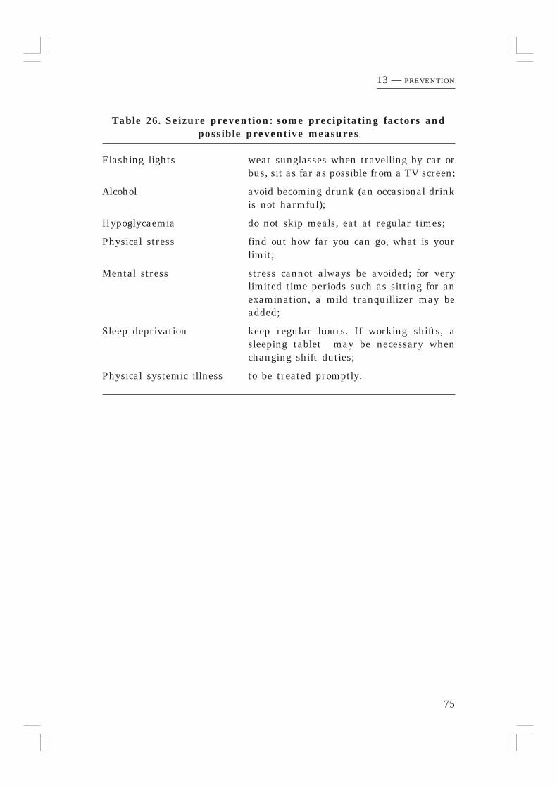
75
13 — PREVENTION
Table 26. Seizure prevention: some precipitating factors andpossible preventive measures
Flashing lights wear sunglasses when travelling by car orbus, sit as far as possible from a TV screen;
Alcohol avoid becoming drunk (an occasional drinkis not harmful);
Hypoglycaemia do not skip meals, eat at regular times;
Physical stress find out how far you can go, what is yourlimit;
Mental stress stress cannot always be avoided; for verylimited time periods such as sitting for anexamination, a mild tranquillizer may beadded;
Sleep deprivation keep regular hours. If working shifts, asleeping tablet may be necessary whenchanging shift duties;
Physical systemic illness to be treated promptly.

76
14 — SOCIAL ASPECTS
14Social aspects
Any seizure is a frightening experience, especially for the parents of achild, and for the close relatives of anyone with epilepsy. And the impactof epilepsy logically is a source of stress.
Throughout the world and through the ages epilepsy has been regardedas a supernatural happening as it is inexplicable and unpredictable. It isoften believed to be a consequence of possession, curse(s), witchcraft orpunishment for some ancestral error.
Epilepsy is often believed to be a contagious disease, and that anyonewho touches the patient, or his excreta, will acquire the disease themselves.These beliefs make the patient and his family very unpopular andisolated. The frightening experience of the seizures can make people tryany available means to lessen their severity, e.g., witchcraft, herbalism,offerings, prayers, and also modern drugs. As complete cure is the realaim, various treatments are tried and/or discarded and others tried,sometimes simultaneously.
Before we start our treatment some points have to be made very clear.The more time the doctor and the Epilepsy Aide take over this the betterwill be the compliance of the patient and the longer the follow-up. Thepoints given in table 27 page 78, have to be made clear.
It is impossible for a doctor to get all these points across to the patientand his parents in five minutes during a visit to the clinic. For this reasonthe Kenya Association for the Welfare of Epileptics has employed peoplecalled “Epilepsy Aides” who are specially trained to deal with theproblems arising from epilepsy. Each clinic has such an Epilepsy Aide(page 79).
When the child with epilepsy is of normal intelligence he can attend anormal school. But often teachers and heads have to be reassured thatthe condition is not dangerous to the staff and other pupils. Teachers andparents should be given appropriate counselling so that they learn toaccept that epilepsy is not a disabling condition but one that can usuallybe controlled and lived with. Families of people with epilepsy benefit fromproper counselling and they can then help the patient feel more acceptedby the family and the community.
Some children, although of normal intelligence, have specific learningdisabilities, which require special attention from the teacher.

77
14 — SOCIAL ASPECTS
When the child with epilepsy is of subnormal intelligence (and we mayexpect 20–30% of our patients to be so), assessment has to be made to seewhat kind of special education is needed, e.g., normal school with specialsupervision, special class in normal school, or special school. For example,the Kenya Institute of Special Education (KISE) has established 40assessment centres throughout the country. The Epilepsy Aide, with thehelp of such a centre, can determine the best available educationalfacility for the child.
If the child has a severe or profound mental retardation (one-third ofthe mentally retarded group), institutional care may be necessary,especially if there is also a physical handicap. There are not yet sufficientinstitutions to accommodate all the children requiring such facilities inAfrica, therefore, the Epilepsy Aide, with the help of a social worker andan occupational therapist, should assist the mother as much as possiblewith the care of the child.
For adults with epilepsy counselling is as necessary as for children,and they may need special advice and help with choice of work. Forexample, they should not become professional drivers or do a job requiringstanding on ladders, climbing trees or using heavy machinery untilseizures have been controlled (no seizures at all) for at least two years.Otherwise nearly any type of work is open to people with epilepsy.However, it is important that the person’s colleagues know that seizuresmay occur sometimes and are instructed how to handle such a situation.
There are situations which the patient learns may precipitate aseizure. Then it is better to avoid these situations, or if they areunavoidable, to ask for an increased dosage of the anticonvulsant drug soas to enjoy as normal a life as possible. Some of the well-knownprecipitating factors and the possible preventive measures are given intable 26, page 75.
When the seizures are not completely controlled, precautions have tobe taken when swimming or bathing. Someone else should always be nearat hand, and when going to the toilet the door should not be locked.
FIRE HAZARDMany mothers cook on jikos (charcoal stoves). These are usually placed onthe ground. Any young child may fall on such a jiko, but children withseizure disorders are even more prone to be burned. To prevent accidentswith fires, a protective shield should be made around the stove or itshould be raised off the ground. Similar precautions should be taken toprotect children and adults with epilepsy from burning themselves onopen fires or electrical appliances.

78
14 — SOCIAL ASPECTS
Table 27. Facts to be stressed to an epilepsy patient andrelatives
– Drugs have to be taken for many years, possibly a life-time;
– Discontinuation of the drugs will result in recurrence of the seizures;
– Sudden discontinuation may result in life-threatening status epilepticus;
– It may take several days to a few weeks before the drugs have anyeffect;
– A combination with herbal treatment might be dangerous as interactionbetween the drugs and the herbs cannot be predicted;
– The disease is not contagious and anyone can touch the person whilehe is having a seizure (e.g., to remove him from the danger of fire orwater), or in between the seizures;
– If the child is of normal intelligence he should be placed in a normalschool;
– Over-protection is not helpful in a child’s upbringing, but reasonableprecautions should be taken if there are still occasional seizures (e.g.,protection from fire, swimming under supervision, not climbing trees);
– Epilepsy has to be talked about in the family, at school or in the worksurroundings;
– Epilepsy is not a reason for being unable to marry and have a family.

79
15 — SPECIAL EPILEPSY CLINICS
15Special epilepsy clinics
As the number of patients with epilepsy who are seeking medicaltreatment is rapidly increasing, they should be seen and treated in theexisting health services. But every patient with epilepsy needs attentionin a different way from a patient with, for example, tonsillitis who willbe cured in a few days. Therefore a clinic especially for epilepsy patientsis advocated where special attention can be given more easily than in abusy general out-patients department.
The patient with epilepsy has to be informed about his/her chronicdisease. Explanations have to be given about the medicines and their use,as compliance with drug taking is essential in seizure control. To suddenlystop taking medicines may result in a dangerous status epilepticus.
Depending on the number of patients attending, a clinic once a weekor once a month is sufficient. An interval longer than a month is notadvisable as a patient has to be seen more often after initiating thetreatment. In the beginning, a monthly visit is necessary to observeseizure control and reactions to the medication. If allergic side-effectsoccur even this interval is too long.
Patients must be informed that if reactions such as skin rash or feveroccur they must visit the medical centre before the appointed date.
When good seizure control has been achieved a three-monthly visitschedule is sufficient.
FIRST VISITThe process of history-taking and counselling of the patient and his familyis very time consuming. Not only are there the medical problems, but thesocial problems also have to be discussed as these are sometimes moreworrying than the actual seizures. Registration cards have to be made out.
THE “EPILEPSY AIDE”Within a clinic it is very valuable to employ an Epilepsy Aide or social
worker. In KAWE clinics these are persons with secondary-school educationwho come from the area where a clinic is situated and speak the samelanguage as the patients in that clinic. They are specially trained to deal withproblems arising from epilepsy such as schooling, unemployment, socialostracism, non-compliance and so on.

80
15 — SPECIAL EPILEPSY CLINICS
If the community already has a Community Health Worker, he/shewill identify the patients with epilepsy in that community and refer themto the Epilepsy Aide, who will take a full medical and social history beforereferring the patient to the Epilepsy Clinic.
If, however, the community does not have a Community HealthWorker, the Epilepsy Aide himself has to identify patients with epilepsyin the community.
The Epilepsy Aide sees the patient before, during and after clinichours so as to be able to answer the many questions arising from thisspecial condition and its treatment. The Aide also gives advice on thenecessity for drug compliance. Moreover, the Aide keeps in contact withthe patient and his/her relatives between visits to the clinic to help withany problems, e.g., school, job, recreation.
To help parents, teachers and health workers, National Epilepsy SupportFoundations have published booklets. If such an organization does not yetexist in the country the International Bureau for Epilepsy can be contacted(see useful addresses page 122).
REGISTRATION CARDS AND RECORD KEEPINGPatients with epilepsy are treated for many years and information has tobe kept for a very long time. Records should be clear and easily accessibleso that anyone can continue the treatment in the absence of the usualmedical worker. Patients and the medical worker must know the presentmedication and what has been given before. If the therapy has beenchanged, what was the reason, e.g., allergy, no response, etc?
At every visit the patient must get a date for the next appointment, sohe realizes that treatment has to be continued. In KAWE clinicsinstructions are given to return left-over tablets. Compliance can bemeasured by counting them and warnings given if necessary.
It may be useful to make out the registration and record-keeping cardsas detailed in table 28.

81
15 — SPECIAL EPILEPSY CLINICS
Table 28. Useful registration and record cards
1. Epilepsy treatment record card (kept in the clinic)The history taken by the Aide should be kept, but a summary with thepositive findings should be entered on this card. Initial treatment andnumber of tablets dispensed is recorded. Progress notes are made here,and the number of seizures noted. Samples of these cards are given inAppendix D. When the epilepsy clinic is held in an established healthfacility, the usual clinic cards can be adapted for use in the epilepsyclinic.
2. Seizure record cardThis card is kept by the patient or by a member of the family. Seizuresare recorded by the day and any unusual symptoms are noted.
3. Epilepsy appointment cardThis card is kept by the patient. Besides the clinic’s and patient’s nameand registration number, the date of the next appointment is given.Present medication is recorded on this card.
For card numbers 2 and 3, an ordinary exercise book cut in half isvery useful.
4. Appointment bookThis book is kept at the clinic.
After every clinic the book is checked to see which patients did notattend, and the Aide can take action accordingly.
5. Epilepsy registerThis register is kept at every epilepsy clinic by the medical workerrunning the clinic. The information entered on this register is used forannual reporting. If every clinic uses the same register uniform reportingis guaranteed and information for epidemiology studies can be collected(see sample in Appendix D).
6. Annual report formInformation from the epilepsy register is entered on this form and sentyearly to the District Medical Officer of Health (see Appendix D).
7. Recording of drugs usedThis can either be done on the Epilepsy Register or on a separate sheetof paper.

82
MEDICINES AND COSTSSupplyWhen an epilepsy clinic is opened it is absolutely essential that aregular drug supply be maintained. This is even more important inepilepsy than for any other medical condition. Irregular treatment is asbad as no treatment at all (indeed it may be worse as status epilepticusmay be precipitated).
CostsA disorder that requires therapy for many years or for life is, in addition
to being a medical and social problem, a great financial burden to the patient,parents and other relatives.
This burden can be alleviated by not charging for consultation. Becausecosts have to be covered, some cost-sharing has to be introduced. Forexample, KAWE has introduced cost-sharing in 1996 and sells drugs at costprice. Still many poor patients can not afford the drugs and those are nowbeing sponsored. KAWE encourages each clinic to form a Community BasedOrganization, consisting of patients, relatives and well-wishers. They havetasks such as awareness creation, counselling, home-visits, helping out inthe clinic and they will put in place modalities to carry together the financialburden for the poor patients.
The costs are dependent on the type of drug used. If phenobarbitone isused alone and bought wholesale from local manufacturers the cost perpatient per year is (prices of the year 2000) between US$ 0.9 - US$1.35(Burkina Faso) and US$ 11.- - US$ 16.- (Togo). But when carbamazepine isused the costs per year per patient may reach at least US$ 18.- (Cameroon)to US$ 146.- (Gabon) and for valproate annual costs for adults would liebetween US$ 237.- (South Africa) and US$ 420.- (Gabon).
Prices do change continuously. In table 29 the relative costs of the defineddaily dose (the usually effective dose/day) of various AEDs is compared withthe cost of phenobarbitone.
Syrups are more expensive than tablets. As most children can taketablets quite easily, syrups are not really necessary. If there are problemswith swallowing, all uncoated tablets (phenobarbitone, carbamazepine,clonazepam, and sometimes phenytoin) can be crushed and administeredin milk, tea or porridge. Only if tablets are coated (valproate andsometimes phenytoin) might syrups be indicated.
If, however, no funds are available for the medicines, cost sharing can be worked out according to the policy of the health institution where theepilepsy clinic is held.
15 — SPECIAL EPILEPSY CLINICS

83
Other costsThe setting up of a special epilepsy clinic need not involve many othercosts. The Medical or Clinical Officer has only to re-allocate his time: heworks in his normal surroundings. A member of staff could be trained asan Epilepsy Aide, or someone has to be employed for this function.A national epilepsy support organization may be able to provide assistanceon request. The Epilepsy Aide should make home visits. The best methodof transport is probably by bicycle/motorcycle.
15 — SPECIAL EPILEPSY CLINICS
Table 29. Relative cost of the daily requirement ofanticonvulsants
Antiepileptic drug Usually effective Price relative todaily dose (mg) Phenobarbitone
Phenobarbitone 100 1,00
Phenytoin 300 1,13
Carbamazepine 800 4,89
Clonazepam 8 5,03
Ethosuximide 1250 6,41
Valproate 1500 7,52
Oxcarbazepine 1500 15,53
Vigabatrin 2000 32,59
Lamotrigine 300 41,54
Gabapentin 1800 44,87
Topiramate 300 52,17
Levetiracetam 2000 59,34
Felbamate 2400 82,43
The relation of the costs are based on a specific set of data from onecountry, therefore the multiplication factor may vary in other places;however, in general this will not influence the ranking very much.

84
“Please give me my daily medicines”
ph
oto
: J.
Lo
eb
er

85
Appendices
APPENDIX A
Review of the epilepsies and theirtreatment according to age-linked groups
Seizures starting in the first three years of life are often symptomatic. Withmodern investigation methods and laboratory techniques it is often possible tofind the cause.
Seizures starting in the 5–15 years age group are often idiopathic, whileseizures beginning in adults are again often symptomatic.
Treatment of seizures in the newbornAll newborns with seizures should be transferred to hospital to determine thecause (hypocalcaemia, hypoglycaemia, meningitis, jaundice, etc.) and treatmentgiven accordingly.To stop the seizures one injection of diazepam 0.5 mg/kg i.v. may begiven followed by phenobarbitone, loading dose 15–20 mg/kg i.v., thenmaintenance dose 3.5 mg/kg daily.N.B. If pyridoxine-dependent epilepsy, then treatment is 100 mg
pyridoxine daily until dietary pyridoxine can take over.If pyridoxine deficiency, supply 5 mg pyridoxine daily.
EPILEPSIES IN THE NEWBORN
(In the first 28 days about 0.5% of neonates have seizures)Idiopathic
– Benign neonatal familial convulsions (prevalence unknown)
Occurrence: on the second and third day of lifeSeizure type: clonic or apnoeic spellsAetiology: it is a rare, dominantly inherited disorder, without a
known aetiological factorPrognosis: about 14% develop epilepsy.
– Benign neonatal convulsions (2%-7% of all neonatal convulsions)
Occurrence: around the fifth daySeizure type: frequently repeated clonic or apnoeic seizuresAetiology: no known aetiological factorPrognosis: good, no recurrence of seizures, psychomotor
development not affected.
APPENDIX A — REVIEW OF THE EPILEPSIES AND THEIR TREATMENT

86
SymptomaticSeizure type: focal-clonic, tonic, clonic, or apnoeic spellsAetiology: birth asphyxia, intracranial haemorrhage, infections,
metabolic disorders, developmental abnormalities,pyridoxine dependency
Prognosis: depends on the degree of brain damage from completerecovery, recovery with lasting neurological sequelae(spastic quadri- and hemiplegia, mental retardation)or death
– Pyridoxine-dependent epilepsy (prevalence rare)
Onset: within the first 24 hours of lifeSeizure type: focal and generalized
child is very irritable, has brisk reflexes and hypotoniaAetiology: an autosomal recessive disorderPrognosis: poor, may die or
survive with mental handicap
– Pyridoxine deficiency (prevalence rare)
Onset: maybe later than Pyridoxine dependencySeizure type: generalizedAetiology: lack of vitamin B6 in the diet
malnutrition, malabsorptioninterference by other drugs such as isoniazid
– Early myoclonic encephalopathy (prevalence rare)
Onset: before 3 months of ageSeizure type: initially fragmentary myoclonus, then erratic partial
seizures, massive myoclonus, or tonic spasmsAetiology: familial cases are frequent and suggest the influence
of one or several congenital metabolic errors.Prognosis: poor, psychomotor development is arrested, death
may occur during the first year
– Early infantile epileptic encephalopathy with suppression burst(Ohtahara’s syndrome; prevalence rare)
Onset: within the first few months of lifeSeizure type: frequent tonic spasms, partial seizures may occur,
myclonic seizures rareAetiology: obscurePrognosis: poor with severe psychomotor retardation, seizure
intractability, often there is evolution to West’ssyndrome at age 4–6 months
APPENDIX A — REVIEW OF THE EPILEPSIES AND THEIR TREATMENT

87
EPILEPSIES STARTING INCHILDREN AGED 3 MONTHS TO 3–4 YEARSMany forms of epilepsy begin in this age group, e.g.,
– The localization-related symptomatic epilepsies (after injury or infection);– The generalized symptomatic epilepsies (often the epilepsies occurring as a
result of inborn errors of metabolism);– The special syndromes (West, Lennox-Gastaut);– The myoclonic epilepsies.
There is a frequent association with neurological signs and/or mental retardation.The outcome is often unfavourable.
The seizures in this age group are characterized by the rarity of generalizedtonic-clonic seizures, most probably due to incomplete maturation of the inter-hemispherical pathways. Generalized tonic seizures and myoclonic seizures arethe main seizure types.
– Infantile spasms (West’s syndrome;hypsarrhythmia; 2.65% of allepilepsies: Loiseau 1991)Onset: between 4 and 7 months, always before 1 yearSeizure type: most often a flexor spasm, a sudden flexion of the
head, bending of the knees, and flexion with abductionof the arms (Salaam attack); sometimes an extensorspasm, like a Moro reflex, with abduction andextension of the arms, neck, trunk and legs; ora mixed picture with arms and head flexed and legsextended;they last only a few seconds, or even less than asecond, but may be repeated many times some secondsapart
Aetiology: most often organic brain damage caused by– infections (pre- or postnatal) such as cytomegalo-
virus, toxoplasmosis, viral encephalitis, rubellaor syphilis;
– birth asphyxia, low birthweight;– congenital malformations and metabolic diseases;– idiopathic, no known cause
Prognosis: depends on the degree of brain damage, about 80%are mentally retarded, a smaller group also havingcerebral palsy;in the idiopathic group, the prognosis is favourablewhen treated early
Treatment: to be carried out in hospital– ACTH, 20–60 int. units daily i.m. for one or more
months followed by prednisolone; or– prednisolone from the start, 2 mg/kg daily;– nitrazepam, clonazepam, or valproate, although
less effective, can also be used.
APPENDIX A — REVIEW OF THE EPILEPSIES AND THEIR TREATMENT

88
– Lennox-Gastaut syndrome (0.78% of all epilepsies: Loiseau 1991)
Onset: usually between 3–5 years (2–7)Seizure type: a combination of absences, myoclonic jerks and atonic
falls, many times daily, often with injuries to chin,teeth, nose, forehead and occiput (a protective helmetis recommended);status epilepticus is frequent
Aetiology: – following infantile spasms– measles encephalitis– subacute sclerosing pan-encephalitis (*see page 92)
– anoxia, e.g., after a prolonged febrile convulsion– lead poisoning– degenerative brain diseases– cryptogenic– idiopathic
Prognosis: very poor, may persist into adult life;90% are mentally retarded;progressive dementia, ataxia may occur;very difficult to treat
Treatment: seizures very resistant to the usual anticonvulsants;many drugs are tried, and often these children areovertreated with a combination of more than threedrugs, without success.
– Epilepsy with myoclonic-astatic seizures (Doose’s syndrome, 0.16% of allepilepsies: Loiseau 1991)Onset: mostly between 2 and 5 years (7 months–6 years)Seizure type: myoclonic, astatic, myoclonic-astatic, absences with
clonic and tonic components;status frequently occurs;difficult to differentiate from the Lennox-Gastautsyndrome
Aetiology: often idiopathic;usually a normal developmental background
Prognosis: course and outcome variableTreatment: valproate, clonazepam.
– Benign myoclonic epilepsy in infancy (1.25% of all epilepsies: Loiseau 1991)Onset: during first or second year of lifeSeizure type: generalized myoclonus in brief burstsAetiology: often a family historyPrognosis: easily controlled by treatment;
may be accompanied by a relative delay of intellectualdevelopment and minor personality disorders
Treatment: valproate.
APPENDIX A — REVIEW OF THE EPILEPSIES AND THEIR TREATMENT

89
– Severe myoclonic epilepsy in infancy(7% of the epilepsies with onset before 3 years: Dalla Bernadina et al. 1982)
Onset: during the first year of lifeSeizure type: generalized or unilateral febrile clonic seizures, then
myoclonic jerks and often partial seizuresAetiology: a family history of epilepsy or febrile convulsions;
normal development before onsetPrognosis: psychomotor development is retarded from the second
year of life onwards;ataxia, pyramidal signs and interictal myoclonusappear
Treatment: very resistant to all forms of treatment.
SEIZURES OCCURRING BETWEEN 6 MONTHS AND 5 YEARSSeizures occurring in children in this age group are often febrile convulsions. Thisis usually a benign condition and is discussed in Chapter 11.
EPILEPSY STARTING IN CHILDREN AGED BETWEEN 3 AND10 YEARSIn this age group the symptomatic epilepsies still start, depending on the age atwhich the disease or injury occurred.
But this age group is more characterized by the emergence of the idiopathicepilepsies. These occur in both groups, the epilepsies with partial onset ofseizures, and the group with generalized onset of seizures. The prognosis is oftenfavourable.
Localization-related epilepsies, idiopathic– Benign childhood epilepsy with centro-temporal spikes
(Rolandic epilepsy; 7.47% of all epilepsies: Loiseau 1991)
Onset: between 3 and 13 years (peak 9–10 years)Seizure type: partial motor, often affecting the face;
related to sleep, no loss of consciousness;may develop into generalized tonic-clonic seizures
Aetiology: idiopathicPrognosis: good, usually disappears with or without treatment
before the age of 16Treatment: if necessary, carbamazepine to be continued until a
two-year seizure free period has been reached;if seizures occur only at night and if side-effects ofcarbamazepine are evident, or in case of adversesocial aspects, e.g., stigmatization, not starting orwithdrawal of treatment can be considered.
APPENDIX A — REVIEW OF THE EPILEPSIES AND THEIR TREATMENT

90
– Childhood epilepsies with occipital paroxysms(0.78% of all epilepsies: Loiseau 1991)
Onset: childhoodSeizure type: seizures start with visual symptoms or hallucin-
ations together with anxiety, often followed by ahemiclonic seizure or automatisms, followed bymigrainous headaches
Aetiology: idiopathicPrognosis: not certainTreatment: as in benign childhood epilepsy, above.
Localization-related epilepsies, symptomatic
(40% of all epilepsies in children younger than 15 years: Vianni et al. 1988)Onset: throughout childhood viral, bacterial and parasitic
diseases may occur, neurological sequelae withseizures may develop months or years after theevent;accidents are also a common cause
Seizure type: partial with or without secondary generalizationAetiology: often cryptogenic;
usually a normal developmental backgroundPrognosis: depends on the brain lesionTreatment: phenytoin, carbamazepine;
if the seizures become secondary generalized,phenobarbitone may be used.
Generalized epilepsies– Childhood absence epilepsy (pyknolepsy; 9.35% of all epilepsies: Loiseau 1991)
Onset: children of school age, with peak manifestation 6–7years
Seizure type: see page 23Aetiology: idiopathicPrognosis: remits in adolescence, but may be replaced by
generalized tonic-clonic seizures (GTCS)Treatment: ethosuximide, valproate.
– Epilepsy with myoclonic absences (0.16% of all epilepsies: Loiseau 1991)
Onset: between 1 and 12 yearsSeizure type: absence accompanied by severe bilateral rhythmical
clonic jerks, often associated with tonic contractions;they may occur many times a day;the patient may be aware of the jerking movement
Aetiology: not defined
APPENDIX A — REVIEW OF THE EPILEPSIES AND THEIR TREATMENT
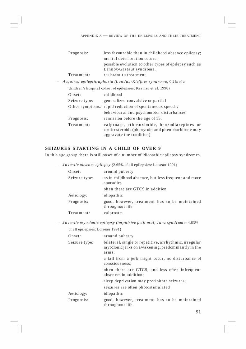
91
Prognosis: less favourable than in childhood absence epilepsy;mental deterioration occurs;possible evolution to other types of epilepsy such asLennox-Gastaut syndrome.
Treatment: resistant to treatment
– Acquired epileptic aphasia (Landau-Kleffner syndrome; 0.2% of a
children’s hospital cohort of epilepsies: Kramer et al. 1998)
Onset: childhoodSeizure type: generalized convulsive or partialOther symptoms: rapid reduction of spontaneous speech;
behavioural and psychomotor disturbancesPrognosis: remission before the age of 15.Treatment: valproate, ethosuximide, benzodiazepines or
corticosteroids (phenytoin and phenobarbitone mayaggravate the condition)
SEIZURES STARTING IN A CHILD OF OVER 9In this age group there is still onset of a number of idiopathic epilepsy syndromes.
– Juvenile absence epilepsy (2.65% of all epilepsies: Loiseau 1991)
Onset: around pubertySeizure type: as in childhood absence, but less frequent and more
sporadic;often there are GTCS in addition
Aetiology: idiopathicPrognosis: good, however, treatment has to be maintained
throughout lifeTreatment: valproate.
– Juvenile myoclonic epilepsy (impulsive petit mal; Janz syndrome; 4.83%
of all epilepsies: Loiseau 1991)
Onset: around pubertySeizure type: bilateral, single or repetitive, arrhythmic, irregular
myoclonic jerks on awakening, predominantly in thearms;a fall from a jerk might occur, no disturbance ofconsciousness;often there are GTCS, and less often infrequentabsences in addition;sleep deprivation may precipitate seizures;seizures are often photostimulated
Aetiology: idiopathicPrognosis: good, however, treatment has to be maintained
throughout life
APPENDIX A — REVIEW OF THE EPILEPSIES AND THEIR TREATMENT

92
Treatment: valproate is the treatment of choice but pheno-barbitone could be tried;night-time sleep should be sufficient.
– Epilepsy with GTCS on awakening (1.87% of all epilepsies: Loiseau 1991)
Onset: second decade of lifeSeizure type: generalized tonic-clonic seizures shortly after
awakening;sometimes they occur in the evening in the period ofrelaxation;may be precipitated by sleep deprivation;significant correlation with photosensitivity;other seizure types like myoclonic jerks and absencemay occur
Aetiology: idiopathicPrognosis: good, however, treatment has to be maintained
throughout lifeTreatment: valproate is the treatment of choice but pheno-
barbitone could be tried;night-time sleep should be sufficient.
– Primary reading epilepsy (prevalence rare)Onset: late pubertySeizure type: simple partial motor involving masticatory muscles
or visual, and if the stimulus is not interrupted,GTCS may occur
Aetiology: this syndrome may be inheritedPrecipitating factor: reading, especially aloud, independent of the
content of the textPrognosis: benign, little tendency to spontaneous seizures; there
are no abnormal neurological findings, but anabnormal EEG pattern.
Treatment: valproate (not always necessary).
* Subacute sclerosing pan-encephalitis (SSPE) is a rare, encephalitis ofdecreasing incidence as its aetiology the measles virus is eradicated. It is aslow virus infection, the encephalitis occurring several years after theprimary measles infection. Prognosis is poor, over a period of severalmonths there is deterioration of intellectual function, motor function, andthere is generalized myoclonic jerking. The patient becomes bedridden, thisphase may last for some years and leads to death. Treatment is supportive;corticosteroids may be tried.
APPENDIX A — REVIEW OF THE EPILEPSIES AND THEIR TREATMENT

93
EPILEPSY AFTER HEAD INJURYThe onset of these seizures depends on the age at the time of the accident,e.g., birth trauma will give seizures in the first year of life.
Not everyone who has had a head injury will develop seizures. Seizuresare more common when the injury has been penetrating, when there was adepressed skull fracture, an intracranial haematoma, or if in the acute statethere was post-traumatic amnesia of more than 24 hours’ duration. Post-traumatic seizures develop in 50% in the first year following the accident,and another 20% will have developed by the end of the second year. The useof prophylactic anticonvulsants after a serious head injury may be considered.Thus far studies on prophylaxis have not been able to demonstrate aneffective approach.
EPILEPSY CAUSED BY A BRAIN TUMOURA tumour, benign, malignant or a metastasis, may occur in any agegroup, but is more common in the older age group.
Epilepsy starting after the age of 20 years is always suspicious and fullinvestigations, preferably with CT scan should be done.
EPILEPSY FOLLOWING CEREBRO-VASCULAR DISEASEIn people over 50 years old, cerebro-vascular disease is a common causeof epilepsy. The seizures may follow a cerebro-vascular accident (stroke),or may develop during the course of a subclinical cerebro-vascular disease.
In all these groups (injury, tumour, cerebro-vascular accident) the type ofseizure depends on the localization of the injury.
Treatment is with phenytoin or carbamazepine.When there are secondary generalized seizures, phenobarbitone may
be used as an alternative.
APPENDIX A — REVIEW OF THE EPILEPSIES AND THEIR TREATMENT

94
APPENDIX B — ANTIEPILEPTIC DRUGS: PHENOBARBITONE
APPENDIX B
Antiepileptic Drugs (AEDs)
PHENOBARBITONEStarting dose
Newborns: 15–20 mg/kg once only (loading dose)Children: 30 mg daily (N.B. children with febrile convulsions
5 mg/kg immediately, usually 45–60 mg daily)Adults: 60 mg daily.
Increments30 mg every 4 weeks.
Maintenance doseNewborns: 3.5 mg/kg/dayChildren: 2–6 mg/kg/dayAdults: 0.5–4 mg/kg/day, maximum 180 mg/day.
Elimination half-life timeNewborns: ± 100 hoursChildren: 30–70 hoursAdults: 60–150 hoursInfants and children metabolize phenobarbitone quicker than adults. Thereforea higher dose per kilogram body weight has to be given. But in newborns, half-life time is longer—up to 100 hours and in pre-term newborns it is up to 200 hours.
Steady state (reached after five times the half-life)Reached in children in 11⁄2 weeks after starting therapyReached in adults in 3–4 weeks after starting therapy.
Dose frequencyIn children and adults: once dailyIn infants: twice daily.
IndicationsFirst drug in:– primary and secondary generalized tonic-clonic seizures– febrile convulsions.
Not indicated in– absence-seizures– seizures which occur mainly during sleep– children with hyperactive behaviour.
95
InteractionsPhenobarbitone decreases the serum levels of:– bilirubin, folate, cortisol, vitamin D and K– carbamazepine, phenytoin, valproate– chloramphenicol in neonates, doxycycline– digitoxin, griseofulvin, warfarin,– contraceptive hormonesPhenobarbitone levels are increased by:– phenytoin, valproate– frusemide.
PregnancyDuring pregnancy the phenobarbitone level tends to fall and it rises again in thepuerperium.
ToxicityLocal effects
– very rare.Dose-determined effects
– during the first few days of treatment drowsiness may occur, but thisdisappears by itself without reducing the dosage
– when increasing the dosage drowsiness may recur, now a sign of toxicity, anddosage should be reduced
– hyperactivity and irritability occur in children but the condition of thechildren should be noted before treatment is started: the irritability andhyperactivity might be due to the organic brain damage rather than thephenobarbitone
– decline in scholastic performance
– lethargy, hypoactivity, ataxia– confusion in the aged.Idiosyncratic effects
– skin rash, exfoliative dermatitis, porphyrinuria;– agranulocytosis, aplastic anaemia, jaundice and hepatitis, but very rarely;– Dupuytren’s contracture and frozen shoulder are more common.Effects on the foetus and newborn
– congenital malformations are sometimes associated with phenobarbitonetherapy especially when caffeine or other anticonvulsants are used in addition
– increased bleeding tendency due to decreased vitamin K levels in the newborn– phenobarbitone-withdrawal syndrome (hypotonia and irritability) in newborns
born to mothers on phenobarbitone treatment—may be prevented bybreastfeeding.
Breast milk
Phenobarbitone is present in breast milk, and sometimes produces drowsinessin infants when the mother is on a high dosage.
APPENDIX B — ANTIEPILEPTIC DRUGS: PHENOBARBITONE
96
PHENYTOIN (DIPHENYL HYDANTOIN)Starting dose
3 mg/kg/dailyFor instance: 1–6 years: 50 mg daily
7–14 years: 100 mg dailyadults: 200 mg daily
Increments25 mg in children every 3–4 weeks50 mg in adults every 3–4 weeks.
Maintenance dose3–8 mg/kg/daily (maximum 400 mg if serum levels are not available. If they areavailable, serum level generally to be kept below 20 µg/ml, but may exceed thislevel if need be and if toxicity is not a problem).
Elimination half-life timeFrom 9–140 hours.The half-life depends on the dosage and the duration of intake. The eliminationtime becomes longer the higher the dosage. On the other hand, when treatmentis started, people metabolize phenytoin slower until more liver enzymes areinduced. The enzyme system is satiable, therefore at higher doses a smallincrease in the dosage could suddenly result in a toxic phenytoin serum level.Patients who, on genetic basis, metabolize phenytoin slowly will get intoxicationmore easily.Children metabolize quicker and therefore need more mg/kg a day than an adult.
Increments should never be 100 mg, but always 50 or 25 mg in adults and, asavailable, 25–30 mg in children.
Steady stateSteady state is reached in 7–30 days after starting therapy.
Dose frequencyIn children: twice daily
In adults: once daily (unless gastrointestinal discomfort, thendivide into two dosages)
AbsorptionAbsorption is different in tablets and capsules from different manufacturers. Ifpossible, do not change suddenly from one make to another. If such a change isunavoidable check sooner than usual.
IndicationsAs first drug in:– partial seizures with or without secondary generalizationAlso active in:– primary generalized tonic-clonic seizures (beware of provocation of absence
seizures)– status epilepticus (see page 66)NB Not indicated in: absence- and myoclonic seizures or febrile convulsions.
APPENDIX B — ANTIEPILEPTIC DRUGS: PHENYTOIN

97
InteractionPhenytoin decreases the serum levels of:– folate, vitamin D, griseofulvin– carbamazepine, clonazepam– contraceptive hormones– vitamin K in newbornsPhenytoin sometimes increases the levels of:– phenobarbitoneThe phenytoin level may be increased by:– INH, rifampicin and ketoconazole.
ToxicityLocal effects
– slight upper abdominal discomfort, nausea, vomiting.Dose-determined effects
– nystagmus, ataxia, diplopia, drowsiness, slurred speech– vomiting, choreiform movements– gingival hypertrophy, can be reduced by good dental hygiene (regular teeth
brushing)– hirsutism, acne, coarse facies– re-occurrence of seizures– cerebellar syndrome.Idiosyncratic effects
– morbilliform rash rarely going into exfoliative dermatitis– lymphadenopathy, fever, eosinophilia– bone-marrow depression– hepatitis.Effects on the foetus and newborn
– there is an increased occurrence of cleft lip and palate, and increasedcongenital heart malformations.
– in the newborn vitamin K deficiency with bleeding may occur.Breast milk
– phenytoin is present in breast milk but in amounts too small to be harmful.So continue breastfeeding.
APPENDIX B — ANTIEPILEPTIC DRUGS: PHENYTOIN

98
CARBAMAZEPINEStarting dose (First week administer half the starting dose)
Under 1 year: 100 mg daily1–5 years : 150 mg daily6–10 years: 200 mg daily11–15 years: 200–300 mg dailyAdult: 200–400 mg daily.
IncrementsChildren: 50 mg weeklyAdults: 100 mg every 1–2 weeks.
Maintenance doseChildren: 10–30 mg/kg/dayAdults: 10–20 mg/kg/day (400–1400 mg).
Elimination half-life timeUp to 36 hours after the first doseDecreasing to up to 12 hours when taken regularly, and even shorter whencombined with phenobarbitone and/or phenytoin.
Steady stateReached in up to 8 days.
Dose frequency2 times daily when it is the only drug
3 times daily when the dosage is high or in combination with other drugs.Indications
– benign childhood epilepsy with centrotemporal spikes– childhood epilepsies with occipital paroxysms– all other partial seizures, with simple and complex symptomatology– primary generalized tonic-clonic seizures (beware of provocation of absence
seizure)– secondary generalized tonic-clonic seizures.NB Not indicated in: absence- and myoclonic seizures or febrile convulsions.
InteractionsCarbamazepine decreases the serum levels of:– folates, warfarin, doxycycline and oral contraceptivesCarbamazepine serum levels are decreased by:– phenytoin and phenobarbitoneCarbamazepine levels are increased by:– erythromycin and INH.
APPENDIX B — ANTIEPILEPTIC DRUGS: CARBAMAZEPINE

99
ToxicityLocal effects
– occasionally anorexia, nausea or vomiting.Dose-determined effects
– headache, dizziness, somnolence, ataxia– disturbed vision, diplopia– overdosage might give tremor, excitation and convulsions.Idiosyncratic effects
– hepatitis, jaundice, fever– skin rashes (especially sunshine induced), generalized erythema, erythema
multiforme exudativum (Stevens-Johnson syndrome), exfoliative dermatitis,lymph-node swelling
– aplastic anaemia, leucopenia, neutropenia.Effects on the foetus and newborn
– Congenital malformations have been reported (spina bifida). Treatment withcarbamazepine can be continued during pregnancy when given as monotherapy.
Breast milk
– Carbamazepine passes into the breast milk, but not in sufficient amounts tostop breastfeeding.
VALPROIC ACID (valproate; dipropylacetic acid)Valproate is manufactured either as the acid, the sodium salt (sodium valproate) orthe magnesium salt (magnesium valproate).Starting dose
Children: 15–20 mg/kg/dayAdults: 10–15 mg/kg/dayFor instance:1–2 years: 150–200 mg daily3–5 years: 200–300 mg daily6–10 years: 300–400 mg daily11–15 years: 450 mg dailyadults: 600 mg daily.
IncrementsIn children: 10 mg/kg daily every 4–7 daysIn adults: 200 mg daily every 4–7 days.
Maintenance dose10–30 mg/kg/day (in adults 600–2400 mg daily).
Elimination half-life timeApproximately 16 hours.
Steady stateReached in 3–4 days.
APPENDIX B — ANTIEPILEPTIC DRUGS: CARBAMAZEPINE, VALPROATE

100
Dose frequencyThree times daily.
Indications– absence-seizures– myoclonic forms of generalized epilepsy– febrile convulsions– all generalized tonic, clonic or tonic-clonic seizures– all varieties of partial seizures– photosensitive epilepsy.NB Not indicated in: Hepatic impairment; infants with severe epilepsy combined
with phenobarbitone and/or phenytoin
InteractionsValproate increases the serum level of:– phenobarbitone, lamotrigine, phenothiazines and antidepressantsValproate levels are decreased by:
– phenytoin and phenobarbitone but not by carbamazepine.Toxicity
Local effects
– mild, like nausea, vomiting, diarrhoea occur, mainly at the beginning oftreatment
– decreased but also increased appetite with weight gain may occur– if the tablets are not enteric-coated gastric distress is frequent.Dose-determined effects
– tremor, weakness, ataxia– excitement, mental stimulation.Idiosyncratic effects
– thrombocytopenia and prolonged bleeding time
– Acquired factor VIII deficiency (Von Willebrand disease)– hair loss (temporary and reversible)– impaired hepatic function, especially in children under two on polytherapy
(but an isolated increase in gamma GT in some cases is not a reason to lowerthe valproate dose)
– polycystic ovaries– pancreatitis.Effects on the foetus and newborn
– as other antiepileptic drugs, in particular spina bifida.Breast milk
– valproate passes into the breast milk. No reason to stop breastfeeding.
APPENDIX B — ANTIEPILEPTIC DRUGS: VALPROATE

101
ETHOSUXIMIDEStarting dose
3–6 years: 250 mg dailyOver 6 years: 500 mg dailyAdults: 500 mg daily.
Increments250 mg per day at weekly intervals.
Maintenance doseChildren: 10–25 mg/kg/dayAdults: 10–20 mg/kg/day (750–1500 mg).
Elimination half-life timeChildren: 10–40 hoursAdults: 40–70 hours.
Steady stateReached:– in children in about 7 days– in adults in 10–14 days.
Dose frequencyChildren under 6 years: 3 times dailyChildren above 6 years: 2 times dailyAdults: once daily is sufficient, but because of gastric irritation
twice daily is better.Indications
Absence seizures (next choice after valproate).Interactions
Ethosuximide levels are increased by:– valproate and isoniazidEthosuximide levels are decreased by:– phenytoin and carbamazepine.
ToxicityLocal effects
– nausea, vomiting or anorexia.Dose-determined effects
– headache, tiredness, drowsiness, ataxia– psychotic states with hallucinations.Idiosyncratic effects
– skin rashes, a lupus erythematosis syndrome– leucopenia, agranulocytosis.
APPENDIX B — ANTIEPILEPTIC DRUGS: ETHOSUXIMIDE

102
CLONAZEPAMStarting dose
0.01–0.03 mg/kg/dayFor instance:up to 1 year: 0.125 mg daily1–5 years: 0.250 mg daily6–12 years: 0.500 mg dailyAdults: 1 mg daily.
IncrementsChildren under 6 years: 0.250 mg daily at weekly intervalsChildren over 6 years: 0.500 mg daily at weekly intervalsAdults: 1 mg daily at weekly intervals.
Maintenance doseUp to 1 year: 0.5–1 mg daily1–5 years: 1–3 mg daily6–12 years: 3–6 mg dailyOver 12 years: 4–8 mg daily.
Elimination half-life time20–60 hours.
Steady stateReached in 8–14 days.
Dose frequencyA once-daily dose is possible in adultsIn children it should be divided into 2–3 doses.
Indications– symptomatic generalized epilepsy– idiopathic epilepsy– status epilepticus– myoclonic seizures.
InteractionsClonazepam serum levels are decreased by:– phenobarbitone, phenytoin and carbamazepineClonazepam increases the effects of alcohol.
ToxicityLocal effects
– almost no local effects.Dose-determined effects
– drowsiness, fatigue, dizziness– muscle weakness, ataxia– increased bronchial excretion and salivation
– paradoxical aggression, irritability, hyperactivity.
APPENDIX B — ANTIEPILEPTIC DRUGS: CLONAZEPAM
103
Idiosyncratic effects
– skin rash (very rarely).Teratogenic effects
– an increased risk of oral clefts has been reported.Breast milk
– lethargy and weight loss may occur in infants.Note:A good measure of tolerance is usually developed so that the dosage has to beincreased over time to give the same antiepileptic effect. If this tolerance hascaused the clonazepam to reach too high a dosage, it is better to withdraw theclonazepam very gradually and to introduce another anticonvulsant.Caution:Never cease clonazepam therapy suddenly, severe withdrawal symptoms mayoccur, and a status epilepticus be introduced.
DIAZEPAMThis drug is mainly used to curb status epilepticus, or prolonged febrile convulsions.It is then given intravenously or rectally (do not give intramuscularly as then actionis unpredictable and absorption slow).Dosage in status epilepticus or prolonged febrile convulsions:
1 mg/minute i.v. until seizures stop, but not more than:5 mg in children under 5 years10 mg in children 5–10 years15–20 mg in older children and adultsThe same solution may be given after 20–30 minutes and once again after 30minutes.
Side-effectsRespiratory depression is possible, especially if patient is on maintenance doseof phenobarbitone.
APPENDIX B — ANTIEPILEPTIC DRUGS: CLONAZEPAM, DIAZEPAM

104
APPENDIX B — ANTIEPILEPTIC DRUGS
Tab
le A
. Su
mm
ary
of t
he
mai
n i
nfo
rmat
ion
giv
en i
n A
pp
end
ix B
Ph
enob
arb
iton
eP
hen
ytoi
nC
arb
amaz
epin
eV
alp
roat
eE
thos
uxi
mid
eC
lon
azep
am
Sta
rtin
g d
ose
new
born
15–2
0 m
g/kg
on
cein
fan
t30
mg/
day
100
mg/
day
0.12
5 m
g/da
y1–
5 yr
30–6
0 m
g/da
y50
mg/
day
150
mg/
day
150
mg/
day
250
mg/
day
0.25
0 m
g/da
y6–
10 y
r60
mg/
day
100
mg/
day
200
mg/
day
300
mg/
day
500
mg/
day
0.50
0 m
g/da
y11
–15
yr60
mg/
day
100
mg/
day
200
mg/
day
450
mg/
day
500
mg/
day
0.50
0 m
g/da
yad
ult
60 m
g/da
y20
0 m
g/da
y20
0 m
g/da
y60
0 m
g/da
y50
0 m
g/da
y1
mg/
day
Incr
emen
t of
dai
ly d
ose
chil
d30
mg
4 w
eekl
y25
mg
3–4
wkl
y50
mg/
wkl
y10
mg/
kg25
0 m
g/w
kly
0.25
0 m
g/w
kly
adu
lt30
mg
4 w
eekl
y50
mg
3–4
wkl
y10
0 m
g/w
kly
200
mg/
4–7
days
250
mg/
wkl
y1
mg/
wkl
y
Mai
nte
nan
ce d
ose
new
born
/infa
nt
3.5
mg/
kg/d
ay0.
5–1
mg/
day
chil
d2–
6 m
g/kg
/day
3–8
mg/
kg/d
ay10
–30
mg/
kg/d
ay10
–30
mg/
kg/d
ay10
–25
mg/
kg/d
ay1–
6 m
g/da
yad
ult
0.5–
4 m
g/kg
/day
3–8
mg/
kg/d
ay10
–20
mg/
kg/d
ay10
–35
mg/
kg/d
ay10
–20
mg/
kg/d
ay4–
8 m
g/da
yin
adu
lt60
–250
mg
max
400
mg
400–
1400
mg
600–
2400
mg
750–
1500
mg
Hal
f-li
fe t
ime
new
born
± 10
0 h
ours
chil
d30
–70
hou
rsfi
rst—
36 h
ours
10–4
0 h
ours
adu
lt60
–150
hou
rs9–
140
hou
rsla
ter—
12 h
ours
± 16
hou
rs40
–70
hou
rs20
–60
hou
rs
Fre
qu
ency
of
dos
age
2 ti
mes
wh
ench
ild
once
dai
ly2
tim
eson
ly d
rug
3 ti
mes
3 ti
mes
dai
ly2–
3 ti
mes
adu
lton
ce d
aily
once
dai
ly3
tim
es w
hen
3 ti
mes
2 ti
mes
dai
lyon
ce d
aily
mor
e dr
ugs

105
APPENDIX B — ANTIEPILEPTIC DRUGS
Ph
enob
arb
iton
eP
hen
ytoi
nC
arb
amaz
epin
eV
alp
roat
eE
thos
uxi
mid
eC
lon
azep
am
Ind
icat
ion
sG
TC
Spa
rtia
l sei
zure
spa
rtia
l sei
zure
sab
sen
ces
abse
nce
sG
TC
S, i
diop
atic
or
febr
ile
con
vuls
ion
sse
c G
TC
Sco
mpl
ex p
arti
alm
yocl
onic
sei
zure
ssy
mpt
omat
icda
ytim
e se
izu
res
noc
turn
al s
eizu
res
seiz
ure
s, s
ec G
TC
Saw
aken
ing
seiz
ure
sm
yocl
onic
sei
zure
sM
ain
sid
e-ef
fect
sdr
owsi
nes
s,dr
owsi
nes
s,dr
owsi
nes
s,n
ause
a, v
omit
ing,
drow
sin
ess,
drow
sin
ess,
hyp
erac
tivi
tygu
m h
yper
trop
hy
atax
iadi
arrh
oea,
hal
luci
nat
ion
sw
eakn
ess,
ata
xia,
in c
hil
dren
alle
rgy:
ras
h +
skin
ras
hin
crea
sed
wei
ght,
incr
ease
d br
onch
ial
lym
phad
enop
ath
yh
air
loss
secr
etio
n,
aggr
essi
ven
ess
Mai
n i
nte
ract
ion
sde
crea
ses
vita
min
D, f
olat
esvi
tam
in D
, fol
ates
fola
tes,
phen
ytoi
n, c
arba
-ca
rbam
azep
ine,
oral
con
trac
epti
ves
maz
epin
e, v
al-
clon
azep
am, o
ral
proa
te, o
ral
con
trac
epti
ves
con
trac
epti
ves
decr
ease
d by
phen
ytoi
nph
enyt
oin
phen
obar
bito
ne
phen
obar
bito
ne
carb
amaz
epin
eca
rbam
azep
ine,
phen
ytoi
n, p
hen
o-ba
rbit
one
incr
ease
sph
enob
arbi
ton
eef
fect
of
alco
hol
phen
obar
bito
ne
effe
ct o
f al
coh
olph
enot
hia
zin
esan
tide
pres
san
tsin
crea
sed
byph
enyt
oin
INH
INH
INH
valp
roat
eri
fam
pici
ner
yth
rom
ycin
phen
ytoi
nfr
use
mid
eva
lpro
ate

106
APPENDIX C
Head circumference graphs
APPENDIX C — HEAD CIRCUMFERENCE GRAPHS
Head circumference graph: boys(adapted from G. Nellhaus 1968)

107
APPENDIX C — HEAD CIRCUMFERENCE GRAPHS
Head circumference graph: girls(adapted from G. Nellhaus 1968)

108
APPENDIX D — RECORD CARDS
APPENDIX D
Record cards
Epilepsy treatment record card
Date first visit Clinic location
Name Patient’s registration No.
Date of birth Patient’s address
Sex Location
Ethnic origin Sublocation
Village
Age ay onset of seizures
Seizure type or description
Epilepsy type
idiopathic febrile convulsions
known/suspected symptomatic others
Aetiology of symptomatic epilepsy
birth injury/asphyxia accident
meningitis/encephalitis tumour
measles/malaria following febrile convulsions
other or unknown disease others
Frequency of seizures before treatment
Number of seizures before treatment
Usual time of seizures
while awake on awakening
while asleep mixed
Trigger to seizures
alcohol fever/stress
pregnancy photostimulation
menstruation others
Birth order
Epilepsy in family
Pregnancy
Delivery
Milestones
Physical examination
Eyes Weight
ENT Height
Skin Head circumference
Chest Investigations
Abdomen
Reflexes If on treatment
medicines
Physical handicaps dosage
Mental retardation effect
General impression Management up till now
Schooling Diagnosis
Job Plan

109
APPENDIX D — RECORD CARDS
Sei
zure
rec
ord
card
NAME:
DOB:
Weight:
KAWE NUMBER:
------------------------------------------------------------------------------------
DRUG:
Phenobarbitone
-------------------------------------------------------------------------
(dailyPhenytoin---------------------------------------------------------------------
dosageCarbamazepine-----------------------------------------------------------------
in mg)Valproate---------------------------------------------------------------------
Clonazepam--------------------------------------------------------------------
------------------------------------------------------------------------------------
80-----------------------------------------------------------------
60-----------------------------------------------------------------
40-----------------------------------------------------------------
20-----------------------------------------------------------------
10-----------------------------------------------------------------
8-----------------------------------------------------------------
6-----------------------------------------------------------------
4-----------------------------------------------------------------
2-----------------------------------------------------------------
0-----------------------------------------------------------------
ATTENDANCE
(tick if seen
during the month)
------------------------------------------------------------------------------------
COMPLIANCE and
COMMENTS
(see also
continuation cards)
NUMBER OF
SEIZURES
(monthly
unless
stated
otherwise)
JANFEBMARAPRMAYJUNJULAUGSEPOCTNOVDECJANFEBMARAPRMAYJUNJULAUGSEPOCTNOVDECJANFEBMARAPRMAYJUNJULAUGSEPOCTNOVDEC

110
APPENDIX D — RECORD CARDS
Epi
leps
y re
gist
er,
firs
t vi
sit
Epi
leps
y re
gist
er, f
irst v
isit
Clin
ic’s
nam
e:R
epor
ting
year
:
Registration number
sex
date first visit
date of birth
age first seizure
seizure type
epilepsy type
aetiology
frequency seizurebefore treatment
number of seizuresbefore treatment
time of seizures
trigger
birth order
epilepsy in family
physical handicaps
occupation
mental retardation
on treatmentnowbefore
new treatment

111
APPENDIX D — RECORD CARDS
Epi
leps
y re
gist
er,
foll
ow u
p
Epi
leps
y re
gist
er, f
ollo
w u
pC
linic
’s n
ame:
Rep
ortin
g ye
ar:
No.
31 Dec first calender year
31 Dec one year later
31 Dec two years later
condition
condition
attendance
treatment
condition
attendance
treatment
condition
attendance
treatment
cond
ition
:c.
c.=
com
plet
e co
ntro
l, no
not i
mpr
oved
:=
no c
hang
e or
wor
seat
tend
ance
:ye
s or
no
seiz
ures
in la
st y
ear
if no
t atte
ndin
g be
caus
ew
.c.
=w
ell c
ontr
olle
d, 1
–12
died
–di
scha
rged
from
trea
tmen
tse
izur
es la
st y
ear
–tr
ansf
erre
dim
prov
ed=
>12
sei
zure
s, b
ut–
mov
ed o
ut o
f are
abe
tter
than
bef
ore
–de
faul
ted
–di
ed

112
Annual report form
Clinic: ............... Calender year: ......................
A. Number of patients under treatment 1 January ...........
B. Added to register: new patients .......................
transfer from other clinics ........
resumed treatment ...................
male
female
age: 0–1 year .............
1–5 years ............
6–10 years ...........
11–15 years ..........
16–20 years ..........
21–30 years ..........
31–40 years ..........
41–50 years ..........
>50 years ............
C. Treatment discontinued
discharged from treatment..............................
transferred to other clinic ...........................
moved out of area .....................................
died....................................................
defaulted...............................................
D.Number of patients on treatment 31 December (= A + B –
C) ..........................................................
E. Number of tablets used
phenobarbitone 30 mg ..........................
phenytoin 50 mg ...............................
phenytoin 100 mg ..............................
carbamazepine 200 mg ..........................
valproate 150 mg ..............................
valproate 200 mg ..............................
valproate 300 mg ..............................
clonazepam 0.5 mg .............................
clonazepam 2 mg ...............................
others.........................................
...............................................
...............................................
APPENDIX D — RECORD CARDS

113
BIBLIOGRAPHY
Bibliography
Avode DG, Capo-Chichi OB, Gandaho P, et al. Epilepsie provoquée par la cysticercose.A propos d’une enquête sociologique et culturelle réalisée à Savalou au Bénin.Bull Soc Path Ex 1996;89:45-47.
Baldwin S, Asindua S, Stanfield P. Survey of childhood disabilities within acommunity-based programme for the rehabilitation of the disabled in KibweziDivision, Kenya. Nairobi: African Medical and Research Foundation (AMREF)/ActionAid, 1990.
Balogou AA, Grunitzky KE, Beketi KA, et al. Cysticercose et épilepsie au nord duTogo dans le Tone. Rev Neurol 2000;156:270-273.
Brown JK. Fits in children. In: Laidlaw J, Richens A, eds. A textbook of epilepsy. 2nded. London: Churchill Livingstone, 1982.
Commission on Classification and Terminology of the International League againstEpilepsy. Proposal for revised clinical and electroencephalographic classificationof epileptic seizures. Epilepsia 1981;22:489-501.
Commission on Classification and Terminology of the International League againstEpilepsy. Proposal for revised classification of epilepsies and epileptic syndromes.Epilepsia 1989;30:389-399.
Consensus development panel. Febrile seizures: long term management of childrenwith fever-associated seizures. Pediatrics 1980;66:1009-1012.
Corbett J. Epilepsy as part of a handicapping condition. In: Ross E, Reynolds E, eds.Paediatric perspectives on epilepsy. Chichester: John Wiley & Sons, 1985.
Dalla Bernardina B, Dulac O, Bureau M, et al. Encéphalopathie myocloniqueprécoce avec épilepsie. Rev EEG Neurophysiol 1982;12:8-14.
Debouverie M, Kabore J, Dumas M, et al. Epidémiologie de l’épilepsie au Burkina-Faso. Dans: Dumas M, Giordano C, Gentilini M, Chieze F, eds. NeurologieTropicale. Paris: John Libbey Eurotext, 1993:57-61.
Dekker-de Kiefte PA. The Kenya Association for the Welfare of Epileptics. TropGeogr Med 1994;46(suppl 3):S20-S21.
Diop AG, Agbohous OL, Ndiaye M, et al. Prévalence de l’épilepsie en milieu scolairesénégalais. Oral communication presented at the 12th congress of the PanAfrican Association of Neurological Sciences, Durban (South Africa), 19-23 May1996.
Dongmo L, Ndo D, Atchou G, Njamnshi A. Epilepsie au Sud-Cameroun: enquêtepréliminaire dans le village Bilomo. Communication orale présentée au 3ème
Congrès International de Neurologie Tropicale, Fort-de-France, Martinique, 30Novembre - 2 Décembre 1998.
Dumas M, Grunitzky E, Deniau M, et al. Epidemiological study of neurocysticercosisin Northern Togo (West Africa). Acta Leidensia 1989;57:191-196.
Farnarier G, Diop S, Coulibaly B, et al. Onchocercose et épilepsie: enquêteépidémiologique au Mali. Med Trop (Mars) 2000;60:151-155.
Goudsmit J, Van der Waals FW, Gajdusek DC. Epilepsy in the Gbawein andWroughbarh clan of Grand Bassa County, Liberia: the endemic occurrence of“See-ee” in the native population. Neuroepidemiology 1983;2:24-34.
Grunitzky EK, Dumas M, Mbella EM, et al. Les épilepsies au Togo. Epilepsies1991;3:295-303.

114
BIBLIOGRAPHY
Grunitzky EK, Balogou AK, Agboli K, et al. Cartographie des épilepsies au Togo.Oral communication presented at the 12th congress of the Pan African Associationof Neurological Sciences, Durban (South Africa), 19-23 May 1996.
Hauser WA, Annegers JF, Kurland LT. Incidence of epilepsy and unprovokedseizures in Rochester, Minnesota: 1935-1984. Epilepsia 1993;34:453-468.
Hauser WA. Incidence and prevalence. In: Engel J Jr, Pedley TA, eds. Epilepsy: acomprehensive textbook. Philadelphia: Lippincott-Raven Publishers, 1997.
Kaamugisha J, Feksi AT. Determining the prevalence of epilepsy in the semi-urbanpopulation of Nakuru, Kenya, comparing two independent methods not apparentlyused before in epilepsy studies. Neuroepidemiology 1988;7:115-121.
Kaiser C, Kipp W, Asaba G, et al. Prevalence of epilepsy follows the distribution ofonchocerciasis in a West Ugandan focus. Bull WHO 1996;74:361-367.
Kouassi B, Koffi JK, Diarra JA, et al. Prévalence de l’épilepsie en milieu ruralivoirien: étude pilote. Pub Méd Afr 1988;89:25-30.
Kramer U, Nevo Y, Neufeld MY, et al. Epidemiology of epilepsy in childhood: a cohortof 440 consecutive patients. Pediatr Neurol 1998;18:46-50.
Livingston S. Comprehensive management of epilepsy in infancy, childhood andadolescence. Springfield, Ill: Charles C. Thomas, 1972.
Loiseau P, Duche B, Loiseau J. Classification of epilepsies and epileptic syndromesin two different samples of patients. Epilepsia 1991;32:303-309.
Longe AC, Osuntokun BO. Prevalence of neurological disorders in Udo, a ruralcommunity in southern Nigeria. Trop Geogr Med 1989;41:36-40.
Matuja WB. Psychological disturbance in African Tanzanian epileptics. Trop GeogrMed 1990;42:359-364.
Ndiaye IP, Mauferon JB, Diagne M. Epidémiologie de l’épilepsie au Sénégal.Communication orale présentée au 7ème Congrès de la PAANS, Abidjan, Côted’Ivoire, 23-30 Avril 1986.
Preux PM. Contribution à la connaissance épidémiologique de l’épilepsie en Afriquesubsaharienne. Thèse, 2000.
Rodin EA. The prognosis of patients with epilepsy. Springfield Ill: Charles C.Thomas Publisher, 1968.
Rwiza HT, Kilonzo GP, Haule J, et al. Prevalence and incidence of epilepsy inUlanga, a rural Tanzanian district: a community-based study. Epilepsia1992;33:1051-1056.
Rwiza HT. The Muhimbili epilepsy project, a three pronged approach. Assessmentof the size of the problem, organization of an epilepsy care system and researchon risk factors. Trop Geogr Med 1994;46(suppl 3):S22-S24.
Sander JWAS, Sillanpää M. Natural history and prognosis. In: Engel J Jr, PedleyTA, eds. Epilepsy: a comprehensive textbook. Philadelphia: Lippincott-RavenPublishers, 1997.
Shorvon SD, Reynolds EH. Early prognosis of epilepsy. BMJ 1982;285:1699-1701.Tekle-Haimanot R, Forsgren L, Abebe M, et al. Clinical and electroencephalographic
characteristics of epilepsy in rural Ethiopia: a community-based study. EpilepsyRes 1990;7:230-239.
Viani F, Beghi E, Atza G, Gulotta MP. Classifications of epileptic syndromes:advantages and limitations for evaluation of childhood epileptic syndromes inclinical practice. Epilepsia 1988;29:440-445.
World Health Organization. Initiative of support to people with epilepsy. Geneva:WHO, Division of Mental Health, 1990.

115
INDEX
IndexA
Abscess 8, 11Absence seizure
19, 23, 26, 29, 32, 33, 36,88, 91, 92, 94, 100, 101
atypical 19, 33provocation 96, 98
Accidents 8, 11, 39, 77, 90, 93Acquired epileptic aphasia (Landau-
Kleffner syndrome 35, 91ACTH 28, 33, 36, 87Aetiology. see CausesAge of onset 19, 31, 33, 38, 39,
85–92, 86Aggression 22, 40, 55, 60, 102, 105AIDS 8Alcohol
8, 16, 32, 39, 40, 47, 50, 66, 75,102, 105, 108
Allergic side-effects. see Idiosyncraticside-effects
Annual report form 81, 112Anoxia 8, 11, 88Antibiotics 7, 63, 67Antiepileptic Drugs (AEDs) 94–103. see
also entries for individual drugsAntimycotics e.g., griseofulvin,
ketoconazole. 63, 97Antituberculosis medicines 63, 97. see
also IsoniazidAnxiety. 22, 47, 90Aphasia 18, 35, 65, 91Apnoea 46, 50Apnoeic spells 85Appointment 80
book 81card 81
Asphyxia, birth asphyxia. see Birthinjury, trauma
Astatic seizures. see Atonic seizuresAtaxia
9, 18, 42, 59, 60, 61, 64, 88, 89,95, 97, 99, 100, 101, 102, 105
Atonic seizures 19, 28, 33, 88Attack 3. see also
Convulsion; Fit; Seizure
Aura 17, 19, 20, 22, 39, 49, 51Automatisms 19, 22, 36, 90Autonomic symptoms 19, 22Awakening:
seizures on 33, 34, 61, 91, 92
B
Bacterial infections 7, 74, 90Bathing 77Batten disease 9Behaviour problems 17, 55, 60, 70, 91Benign childhood epilepsy with centro-
temporal spikes 34, 89Benign focal epilepsy 33. see also
Benign childhood epilepsy withcentro-temporal spikes; Childhoodepilepsies with occipital paroxysms
Benign myoclonic epilepsy in infancy34, 88
Benign neonatal convulsions 34, 85Benign neonatal familial convulsions
34, 85Benzodiazepines
32, 36, 55, 102, 103. see alsoClonazepam and Diazepam
Bibliography 113Bielschowsky 9Bilirubin 8, 74, 95Birth injury, trauma
8, 11, 12, 13, 33, 53, 54, 74, 86, 87,93, 108
Bleeding tendency 64, 95, 97, 100Blood
glucose 7, 43, 66tests 43, 66
Bourneville disease 8, 9Brain
damage11, 13, 16, 29, 32, 53, 54, 62, 74, 86,87, 95
lesion. see Brain: damagematuration 15tumour 8, 11, 13, 29, 43, 67, 108
Breastfeeding 65milk and AED's 65. see also entries
for individual drugsBreath-holding spells 46, 50Bronchial excretion 102Burns 77

116
INDEX
C
Calcifications 11, 43Carbamazepine
28, 29, 32, 33, 36, 38, 52, 58, 61, 62,63, 70, 89–92, 97–103
Carbon monoxide poisoning 8Causes
of a seizure 7, 8of epilepsy
11, 12, 13, 14, 31, 33, 38Cerebellar signs and syndrome 60, 97Cerebral
atrophy 69blood flow 47, 51malaria 8, 10, 11, 52necrosis 29oedema 8palsy 53, 87
Cerebro-vascular accident (disease)8, 13, 93
Cerebromacular degeneration 8. seealso Tay-Sachs; Bielschowsky,Spielmeyer-Vogt;Batten
ChildhoodAbsence Epilepsy 2, 4, 23, 34, 90epilepsies with occipital paroxysms
34, 90, 98infections 14, 54, 70
Chlorpromazine 55Chronic progressive epilepsia partialis
continua 34Circulatory disturbances 8Classification of
Epilepsies and Epileptic Syndromes31–38
epileptic seizures 19, 20–25status epilepticus 30
Cleft lip/palate 97, 103Clonazepam
56, 61, 82, 83, 87, 88, 97, 102, 103, 104,105, 109, 112
Clonic seizures 19, 25, 88, 89Cold injury in newborns 8Community Health Worker 80Complex partial seizures
19, 22, 23, 26, 29, 33, 39, 50, 61, 72Complex partial status 30Compliance of patient
59, 65, 68, 76, 79, 80, 109Computer-assisted tomography (CT)
scan 44, 67
Conditions co-existing with epilepsy 53–56
Congenital malformations. seeDevelopmental abnormalities/congenital malformations
Contraceptive hormones (the pill)63, 95, 97, 98
Contributory factors 15Convulex. see ValproateConvulsion 3. see also SeizureCosts
for epilepsy clinic 83of medicines 82, 82–83
Counselling 45, 76–77, 79Cryptogenic epilepsies, seizures
12, 14, 31, 34, 36, 72, 88Cysticercosis. see NeurocysticercosisCytomegalovirus 43, 87
D
Definitions ofdegree of mental retardation 53epilepsy 3seizure 3
Degenerative brain diseases8, 9, 31, 88
Déjà vu 22Delivery (partus) 40Dementia 8, 9, 56, 88Depakine. see ValproateDevelopment of child 40, 45, 69, 88Developmental abnormalities/congenital
malformations8, 86, 87, 95, 97, 99
Diazepam30, 59, 60, 61, 66, 67, 70, 85, 103
Diet (ketogenic) 65Differential diagnosis. see Disorders to
be distinguished from epilepsyDilantin. see PhenytoinDiphenyl hydantoin. see PhenytoinDiplopia 64, 97, 99Dipropylacetic Acid. see ValproateDiscontinuation of drugs 58, 68, 78, 79Disorders to be distinguished from
epilepsy 46–52Dizziness 46, 47, 51, 59, 61, 99, 102Dose frequency 58. see also
Antiepileptic Drugs (AEDs)Dose-determined effects 64. see also
Antiepileptic Drugs (AEDs)
117
INDEX
Down’s syndrome 54Drop attacks 28, 61Drowsiness
42, 49, 60, 95, 97, 101, 102, 105Drug therapy 57–65. see also
Antiepileptic Drugs (AEDs)Duration of the seizure 19, 26, 69Duration of treatment 58
E
Early infantile epileptic encephalopathywith suppression bursts 34, 86
Early myoclonic encephalopathy 34, 86Eclampsia 8Education 76–77Effect of drugs on foetus and
newborn. see entries for individualdrugs
Electroencephalography (EEG)16, 20, 23, 26, 28, 30, 43, 44, 45, 67, 92
Electrolyte imbalance7, 8, 43, 52, 66, 74
Elimination half-life time. see Half-lifetime
Emeside. see EthosuximideEmotional factors
16, 18, 32, 39, 47, 49, 50, 55, 57Encephalitis 8, 10, 87, 88, 92, 108Encephalo-trigeminal facial angiomatosis
8Encephalopathy 8Epanutin. Zie PhenytoinEpidemiology 4Epilepsy
after a brain tumour 13after cerebro-vascular disease 13after head injury 13Aide 62, 72, 76, 79, 83clinics 79–83definition 3in the newborn 85partialis continua 30register 81treatment record 108with continuous spike waves during
slow wave sleep 35with grand mal seizures (GTCS) on
awakening 34, 92with myoclonic absences 34, 90with myoclonic-astatic seizures
34, 88Epileptic Syndromes, 31–38, 85–92
Episodic confusion 50Ethosuximide
29, 32, 36, 61, 83, 90, 91, 101, 104
F
Fainting attack 46, 47, 49, 65Family
aspects29, 41, 45, 64, 68, 76, 78, 79, 81
history27, 32, 36, 40, 45, 70, 88, 89, 108, 110
Fatigue 32, 59, 65, 102Febrile convulsions
8, 10, 32, 35, 36, 69, 70, 74, 88, 89,94, 100, 103
febrile status epilepticus30, 61, 69, 103
Fire hazard 77Fit 3, 49Flashing lights 16, 39, 51, 75Flexor spasms 28, 87Focal. see PartialFocus 3, 11, 17, 22, 23, 32Folate 61, 63, 95, 97, 98, 105Frequency of seizures
19, 25, 26, 40, 58, 72, 108, 110Frontal lobe epilepsies
23, 34, 36, 47, 49Frusemide 95, 105
G
Gastric irritation 61, 100, 101Generalized epilepsies 32–38, 60
treatment 85–92Generalized seizures 3, 19, 20, 89–92
Absence seizures19, 23, 26, 29, 32, 32–38treatment 60, 90, 91
Generalized tonic-clonic seizures(GTCS)19, 23, 25, 27, 28, 58, 89–92
Geneticcounselling 74factors 12, 14, 15, 31, 96trait in febrile convulsions 69
Gingival hypertrophy 60, 97Glossary 1Glucose solution 7, 66Grand mal 28
seizures 34status 30
Griseofulvin 95, 97

118
INDEX
H
Haemorrhage 8, 11, 44, 86Hair loss 100, 105Half-life time 2, 60–62Hallucinations 22, 36, 90, 101, 105Haloperidol 55Head circumference 42, 106Head injury 3, 8, 11, 13, 15, 93. see
also InjuryHeart disease 47Hemiconvulsions 69Hemiplegia 54, 69, 86Hepatic impairment 100HHE syndrome 69Hirsutism 60, 97History taking 39-41, 45, 46, 66, 69,
70, 80, 81Hormonal changes 16Hydrocephalus 8Hyperactivity 9, 55, 60, 95, 102, 105Hyperbilirubinaemia 8, 74Hyperkinetic. see HyperactivityHypertensive encephalopathy 8Hyperventilation 16, 23, 44Hypocalcaemia 7, 8, 11, 85Hypoglycaemia
7, 8, 39, 46, 52, 58, 74, 75, 85Hypomagnesaemia 8Hypothermia 8Hypsarrhythmia; 87Hysteria 47
I
Idiopathic epilepsy2, 3, 4, 11, 12, 14, 15, 26, 27, 31, 32,33, 34, 36, 58, 60, 72, 85, 87, 88, 89,90, 91, 92, 102, 108
Idiosyncratic side-effects64, 95, 97, 99, 100, 101, 103
Impulsive petit mal. see Juvenile:myoclonic epilepsy
Inborn errors of metabolism 33, 87Incidence
of cerbral palsy 53of epilepsy 4, 5of febrile convulsions 69
Increments in dosage 62. see alsoentries for individual drugs
Infantile spasms9, 28, 33, 34, 36, 72, 87, 88
Infections and infectious diseases7, 8, 10, 11, 14, 52, 54, 69, 70, 86, 87, 92
INH. see IsoniazidInjury
2, 8, 13, 15, 49, 87, 89, 93, 108. seealso Head injury
Intelligence 45, 53, 56, 76, 78Interaction 63, 78. see also entries for
individual drugsInternational Bureau for Epilepsy 80International classification of epilepsies
and epileptic syndromes 31, 34International classification of epileptic
seizures 19, 20Investigations
7, 13, 39, 43, 66, 67, 93, 108IQ 53Irritability 17, 18, 55, 95, 102Isolated seizures 35Isolated status epilepticus 35Isoniazid 86, 101
J
Jacksonian March 22, 36Janz syndrome 34, 91Jaundice 85, 95, 99Jikos 77Juvenile
absence epilepsy 23, 34, 91myoclonic epilepsy 4, 34, 91
K
Kenya Association for the Welfare ofEpileptics 1, 76
Kenya Institute of Special Education(KISE) 77
Kernicterus 8, 74Ketogenic diet 65Kojewnikow syndrome 30, 34
L
Laboratory investigations 7, 43, 66, 67Labyrinthitis 51Lack of
concentration 55, 56sleep 16, 39
Landau-Kleffner syndrome 35, 91Lead poisoning 8, 88Learning disorders 56, 57, 76Lennox-Gastaut syndrome
34, 36, 87, 88, 91

119
INDEX
Liverand neurodegenerative disorders 9enzyme induction 63, 96function tests 43
Local side-effects 64Localization-related epilepsy
4, 31, 33, 34, 36, 87, 89, 90, 96, 98Lumbar puncture 7, 43, 66, 70
M
Malabsorption 86Malaria 7, 8, 10, 11, 52, 70, 74, 108Malnutrition 86Management
of febrile convulsions 70of seizures 45, 57of status epilepticus 66
Marriage 78Measles
encephalitis 88febrile convulsions 10, 70, 108vaccination 74virus 92
Measurements 42, 45Medical history
20, 28, 38, 39, 45, 46, 79Memory 17, 22, 23, 25, 36Menière’s disease, 51Meningitis
3, 7, 8, 10, 15, 43, 52, 67, 70,74, 85, 108
Mental deterioration 91, 92Mental retardation
9, 33, 45, 53, 55, 77, 86, 87, 108, 110definitions:mild, moderate, severe, profound 53
Mental stress 75Mesial temporal sclerosis 69Metabolic disorders
7, 11, 35, 74, 86, 87Microcephaly 8Migraine 2, 46, 51Milestones 40, 45, 108Monotherapy 62, 99Motor symptoms 19, 22, 23, 30, 36Multiple febrile convulsions 69Multiple handicaps 33, 53, 54Myoclonic seizures
19, 23, 25, 28, 33, 61, 87, 96, 100, 105Myoclonic-astatic seizures. 9, 34, 88
N
Narcolepsy 46, 50Nausea 47, 51, 97, 99, 100, 101, 105Neonatal seizures 35, 72, 85Neurocysticercosis 8, 11, 43, 74Neurofibromatosis 8, 9Neurological
deficits 32, 69, 72examination 42, 45sequelae 7, 86, 90signs 87
Newborn7, 8, 15, 28, 46, 63, 64, 74, 85, 94,95, 97, 104
Niemann-Pick disease 8, 9Night terrors 46, 50Nightmares 46, 50Nitrazepam 87Nocturnal seizures 33, 38, 60, 105Nystagmus 42, 60, 64, 97
O
Observation 42, 45Occipital lobe epilepsies 34, 36Onchocerciasis 11Oral steroids 36Organo-phosphorus poisoning 8Over-protection of child 78Overactivity. see HyperactivityOverhydration 16
P
Pancreatitis 100Panic attacks 50Paracetamol 70Paraldehyde 67, 70Parasitic infections 7, 11, 90Parietal lobe epilepsies 34, 36Partial
epilepsy 31, 32motor status 30seizures 20, 21, 60, 61, 86seizures secondary generalized
3, 19, 23, 25, 27, 33, 36, 58, 90,94, 96, 98
Penicillin 8Perinatal history 40, 45Pertussis 8
immunization 8Petit mal 23, 34, 91

120
INDEX
Phenobarbitone28, 32, 33, 36, 38, 56, 58, 62, 63, 64,65, 67, 70, 82, 83, 85, 94, 104
Phenylketonuria 8, 9Phenytoin
28, 32, 33, 36, 38, 58, 60, 62, 63, 65,67, 70, 82, 90, 95, 96, 100, 104
Photic stimulation 44Photosensitivity 92, 100Physical
examination 38, 42, 45, 108handicap/disability
45, 54, 55, 77, 108, 110stress 16, 75
Poisoning 8, 11, 52, 88Porphyria 8Post-ictal phase 17, 18, 26, 28, 49Precautions 77Precipitating factors
15, 16, 31, 39, 47, 75, 77, 92Prednisolone 28, 33, 36, 87Pregnancy 40, 64, 65, 95, 99, 108Prevalence of epilepsy 4Prevention 74, 77Primary generalized seizure
3, 20, 23, 27, 28, 33, 58, 94, 96, 98Primary reading epilepsy 34, 92Prodromal phase 17, 39Prognosis 31, 36, 72Prolonged febrile convulsions
30, 61, 69, 70Pseudo-epileptic seizures
46, 47, 49, 50, 65Psychiatric
abnormalities 72problems 55
Psychic symptoms 19, 22Psychogenic pseudoepileptic seizures
36, 46, 47, 49Psychological evaluation 45Psychomotor
development 85, 86, 89seizure 22, 29, 30
Pyknolepsy 2, 23, 90Pyramidal signs 89Pyridoxine
deficiency 8, 85, 86dependency 8, 85, 86
Pyridoxine-dependent epilepsy 85, 86
Q
Quality of Life 72
R
Rabies 8Rash 64, 79, 95, 97, 99, 101, 103Recklinghausen disease 8, 9Record cards 80, 108Rectal diazepam 70Reflex epilepsy 16Registration cards 80, 108Rolandic epilepsy 89Roseola infantum 10Rubella 87
S
Salaam attack 87Scar tissue 11Scholastic performance 60, 95School 45, 76, 80Secondary generalized seizures. see
Partial/ seizures secondarygeneralized
Seizure 3, 17–25, 39, 45. see alsoFit and Convulsion
aura 17, 20causes of 7, 8, 9classification 19components of 17definition of 3description of 20–29, 39duration of 19, 39frequency of 19, 25, 40ictus 17, 39neonatal 85onset 39post-ictal phase 18, 19, 40pre-ictal phase 39prodromal phase 17, 39
record card 81, 109resistance to 15Rolandic 89threshold 15time of day 19, 31, 33, 40type 19, 31, 32, 38unclassified 19, 28
Senile falls 46, 51Sensory and somatosensory seizures
and symptoms 19, 22, 36Severe myoclonic epilepsy in infancy
35, 89Shigellosis 10Sickle-cell crisis 8Side-effects of drugs 58, 59, 64

121
INDEX
Simple partial seizure20, 22, 23, 29, 36, 92, 96, 98
Situation-related seizures 35, 36Skull X-ray 43, 44, 67Sleep 16, 19, 60, 61
apnoea 46, 50deprivation 16, 32, 44, 75, 91walking 46, 50
Social aspects and difficulties72, 75, 76, 77
Social history 41, 45, 79Space-occupying lesion 8Spasticity 33, 42, 53, 86Speech 18, 42, 91, 97Spielmeyer-Vogt disease 9Spina bifida 61, 99, 100Starting dose 62Status epilepticus
29, 30, 36, 56, 58, 61, 66Steady state 62Steroids 36Stress 75, 76Strychnine 8Sturge-Weber syndrome 8, 9, 11, 43Subacute sclerosing
pan-encephalitis(SSPE) 8, 88, 92Surgery 65Swimming 77, 78Swoon 50Symptomatic epilepsy
3, 11, 14, 31, 85, 86, 87, 89, 102Syncope 47, 49Syphilis 87
T
Tay-Sachs’ disease 9Temper tantrum 46Temporal lobe
epilepsy22, 23, 29, 32, 34, 36, 55, 69
lesions 57Teratogenicity
64, 95, 97, 99, 100, 103Tetanus 8, 46, 52Therapeutic level 60, 62, 65Threshold for seizures 15, 55Time of seizures 19, 31, 33, 38, 40Todd’s paralysis/paresis 18 40Tongue bite 49Tonic seizures 19, 25, 33, 86, 100Tonic-clonic seizures 19, 25Toxic dose/toxic level 65
Toxoplasmosis 8, 11, 43, 87Traditional medicines, 66, 78Transient ischaemic attacks 46, 51Trauma 8, 74Treatment 28, 32, 36, 57, 94
consequence of early start 72consequence of late start 72management during a seizure 57of febrile convulsions 70of neonatal seizures 85of status epilepticus 66
Trigeminal neuralgia 51Tuberculoma 8, 43Tuberculosis 11, 43Tuberous sclerosis 8, 9, 11, 43, 54Tumour 8, 11, 44, 93Type of seizure 19, 38
U
Unclassified epileptic seizures 19, 28Unilateral seizures 89Uraemia 8Urinary tract infection 10
V
Valproate28, 29, 32, 58, 61, 62, 70, 87, 99
Vascular anomalies 8, 11, 44Vertigo 51Vitamin B6 86Vitamin D 63, 95, 97Vitamin K 64, 95, 97Vomiting 97, 99Von Recklinghausen disease 8, 9
W
West syndrome 34, 87Withdrawal of treatment 58Work by patient 77, 80
X
X-ray investigation 43, 45

122
Useful addresses.
It is important to offer patients and their families support from peoplewho have gone through similar experiences or for other reasons have aspecial interest in epilepsy. In many countries these people have formedorganisations for this purpose and to guard the interests of people withepilepsy. In a number of countries there are also professional organisationswhose members wish to improve care and knowledge about epilepsythrough research, study, education and networking. These nationalorganisations are members (chapters) of umbrella organisations that acton a global scale, respectively the International Bureau for Epilepsy (IBE)and the International League Against Epilepsy (ILAE).In 1997 ILAE, IBE, and the World Health Organization started a jointinitiative to bring epilepsy “Out of the Shadows” by improving thediagnosis, treatment, prevention and social acceptability of the disorderworld-wide. This initiative has the alias: Global Campaign AgainstEpilepsy.Addresses are presented of national organisations as known at the timeof printing of this book. National organisations often do not have apermanent office. If the address is not functional, the office of IBE orILAE may provide an update. From IBE and ILAE it is also possible toobtain addresses of people with an interest in epilepsy in countries thathave not yet established a formal organisation. Furthermore IBE andILAE have websites with useful information.http://www.ibe-epilepsy.org and http://www.ilae-epilepsy.org
USEFUL ADDRESSES

123
Offices IBEIBE Dublin Office253, Crumlin road, Dublin 12,Ireland.Tel: +353 1 456 0298Fax: +353 1 455 4648e-mail: [email protected]
IBE Heemstede Office (alsosecretariat ILAE/IBE/WHO GlobalCampaign Against Epilepsy)Achterweg 5, 2103 SW Heemstede,the Netherlands.Tel: +31 23 523 7411Fax: +31 23 547 0119e-mail: [email protected]
Headquarters Office ILAEAvenue Marcel Thiry 204, B-1200Brussels, BelgiumTel: + 32 2 774 9547Fax: + 32 2 774 9690Website:http://www.ilae-epilepsy.org/about/offices.cfmNele DevolderMembership Services E-mail:[email protected]
Epilepsy Support FoundationMr. T. ChihotaPrivate Bag BO91GabaroneBOTSWANA
Egyptian Society Against EpilepsyFarouk KouraPresidentCairo University13 Yossef El GindyFalaki Sq.Cairo, EGYPTTel: +20 2 585 2684Fax: +20 2 336 5920
Egyptian Chapter AgainstEpilepsyProf. Ahmad Talaat El GhonemyGeneral SecretaryCairo UniversityDept. of Neurology32 Falak St.Cairo, EGYPTTel: +20 2 348 0388Fax: +20 2 361 3228
Epilepsy Support Association ofEthiopiaDr Zenebe Gedlie DamtiePO Box 25516Addis Ababa – Code 1000ETHIOPIATel: +251 155 3617Fax: +251 155 1981e-mail:[email protected]
International organisations
National organisations
USEFUL ADDRESSES

124
Ghana Epilepsy AssociationMr Paul AyisuPO Box M230AccraGHANATel: +233 21 665421 ext4420e-mail: [email protected]
Kenya Association for the Welfareof EpilepticsMr. Gachegu Gitonga,Programme Co-ordinatorPO Box 60790NairobiKENYA - 00200Tel: +254 2 570885Fax: +254 2 574871e-mail: [email protected]
Kenya Society for EpilepsyChairman: Prof Renato RubertiCapital Towers 2nd FloorPO Box 20413NairobiKENYATel: +254 2 722487Fax: +254 2 725776e-mail: [email protected]
Epilepsy South AfricaMrs. Kathy PahlPO Box 737935 ObservatorySOUTH AFRICATel: +27 21 447 3014Fax: +27 21 448 5053e-mail: [email protected]
Parents Organisation for Childrenwith Epilepsy in TanzaniaPO Box 65293Dar Es SalaamTANZANIA
Tanzania Epilepsy Associationprof. dr. W. MatujaChairperson Tanzanian EpilepsyAssociationP.O. Box 3148; Dar Es SalaamTANZANIA
Epilepsy Support AssociationMbarara University TeachingHospitalDept. Psychiatry, Epilepsy ProjectMr. Auustine MugaruraPOBox 1410Mbarara, UGANDATel/fax: +256 485 20596e-mail: [email protected]
Zimbabwe Epilepsy SupportFoundationMrs. F. Sigudu, ChairpersonPO Box A104AvondaleHarareZIMBABWETel/fax: +263 4 571225
Zimbabwe League againstEpilepsy,Prof. L. Levyc/o Dept. of Pharmacy,P.O. Box A 178Avondale, HarareZIMBABWETel: +263 4 790233Fax: +263 4 795019
USEFUL ADDRESSES