Embed Size (px)
Citation preview
Epigenetic inactivation of the p53-induced longnoncoding RNA TP53 target 1 in human cancerAngel Diaz-Lagaresa, Ana B. Crujeirasa,b,c, Paula Lopez-Serraa, Marta Solera, Fernando Setiena, Ashish Goyald,e,Juan Sandovala, Yutaka Hashimotoa, Anna Martinez-Cardúsa, Antonio Gomeza, Holger Heyna, Catia Moutinhoa,Jesús Espadaf,g, August Vidalh, Maria Paúlesh, Maica Galáni, Núria Salaj, Yoshimitsu Akiyamak, María Martínez-Iniestal,Lourdes Farrél,m, Alberto Villanueval, Matthias Grossd,e, Sven Diederichsd,e,n,o,p, Sonia Guila,1, and Manel Estellera,q,r,1
aCancer Epigenetics and Biology Program (PEBC), Bellvitge Biomedical Research Institute (IDIBELL), L’Hospitalet de Llobregat, 08908 Barcelona, Catalonia,Spain; bCentro de Investigación Biomédica en Red (CIBER) Fisiopatología de la Obesidad y Nutrición (CIBERobn), Instituto Salud Carlos III, 28029 Madrid,Spain; cEndocrine Division, Complejo Hospitalario Universitario de Santiago, 15706 Santiago de Compostela, Spain; dDivision of RNA Biology and Cancer(B150), German Cancer Research Center (DKFZ), 69120 Heidelberg, Germany; eInstitute of Pathology, University Hospital Heidelberg, 69120 Heidelberg,Germany; fExperimental Dermatology and Skin Biology Group, Ramón y Cajal Institute for Biomedical Research (IRYCIS), Ramón y Cajal University Hospital,28034 Madrid, Spain; gBionanotechnology Laboratory, Bernardo O’Higgins University, Santiago 8370854, Chile; hPathology Department, HospitalUniversitari de Bellvitge, IDIBELL, L’Hospitalet de Llobregat, 08907 Barcelona, Catalonia, Spain; iDepartment of Medical Oncology, Catalan Institute ofOncology (ICO), IDIBELL, L’Hospitalet de Llobregat, 08908 Barcelona, Catalonia, Spain; jUnit of Nutrition and Cancer, Cancer Epidemiology ResearchProgram, ICO, IDIBELL, 08908 Barcelona, Catalonia, Spain; kDepartment of Molecular Oncology, Graduate School of Medical and Dental Sciences, TokyoMedical and Dental University, 113-8510 Tokyo, Japan; lProgram Against Cancer Therapeutic Resistance (ProCURE), ICO, IDIBELL, L’Hospitalet del Llobregat,08908 Barcelona, Catalonia, Spain; mLaboratory of Experimental Pathology (LAPEX), Gonçalo Moniz Research Center, Oswaldo Cruz Foundation (CPQGM/FIOCRUZ) and National Institute of Science and Technology of Tropical Diseases (INCT/DT), 40296710 Salvador, Bahia, Brazil; nDivision of Cancer Research,Department of Thoracic Surgery, Medical Center–University of Freiburg, 79106 Freiburg, Germany; oFaculty of Medicine, University of Freiburg, 79106Freiburg, Germany; pGerman Cancer Consortium (DKTK), 79106 Freiburg, Germany; qDepartament de Ciències Fisiològiques II, Escola de Medicina,Universitat de Barcelona, 08907 Barcelona, Catalonia, Spain; and rInstitució Catalana de Recerca i Estudis Avançats (ICREA), 08010 Barcelona, Catalonia,Spain
Edited by Mariano Barbacid, Spanish National Cancer Research Center (CNIO), Madrid, Spain, and approved October 12, 2016 (received for review May27, 2016)
Long noncoding RNAs (lncRNAs) are important regulators of cellularhomeostasis. However, their contribution to the cancer phenotypestill needs to be established. Herein, we have identified a p53-induced lncRNA, TP53TG1, that undergoes cancer-specific promoterhypermethylation-associated silencing. In vitro and in vivo assaysidentify a tumor-suppressor activity for TP53TG1 and a role in the p53response to DNA damage. Importantly, we show that TP53TG1 bindsto the multifaceted DNA/RNA binding protein YBX1 to prevent itsnuclear localization and thus the YBX1-mediated activation of onco-genes. TP53TG1 epigenetic inactivation in cancer cells releases thetranscriptional repression of YBX1-targeted growth-promoting genesand creates a chemoresistant tumor. TP53TG1 hypermethylation inprimary tumors is shown to be associated with poor outcome. Theepigenetic loss of TP53TG1 therefore represents an altered event in anlncRNA that is linked to classical tumoral pathways, such as p53 sig-naling, but is also connected to regulatory networks of the cancer cell.
DNA methylation | long noncoding RNA | epigenetics | cancer
It has been estimated that only 2% of the genome is transcribedinto protein-coding RNAs (1), so most of the transcriptome
corresponds to noncoding transcripts, which are classifiedaccording to their length and structural properties (2, 3). Amongthe small noncoding RNAs (ncRNAs), the abundant class ofmiRNAs (19–25 nt) regulates gene expression through interac-tions based on their complementarity with target mRNAs (4).These ncRNAs have been widely studied in the context of cancercells (5, 6). The other main group of ncRNAs is the long ncRNAs(lncRNAs; arbitrarily >200 nt), which are defined as lackingprotein-coding potential but otherwise often display mRNA-likeproperties, including multiexonic gene structures and poly(A) tails(2, 3). LncRNAs are associated with a variety of regulatory functions,including chromatin-related roles, splicing control, and transcrip-tional regulation (7–9). Despite recent reports describing aberrantexpression of lncRNAs in human tumors (8–11), few of these mol-ecules have been carefully characterized with respect to their func-tional roles in the promotion or inhibition of carcinogenesis (12,13). However, significant exceptions exist, and mechanisms ofaction and downstream cellular effects have been demonstratedfor lncRNAs acting as oncogenes, such as HOTAIR (14) and
MALAT1 (15), or as tumor suppressors, such as LincRNA-p21 (16)and Uc.283+A (17).Several recent studies have demonstrated that lncRNAs are
deregulated in cancer tissues. Causes of this deregulation includegenetic events leading to abnormal expression, such as copynumber alterations or single-nucleotide polymorphisms located inlncRNA promoter regions (8–11). However, another possibility isthat lncRNAs may themselves be targets of epigenetic disruption.In this regard, the promoter CpG island hypermethylation-asso-ciated silencing of coding tumor-suppressor genes and micro-RNAs is a well-established hallmark of human cancer (18–21).In recent times, a relatively small number of lncRNAs, such as
Significance
Long noncoding RNAs (lncRNAs) are starting to be recognizedas critical molecules for cellular transformation, although onlya few candidates have so far been characterized. Here we re-port that TP53TG1 is an lncRNA that is critical for the correctresponse of p53 to DNA damage. The cancer growth suppres-sor features of TP53TG1 are linked to its ability to block thetumorigenic activity of the RNA binding protein YBX1. The DNAmethylation-associated silencing of TP53TG1 produces aggres-sive tumors that are resistant to cellular death when DNA-damaging agents and small targeted molecules are used. Ourstudy provides an example of a tumor suppressor lncRNA un-dergoing an epigenetic lesion in cancer that is placed at thecrossroads of DNA damage and oncogenic pathways.
Author contributions: A.D.-L., S.G., and M.E. designed research; A.D.-L., A.B.C., P.L.-S.,M.S., F.S., A. Goyal, J.S., Y.H., A.M.-C., A. Gomez, H.H., C.M., J.E., A. Vidal, M.P., M. Galan,N.S., Y.A., M.M.-I., L.F., A. Villanueva, M. Gross, S.D., and S.G. performed research; A.D.-L.,A.B.C., P.L.-S., M.S., F.S., A. Goyal, J.S., Y.H., A.M.-C., A. Gomez, H.H., C.M., J.E., A. Vidal,M.P., M. Galan, N.S., Y.A., M.M.-I., L.F., A. Villanueva, M. Gross, S.D., S.G., and M.E. ana-lyzed data; and A.D.-L., S.G., and M.E. wrote the paper.
The authors declare no conflict of interest.
This article is a PNAS Direct Submission.
Freely available online through the PNAS open access option.1To whom correspondence may be addressed. Email: [email protected] or [email protected].
This article contains supporting information online at www.pnas.org/lookup/suppl/doi:10.1073/pnas.1608585113/-/DCSupplemental.
www.pnas.org/cgi/doi/10.1073/pnas.1608585113 PNAS | Published online November 7, 2016 | E7535–E7544
MED
ICALSC
IENCE
SPN
ASPL
US
transcribed–ultraconserved regions (22), antisense RNAs (23),and small nucleolar RNAs (24), have been shown to undergocancer-specific epigenetic inactivation, but the field of DNAmethylation-dependent silencing of lncRNAs remains largely un-explored. In this study, we have found that the epigenetic loss ofthe lncRNA TP53TG1 enhances tumor progression, prevents aneffective p53 response to DNA damage, creates a chemoresistantphenotype, and unleashes the transforming capacities of its part-ner, identified herein, the RNA binding protein YBX1.
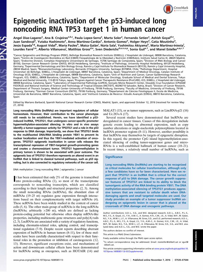
ResultsTP53TG1 Shows Cancer-Specific DNAMethylation-Associated TranscriptionalSilencing. To characterize cancer-specific DNA methylation changesthat affect lncRNA expression, we followed the experimental work-flow shown in Fig. 1A. We took advantage of the existence of anisogenic cell line derived from the colon cancer cell line HCT-116,which shows genetic disruption by homologous recombination of the
DNA methyltransferase 1 (DNMT1) and DNMT3b (double knock-out, DKO) (25). DKO cells have drastically reduced DNMT activity,5-methylcytosine DNA content, and, above all, a release of codinggenes and microRNA silencing associated with promoter CpG islandhypomethylation (25). We wanted to identify cancer-specific DNAmethylation events in lncRNA loci, so we also included two samplesof normal colon mucosa in our study design. We obtained theDNA methylation profile of the wild-type HCT-116 cells, the derivedDKO cells, and the aforementioned normal colon using the InfiniumHumanMethylation450 (450K) microarray, which interrogates 482,422CpG sites (26). Noting that the platform includes 104 annotatedlncRNAs with a 5′-CpG island (SI Appendix, Table S1) enabled us tonarrow our search to the candidate methylated positions with a possibleeffect on lncRNA expression. To identify from among these candidatesthose lncRNAs with cancer-specific differential methylation, we im-posed stringent criteria to consider only those CpG island sites witha ≥70% change in the CpG methylation level between the cancer cell
A B
DC
Fig. 1. Epigenetic silencing of the lncRNA TP53TG1 in cancer cells. (A) Schematic strategy used to identify tumor-specific DNA methylation events in lncRNAs.(B) Bisulfite genomic sequencing analysis of TP53TG1 promoter CpG island in cancer cell lines and normal tissues. Locations of bisulfite genomic sequencing PCRprimers (black arrows), CpG dinucleotides (vertical lines), and the TSS (long black arrow) are shown. Ten single clones are represented for each sample. The presence ofunmethylated and methylated CpGs is indicated by white and black squares, respectively. Red circles indicate the CpGs detected by the DNA methylation 450Kmicroarray. (C) DNAmethylation-associated transcriptional silencing of TP53TG1 in cancer cells. (Upper) TP53TG1 expression levels in methylated (HCT-116, KM12, andKATOIII) and unmethylated (SW480, HT29, MKN-7, SNU-1, MKN-45, NUGC-3, and GCIY) cancer cell lines determined by qRT-PCR. (Lower) Restored TP53TG1 expressionafter treatment with the DNA demethylating agent 5-aza-2´-deoxycytidine (AZA) or upon genetic depletion (DKO) in the originally methylated cell lines. Values weredetermined from triplicates and are expressed as the mean ± SEM. (D) TP53TG1 RNA-FISH and intracellular localization. (Upper) TP53TG1 subcellular distribution inDKO by qRT-PCR. RNAU6b and GAPDH genes were used as controls for the nuclear and cytoplasmic fractions, respectively. Values were determined from triplicatesand are expressed as the mean ± SEM. The effectiveness of cell fractionation was evaluated with lamin B1 (nuclear) and tubulin (cytoplasmic) by Western blot. C,cytoplasm; N, nucleus. (Lower) Single-molecule visualization of TP53TG1 (red spots) in HCT-116 and DKO cell lines by FISH.
E7536 | www.pnas.org/cgi/doi/10.1073/pnas.1608585113 Diaz-Lagares et al.
line HCT-116 compared with the demethylated DKO cell line andthe normal colon cell line. Under these conditions, we identified 10targets: TP53TG1 (TP53 target gene 1), NCRNA00028, LOC157627,MIR155HG, WT1AS, LINC00473, C20orf200, EMX2OS, MIAT,and MYCNOS (Fig. 1A and SI Appendix, Table S2). QuantitativeRT-PCR analyses confirmed transcriptional silencing in HCT-116cells and demethylation-associated reactivation in DKO cellsfor seven (70%) of the lncRNAs: TP53TG1, NCRNA00028,LOC157627, MIR155HG, WT1AS, C20orf200, and MYCNOS(SI Appendix, Fig. S1). Although all of them were promising candi-dates for further analysis, we decided to focus our interest onTP53TG1 because of its high-ranking order in the differential meth-ylation (SI Appendix, Table S2) and expression (SI Appendix, Fig. S1)data and because of its proposed role in central cancer pathways suchas the p53 network (27). With respect to the latter, TP53TG1 ex-pression is induced in a p53-dependent manner upon conditions ofcellular stress that involve the induction of double-strand breaks, suchas UV irradiation (27, 28), or exposure to bleomycin or cisplatin (28).To further demonstrate the silencing of TP53TG1 in cancer cells
in association with the presence of promoter CpG island hyper-methylation, we proceeded to characterize its transcription start site(TSS) and DNA methylation patterns. Using rapid amplification ofcDNA ends (RACE), we confirmed that the TP53TG1 transcriptoriginates within the CpG island in the DNAmethylation microarray,confirming the Refseq database annotation in UCSC (NR_015381.1from GRCh38/hg38). Bisulfite genomic sequencing of multipleclones confirmed the dense methylation of the CpG island in HCT-116 cells and its unmethylated status in normal colorectal mucosa(Fig. 1B). We extended our DNA methylation analyses to another12 gastrointestinal cancer cell lines (Fig. 1B). In addition to HCT-116, TP53TG1 5′-end CpG island hypermethylation was also foundin the colorectal cancer cell line KM12 and in the gastric cancer celllines KATO-III and TGBC11TKB. All of the other cell lines wereunmethylated at this locus (Fig. 1B). Normal gastric mucosa andnontumorigenic human colon epithelial cells (HCECs) (29) wereunmethylated at the TP53TG1 CpG island (Fig. 1B). QuantitativeRT-PCR revealed a loss of TP53TG1 expression in all of thehypermethylated cells, whereas lncRNA was expressed in theunmethylated cases (Fig. 1C). Importantly, the use of the deme-thylating agent 5-aza-2’-deoxycytidine restored TP53TG1 expres-sion in the hypermethylated HCT-116, KM12, KATO-III, andTGBC11TKB cell lines (Fig. 1C). Subcellular fractioning showedthat the TP53TG1 transcript in DKO cells, in addition to beingpresent in both the cytoplasm and the nucleus, was particularlyenriched in the cytosolic compartment (Fig. 1D). RNA fluorescencein situ hybridization (RNA-FISH) corroborated this intracellular lo-calization (Fig. 1D). We confirmed the noncoding nature of theTP53TG1 RNA transcript by using an artificially created recombi-nant protein followed byWestern blot to demonstrate the lack of anyTP53TG1-derived protein in the unmethylated HCEC and DKO celllines (SI Appendix, Fig. S2). In vitro transcription/translation assaysconfirmed the absence of TP53TG1 coding potential (SI Ap-pendix, Fig. S2). TP53TG1’s lack of coding capacity was alsoapparent from the negative codon substitution frequency (CSF)score (30) of –402.4488, which was similar to that obtained fromthe well-characterized lncRNA HOTAIR (–354.5062). Overall,these results indicate that tumor-specific promoter CpG islandhypermethylation-associated silencing of the lncRNA TP53TG1occurs in colorectal and gastric cancer cells.
TP53TG1 Exhibits Tumor Suppressor-Like Features in Cancer Cells.Having observed the CpG island hypermethylation-associated si-lencing of TP53TG1 in colorectal and gastric cancer cells, we usedin vitro and in vivo approaches to assess the ability of the lncRNAto suppress tumor growth. For the in vitro approach, we stablytransfected the HCT-116 cell line, hypermethylated and silencedfor TP53TG1, with a plasmid encoding the full-length lncRNAtranscript. The efficiency of transfection was assessed by measuring
TP53TG1 expression by quantitative RT-PCR (Fig. 2A). UponTP53TG1 transfection, the cells proved to be significantly lessproliferative in the 3-(4,5-dimethylthiazol-2-yl)-2,5-diphenyltetrazoliumbromide (MTT) assay (Fig. 2B) and had a significantly lower per-centage colony formation density (Fig. 2B) than empty vector-transfected cells. The reduced growth of the TP53TG1-transfectedHCT-116 cell line was associated with apoptosis induction, basedon the assessment of the sub-G1 cell population, caspase 3/7 levels,and the annexin V assay (Fig. 2C). The restoration of TP53TG1expression upon transfection in HCT-116 was also associated witha decreased invasion and migration potential of these cells, asmeasured by the Matrigel-based xCELLigence real-time assay(31) (Fig. 2D) and the wound-healing assay (Fig. 2D).For the in vivo approach, we first used tumor-formation assays in
nude mice. HCT-116 cells transfected with either the empty or theTP53TG1 vector were s.c. injected into nude mice. Tumors origi-nating from TP53TG1-transfected HCT-116 cells had a significantlylower volume and weight than empty vector-transfected/derivedtumors (Fig. 2E), in addition to a higher apoptosis rate (Fig. 2E).We also did an orthotopic growth study, implanting equal-sizedtumor pieces from the s.c. model into the colon tract. We observedthat orthotopic TP53TG1-transfected tumors were significantlysmaller than the empty vector-transfected tumors (Fig. 2F). We alsoproceeded with the converse experiment in which we analyzed theeffect of TP53TG1 depletion in nontransformed immortalizedcolonocytes (HCECs), which have an unmethylated CpG island andexpress the lncRNA transcript. Upon efficient small interferenceRNA (siRNA)-mediated down-regulation of TP53TG1 (Fig. 2G),we observed a significant enhancement of cell viability, as measuredby the MTT assay, relative to control siRNA-transfected cells (Fig.2G), and a reduced apoptosis rate (Fig. 2G). Overall, our findingssuggest that TP53TG1 has a tumor-suppressor role.
TP53TG1 Contributes to p53 Response to DNA Damage. p53 is known toactivate TP53TG1 expression upon the induction of double-strandbreaks in DNA caused by ionizing irradiation (27, 28) or treatmentwith DNA-damaging agents (27). This prompted us to considerwhether TP53TG1 hypermethylation-associated silencing in cancercells affected the sensitivity of chemotherapy drugs that directlytarget DNA. Using the TP53TG1-unmethylated HCEC line, whichcarries a wild-type p53 gene, we observed that treatment with theDNA-damaging agents caused not only the expected induction ofp53 but also enhanced TP53TG1 expression (Fig. 3A). The sameeffect was observed in two other TP53TG1-unmethylated andp53 wild-type gastrointestinal cell lines: SW48 (colon) and SNU-1(stomach) (Fig. 3B). However, we did not find increased TP53TG1expression when using DNA-damaging agents in the unmethylated,but p53 mutant, SW-620 colorectal cancer cell line (Fig. 3B). Instead,we found that the induction of TP53TG1 upon DNA damage wasdependent on the presence of a functional p53: siRNA-mediateddepletion of p53 in HCECs impaired the overexpression ofTP53TG1 when the DNA-damaging drug was used (Fig. 3C). Wethen attempted to determine how p53 exerted its control overTP53TG1 transcription at the molecular level. The chromatin im-munoprecipitation (ChIP) assay revealed that p53 binds to a pre-viously identified enhancer sequence located in intron 2 of TP53TG1(32, 33) (Fig. 3D). The association of p53 to the TP53TG1 locus hasbeen previously reported using ChIP followed by deep sequencing(ChIP-seq) (34). The described enhancer intronic region interactswith the promoter of TP53TG1, as shown by chromatin interactionanalysis by paired-end tag sequencing (ChIA-PET) (35) (SI Appen-dix, Fig. S3). p53 binds to this regulatory region of TP53TG1 andtranscriptionally activates it when the DNA-damaging agent is used(Fig. 3D). We observed the same binding pattern for the classicalp53-responding gene p21WAF1 (SI Appendix, Fig. S4).Having discovered that TP53TG1 restoration in an initially
methylation-silenced cell induces apoptosis and that p53 activatesTP53TG1 transcription upon DNA damage, we wondered whether
Diaz-Lagares et al. PNAS | Published online November 7, 2016 | E7537
MED
ICALSC
IENCE
SPN
ASPL
US

the recovery of TP53TG1 expression sensitizes to DNA-damagingagents. To address this, we measured the IC50 of the HCT-116,TP53TG1-unmethylated, and p53 wild-type cell line, transfectedwith an empty vector or with the studied lncRNA for four differentsingle-treatment DNA-damaging agents (doxorubicin, paclitaxel,carboplatin, and cisplatin) and the drug combinations used in theclinical context of colorectal cancer, 5-fluorouracil + oxaliplatin and5-fluorouracil + irinotecan. We found that, in all of the cases, thetransfection-mediated recovery of TP53TG1 expression significantlyincreased the sensitivity to these agents (Fig. 3E). We also extendedthe in vitro cell viability experiments to an in vivo mouse model oforthotopic growth. We observed that orthotopic colorectal tumorsderived from TP53TG1-transfected HCT-116 cells were signifi-cantly more responsive to the common colorectal chemotherapeuticregimen of 5-fluorouracil + oxaliplatin than empty vector-derivedtumors (Fig. 3F). These results indicate that TP53TG1 probablyplays an important role in the p53-mediated antitumoral effects ofDNA-damaging agents.
TP53TG1 Binds to the RNA Binding Protein YBX1 and Prevents ItsNuclear Localization. With the exception of its role as a transcriptinduced by p53 upon DNA damage (27, 28), nothing is knownabout the functions of TP53TG1. To identify protein partnersof TP53TG1 that could characterize its activity in the processesdescribed here, we performed RNA pull-down assays combinedwith mass spectrometry (MS). In vitro-synthesized full-length
TP53TG1 RNA, or its antisense version, was incubated in thepresence of HCEC extracts and the retrieved proteins were ana-lyzed by SDS/PAGE. As shown in Fig. 4A, a protein band of ∼45kDa was specifically pulled down by TP53TG1 RNA but not byother control RNAs. This band was cut out from the gel, trypsin-digested, and further characterized by MS, which identified theisolated band as the Y-box binding protein 1 (YBX1, also known asYB-1) through the identification of four distinct peptides (Fig. 4Band SI Appendix, Table S3). YBX1 functions as both a DNA andRNA binding protein and has been implicated in numerous cellularprocesses, including regulation of transcription and translation, pre-mRNA splicing, DNA reparation, and mRNA packaging (36).YBX1 activation has been associated with cancer progression (37,38), the epithelial–mesenchymal transition (39), metastasis (40), anddrug resistance (41, 42). Western blot with a specific antibody con-firmed that YBX1 is enriched in the TP53TG1 pull-down (Fig. 4C).The reciprocal experiment showed that immunoprecipitation ofYBX1 from HCECs can copurify the TP53TG1 lncRNA as abinding partner (Fig. 4D). Control IgG was used as a negative re-action and the YBX1 partner G3BP1 mRNA (43) was used as apositive control for YBX1 binding (Fig. 4D). Bioinformatic predic-tion of protein partners for TP53TG1 using the catRAPID system(44) also indicated that YBX1 was a likely target of the lncRNAstudied here. We further characterized the specific RNA sequence inthe TP53TG1 transcript that was responsible for the binding to theYBX1 protein. Using various YBX1 deletion mutants, followed by
A
D
E
G
F
B C
Fig. 2. TP53TG1 exhibits tumor-suppressor features in vitro and in vivo. (A) Efficient restoration of TP53TG1 upon transfection in HCT-116 cells according to qRT-PCR. Expression of TP53TG1 in cellular pools (four different clones) of HCT-116 stable transfected cells in comparison with the empty vector. Values were analyzedfrom triplicates and expressed as the mean ± SEM. (B) MTT and colony formation assays. TP53TG1 transfection reduces cell viability and the number of colonies inHCT-116 cells. (C) TP53TG1 restoration induces apoptosis in HCT-116 cells. Apoptotic cells were evaluated in TP53TG1 stably transfected HCT-116 cells after 24 h asthe sub-G1 cell population and annexin V-positive/IP-negative cells by FACS analysis. Caspase 3/7 activity was determined after 24 h by a luminometric assay.Values are expressed as the mean ± SEM (n = 6). (D) TP53TG1 reduces invasion and migration properties in HCT-116 cells evaluated by the xCELLigence Real-Time(cell index) approach (Left and Middle) and the wound-healing assays (n = 3) (Right), respectively. (E) Growth inhibitory effect of TP53TG1 restitution in HCT-116mouse tumor xenografts. (Left) Tumor volume (n = 8) was monitored over time. (Middle Left) Tumors were excised and weighed at 34 d. (Middle) Representativeimages of the confluence of HCT-116 stable transfected cells maintained in vitro for 7 d after implantation in mice and the size of the tumors at the end of theanalyses. (Middle Right) Detection and (Right) quantification of apoptotic cells (in brown) from s.c. tumors by TUNEL assay (n = 5). (Scale bar, 100 μm.) (F) Growth-inhibitory effect of TP53TG1 restitution in HCT-116 cells in a colorectal orthotopic mouse model. Small pieces of the s.c. tumor model were implanted in the colonof nude mice, and tumor weight was measured after 30 d (n = 4–5). (G) Effect of TP53TG1 RNAi-mediated knockdown in the nontumorigenic HCEC line. (Left)Values were obtained by qRT-PCR and expressed as the mean ± SEM (n = 3). The photograph shows the transfection control efficiency (green). TP53TG1 down-regulation after 72 h reduces cell viability and apoptosis. Cell viability (Middle Left) was determined by MTT (n = 3) and the frequency of apoptotic cells wasdetermined by FACS analysis of the sub-G1 cell population (n = 3) (Middle Right) and annexin V-positive/IP-negative cells (Right) (n = 2 for each independentsiRNA). **P < 0.01; *P < 0.05.
E7538 | www.pnas.org/cgi/doi/10.1073/pnas.1608585113 Diaz-Lagares et al.
YBX1Western blot in the RNA pull-down assays, we identified that,from the 710 nucleotides of the full-length transcript, a middle re-gion of 253 nucleotides was responsible for YBX1 binding (Fig. 4E).This region included five CACC YBX1 binding motifs (45, 46)(Fig. 4E). Most importantly, when we mutated the five CACCmotifs,the binding of TP53TG1 to YBX1 was notably diminished (Fig. 4E).In addition, mobility shift assays showed that the recombinant YBX1protein was able to bind specifically to this middle region, indicating adirect interaction with the lncRNA (Fig. 4E). The presence of theintact CACC YBX1-binding sites was necessary to ensure the tumor-suppressor function of TP53TG1 observed here, as transfection ofthe lncRNA form mutated at the five CACC motifs was unable toinhibit HCT-116 cell growth, as determined by the MTT and colony-formation assay (Fig. 4F). In addition, the cotransfection of the full-length wild-type YBX1 protein in TP53TG1-transfected HCT-116cells increased their growth and invasion potential, thus reversing inpart the tumor suppressor role of TP53TG1 (Fig. 4G). Our resultstherefore imply that the multitasked YBX1 protein is a direct targetof TP53TG1 involved in its associated tumor suppressor features.
We then examined the effect that TP53TG1 could have on YBX1.The transfection-mediated restoration of TP53TG1 expression inHCT-116 did not affect the total mRNA or protein levels of YBX1(Fig. 5A). In a similar manner, the siRNA-mediated depletion ofTP53TG1 in HCECs affected neither the total transcript nor theamount of the YBX1 protein (Fig. 5A). There was also no asso-ciation between the presence of TP53TG1 methylation-associatedsilencing in gastrointestinal cancer cell lines and YBX1 RNAlevels (Fig. 5A). Thus, TP53TG1 does not seem to have a signif-icant role in determining the overall levels of YBX1 in a particularcell or tumor. However, a key component of YBX1 activity residesin its controlled trafficking between the nucleus and the cytosolrelated to its dual activities as a DNA- and an RNA-bindingprotein (36, 41, 42). To assess the impact of TP53TG1 in YBX1intracellular compartmentalization, we isolated and studied thenuclear and cytosolic fraction in our experimental system in detail.We found that YBX1 was present in the nuclear and cytosoliccompartments of TP53TG1-methylated HCT-116 cells (Fig. 5B), butthe transfection-mediated recovery of TP53TG1 expression in thesecells caused the exclusion of YBX1 from the nucleus (Fig. 5B).
A
D E F
B C
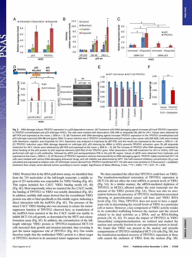
Fig. 3. DNA damage induces TP53TG1 expression in a p53-dependent manner. (A) Treatment with DNA-damaging agents increases p53 and TP53TG1 expressionin TP53TG1-unmethylated and p53 wild-type HCECs. The cells were treated with doxorubicin (100 nM) or etoposide (50 μM) for 24 h. Values were obtained byqRT-PCR and expressed as the mean ± SEM (n = 3). (B) Treatment with DNA-damaging agents increases TP53TG1 expression in the TP53TG1-unmethylated andp53 wild-type colorectal (SW-48) and gastric (SNU-1) cancer cells but not in TP53TG1-unmethylated and p53 mutant colon cancer cells (SW-620). Cells were treatedwith bleomycin, cisplatin, and etoposide for 24 h. Expression was analyzed in triplicates by qRT-PCR, and the results are expressed as the mean ± SEM (n = 4).(C) TP53TG1 induction upon DNA damage depends on wild-type p53. p53 silencing by siRNA in HCECs prevents TP53TG1 activation upon 50 μM etoposidetreatment for 24 h. Values were obtained by qRT-PCR and expressed as the mean ± SEM (n = 3). (D) The increase of TP53TG1 after DNA damage is mediated bydirect binding of the p53 protein to p53 response elements (p53 REs) of the TP53TG1 gene. After doxorubicin (100 nM) treatment for 24 h in HCECs, ChiP wasperformed with IgG or p53 antibodies, followed by qPCR and semiquantitative PCR in the p53 RE region. Values of qPCR were obtained from triplicates andexpressed as themean ± SEM (n = 3). (E) Recovery of TP53TG1 expression restores chemosensitivity to DNA-damaging agents. TP53TG1 stably transfected HCT-116cells were treated with various DNA-damaging anticancer drugs, and cell viability was determined by MTT. The half-maximal inhibitory concentration (IC50) wascalculated and expressed as relative units. (F) Orthotopic tumors derived from TP53TG1-transfected HCT-116 cells were more sensitive to 5-fluorouracil + oxaliplatintreatment than empty vector-derived tumors according to tumor weight. Significance of Mann-Whitney U test, ***P < 0.001; **P < 0.01; *P < 0.05.
Diaz-Lagares et al. PNAS | Published online November 7, 2016 | E7539
MED
ICALSC
IENCE
SPN
ASPL
US
Immunofluorescence assays confirmed the localization of YBX1 inthe cytosol and nucleus of HCT-116 cells (Fig. 5B) and its exclusivelycytosolic presence upon the transfection-related restoration ofTP53TG1 expression (Fig. 5B). Using a panel of TP53TG1-meth-ylated (KM12, TGBC11TKB, and KATO-III) and -unmethylated(HT-29, SW480, HCEC, GCIY, and NUG-3) gastrointestinal cells,we confirmed that nuclear staining was almost absent in the unme-thylated cell lines and the clear detection of both nuclear and cy-tosolic localization in the hypermethylated samples (Fig. 5 C and D).These results suggest that the tumor-suppressor role of
TP53TG1 identified here could be mediated by preventing thewidely described protumorigenic nuclear functions of YBX1 (36–42). YBX1 is known to bind to the promoter of the phosphatidy-
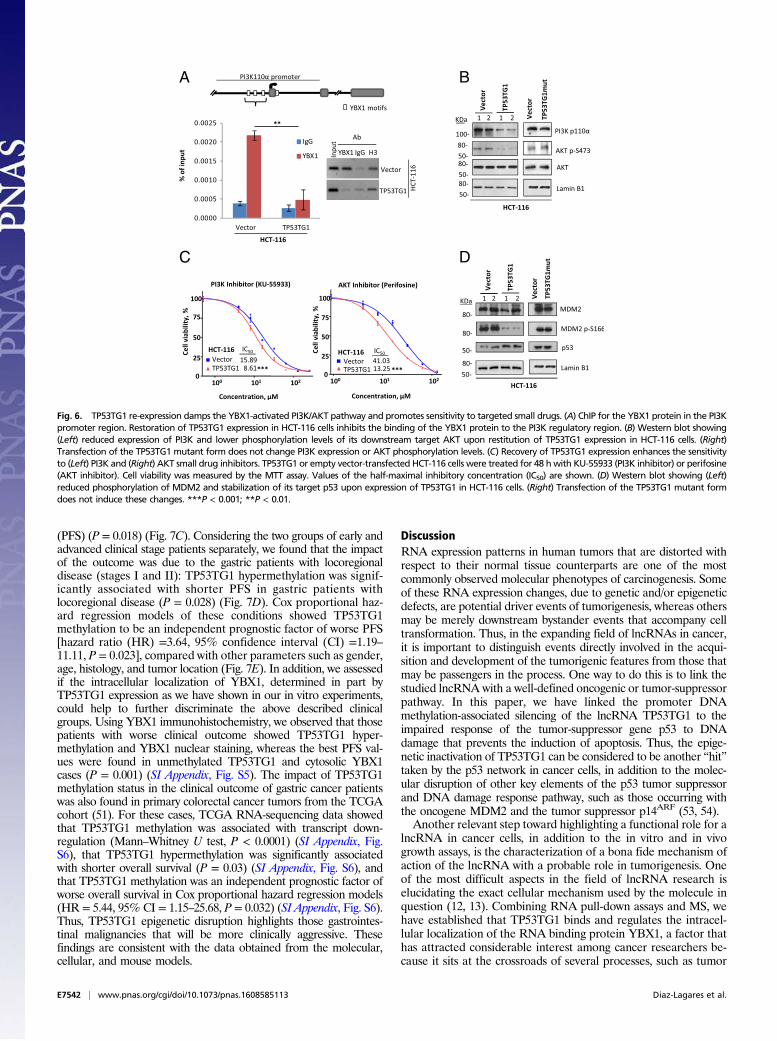
linositol-4,5-bisphosphate 3-kinase (PI3K) gene and to stimulateits transcription (47). Importantly for our observed chemoresistantphenotypes, it is known that PI3K stimulates AKT and MDM2phosphorylation, thus facilitating p53 degradation (48). In oursystem, ChIP demonstrated that the transfection-mediated recoveryof TP53TG1 expression in HCT-116 cells reduced the binding ofthe YBX1 protein to the PI3K promoter (Fig. 6A). Most importantly,the diminished binding of YBX1 to the PI3K promoter was associ-ated with the down-regulation of the PI3K protein and thereduced phosphorylation of AKT in comparison with empty vector-transfected cells (Fig. 6B). Transfection of the TP53TG1 transcriptmutated at the five CACC YBX1 binding sites did not affect PI3Kprotein levels or AKT phosphorylation (Fig. 6B). In addition, the
A
E F G
B C D
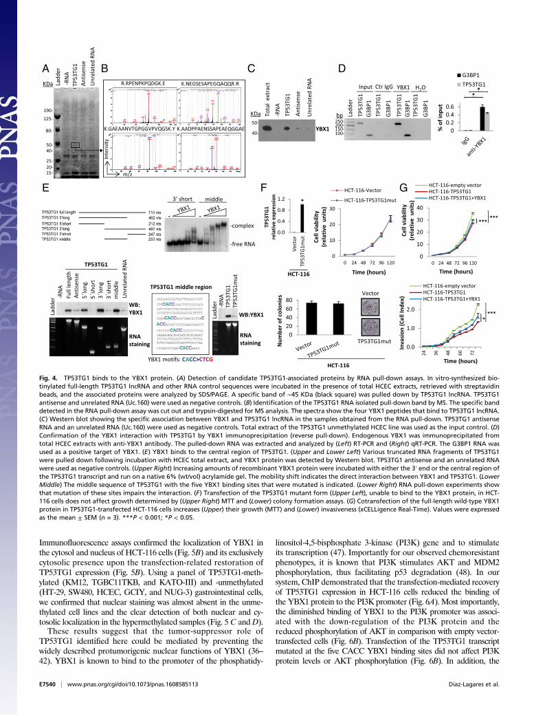
Fig. 4. TP53TG1 binds to the YBX1 protein. (A) Detection of candidate TP53TG1-associated proteins by RNA pull-down assays. In vitro-synthesized bio-tinylated full-length TP53TG1 lncRNA and other RNA control sequences were incubated in the presence of total HCEC extracts, retrieved with streptavidinbeads, and the associated proteins were analyzed by SDS/PAGE. A specific band of ∼45 KDa (black square) was pulled down by TP53TG1 lncRNA. TP53TG1antisense and unrelated RNA (Uc.160) were used as negative controls. (B) Identification of the TP53TG1 RNA isolated pull-down band by MS. The specific banddetected in the RNA pull-down assay was cut out and trypsin-digested for MS analysis. The spectra show the four YBX1 peptides that bind to TP53TG1 lncRNA.(C) Western blot showing the specific association between YBX1 and TP53TG1 lncRNA in the samples obtained from the RNA pull-down. TP53TG1 antisenseRNA and an unrelated RNA (Uc.160) were used as negative controls. Total extract of the TP53TG1 unmethylated HCEC line was used as the input control. (D)Confirmation of the YBX1 interaction with TP53TG1 by YBX1 immunoprecipitation (reverse pull-down). Endogenous YBX1 was immunoprecipitated fromtotal HCEC extracts with anti-YBX1 antibody. The pulled-down RNA was extracted and analyzed by (Left) RT-PCR and (Right) qRT-PCR. The G3BP1 RNA wasused as a positive target of YBX1. (E) YBX1 binds to the central region of TP53TG1. (Upper and Lower Left) Various truncated RNA fragments of TP53TG1were pulled down following incubation with HCEC total extract, and YBX1 protein was detected by Western blot. TP53TG1 antisense and an unrelated RNAwere used as negative controls. (Upper Right) Increasing amounts of recombinant YBX1 protein were incubated with either the 3′ end or the central region ofthe TP53TG1 transcript and run on a native 6% (wt/vol) acrylamide gel. The mobility shift indicates the direct interaction between YBX1 and TP53TG1. (LowerMiddle) The middle sequence of TP53TG1 with the five YBX1 binding sites that were mutated is indicated. (Lower Right) RNA pull-down experiments showthat mutation of these sites impairs the interaction. (F) Transfection of the TP53TG1 mutant form (Upper Left), unable to bind to the YBX1 protein, in HCT-116 cells does not affect growth determined by (Upper Right) MTT and (Lower) colony formation assays. (G) Cotransfection of the full-length wild-type YBX1protein in TP53TG1-transfected HCT-116 cells increases (Upper) their growth (MTT) and (Lower) invasiveness (xCELLigence Real-Time). Values were expressedas the mean ± SEM (n = 3). ***P < 0.001; *P < 0.05.
E7540 | www.pnas.org/cgi/doi/10.1073/pnas.1608585113 Diaz-Lagares et al.
diminished PI3K/AKT signaling seen when TP53TG1 expression wasrestored was associated with greater sensitivity to PI3K and AKTinhibitors (Fig. 6C). Most importantly, the inhibition of the PI3K/AKT pathway upon TP53TG1 restitution also decreased MDM2phosphorylation and estabilized p53 protein levels (Fig. 6D). Theseresults therefore suggest that the epigenetic silencing of TP53TG1in cancer cells promotes the YBX1-mediated activation of the PI3K/AKT pathway, which then creates further resistance not only tocommon chemotherapy DNA-damaging agents but also to smalldrug-targeted inhibitors. This finding relates to the broad spectrum ofchemoresistance that is customarily associated with the YBX1 pro-tein (36, 41, 42).
TP53TG1 Hypermethylation Occurs in Gastrointestinal Tumors inAssociation with Poor Outcome. The presence of TP53TG1 cancer-specific promoter CpG island hypermethylation was not an in vitrocharacteristic restricted to colorectal and gastric cancer cell lines.Data mining of large collections of primary human tumors inter-rogated by the same DNA methylation microarray platform as usedhere (26) confirmed the existence of TP53TG1 hypermethylation in
6% (12 of 180) and 10% (9 of 94) of colorectal (49) and gastric (50)tumors, respectively (Fig. 7A). The DNAmethylation data availablefrom The Cancer Genome Atlas (TCGA) for colorectal (51) andgastric (52) primary tumors also show the presence of TP53TG1hypermethylation in 4% (14 of 369) and 13% (38 of 298) of thesecases, respectively (Fig. 7A). The higher frequency of aberrantTP53TG1 DNA methylation noted in the primary stomach neo-plasias prompted us to focus our efforts on this tumor type. TCGARNA-sequencing data in gastric carcinomas (52) show that TP53TG1methylation was associated with transcript down-regulation (Mann–Whitney U test, P < 0.0001) (Fig. 7B). Thus, we determined bymethylation-specific PCR (MSP) the DNA methylation status of the5′-end promoter CpG island of TP53TG1 in a collection of 173primary gastric tumors. We found TP53TG1 hypermethylation in26% (45 of 173) of the gastric neoplasms studied. We studied ingreater depth the possible clinical impact of the epigenetic alterationin TP53TG1 in the 63 gastric cancer patients for whom we had de-tailed information about pathological characteristics and diseaseoutcome.We found that the presence of TP53TG1 hypermethylationwas significantly associated with shorter progression-free survival
A
B C D
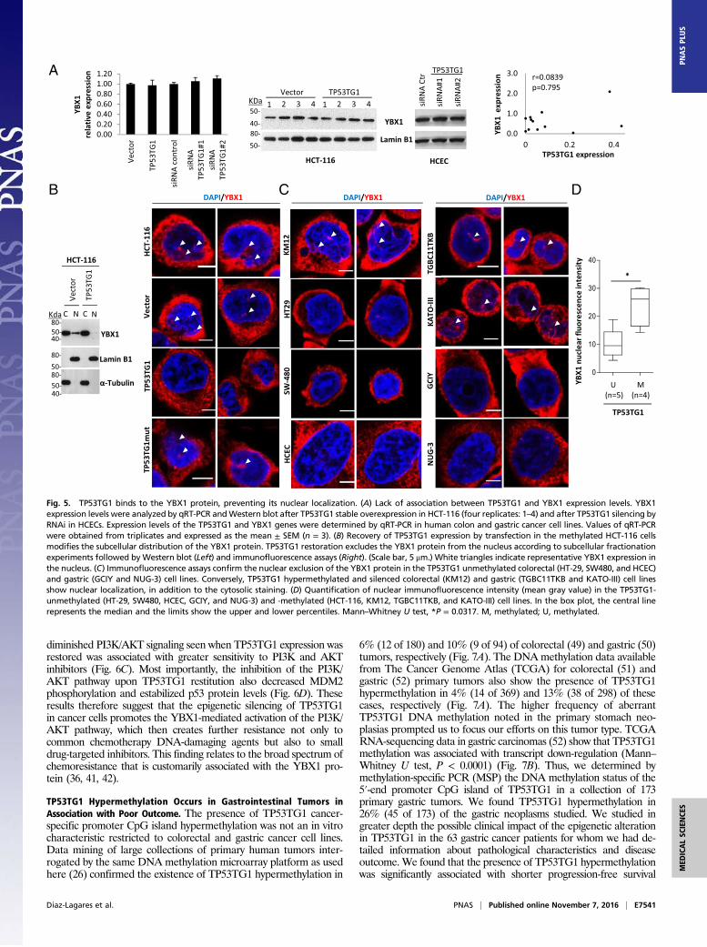
Fig. 5. TP53TG1 binds to the YBX1 protein, preventing its nuclear localization. (A) Lack of association between TP53TG1 and YBX1 expression levels. YBX1expression levels were analyzed by qRT-PCR andWestern blot after TP53TG1 stable overexpression in HCT-116 (four replicates: 1–4) and after TP53TG1 silencing byRNAi in HCECs. Expression levels of the TP53TG1 and YBX1 genes were determined by qRT-PCR in human colon and gastric cancer cell lines. Values of qRT-PCRwere obtained from triplicates and expressed as the mean ± SEM (n = 3). (B) Recovery of TP53TG1 expression by transfection in the methylated HCT-116 cellsmodifies the subcellular distribution of the YBX1 protein. TP53TG1 restoration excludes the YBX1 protein from the nucleus according to subcellular fractionationexperiments followed byWestern blot (Left) and immunofluorescence assays (Right). (Scale bar, 5 μm.) White triangles indicate representative YBX1 expression inthe nucleus. (C) Immunofluorescence assays confirm the nuclear exclusion of the YBX1 protein in the TP53TG1 unmethylated colorectal (HT-29, SW480, and HCEC)and gastric (GCIY and NUG-3) cell lines. Conversely, TP53TG1 hypermethylated and silenced colorectal (KM12) and gastric (TGBC11TKB and KATO-III) cell linesshow nuclear localization, in addition to the cytosolic staining. (D) Quantification of nuclear immunofluorescence intensity (mean gray value) in the TP53TG1-unmethylated (HT-29, SW480, HCEC, GCIY, and NUG-3) and -methylated (HCT-116, KM12, TGBC11TKB, and KATO-III) cell lines. In the box plot, the central linerepresents the median and the limits show the upper and lower percentiles. Mann–Whitney U test, *P = 0.0317. M, methylated; U, methylated.
Diaz-Lagares et al. PNAS | Published online November 7, 2016 | E7541
MED
ICALSC
IENCE
SPN
ASPL
US
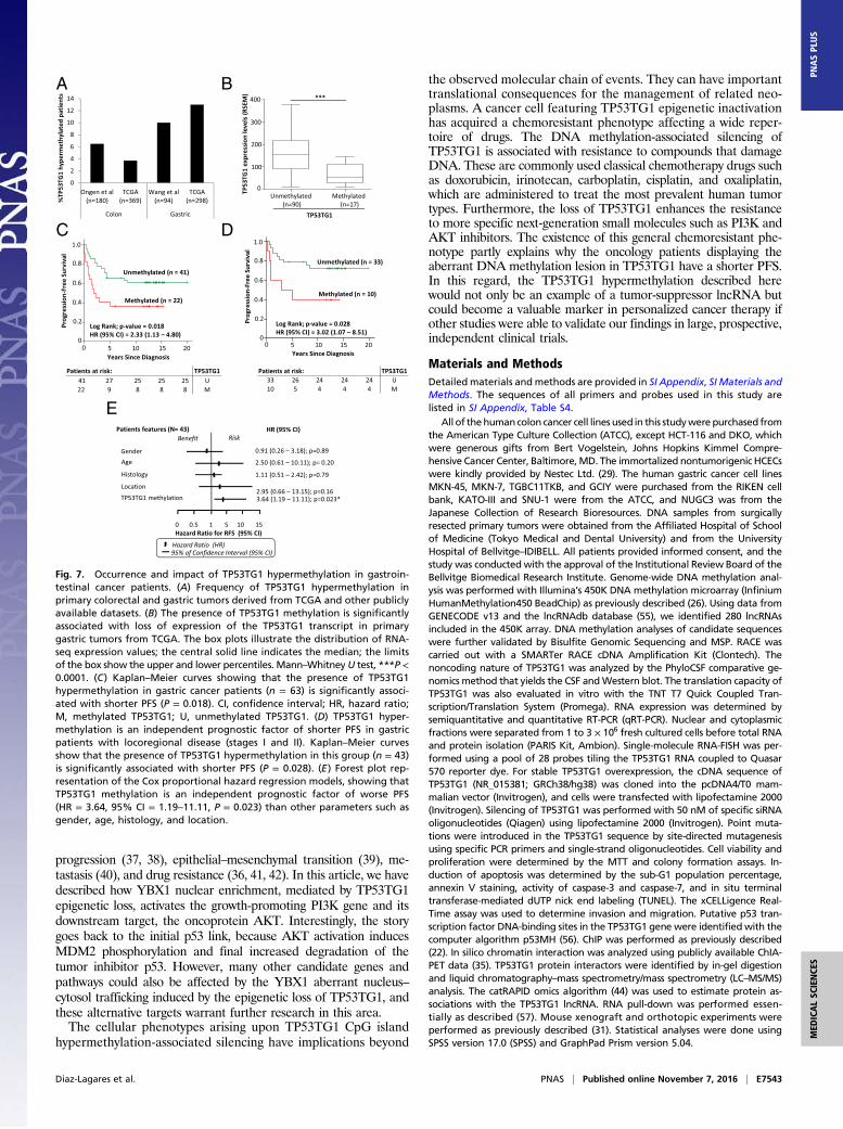
(PFS) (P = 0.018) (Fig. 7C). Considering the two groups of early andadvanced clinical stage patients separately, we found that the impactof the outcome was due to the gastric patients with locoregionaldisease (stages I and II): TP53TG1 hypermethylation was signif-icantly associated with shorter PFS in gastric patients withlocoregional disease (P = 0.028) (Fig. 7D). Cox proportional haz-ard regression models of these conditions showed TP53TG1methylation to be an independent prognostic factor of worse PFS[hazard ratio (HR) =3.64, 95% confidence interval (CI) =1.19–11.11, P = 0.023], compared with other parameters such as gender,age, histology, and tumor location (Fig. 7E). In addition, we assessedif the intracellular localization of YBX1, determined in part byTP53TG1 expression as we have shown in our in vitro experiments,could help to further discriminate the above described clinicalgroups. Using YBX1 immunohistochemistry, we observed that thosepatients with worse clinical outcome showed TP53TG1 hyper-methylation and YBX1 nuclear staining, whereas the best PFS val-ues were found in unmethylated TP53TG1 and cytosolic YBX1cases (P = 0.001) (SI Appendix, Fig. S5). The impact of TP53TG1methylation status in the clinical outcome of gastric cancer patientswas also found in primary colorectal cancer tumors from the TCGAcohort (51). For these cases, TCGA RNA-sequencing data showedthat TP53TG1 methylation was associated with transcript down-regulation (Mann–Whitney U test, P < 0.0001) (SI Appendix, Fig.S6), that TP53TG1 hypermethylation was significantly associatedwith shorter overall survival (P = 0.03) (SI Appendix, Fig. S6), andthat TP53TG1 methylation was an independent prognostic factor ofworse overall survival in Cox proportional hazard regression models(HR = 5.44, 95% CI = 1.15–25.68, P = 0.032) (SI Appendix, Fig. S6).Thus, TP53TG1 epigenetic disruption highlights those gastrointes-tinal malignancies that will be more clinically aggressive. Thesefindings are consistent with the data obtained from the molecular,cellular, and mouse models.
DiscussionRNA expression patterns in human tumors that are distorted withrespect to their normal tissue counterparts are one of the mostcommonly observed molecular phenotypes of carcinogenesis. Someof these RNA expression changes, due to genetic and/or epigeneticdefects, are potential driver events of tumorigenesis, whereas othersmay be merely downstream bystander events that accompany celltransformation. Thus, in the expanding field of lncRNAs in cancer,it is important to distinguish events directly involved in the acqui-sition and development of the tumorigenic features from those thatmay be passengers in the process. One way to do this is to link thestudied lncRNA with a well-defined oncogenic or tumor-suppressorpathway. In this paper, we have linked the promoter DNAmethylation-associated silencing of the lncRNA TP53TG1 to theimpaired response of the tumor-suppressor gene p53 to DNAdamage that prevents the induction of apoptosis. Thus, the epige-netic inactivation of TP53TG1 can be considered to be another “hit”taken by the p53 network in cancer cells, in addition to the molec-ular disruption of other key elements of the p53 tumor suppressorand DNA damage response pathway, such as those occurring withthe oncogene MDM2 and the tumor suppressor p14ARF (53, 54).Another relevant step toward highlighting a functional role for a
lncRNA in cancer cells, in addition to the in vitro and in vivogrowth assays, is the characterization of a bona fide mechanism ofaction of the lncRNA with a probable role in tumorigenesis. Oneof the most difficult aspects in the field of lncRNA research iselucidating the exact cellular mechanism used by the molecule inquestion (12, 13). Combining RNA pull-down assays and MS, wehave established that TP53TG1 binds and regulates the intracel-lular localization of the RNA binding protein YBX1, a factor thathas attracted considerable interest among cancer researchers be-cause it sits at the crossroads of several processes, such as tumor
A B
C D
Fig. 6. TP53TG1 re-expression damps the YBX1-activated PI3K/AKT pathway and promotes sensitivity to targeted small drugs. (A) ChIP for the YBX1 protein in the PI3Kpromoter region. Restoration of TP53TG1 expression in HCT-116 cells inhibits the binding of the YBX1 protein to the PI3K regulatory region. (B) Western blot showing(Left) reduced expression of PI3K and lower phosphorylation levels of its downstream target AKT upon restitution of TP53TG1 expression in HCT-116 cells. (Right)Transfection of the TP53TG1 mutant form does not change PI3K expression or AKT phosphorylation levels. (C) Recovery of TP53TG1 expression enhances the sensitivityto (Left) PI3K and (Right) AKT small drug inhibitors. TP53TG1 or empty vector-transfected HCT-116 cells were treated for 48 hwith KU-55933 (PI3K inhibitor) or perifosine(AKT inhibitor). Cell viability was measured by the MTT assay. Values of the half-maximal inhibitory concentration (IC50) are shown. (D) Western blot showing (Left)reduced phosphorylation of MDM2 and stabilization of its target p53 upon expression of TP53TG1 in HCT-116 cells. (Right) Transfection of the TP53TG1 mutant formdoes not induce these changes. ***P < 0.001; **P < 0.01.
E7542 | www.pnas.org/cgi/doi/10.1073/pnas.1608585113 Diaz-Lagares et al.
progression (37, 38), epithelial–mesenchymal transition (39), me-tastasis (40), and drug resistance (36, 41, 42). In this article, we havedescribed how YBX1 nuclear enrichment, mediated by TP53TG1epigenetic loss, activates the growth-promoting PI3K gene and itsdownstream target, the oncoprotein AKT. Interestingly, the storygoes back to the initial p53 link, because AKT activation inducesMDM2 phosphorylation and final increased degradation of thetumor inhibitor p53. However, many other candidate genes andpathways could also be affected by the YBX1 aberrant nucleus–cytosol trafficking induced by the epigenetic loss of TP53TG1, andthese alternative targets warrant further research in this area.The cellular phenotypes arising upon TP53TG1 CpG island
hypermethylation-associated silencing have implications beyond
the observed molecular chain of events. They can have importanttranslational consequences for the management of related neo-plasms. A cancer cell featuring TP53TG1 epigenetic inactivationhas acquired a chemoresistant phenotype affecting a wide reper-toire of drugs. The DNA methylation-associated silencing ofTP53TG1 is associated with resistance to compounds that damageDNA. These are commonly used classical chemotherapy drugs suchas doxorubicin, irinotecan, carboplatin, cisplatin, and oxaliplatin,which are administered to treat the most prevalent human tumortypes. Furthermore, the loss of TP53TG1 enhances the resistanceto more specific next-generation small molecules such as PI3K andAKT inhibitors. The existence of this general chemoresistant phe-notype partly explains why the oncology patients displaying theaberrant DNA methylation lesion in TP53TG1 have a shorter PFS.In this regard, the TP53TG1 hypermethylation described herewould not only be an example of a tumor-suppressor lncRNA butcould become a valuable marker in personalized cancer therapy ifother studies were able to validate our findings in large, prospective,independent clinical trials.
Materials and MethodsDetailed materials andmethods are provided in SI Appendix, SI Materials andMethods. The sequences of all primers and probes used in this study arelisted in SI Appendix, Table S4.
All of thehumancolon cancer cell lines used in this studywere purchased fromthe American Type Culture Collection (ATCC), except HCT-116 and DKO, whichwere generous gifts from Bert Vogelstein, Johns Hopkins Kimmel Compre-hensive Cancer Center, Baltimore,MD. The immortalized nontumorigenic HCECswere kindly provided by Nestec Ltd. (29). The human gastric cancer cell linesMKN-45, MKN-7, TGBC11TKB, and GCIY were purchased from the RIKEN cellbank, KATO-III and SNU-1 were from the ATCC, and NUGC3 was from theJapanese Collection of Research Bioresources. DNA samples from surgicallyresected primary tumors were obtained from the Affiliated Hospital of Schoolof Medicine (Tokyo Medical and Dental University) and from the UniversityHospital of Bellvitge–IDIBELL. All patients provided informed consent, and thestudy was conducted with the approval of the Institutional Review Board of theBellvitge Biomedical Research Institute. Genome-wide DNA methylation anal-ysis was performed with Illumina’s 450K DNA methylation microarray (InfiniumHumanMethylation450 BeadChip) as previously described (26). Using data fromGENECODE v13 and the lncRNAdb database (55), we identified 280 lncRNAsincluded in the 450K array. DNA methylation analyses of candidate sequenceswere further validated by Bisulfite Genomic Sequencing and MSP. RACE wascarried out with a SMARTer RACE cDNA Amplification Kit (Clontech). Thenoncoding nature of TP53TG1 was analyzed by the PhyloCSF comparative ge-nomics method that yields the CSF andWestern blot. The translation capacity ofTP53TG1 was also evaluated in vitro with the TNT T7 Quick Coupled Tran-scription/Translation System (Promega). RNA expression was determined bysemiquantitative and quantitative RT-PCR (qRT-PCR). Nuclear and cytoplasmicfractions were separated from 1 to 3 × 10⁶ fresh cultured cells before total RNAand protein isolation (PARIS Kit, Ambion). Single-molecule RNA-FISH was per-formed using a pool of 28 probes tiling the TP53TG1 RNA coupled to Quasar570 reporter dye. For stable TP53TG1 overexpression, the cDNA sequence ofTP53TG1 (NR_015381; GRCh38/hg38) was cloned into the pcDNA4/T0 mam-malian vector (Invitrogen), and cells were transfected with lipofectamine 2000(Invitrogen). Silencing of TP53TG1 was performed with 50 nM of specific siRNAoligonucleotides (Qiagen) using lipofectamine 2000 (Invitrogen). Point muta-tions were introduced in the TP53TG1 sequence by site-directed mutagenesisusing specific PCR primers and single-strand oligonucleotides. Cell viability andproliferation were determined by the MTT and colony formation assays. In-duction of apoptosis was determined by the sub-G1 population percentage,annexin V staining, activity of caspase-3 and caspase-7, and in situ terminaltransferase-mediated dUTP nick end labeling (TUNEL). The xCELLigence Real-Time assay was used to determine invasion and migration. Putative p53 tran-scription factor DNA-binding sites in the TP53TG1 gene were identified with thecomputer algorithm p53MH (56). ChIP was performed as previously described(22). In silico chromatin interaction was analyzed using publicly available ChIA-PET data (35). TP53TG1 protein interactors were identified by in-gel digestionand liquid chromatography–mass spectrometry/mass spectrometry (LC–MS/MS)analysis. The catRAPID omics algorithm (44) was used to estimate protein as-sociations with the TP53TG1 lncRNA. RNA pull-down was performed essen-tially as described (57). Mouse xenograft and orthotopic experiments wereperformed as previously described (31). Statistical analyses were done usingSPSS version 17.0 (SPSS) and GraphPad Prism version 5.04.
A
C
E
D
B
Fig. 7. Occurrence and impact of TP53TG1 hypermethylation in gastroin-testinal cancer patients. (A) Frequency of TP53TG1 hypermethylation inprimary colorectal and gastric tumors derived from TCGA and other publiclyavailable datasets. (B) The presence of TP53TG1 methylation is significantlyassociated with loss of expression of the TP53TG1 transcript in primarygastric tumors from TCGA. The box plots illustrate the distribution of RNA-seq expression values; the central solid line indicates the median; the limitsof the box show the upper and lower percentiles. Mann–Whitney U test, ***P <0.0001. (C) Kaplan–Meier curves showing that the presence of TP53TG1hypermethylation in gastric cancer patients (n = 63) is significantly associ-ated with shorter PFS (P = 0.018). CI, confidence interval; HR, hazard ratio;M, methylated TP53TG1; U, unmethylated TP53TG1. (D) TP53TG1 hyper-methylation is an independent prognostic factor of shorter PFS in gastricpatients with locoregional disease (stages I and II). Kaplan–Meier curvesshow that the presence of TP53TG1 hypermethylation in this group (n = 43)is significantly associated with shorter PFS (P = 0.028). (E) Forest plot rep-resentation of the Cox proportional hazard regression models, showing thatTP53TG1 methylation is an independent prognostic factor of worse PFS(HR = 3.64, 95% CI = 1.19–11.11, P = 0.023) than other parameters such asgender, age, histology, and location.
Diaz-Lagares et al. PNAS | Published online November 7, 2016 | E7543
MED
ICALSC
IENCE
SPN
ASPL
US
ACKNOWLEDGMENTS.We thank Diana Garcia, Carles Arribas, and SebastianMoran [Genomics and Epigenomics Service, Cancer Epigenetics and BiologyProgram (PEBC), Bellvitge Biomedical Research Institute (IDIBELL)]; NadiaGarcía [Unit of Nutrition and Cancer, Catalan Institute of Oncology (ICO),IDIBELL]; Carolina de la Torre and Silvia Barceló (Proteomics Service, IDIBELL);Esther Castaño [Centres Científics i Tecnològics de la Universitat de Barce-lona (CCiTUB), Bellvitge, Universitat de Barcelona–IDIBELL]; and Carme Casal(Microscopy Service, PEBC, IDIBELL) for their technical support. This work wassupported by the European Research Council under the European Commun-ity’s Seventh Framework Programme (FP7/2007-2013)/ERC Grant Agreement268626/EPINORC project; the Spanish Ministry of Economy and Competitive-ness (MINECO Projects SAF2011-22803, PI13-01339, SAF2014-55000-R, and
SAF2014-55000-R); the Instituto de Salud Carlos III (ISCIII), co-financed bythe ERDF Fund “A Way to Achieve Europe,” under the Integrated Projectof Excellence PIE13/00022 (ONCOPROFILE); and Spanish Cancer Research Net-work (RTICC) Grant RD12/0036/0039, La Marató de TV3 Foundation Grant20131610, the Cellex Foundation, and the Health and Science Departmentsof the Catalan Government (Generalitat de Catalunya) AGAUR Projects2009SGR1315 and 2014SGR633. A.D.-L. is funded by Río Hortega GrantCM14/00067 from ISCIII. A.B.C. was funded by a research contract ”SaraBorrell“ (C09/00365) and CIBERobn from the ISCIII, Spain. J.S. is a MiguelServet researcher at ISCIII. S.D. is funded by the German Research Founda-tion (DFG Di 1421/7-1) and the German Cancer Research Center (DKFZ). M.E.is an ICREA Research Professor.
1. Kapranov P, et al. (2007) RNA maps reveal new RNA classes and a possible function forpervasive transcription. Science 316(5830):1484–1488.
2. Esteller M (2011) Non-coding RNAs in human disease. Nat Rev Genet 12(12):861–874.3. Djebali S, et al. (2012) Landscape of transcription in human cells. Nature 489(7414):
101–108.4. Jonas S, Izaurralde E (2015) Towards a molecular understanding of microRNA-mediated
gene silencing. Nat Rev Genet 16(7):421–433.5. Adams BD, Kasinski AL, Slack FJ (2014) Aberrant regulation and function of microRNAs
in cancer. Curr Biol 24(16):R762–R776.6. Nana-Sinkam SP, Croce CM (2014) MicroRNA regulation of tumorigenesis, cancer
progression and interpatient heterogeneity: Towards clinical use. Genome Biol 15(9):445.
7. Rinn JL, Chang HY (2012) Genome regulation by long noncoding RNAs. Annu RevBiochem 81:145–166.
8. Huarte M (2015) The emerging role of lncRNAs in cancer. Nat Med 21(11):1253–1261.9. Liz J, Esteller M (2016) lncRNAs and microRNAs with a role in cancer development.
Biochim Biophys Acta 1859(1):169–176.10. Ling H, et al. (2015) Junk DNA and the long non-coding RNA twist in cancer genetics.
Oncogene 34(39):5003–5011.11. Kim J, et al. (2016) Long noncoding RNAs in diseases of aging. Biochim Biophys Acta
1859(1):209–221.12. Bassett AR, et al. (2014) Considerations when investigating lncRNA function in vivo.
eLife 3:e03058.13. Guil S, Esteller M (2015) RNA-RNA interactions in gene regulation: The coding and
noncoding players. Trends Biochem Sci 40(5):248–256.14. Gupta RA, et al. (2010) Long non-coding RNA HOTAIR reprograms chromatin state to
promote cancer metastasis. Nature 464(7291):1071–1076.15. Gutschner T, et al. (2013) The noncoding RNA MALAT1 is a critical regulator of the
metastasis phenotype of lung cancer cells. Cancer Res 73(3):1180–1189.16. Huarte M, et al. (2010) A large intergenic noncoding RNA induced by p53 mediates
global gene repression in the p53 response. Cell 142(3):409–419.17. Liz J, et al. (2014) Regulation of pri-miRNA processing by a long noncoding RNA
transcribed from an ultraconserved region. Mol Cell 55(1):138–147.18. Feinberg AP (2007) Phenotypic plasticity and the epigenetics of human disease.
Nature 447(7143):433–440.19. Baylin SB, Jones PA (2011) A decade of exploring the cancer epigenome—Biological
and translational implications. Nat Rev Cancer 11(10):726–734.20. Weichenhan D, Plass C (2013) The evolving epigenome. HumMol Genet 22(R1):R1–R6.21. Heyn H, Esteller M (2012) DNA methylation profiling in the clinic: Applications and
challenges. Nat Rev Genet 13(10):679–692.22. Lujambio A, et al. (2010) CpG island hypermethylation-associated silencing of non-
coding RNAs transcribed from ultraconserved regions in human cancer. Oncogene29(48):6390–6401.
23. Boque-Sastre R, et al. (2015) Head-to-head antisense transcription and R-loop for-mation promotes transcriptional activation. Proc Natl Acad Sci USA 112(18):5785–5790.
24. Ferreira HJ, Heyn H, Moutinho C, Esteller M (2012) CpG island hypermethylation-associated silencing of small nucleolar RNAs in human cancer. RNA Biol 9(6):881–890.
25. Rhee I, et al. (2002) DNMT1 and DNMT3b cooperate to silence genes in human cancercells. Nature 416(6880):552–556.
26. Sandoval J, et al. (2011) Validation of a DNA methylation microarray for 450,000 CpGsites in the human genome. Epigenetics 6(6):692–702.
27. Takei Y, Ishikawa S, Tokino T, Muto T, Nakamura Y (1998) Isolation of a novel TP53target gene from a colon cancer cell line carrying a highly regulated wild-type TP53expression system. Genes Chromosomes Cancer 23(1):1–9.
28. Kabacik S, Manning G, Raffy C, Bouffler S, Badie C (2015) Time, dose and ataxiatelangiectasia mutated (ATM) status dependency of coding and noncoding RNA ex-pression after ionizing radiation exposure. Radiat Res 183(3):325–337.
29. Poehlmann A, et al. (2013) Non-apoptotic function of caspases in a cellular model ofhydrogen peroxide-associated colitis. J Cell Mol Med 17(7):901–913.
30. Lin MF, Jungreis I, Kellis M (2011) PhyloCSF: A comparative genomics method todistinguish protein coding and non-coding regions. Bioinformatics 27(13):i275–i282.
31. Vizoso M, et al. (2015) Epigenetic activation of a cryptic TBC1D16 transcript enhancesmelanoma progression by targeting EGFR. Nat Med 21(7):741–750.
32. Ernst J, et al. (2011) Mapping and analysis of chromatin state dynamics in nine humancell types. Nature 473(7345):43–49.
33. ENCODE Project Consortium (2012) An integrated encyclopedia of DNA elements inthe human genome. Nature 489(7414):57–74.
34. Nikulenkov F, et al. (2012) Insights into p53 transcriptional function via genome-widechromatin occupancy and gene expression analysis. Cell Death Differ 19(12):1992–2002.
35. Li G, et al. (2012) Extensive promoter-centered chromatin interactions provide a to-pological basis for transcription regulation. Cell 148(1-2):84–98.
36. Kosnopfel C, Sinnberg T, Schittek B (2014) Y-box binding protein 1—A prognosticmarker and target in tumour therapy. Eur J Cell Biol 93(1-2):61–70.
37. Jürchott K, et al. (2010) Identification of Y-box binding protein 1 as a core regulatorof MEK/ERK pathway-dependent gene signatures in colorectal cancer cells. PLoSGenet 6(12):e1001231.
38. Goodarzi H, et al. (2015) Endogenous tRNA-derived fragments suppress breast cancerprogression via YBX1 displacement. Cell 161(4):790–802.
39. Evdokimova V, et al. (2009) Translational activation of snail1 and other developmentallyregulated transcription factors by YB-1 promotes an epithelial-mesenchymal transition.Cancer Cell 15(5):402–415.
40. El-Naggar AM, et al. (2015) Translational activation of HIF1α by YB-1 promotes sar-coma metastasis. Cancer Cell 27(5):682–697.
41. Bargou RC, et al. (1997) Nuclear localization and increased levels of transcriptionfactor YB-1 in primary human breast cancers are associated with intrinsic MDR1 geneexpression. Nat Med 3(4):447–450.
42. Dolfini D, Mantovani R (2013) Targeting the Y/CCAAT box in cancer: YB-1 (YBX1) orNF-Y? Cell Death Differ 20(5):676–685.
43. Somasekharan SP, et al. (2015) YB-1 regulates stress granule formation and tumorprogression by translationally activating G3BP1. J Cell Biol 208(7):913–929.
44. Livi CM, Klus P, Delli Ponti R, Tartaglia GG (2016) catRAPID signature: Identification ofribonucleoproteins and RNA-binding regions. Bioinformatics 32(5):773–775.
45. Wei WJ, et al. (2012) YB-1 binds to CAUC motifs and stimulates exon inclusion byenhancing the recruitment of U2AF to weak polypyrimidine tracts. Nucleic Acids Res40(17):8622–8636.
46. Ray D, et al. (2013) A compendium of RNA-binding motifs for decoding gene regu-lation. Nature 499(7457):172–177.
47. Astanehe A, et al. (2009) The transcriptional induction of PIK3CA in tumor cells isdependent on the oncoprotein Y-box binding protein-1.Oncogene 28(25):2406–2418.
48. Mayo LD, Dixon JE, Durden DL, Tonks NK, Donner DB (2002) PTEN protects p53 fromMdm2 and sensitizes cancer cells to chemotherapy. J Biol Chem 277(7):5484–5489.
49. Ongen H, et al. (2014) Putative cis-regulatory drivers in colorectal cancer. Nature512(7512):87–90.
50. Wang K, et al. (2014) Whole-genome sequencing and comprehensive molecularprofiling identify new driver mutations in gastric cancer. Nat Genet 46(6):573–582.
51. Cancer Genome Atlas Network (2012) Comprehensive molecular characterization ofhuman colon and rectal cancer. Nature 487(7407):330–337.
52. Cancer Genome Atlas Research Network (2014) Comprehensive molecular charac-terization of gastric adenocarcinoma. Nature 513(7517):202–209.
53. Meek DW (2009) Tumour suppression by p53: A role for the DNA damage response?Nat Rev Cancer 9(10):714–723.
54. Li J, Kurokawa M (2015) Regulation of MDM2 stability after DNA damage. J CellPhysiol 230(10):2318–2327.
55. Amaral PP, Clark MB, Gascoigne DK, Dinger ME, Mattick JS (2011) lncRNAdb: A ref-erence database for long noncoding RNAs. Nucleic Acids Res 39(Database issue):D146–D151.
56. Hoh J, et al. (2002) The p53MH algorithm and its application in detecting p53-responsive genes. Proc Natl Acad Sci USA 99(13):8467–8472.
57. Maamar H, Cabili MN, Rinn J, Raj A (2013) linc-HOXA1 is a noncoding RNA that re-presses Hoxa1 transcription in cis. Genes Dev 27(11):1260–1271.
E7544 | www.pnas.org/cgi/doi/10.1073/pnas.1608585113 Diaz-Lagares et al.


![P53 pseudogene: potential role in heat shock induced ... · suppressor activity [3,4]. The p53 gene mutation, dele-tion, insertion or protein sequestration etc are often found in](https://img.dokumen.tips/doc/110x75/5f2e3c9730622c248c578c16/p53-pseudogene-potential-role-in-heat-shock-induced-suppressor-activity-34.jpg)