Embed Size (px)
Citation preview
Research ArticleEpigallocatechin-3-Gallate (EGCG) PromotesAutophagy-Dependent Survival via Influencing the Balance ofmTOR-AMPK Pathways upon Endoplasmic Reticulum Stress
Marianna Holczer,1 Boglárka Besze,1 Veronika Zámbó,1 Miklós Csala ,1,2
Gábor Bánhegyi ,1,2 and Orsolya Kapuy 1
1Department of Medical Chemistry, Molecular Biology and Pathobiochemistry, Semmelweis University, Budapest, Hungary2Pathobiochemistry Research Group of the Hungarian Academy of Sciences and Semmelweis University, Budapest, Hungary
Correspondence should be addressed to Orsolya Kapuy; [email protected]
Received 22 September 2017; Accepted 6 December 2017; Published 31 January 2018
Academic Editor: Maria C. Albertini
Copyright © 2018 Marianna Holczer et al. This is an open access article distributed under the Creative Commons AttributionLicense, which permits unrestricted use, distribution, and reproduction in any medium, provided the original work isproperly cited.
The maintenance of cellular homeostasis is largely dependent on the ability of cells to give an adequate response to various internaland external stimuli. We have recently proposed that the life-and-death decision in endoplasmic reticulum (ER) stress response isdefined by a crosstalk between autophagy, apoptosis, and mTOR-AMPK pathways, where the transient switch from autophagy-dependent survival to apoptotic cell death is controlled by GADD34. The aim of the present study was to investigate the role ofepigallocatechin-3-gallate (EGCG), the major polyphenol of green tea, in promoting autophagy-dependent survival and to verifythe key role in connecting GADD34 with mTOR-AMPK pathways upon prolonged ER stress. Our findings, obtained byusing HEK293T cells, revealed that EGCG treatment is able to extend cell viability by inducing autophagy. We confirmedthat EGCG-induced autophagy is mTOR-dependent and PKA-independent; furthermore, it also required ULK1. We showthat pretreatment of cells with EGCG diminishes the negative effect of GADD34 inhibition (by guanabenz or siGADD34treatment) on autophagy. EGCG was able to delay apoptotic cell death by upregulating autophagy-dependent survival evenin the absence of GADD34. Our data suggest a novel role for EGCG in promoting cell survival via shifting the balance ofmTOR-AMPK pathways in ER stress.
1. Introduction
Green tea is a type of traditional Chinese tea made fromCamellia sinensis leaves, and it has been demonstrated topossess profound biochemical and pharmacological activi-ties, including antioxidative, anti-inflammatory, and anti-carcinogenic properties [1–3]. Green tea contains severalpolyphenolic components, for example, catechin, epicate-chin, and epigallocatechin-3-gallate (EGCG). Effects of themost abundant green tea polyphenol EGCG have beenshown in various pathophysiological conditions, includinginsulin resistance, endothelial dysfunction, and ischemia-reperfusion injuries [4–6]. Many scientific reports proposedthat green tea is able to influence several biological processes
by inhibiting telomerase, mitogen-activated protein kinase(MAPK), activator protein-1, or nuclear factor- (NF-) κB[7]. It has been also shown that EGCG is able to extendlongevity significantly under several stress conditions bypostponing aging and age-related diseases [8–10].
Cellular homeostasis is finely controlled by an evolu-tionarily conserved cytoprotective cellular digestive process,called autophagy [11]. Cells have residual autophagic activityeven under physiological conditions; however, the processgets more efficient during various stress events (i.e., starva-tion and growth factor deprivation) [12]. During autophagy,cellular components become sequestered into a double-membrane vesicle, whose contents are then delivered to anddegraded by lysosomes [13, 14]. Due to the crucial role of
HindawiOxidative Medicine and Cellular LongevityVolume 2018, Article ID 6721530, 15 pageshttps://doi.org/10.1155/2018/6721530

autophagy in maintaining cellular homeostasis, this self-eating process is precisely regulated [11]. Interestingly, anexcessive level of autophagy is also known to cause celldeath [15].
One of the key roles of autophagy is to maintain essentialcellular activity and viability under limited nutrient avail-ability [13]. Therefore, autophagy is tightly controlled bythe two sensors of nutritional conditions, called mTOR andAMPK [16–18]. mTOR (mammalian target of rapamycin)is a serine/threonine protein kinase in the mTORC1 com-plex, which is the main component of the mTOR pathway[18, 19]. This complex is a master regulator by integratinginputs from external and internal signals, such as growthfactors, amino acids, glucose, and energy status, to controlgrowth and metabolism [20]. Besides mTOR, AMPK (AMP-activated protein kinase) also senses cellular energy statusand has a crucial role in maintaining energy homeostasis[21]. AMPK tightly controls ATP-consuming processes,such as glycogen or protein syntheses, and it upregulatesprocesses that yield ATP (i.e., glycolysis) [22, 23]. AMPKis able to promote self-eating by phosphorylating ULK1,one of the main inducers of autophagosome formation[17, 21]. However, ULK1 is also regulated by an mTOR-dependent inhibitory phosphorylation under nutrient-richcondition [21]. In addition, AMPK directly inhibits mTORC1complex via phosphorylation [17, 21] indicating that aproper balance of AMPK-mTOR pathways is essential atphysiological conditions.
It has been lately suggested that EGCG might inducea cytoprotective autophagy in various stress events. Treat-ment with EGCG promotes the formation of autophago-somes both in primary bovine endothelial and humanhepatoma (HepG2) cells [24, 25]. EGCG abolishes thepalmitate-induced accumulation of lipid droplets via facili-tated autophagic flux [24]. Autophagy enhancement uponEGCG administration is unfavourable for hepatitis B virusreplication, and hence it is considered as a potential thera-peutic strategy [25]. EGCG also has neuroprotective effectby activating autophagy and inhibiting Bax and cyto-chrome c translocation in prion-protein-induced damages[26]. Although the positive role of EGCG in enhancingautophagy at various diseases has been already suggested,details of the regulatory mechanisms induced by this naturalcompound are yet to be revealed.
Huang et al. have suggested that EGCG upregulatesAMPK activity in a dose-dependent manner, while mTORpathway gets inhibited in hepatoma cells [27]. A dockingexperiment has also shown that EGCG is an ATP-competitive inhibitor of mTOR [28]. Kim et al. suggestedthat EGCG enhances autophagy through an AMPK-mediated mechanism [24]. Interestingly, EGCG stimulatedboth AMPK and ULK1, but not mTOR, indicating that thepolyphenol-induced autophagy is independent from mTORpathway [24]. These results suggest that EGCG acts as anenhancer on AMPK; however, its effect on mTOR pathwayis still contradictory.
Recently, we have confirmed that activation of autophagyhas a cytoprotective role upon high level of endoplasmicreticulum (ER) stress [29, 30]. This transient elevation of
autophagy is characterized by downregulation of mTORand upregulation of AMPK. Therefore, mTOR inhibitorsand/or AMPK activators (such as rapamycin, resveratrol,and metyrapone) are able to postpone apoptotic cell deathduring excessive ER stress [29, 31]. EGCG is able to restoreCa2+ homeostasis suggesting its cytoprotective effect in ERstress [32]; however, the detailed mechanism of the EGCG-modulated ER stress response remains to be elucidated.
In this study, we investigate the mechanism of EGCG-dependent autophagy and its role in ER stress by using ahuman cell line. We propose that the cytoprotective autoph-agy stimulated by EGCG is regulated via both mTOR andULK1. We also show that EGCG-induced self-eating processis independent from PKA. Here, we present that EGCGaffects the balance of mTOR-AMPK, which delays apoptoticcell death by upregulating autophagy upon ER stress. Ourdata demonstrate a novel mechanism underlying the effectof EGCG on life-and-death decision in ER stress.
2. Materials and Methods
2.1. Materials. Thapsigargin (Sigma-Aldrich, T9033), tunica-mycin (Sigma-Aldrich, T7765), rapamycin (Sigma-Aldrich,R0395), guanabenz (Sigma-Aldrich, G110), H89 (Adipo-gen, AG-CR1-0002), and epigallocatechin gallate (Sigma-Aldrich, E4143) were purchased. All other chemicals wereof reagent grade.
2.2. Cell Culture and Maintenance. A human embryonickidney cell line (HEK293T, ATCC, and CRL-3216) was usedas a model system. It was maintained in DMEM (Life Tech-nologies, 41965039) medium supplemented with 10% fetalbovine serum (Life Technologies, 10500064) and 1% antibi-otics/antimycotics (Life Technologies, 15240062). Culturedishes and cell treatment plates were kept in a humidifiedincubator at 37°C in 95% air and 5% CO2.
2.3. SDS-PAGE and Western Blot Analysis. Cells wereharvested and lysed with 20mM Tris, 135mM NaCl, 10%glycerol, and 1% NP40, pH6.8. Protein content of cell lysateswas measured using Pierce BCA Protein Assay (Thermo Sci-entific, 23225), and equal amounts of proteins were used inthe analysis. SDS-PAGE was done by using Hoefer miniVE(Amersham). Proteins were transferred onto Millipore0.45μM PVDF membrane. Immunoblotting was performedusing TBS Tween (0.1%), containing 5% nonfat dry milk,1% bovine serum albumin (Sigma-Aldrich, A9647), or gela-tin buffer (Sigma-Aldrich, G8327) for blocking membraneand for antibody solutions. Loading was controlled bydeveloping membranes for GAPDH or by dying them withPonceau S in all experiments. At least three independentmeasurements were carried out in each experiment. Thefollowing antibodies were applied: antiLC3B (SantaCruz,sc-16755), antiCaspase3 (SantaCruz, sc-7272), antiPARP(Cell Signaling, 9542S), antiULK-555-P (Cell Signaling,5869S), antiULK (Cell Signaling, 8054S), antip70S6-P (CellSignaling, 9234S), antip70S6 (SantaCruz, sc-9202), anti4-EBP1-P (Cell Signaling, 9459S), anti4-EBP1 (Cell Signaling,9644S), antiGADD34 (SantaCruz, sc-8327), antieiF2α-P
2 Oxidative Medicine and Cellular Longevity

(Cell Signaling, 9721S), antieiF2α (Cell Signaling, 9722S),antiAMPK-P (Cell Signaling, 2531S), antiAMPK (CellSignaling, 2603S) and antiGAPDH (Santa Cruz, 6C5), andHRP-conjugated secondary antibodies (SantaCruz, sc-2354and Cell Signaling, 7074S and 7076S). The bands werevisualised using chemiluminescence detection kit (ThermoScientific, 32106).
2.4. RNA Interference. RNA interference experiments wereperformed using Lipofectamine RNAi Max (Invitrogen)in GIBCO™ Opti-MEM I (GlutaMAX™-I) reduced-serummedium liquid (Invitrogen) and 20pmol/ml siRNA. ThesiGADD34 oligonucleotides were purchased from Ther-moFisher (HSS177543), and the siULK oligonucleotideswere purchased fromAmbion (AM16708). 200000HEK293Tcells were incubated at 37°C in a CO2 incubator inantiobiotic-free medium for 16 hours, and then the RNAiduplex-Lipofectamine™ RNAiMAX (Invitrogen, 13778-075)complexes were added to the cells for overnight. Thenfresh medium was added to the cells, and the appropriatetreatment was carried out. To check the efficiency ofGADD34 silencing, Western blot was used with GADD34monoclonal antibody (SantaCruz, sc-373815).
2.5. RNA Extraction and Real-Time PCR. Total RNA contentof cells was extracted using TRIzol RNA isolation reagent(Invitrogen) [33]. Retrotranscription was performed usingSuperScriptII First-Strand Synthesis System (Invitrogen).Nucleic acid levels were measured using GenQuant proRNA/DNA calculator. Equal amounts of cDNA were usedfor real-time PCR to check the efficiency of GADD34 silenc-ing. PCR reaction and real-time detection were performedusing GoTaq(R) qPCR Master Mix (Promega, A6002) andSTRATAGENEMx3005P Real-Time PCR Detection System.The real-time PCR thermocycles were the following: 95°C10min (1x), 95°C 30 sec, 58°C 45 sec, 72°C 30 sec (40x),95°C 5min, 55°C 1min, and 97°C 30 sec (1x). The appropri-ate forward and reverse real-time PCR primers were used forGADD34 and GAPDH.
2.6. Cell Viability Assays. The relative amount of viable cellswas calculated by Burker chambers. Cell viability wasdetected using CellTiter-Blue assay (Promega, G8080). Cellswere grown and treated on 96-well plates and were incubatedwith resazurin for 2 h at 37°C. Absorbance was measured at620nm and expressed in arbitrary unit, being proportionalto cell toxicity. At least three parallel measurements werecarried out for each of these experiments.
2.7. Statistics. For densitometry analysis, Western blot datawere acquired using ImageJ software. The relative banddensities were shown and normalized to an appropriate totalprotein or GAPDH band used as reference protein (seeSupplementary Information available here). For each of theexperiments, three independent measurements were car-ried out. Results are presented as mean values± S.D.and were compared using ANOVA with Tukey’s multi-ple comparison post hoc test. Asterisks indicate statisti-cally significant difference from the appropriate control:∗p < 0 05; ∗∗p < 0 01.
3. Results
3.1. EGCG Affects Autophagy and Apoptosis in a Dose-Dependent Manner. The beneficial health effect of EGCGhas been widely studied. It has been shown that low concen-trations of EGCG enhance viability of HepG2 cells; however,its high concentration causes a significant decrease in thenumber of viable cells [34]. Experimental data have alsorevealed that EGCG is able to enhance both autophagy andapoptosis [35]. In order to figure out whether these cellularprocesses are correlated to cell viability during EGCG treat-ment, we further explored the role of green tea polyphenolin cellular decision-making process between life and death.First, human embryonic kidney cells (HEK293T) weretreated with various concentrations (10, 20, 40, and 80μM)of EGCG for 24 h, and we monitored both the relativenumber of viable cells and cell viability (Figure 1(a)). Corre-sponding to the already published data, we could confirmthat low concentrations of EGCG (i.e., 10 and 20μM)resulted in a slight increase in cell viability, while excessivelevel of the polyphenol (i.e., 80μM) reduced the amount ofviable cells by ≈50%.
To detect the activation profile or level of the key indica-tors of autophagy (such as LC3II and ULK-555-P) andapoptosis (procaspase-3, cleaved PARP) during EGCG treat-ment, immunoblotting was performed (Figures 1(b) and S1).At low concentration of EGCG (i.e., 10 and 20μM), a highratio of LC3II/LC3I and an intensive phosphorylation ofULK-555-P were observed indicating that cell viability ismaintained in an autophagy-dependent manner by EGCGin a well-defined concentration range. However, a highconcentration of EGCG (80μM) slightly decreased the activ-ity of autophagy, which was accompanied by a decrease inprocaspase-3. Active capase-3 is able to cleave PARP; how-ever, we did not observe any PARP cleavage suggesting thatEGCG-dependent apoptosis might occur at a higher concen-tration of the polyphenol. Interestingly, ER stress was alreadyobserved at low concentration of EGCG (see eiF2α-P andGADD34 level in Figures 1(b) and S1), although the amountof phosphorylated eiF2α was reduced at an excessive level ofthis natural compound.
It is well-known that EGCG enhances AMPK, but itsnegative effect on mTOR has not been thoroughly studiedyet. To investigate the role of EGCG in modifying thecellular balance of AMPK-mTOR pathways, we detectedthe key markers of AMPK (AMPK-P, ULK-555-P) andmTOR pathways (such as 4-EBP1-P and p70S6-P) byimmunoblotting (Figures 1(b) and S1). EGCG treatmentsignificantly enhances AMPK (see the phosphorylated sta-tus of both AMPK and ULK in Figures 1(b) and S1)while mTOR became inactivated. This was detected byboth the dephosphorylation of p70S6 and the appear-ance of the lowest phosphorylation band of 4-EBP1(Figures 1(b) and S1).
Taken together, these results further confirm that EGCG-induced autophagy is not hazardous for human cells butrather helps maintain cell viability; however, excessive levelof this polyphenol might promote an apoptotic cell death.Low dose of EGCG is sufficient to activate AMPK and inhibit
3Oxidative Medicine and Cellular Longevity

mTOR suggesting that EGCG has a key role in unbalancingAMPK-mTOR pathways.
3.2. EGCG Induces Autophagy through mTOR-AMPKPathways. To further explore that EGCG-induced autoph-agy via unbalancing AMPK-mTOR pathways, we usedvarious drugs to enhance autophagy. It is well knownthat rapamycin (Rap) treatment induces autophagy viamTOR downregulation [19], while H-89 is a PKA inhib-itor and promotes an mTOR-independent autophagy [36](Figure S2). In order to understand EGCG-inducedautophagy, we treated HEK293T cells with either Rap(100 nM, 2 h) or H-89 (2.5μM, 2h) and EGCG (20μM,24h) without/with a subsequent Rap (100 nM, 2 h) or H-89(2.5μM, 2h) addition.
We found that combined treatments (i.e., H-89 +EGCGand Rap+EGCG) did not cause a remarkable decrease in
either cell viability or the relative amount of viable cells(Figure S3). Next, the key markers of autophagy, AMPKand mTOR pathways, were detected by immunoblotting(Figure 2). The Rap+EGCG treatment did not cause anyadditive effect on autophagy induction, AMPK activation,and mTOR downregulation, suggesting that both EGCGand Rap act via the same pathway to induce autophagy.Interestingly, the combined treatment with EGCG andH-89 had a significant additive effect on autophagy induc-tion, indicating that EGCG and H-89 employ different path-ways to promote autophagy. Although H-89 itself did notmodify the balance of AMPK-mTOR pathways, EGCG wasable to activate AMPK (see the phosphorylation of AMPKin Figure 2) and downregulate mTOR (see 4-EBP1-P inFigure 2) in the combined treatment.
Our combinatory treatment experiments suggest thatEGCG does not activate autophagy in a PKA-dependent
0
0.2
0.4
0.6
0.8
1
1.2
10 20 40 80
Relat
ive n
umbe
r of c
ells
ns ns
ns
⁎
Ctrl
0
0.2
0.4
0.6
0.8
1
1.2
Relat
ive v
iabi
lity
of ce
lls
ns ns ns
⁎
EGCG concentration (�휇M)
10 20 40 80CtrlEGCG concentration (�휇M)
(a)
Procaspase-3
18LC3 II
GAPDH
4-EBP1-P
GADD34
AMPK-P
Ctrl 10 20 40 80
4-EBP1-T
eiF2�훼-P
AMPK-T
eiF2�훼-T
p70S6-P
P70S6-T
LC3 I16
89
20
72
37
34
140ULK-T
ULK-555-P 140
20
70
70
38
38
62
62
Cleaved PARPPARP
(kD
a)
(�휇M)
(b)
Figure 1: EGCG induces autophagy in a concentration-dependent manner. HEK293T cells were treated with 10, 20, 40, and 80μMEGCG for24 h. (a) Meanwhile, the relative number of viable cells (upper panel) and relative cell viability (lower panel) were denoted. (b) During EGCGtreatment, the markers of autophagy (LC3, ULK-555-P), apoptosis (procaspase-3, PARP), AMPK (AMPK-P), and mTOR (4-EBP1-P,p70S6-P), as well as ER stress markers (i.e., eiF2α-P and GADD34) were followed by immunoblotting. GAPDH was used as loadingcontrol. For each of the experiments, three independent measurements were carried out. Error bars represent standard deviation, andasterisks indicate statistically significant difference from the control: ∗p < 0 05.
4 Oxidative Medicine and Cellular Longevity

manner. Similarly to Rap, EGCG rather induces autophagyvia unbalancing AMPK-mTOR pathways.
3.3. ULK1 Is Essential for the EGCG-Induced Autophagy. It iswell-known that both AMPK and mTOR regulate autophagythrough the phosphorylation of ULK1, one of the key control
elements of this cellular process [21]. While AMPK stimu-lates ULK1 via phosphorylating its Ser-555 and Ser-777,mTOR inhibits autophagy by phosphorylation of differentSer residues in ULK1 (i.e., Ser-757) [21]. Therefore, to furtherconfirm the role of AMPK-mTOR pathways in EGCG-induced autophagy, the effect of EGCG- (20μM, 24h)
Ctrl Rap H-89 EGCG Rap H-89+ EGCG
LC3 I
LC3 II
Procaspase-3
Cleaved PARPPARP
GAPDH
4-EBP1-P
AMPK-P
4-EBP1-T
AMPK-T
16
89
20
37
18
34
20
62
62(k
Da)
(a)
Ctrl
Rap
H-8
9
EGCG
EGCG
+ R
ap
EGCG
+ H
-89
Ctrl
Rap
H-8
9
EGCG
EGCG
+ R
ap
EGCG
+ H
-890
0.20.40.60.8
11.21.4
Procaspase-3/GAPDH
ns ns nsns nsns
ns
Ctrl
Rap
H-8
9
EGCG
EGCG
+ R
ap
EGCG
+ H
-89
Ctrl
Rap
H-8
9
EGCG
EGCG
+ R
ap
EGCG
+ H
-890
0.51
1.52
2.53
3.54
AMPK-P/AMPK-T
⁎⁎
⁎⁎
⁎⁎
⁎⁎
ns
ns⁎⁎
00.20.40.60.8
11.21.4
Cleaved PARP/GAPDH
nsns
ns nsns
nsns
Ctrl
Rap
H-8
9
EGCG
EGCG
+ R
ap
EGCG
+ H
-890
0.20.40.60.8
11.21.41.6
4-EBP1-P/4-EBP1-T
ns⁎⁎
⁎⁎⁎⁎
⁎⁎
ns⁎⁎
⁎⁎⁎⁎ ⁎⁎
⁎⁎
⁎⁎ns
⁎⁎
02468
1012141618
LC3II/LC3I
(b)
Figure 2: mTOR pathway is essential for EGCG-dependent autophagy induction. HEK293T cells were treated with rapamycin(Rap—100 nM, 2 h), H-89 (2.5 μM, 2 h), and EGCG (20 μM, 24 h) without/with followed by Rap (100 nM, 2 h) or H-89 (2.5 μM, 2 h)addition. (a) The markers of autophagy (LC3), apoptosis (procaspase-3, PARP), AMPK (AMPK-P), and mTOR (4-EBP1-P) were followedby immunoblotting. GAPDH was used as loading control. (b) Densitometry data represent the intensity of procaspase-3, cleaved PARPnormalized for GAPDH, LC3II normalized for LC3I, AMPK-P normalized for total level of AMPK, and 4-EBP1-P normalized for totallevel of 4-EBP1. For each of the experiments, three independent measurements were carried out. Error bars represent standard deviation,and asterisks indicate statistically significant difference from the control: ∗∗p < 0 01.
5Oxidative Medicine and Cellular Longevity

induced autophagy was detected in the presence or absenceof ULK1 (Figure 3). We carried out Rap (100 nM, 2 h)and H-89 (2.5μM, 2h) treatments for controls. ULK1
knockdown using siULK did not affect the relative amountof viable cells suggesting that ULK depletion did notinduce cell death (Figure 3(a)).
0
0.2
0.4
0.6
0.8
1
1.2
Ctrl Rap H-89 EGCG Ctrl Rap H-89 EGCG
Relat
ive n
umbe
r of c
ells
siULK
ns nsns
(a)
Ctrl Rap H-89 EGCG Ctrl Rap H-89 EGCGsiULK
LC3 ILC3 II
GAPDH
ULK-555-P
ULK-T
16
140
18
Procaspase-3
Cleaved PARPPARP
89
34
140
37
(kD
a)
(b)
Ctrl
Rap
H-8
9EG
CG Ctrl
Rap
H-8
9EG
CG Ctrl
Rap
H-8
9EG
CG Ctrl
Rap
H-8
9EG
CG
Ctrl
Rap
H-8
9EG
CG Ctrl
Rap
H-8
9EG
CG Ctrl
Rap
H-8
9EG
CG Ctrl
Rap
H-8
9EG
CG
Ctrl
Rap
H-8
9EG
CG Ctrl
Rap
H-8
9EG
CG Ctrl
Rap
H-8
9EG
CG Ctrl
Rap
H-8
9EG
CG
00.5
11.5
22.5
ULK-555-P/ULK-T
⁎⁎⁎
siULK
⁎⁎
00.20.40.60.8
11.21.4
Cleaved PARP/GAPDHsiULKns ns ns
00.20.40.60.8
11.21.41.6
ULK-T/GAPDH
⁎⁎ ⁎⁎⁎⁎
siULK
00.5
11.5
22.5
33.5
ULK-555-P/GAPDH
⁎⁎⁎⁎⁎⁎
siULK
0
0.5
1
1.5siULK
Procaspase-3/GAPDH
ns nsns
siULK
00.5
11.5
22.5
33.5
LC3II/LC3I
⁎⁎
⁎⁎
ns
(c)
Figure 3: ULK1 is essential for EGCG-dependent autophagy induction. HEK293T cells were treated with rapamycin (Rap—100 nM, 2 h),H-89 (2.5 μM, 2 h), and EGCG (20 μM, 24 h) without/with followed by Rap (100 nM, 2 h) or H-89 (2.5μM, 2 h) addition. (a) Meanwhile,the relative number of viable cells was denoted. (b) The markers of autophagy (LC3, ULK-555-P) and apoptosis (procaspase-3, PARP)were followed by immunoblotting. GAPDH was used as loading control. (c) Densitometry data represent the intensity of procaspase-3,cleaved PARP, and ULK-555-P and total level of ULK1 normalized for GAPDH, LC3II normalized for LC3I, and ULK-555-Pnormalized for total level of ULK. For each of the experiments, three independent measurements were carried out. Error bars representstandard deviation, and asterisks indicate statistically significant difference from the control: ∗p < 0 05; ∗∗p < 0 01.
6 Oxidative Medicine and Cellular Longevity

Depletion of ULK1 abolished EGCG-induced autoph-agy (see the low LC3II/I ratio in Figures 3(b) and 3(c)).Similarly to EGCG, Rap was not able to promote autoph-agy in the absence of ULK1, while siULK did not affectthe H-89-induced autophagy (see the LC3II/I ratios inFigures 3(b) and 3(c)). These results further confirm thatAMPK-mTOR-regulated autophagy is independent fromthe PKA pathway.
Taken together, we could conclude that ULK1 is involvedin EGCG-induced autophagy and in shifting the balance ofmTOR-AMPK pathways.
3.4. EGCG Delays Apoptotic Cell Death at an Excessive Levelof ER Stress. We have recently identified various drugs (suchas metyrapone and resveratrol), which imbalance mTOR-AMPK pathways and thus induce autophagy-dependentsurvival in ER stress [31, 37]. Since EGCG affects the activa-tion of AMPK and mTOR, we examined whether EGCG alsohas a positive effect on cell survival during ER stress.
In order to verify the role of EGCG in ER stress,HEK293T cells were pretreated with EGCG (20μM, 24h)and then an ER stressor was added, such as thapsigargin(10μM, 2h) or tunicamycin (25μM, 2h). While thapsigargin(TG) disrupts the calcium storage of the ER, tunicamycin(TM) inhibits N-linked glycosylation of secretory andmembrane proteins in the ER [38, 39]. We have alreadyshown that TM- or TG-induced ER stress occurs a tran-sient peak of autophagy-dependent survival followed byapoptotic cell death [29]. To explore whether EGCG iscapable to maintain cell viability upon ER stress, boththe relative amount of viable cells and relative cell via-bility were detected during EGCG+TG or EGCG+TMtreatments (Figures 4(a) and 5(a)). Addition of EGCGprior to TG or TM significantly extended cell viabilityand postponed cell death even at continuous treatmentswith an excessive level of the ER stressor. Our resultsuggests that this polyphenol is capable of improvingcell viability.
0
0.2
0.4
0.6
0.8
1
1.2
0.5 1 1.5 2
Relat
ive n
umbe
r of c
ells
Time (h)
0.5 1 1.5 2Time (h)
ns ns ⁎⁎
⁎⁎ ⁎
⁎⁎
ns ns
00.10.20.30.40.50.60.70.80.9
1
Relat
ive v
iabi
lity
of ce
lls
TGEGCG + TG
TGEGCG + TG
(a)
Ctrl 15 30 45 60 75 90 120105
GAPDH
ULK-T
ULK-555-P
4-EBP1-P
GADD34
AMPK-P
4-EBP1-T
eiF2�훼-P
AMPK-T
eiF2�훼-T
16
140
20
72
37
18
Cleaved PARPPARP
Procaspase-3
89
34
140
20
38
38
62
62
LC3 IILC3 I
(kD
a)
(min)
(b)
Figure 4: EGCG pretreatment extends autophagy-dependent survival with respect to TG-induced ER stress. HEK293T cells were pretreatedwith 20μM EGCG for 24 h followed by TG (10 μM) treatment for 2 h. (a) Meanwhile, the relative number of viable cells (upper panel) andrelative cell viability (lower panel) were denoted in time. (b) The markers of autophagy (LC3, ULK-555-P), apoptosis (procaspase-3, PARP),AMPK (AMPK-P), and mTOR (4-EBP1-P), as well as ER stress markers (i.e., eiF2α-P and GADD34) were followed by immunoblotting intime. GAPDH was used as loading control. For each of the experiments, three independent measurements were carried out. Error barsrepresent standard deviation, and asterisks indicate statistically significant difference from the control: ∗p < 0 05; ∗∗p < 0 01.
7Oxidative Medicine and Cellular Longevity

In order to detect the effect of EGCG with respect to ERstress, autophagy, apoptosis, AMPK, and mTOR markerswere followed during EGCG+TG or EGCG+TM treatmentsin time by immunoblotting (Figures 4(b), 5(b), S4, and S5). Aremarkably high level of LC3II/I suggested that autophagyremained active even after a two-hour-long TG or TM treat-ment, while neither a drop in procaspase-3 nor the cleavageof PARP was observed. These results indicate that EGCGis able to postpone apoptotic cell death via autophagyinduction upon an excessive level of ER stress.
The intensive phosphorylation of both AMPK and ULK(on its Ser-555 residue) suggests that AMPK got stimulatedand remained active until the end of the combined treatment(Figures 4(b), 5(b), S4, and S5). The mTOR pathway gotdownregulated when ER stress was preceded with EGCGaddition (see the 4-EBP1-P in Figures 4(b), 5(b), S4, and S5).
These results indicate that EGCG induces autophagy viaunbalancing mTOR-AMPK pathways, and by this means itdelays apoptotic cell death in ER stress.
3.5. Addition of EGCG Can Rescue GADD34 Inhibition withRespect to ER Stress. Recently, we have suggested that oneof the key elements of ER stress response mechanism, calledGADD34 (the growth arrest and DNA damage-inducibleprotein) associated with PP1 (protein phosphatase 1), con-stitutes a mechanistic link between ER stress and mTORactivation [37]. It has been also suggested that GADD34promotes autophagy-dependent survival via downregulat-ing mTOR in ER stress or in the stress caused by theexpression of mutant huntingtin proteins [37, 40]. Inhibi-tion of GADD34 by a PP1 inhibitor (i.e., guanabenz) ortransfection with siGADD34 results in a downregulation
0
0.2
0.4
0.6
0.8
1
1.2
0.5 1 1.5 2
Relat
ive v
iabi
lity
of ce
lls
Time (h)
TMEGCG + TM
⁎⁎ns ⁎ ⁎
ns ⁎⁎⁎⁎ ⁎⁎
00.10.20.30.40.50.60.70.80.9
1
Relat
ive n
umbe
r of c
ells
0.5 1 1.5 2Time (h)
TMEGCG + TM
(a)
Ctrl 15 30 45 60 75 90 120105
LC3 II
GAPDH
ULK-T
ULK-555-P
4-EBP1-P
GADD34
AMPK-P
4-EBP1-T
eiF2�훼-P
AMPK-T
eiF2�훼-T
LC3 I16
140
20
72
37
18
Cleaved PARP
PARP
Procaspase-3
89
34
140
20
38
38
62
62
(kD
a)
(min)
(b)
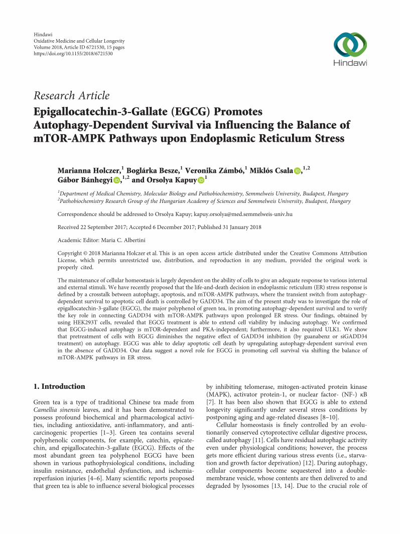
Figure 5: EGCG pretreatment extends autophagy-dependent survival with respect to TM-induced ER stress. HEK293T cells were treatedwith 20μM EGCG for 24 h followed by TM (25 μM) treatment for 2 h. (a) Meanwhile, the relative number of viable cells (upper panel)and relative cell viability (lower panel) were denoted in time. (b) The markers of autophagy (LC3, ULK-555-P), apoptosis (procaspase-3,PARP), AMPK (AMPK-P), and mTOR (4-EBP1-P), as well as ER stress markers (i.e., eiF2α-P and GADD34) were followed byimmunoblotting in time. GAPDH was used as loading control. For each of the experiments, three independent measurementswere carried out. Error bars represent standard deviation, and asterisks indicate statistically significant difference from the control:∗p < 0 05; ∗∗p < 0 01.
8 Oxidative Medicine and Cellular Longevity

of autophagy-dependent survival and a quick activation ofmTOR pathway, followed by apoptotic cell death duringER stress [37]. Both rapamycin and resveratrol treatmentsare able to diminish the negative effect of GADD34 down-regulation by promoting autophagy induction, AMPKupregulation, and mTOR inhibition [37]. Since EGCG seemsto be a potential regulator of mTOR-AMPK balance uponcellular stress, the polyphenol might protect the cells viaautophagy induction even in the absence of GADD34 underER stress.
GADD34 protein level got activated quickly when ERstress was preceded by EGCG addition (Figures 4(b), 5(b),S4, and S5). We observed that GADD34 level remained higheven after 2 h long treatment with TM or TG supposing itsimportant role in EGCG-induced autophagy with respect toER stress.
To explore whether EGCG pretreatment can rescueGADD34 downregulation-induced apoptotic cell death uponER stress, we carried out a combined treatment. First,HEK293T cells were treated with a GADD34 inhibitor,called guanabenz (GB—5μM, 1h), followed by EGCGaddition (20μM) for 24 h. Then ER stress was inducedby TG (10μM, 2h) or TM (25μM, 2h). EGCG pretreat-ment was able to extend cell viability and increase therelative amount of viable cells in GB-pretreated cells underER stress (Figure S6).
We analysed the effect of GADD34 inhibition during ERstress combined with/without EGCG addition via detectingautophagy, apoptosis, AMPK, and mTORmarkers by immu-noblotting (Figures 6 and 7). The inactivation of GADD34was detected by eiF2α-P. In the absence of EGCG, GB quicklydownregulates autophagy and induces apoptotic cell deathduring ER stress. However, when cells were pretreated withEGCG, the high ratio of LC3II/I indicates an intensiveautophagy until the end of the treatment; meanwhile,apoptosis remains inactive. Neither procaspase-3 depletionnor PARP cleavage was detected in the presence of EGCG.Interestingly, EGCG was able to induce AMPK (see theintensive phosphorylation of both AMPK and ULK1 inFigures 6 and 7) and downregulate mTOR (see 4-EBP1-Pin Figures 6 and 7) even if GADD34 was inhibited by GBduring ER stress.
These data suggest that EGCG is able to maintain cellviability via autophagy-dependent survival even in theabsence of GADD34 upon ER stress. Our experimentsindicate that the negative effect of GADD34 inhibition byGB can be suppressed by EGCG-induced imbalance ofmTOR-AMPK pathways with respect to ER stress.
3.6. GADD34 Silencing by siRNA Has Similar Effects toGB Treatment with Respect to ER Stress. To confirm thatEGCG postpones ER stress-induced apoptotic cell deathvia GADD34, the combined treatment of ER stressor andEGCG was done in cells where GADD34 was silenced withsiRNA (Figures 8 and S7). First, we tested the efficiency ofsiGADD34 both on mRNA (data not shown) and protein(Figure S7A) levels. Similar to addition of GB, GADD34silencing drastically decreased the amount of viable cellsduring TG treatment, while pretreatment with 20μM EGCG
for 24 h was able to maintain cell viability (Figure S7B). Addi-tion of TG in HEK293T cells expressing siGADD34 resultedin a short and dumped autophagic response (see the weakLC3II/I ratio and ULK1 phosphorylation in Figure 8), whilean early apoptosis induction was observed, that is, depletionof procaspase-3 and appearance of cleaved PARP werealready detected after 1.5 h long TG treatment. By contrast,EGCG pretreatment could maintain autophagy-dependentsurvival and delay apoptotic cell death even in the absenceof GADD34 (Figure 8). Both LC3II/LC3I and ULK-555-Plevels remain high; meanwhile, no caspase-3 activation wasnoticed. In these combined treatments, the AMPK alsomaintained its active state (see the constant phosphorylationof both AMPK and ULK1 in Figure 8), while the mTORpathway remained blocked (see 4-EBP1-P in Figure 8).Similar effects were observed by using TM (data not shown).
These data further confirm that the negative effect ofGADD34 silencing during ER stress can be rescued byEGCG addition. This natural compound is able to imbal-ance the AMPK-mTOR pathways and promote autophagy-dependent survival in the absence of GADD34.
4. Discussion
ER has a key function to maintain cellular homeostasis bycontaining some of the main regulatory elements of life-and-death decision. Consequently, ER stress-induced dam-ages appear in lots of different human pathologies such asneurodegenerative diseases, obesity, type two diabetes, andmany others [41–43]. Using both molecular and theoreticalbiological techniques, we have shown previously that apopto-tic cell death is always preceded by autophagy-dependentsurvival upon excessive level of ER stress [29, 31]. Therefore,newly identified autophagy inducers might become potentdrugs in the future by postponing the injurious effects ofER stress. We have recently confirmed that the “survivalwindow” of autophagy can be expanded by pretreatmentwith mTOR inhibitors and/or AMPK activators (such asmetyrapone and resveratrol) upon ER stress [31, 37]. Here,we introduce a new candidate for extending cell viability,namely, epigallocatechin-3-gallate (EGCG). Plant polyphe-nols, including green tea flavanols, have pleiotropic effects;however, many of their specific molecular targets have beenrecently identified. Flavanols are widely known as antioxi-dants, but under certain conditions (e.g., in the presence offerric iron) behave as prooxidants [44]. Since they act mainlyon cellular membranes, green tea flavanols are known tomodulate various functions of the ER [45], includingluminal enzyme activities [46, 47], membrane transportprocesses [48, 49], and redox homeostasis [47]. It has beenalso demonstrated that EGCG extends life expectanciessignificantly, which was attributed either to decreasedoxidative stress and inflammation [10] or to the inducedproduction of reactive oxygen species [50]. However, theinvolvement of the AMPK/SIRT1/FOXO axis seems to befirmly established.
Our data demonstrate that a low concentration of EGCGis able to induce autophagy (Figures 1(b) and S1) con-comitantly with rise in cell viability, suggesting this
9Oxidative Medicine and Cellular Longevity

activation of self-eating process induced by the polyphe-nol is not harmful for the cells (Figure 1(a)). Pretreat-ment with low concentration of EGCG followed byaddition of ER stressor (TG or TM) could extendautophagy-dependent survival (Figures 4(b), 5(b), S4, andS5); meanwhile, cell viability did not change (Figures 4(a)and 5(a)) and apoptosis (e.g., PARP cleavage) was notobserved upon ER stress (Figures 4(b), 5(b), S4, and S5).Interestingly, Ahn et al. have indicated that the cytotoxiceffect of excessive level of EGCG is due to the expression of
ER stress response proteins, such as CHOP, GADD34, andATF3 [34]. Here, we show that the translational initiationfactor, eiF2α, gets phosphorylated even at low level of EGCG(Figures 1(b) and S1). Although eiF2α-P has a key role inshutting down the global protein translation upon ER stress,no cell death is observed suggesting that activation of ERstress response mechanism is not fatal. Rather this eiF2αphosphorylation induced by EGCG is essential to upregulateGADD34 level. In this study, we assume that GADD34 levelis increased parallel to autophagy induction upon EGCG
(kD
a)
140
140
Ctrl 0.5 1 1.5 2 0.5 1 2 h1.5+ GB + GB + EGCG
16
20
37
18
20
38
38
62
62
LC3 II
GAPDH
ULK-T
ULK-555-P
4-EBP1-P
AMPK-P
4-EBP1-T
eiF2�훼-P
AMPK-T
eiF2�훼-T
LC3 I
89
34
Cleaved PARPPARP
Procaspase-3
+ TG
(a)
0
5
10
15
20ULK-555-P/ULK-T
⁎⁎ ⁎⁎⁎⁎
⁎⁎
012345
Cleaved PARP/GAPDH
⁎
⁎⁎⁎⁎
⁎⁎
05
10152025
Ctrl 0.5 1 1.5 2 0.5 1 1.5 2
LC3II/LC3I
⁎⁎
⁎⁎
⁎⁎
⁎⁎
Time (h)Ctrl 0.5 1 1.5 2 0.5 1 1.5 2
Time (h)
Ctrl 0.5 1 1.5 2 0.5 1 1.5 2Time (h)
Ctrl 0.5 1 1.5 2 0.5 1 1.5 2Time (h)
Ctrl 0.5 1 1.5 2 0.5 1 1.5 2Time (h)
Ctrl 0.5 1 1.5 2 0.5 1 1.5 2Time (h)
Ctrl 0.5 1 1.5 2 0.5 1 1.5 2Time (h)
0.5
1
1.5Procaspase-3/GAPDH
⁎
⁎⁎ ⁎⁎
⁎⁎
05
10152025
AMPK-P/AMPK-T
⁎⁎
⁎⁎
⁎⁎
⁎⁎
0
0.5
1
1.5
24-EBP1-P/4-EBP1-T
ns ⁎⁎⁎⁎
⁎⁎
0
10
20
30eiF2�훼-P/eiF2�훼-T
ns ns nsns
0
(b)
Figure 6: EGCG-dependent effect on mTOR-AMPK pathways rescues GADD34 inhibition with respect to TG-induced ER stress. HEK293Tcells were pretreated with GB (5 μM, 1 h) then without/with EGCG (20 μM, 24 h) followed by TG addition (10 μM, 2 h). The GB level was kepthigh until the end of the cell treatment. (a) After TG treatment, the markers of autophagy (LC3, ULK-555-P), apoptosis (procaspase-3,PARP), AMPK (AMPK-P), and mTOR (4-EBP1-P), as well as ER stress markers (eiF2α-P) were followed by immunoblotting. GAPDHwas used as loading control. (b) Densitometry data represent the intensity of procaspase-3, cleaved PARP normalized for GAPDH, LC3IInormalized for LC3I, ULK-555-P normalized for total level of ULK1, AMPK-P normalized for total level of AMPK, 4-EBP1-Pnormalized for total level of 4-EBP1, and eiF2α-P normalized for total level of eiF2α. For each of the experiments, three independentmeasurements were carried out. Error bars represent standard deviation, and asterisks indicate statistically significant difference from thecontrol: ∗p < 0 05; ∗∗p < 0 01.
10 Oxidative Medicine and Cellular Longevity
treatment (Figures 1(b) and S1), indicative of its importantrole in green tea polyphenol-induced cell survival.
Previously, we have shown that mTOR is downregulatedwith response to ER stress via GADD34 [37]. We haverecently confirmed that blocking GADD34 results in a quickactivation of both mTOR pathway and apoptotic cell death;meanwhile, AMPK gets downregulated and the period ofautophagy-dependent survival is much shorter upon ERstress [37]. We also supposed that the negative effect ofGADD34 depletion is successfully suppressed with mTOR
inhibitors and/or AMPK activators (such as rapamycin andresveratrol) during ER stress [37]. To further confirm the roleof EGCG in unbalancing mTOR-AMPK pathways, a phar-macological inhibitor (GB) or an siRNA was used to blockGADD34 and then cells were pretreated with EGCG followedby addition of an ER stressor. In this study, we show thata 24-hour long pretreatment with a low concentration ofgreen tea polyphenol followed by TG or TM additionwas able to extend cell viability via intensive activationof both AMPK and autophagy; meanwhile, mTOR and
+ GB + GB + EGCG
ULK-T
(kD
a)
LC3 II
GAPDH
ULK-555-P
4-EBP1-P
AMPK-P
4-EBP1-T
eiF2�훼-P
AMPK-T
eiF2�훼-T
LC3 ICtrl 0.5 1 1.5 2 0.5 1 2 h1.5
16140
20
37
18
Procaspase-3
Cleaved PARPPARP
89
34
140
20
38
38
62
62
+ TM
(a)
0
2
4
6⁎⁎
⁎⁎
⁎⁎ ⁎⁎
0
0.5
1
1.5
2⁎⁎ ⁎⁎ ⁎⁎
⁎⁎
0
20
40
60
ns
ns
nsns
0
1
2
3
4
ns
⁎⁎⁎
⁎⁎
0
2
4
6⁎⁎
⁎
⁎⁎
⁎⁎
0
0.5
1
1.5
⁎⁎
⁎⁎
⁎
⁎⁎
0
0.5
1
2
1.5
⁎⁎⁎⁎
⁎⁎
⁎⁎
Ctrl 0.5 1 1.5 2 0.5 1 1.5 2Time (h)
Ctrl 0.5 1 1.5 2 0.5 1 1.5 2Time (h)
Ctrl 0.5 1 1.5 2 0.5 1 1.5 2Time (h)
Ctrl 0.5 1 1.5 2 0.5 1 1.5 2Time (h)
Ctrl 0.5 1 1.5 2 0.5 1 1.5 2Time (h)
Ctrl 0.5 1 1.5 2 0.5 1 1.5 2Time (h)
Ctrl 0.5 1 1.5 2 0.5 1 1.5 2Time (h)
ULK-555-P/ULK-T
Cleaved PARP/GAPDH
LC3II/LC3I
Procaspase-3/GAPDH
AMPK-P/AMPK-T 4-EBP1-P/4-EBP1-T
eiF2�훼-P/eiF2�훼-T
(b)
Figure 7: EGCG-dependent effect on mTOR-AMPK pathways rescues GADD34 inhibition with respect to TM-induced ER stress. HEK293Tcells were pretreated with GB (5 μM, 1 h) then without/with EGCG (20 μM, 24 h) followed by TM addition (25 μM, 2 h). The GB levelwas kept high until the end of the cell treatment. (a) After TM treatment, the markers of autophagy (LC3, ULK-555-P), apoptosis(procaspase-3, PARP), AMPK (AMPK-P), and mTOR (4-EBP1-P), as well as ER stress markers (eiF2α-P) were followed byimmunoblotting. GAPDH was used as loading control. (b) Densitometry data represent the intensity of procaspase-3, cleaved PARPnormalized for GAPDH, LC3II normalized for LC3I, ULK-555-P normalized for total level of ULK1, AMPK-P normalized for total levelof AMPK, 4-EBP1-P normalized for total level of 4-EBP1, and eiF2α-P normalized for total level of eiF2α. For each of the experiments,three independent measurements were carried out. Error bars represent standard deviation, and asterisks indicate statistically significantdifference from the control: ∗p < 0 05; ∗∗p < 0 01.
11Oxidative Medicine and Cellular Longevity

ER stressor-induced apoptotic cell death were downregulated(Figures 6, 7, and 8). Here, we suggest that EGCG treatmentsuccessfully modifies the balance of mTOR-AMPK pathwaysand thus the negative effect of GADD34 depletion waseffectively suppressed. These results further confirm thatEGCG-dependent fine-tuning of mTOR-APMK pathwayshas a crucial effect to maintain the precise balance of life-and-death decision under ER stress.
Since the effect of EGCG on mTOR pathway seems to becontradictory in the literature, EGCG treatment was com-bined with either mTOR-dependent (rapamycin) or PKA-
dependent (H-89) autophagy promoter to identify whichpathway is involved in autophagy induction in case of EGCGaddition (Figure 2). Autophagy got similarly enhanced bothin EGCG and EGCG+Rap treatments revealing that EGCGand Rap regulate the self-eating process via the same mTORpathway. However, EGCG combined with H-89 significantlypromoted autophagy compared to simple H-89 treatment(Figure 2), suggesting that EGCG-induced autophagy isnot PKA-dependent. Similarly to rapamycin treatment,transfection with siULK drastically inhibited autophagyduring EGCG treatment (Figure 3) confirming that green
(kD
a)
Ctrl 0.5 1 1.5 2 0.5 1 2 h1.5TG TG + EGCG
LC3 II
GAPDH
ULK-T
ULK-555-P
4-EBP1-P
AMPK-P
4-EBP1-T
eiF2�훼-P
AMPK-T
eiF2�훼-T
LC3 I16140
20
37
18
Cleaved PARPPARP
Procaspase-3
89
34
140
20
38
38
62
62
siGadd34
(a)
0
0.5
1
1.5
2⁎⁎⁎⁎
⁎⁎
⁎
0
0.5
1
1.5
ns ns ⁎⁎⁎⁎
0
0.5
1
1.5
2
⁎⁎ ⁎⁎⁎⁎ ⁎⁎
0
1
2
3 ⁎⁎⁎⁎ ⁎⁎ ⁎⁎
0
2
4
6
8
⁎
⁎⁎
⁎⁎
⁎⁎
012345
⁎⁎ ⁎⁎
⁎⁎
⁎⁎
0
2
4
6
8 ns
nsns
⁎
ULK-555-P/ULK-T
Cleaved PARP/GAPDH
LC3II/LC3I
Procaspase-3/GAPDH
AMPK-P/AMPK-T 4-EBP1-P/4-EBP1-T
eiF2�훼-P/eiF2�훼-T
Ctrl 0.5 1 1.5 2 0.5 1 1.5 2Time (h)
Ctrl 0.5 1 1.5 2 0.5 1 1.5 2Time (h)
Ctrl 0.5 1 1.5 2 0.5 1 1.5 2Time (h)
Ctrl 0.5 1 1.5 2 0.5 1 1.5 2Time (h)
Ctrl 0.5 1 1.5 2 0.5 1 1.5 2Time (h)
Ctrl 0.5 1 1.5 2 0.5 1 1.5 2Time (h)
Ctrl 0.5 1 1.5 2 0.5 1 1.5 2Time (h)
(b)
Figure 8: EGCG-dependent effect on mTOR-AMPK pathways rescues GADD34 depletion with respect to ER stress. GADD34 was silencedin HEK293T cells, and then cells were treated with 10 μMTG for 2 h or pretreated with EGCG (20 μM, 24 h) followed by TG addition (10 μM,2 h). (a) After TG treatment, the markers of autophagy (LC3, ULK-555-P), apoptosis (procaspase-3, PARP), AMPK (AMPK-P), and mTOR(4-EBP1-P), as well as ER stress markers (eiF2α-P) were followed by immunoblotting. GAPDHwas used as loading control. (b) Densitometrydata represent the intensity of procaspase-3, cleaved PARP normalized for GAPDH, LC3II normalized for LC3I, ULK555-P normalized fortotal level of ULK1, AMPK-P normalized for total level of AMPK, 4-EBP1-P normalized for total level of 4-EBP1, and eiF2α-P normalized fortotal level of eiF2α. For each of the experiments, three independent measurements were carried out. Error bars represent standard deviation,and asterisks indicate statistically significant difference from the control: ∗p < 0 05; ∗∗p < 0 01.
12 Oxidative Medicine and Cellular Longevity

tea polyphenol induces the self-eating process throughULK1-AMPK-mTOR regulatory network. Since AMPKdownregulates mTOR pathway via direct phosphorylation,we cannot rule out that EGCG has both direct and indirect(through AMPK) negative effects on mTOR. Therefore, fur-ther studies are needed to identify the exact targets of EGCG.
In conclusion, the positive effects with pretreatment ofprecisely chosen concentration of EGCG in a human cell lineare achieved via promoting autophagy-dependent survival.Therefore, green tea consumption or use of EGCG-loadednanoparticles or capsules might have therapeutic role in thenear future not only in the amelioration of the patients’symptoms suffering from ER stress-related diseases, andin the regulation of body weight as caloric restrictionmimetic, but also—obviously not independently from theformer effects—to expand lifespan of people. Our interest-ing findings highlight the potential of EGCG to extend lifeexpectancy by unbalancing mTOR-AMPK pathways viaGADD34 upon ER stress.
Abbreviations
EGCG: Epigallocatechin-3-gallatemTOR: Mammalian target of rapamycinAMPK: 5′ AMP-activated protein kinaseRap: RapamycinGB: Guanabenz.
Conflicts of Interest
The authors declare that there is no conflict of interestregarding the publication of this article.
Acknowledgments
The authors are thankful to M. Márton. This work wassupported by the BaronMunchausen Program of the Depart-ment of Medical Chemistry, Molecular Biology and Patho-biochemistry of Semmelweis University, Budapest, by theÚNKP-17-4-III-SE-75 New National Excellence Program ofthe Ministry of Human Capacities, by the National Research,Development and Innovation Office (K 112696, 124813,and 125201), and by a MedInProt grant of the HungarianAcademy of Sciences.
Supplementary Materials
Figure S1: EGCG induces autophagy in a concentration-dependent manner. Figure S2: the effect of H-89 treatmenton cell viability. Figure S3: mTOR pathway is essential forEGCG-dependent autophagy induction. Figure S4: EGCGpretreatment extends autophagy-dependent survival withrespect to TG-induced ER stress. Figure S5: EGCG pretreat-ment extends autophagy-dependent survival with respect toTM-induced ER stress. Figure S6: EGCG-dependent imbal-ance of mTOR/AMPK rescues GADD34 inhibition withrespect to ER stress. Figure S7: EGCG-dependent imbalanceof mTOR/AMPK rescues GADD34 depletion with respect toER stress. (Supplementary Materials)
References
[1] R. A. Isbrucker, J. Bausch, J. A. Edwards, and E. Wolz, “Safetystudies on epigallocatechin gallate (EGCG) preparations.Part 1: genotoxicity,” Food and Chemical Toxicology, vol. 44,no. 5, pp. 626–635, 2006.
[2] R. A. Isbrucker, J. A. Edwards, E. Wolz, A. Davidovich, andJ. Bausch, “Safety studies on epigallocatechin gallate (EGCG)preparations. Part 2: dermal, acute and short-term toxicitystudies,” Food and Chemical Toxicology, vol. 44, no. 5,pp. 636–650, 2006.
[3] R. A. Isbrucker, J. A. Edwards, E. Wolz, A. Davidovich, andJ. Bausch, “Safety studies on epigallocatechin gallate (EGCG)preparations. Part 3: teratogenicity and reproductive toxicitystudies in rats,” Food and Chemical Toxicology, vol. 44, no. 5,pp. 651–661, 2006.
[4] M. Bose, J. D. Lambert, J. Ju, K. R. Reuhl, S. A. Shapses, andC. S. Yang, “The major green tea polyphenol, (−)-epigallocat-echin-3-gallate, inhibits obesity, metabolic syndrome, andfatty liver disease in high-fat-fed mice,” The Journal of Nutri-tion, vol. 138, no. 9, pp. 1677–1683, 2008.
[5] M. A. Potenza, F. L. Marasciulo, M. Tarquinio et al., “EGCG, agreen tea polyphenol, improves endothelial function and insu-lin sensitivity, reduces blood pressure, and protects againstmyocardial I/R injury in SHR,” American Journal of PhysiologyEndocrinology and Metabolism, vol. 292, no. 5, pp. E1378–E1387, 2007.
[6] J. A. Kim, G. Formoso, Y. Li et al., “Epigallocatechin gallate, agreen tea polyphenol, mediates NO-dependent vasodilationusing signaling pathways in vascular endothelium requiringreactive oxygen species and Fyn,” The Journal of BiologicalChemistry, vol. 282, no. 18, pp. 13736–13745, 2007.
[7] R. Vittal, Z. E. Selvanayagam, Y. Sun et al., “Gene expressionchanges induced by green tea polyphenol (−)-epigallocate-chin-3-gallate in human bronchial epithelial 21BES cells ana-lyzed by DNA microarray,” Molecular Cancer Therapeutics,vol. 3, no. 9, pp. 1091–1099, 2004.
[8] I. Sadowska-Bartosz and G. Bartosz, “Effect of antioxidantssupplementation on aging and longevity,” BioMed ResearchInternational, vol. 2014, Article ID 404680, 17 pages, 2014.
[9] L. Zhang, G. Jie, J. Zhang, and B. Zhao, “Significant longevity-extending effects of EGCG on Caenorhabditis elegans understress,” Free Radical Biology & Medicine, vol. 46, no. 3,pp. 414–421, 2009.
[10] Y.Niu,L.Na,R. Fenget al., “Thephytochemical, EGCG,extendslifespan by reducing liver and kidney function damage andimproving age-associated inflammation and oxidative stress inhealthy rats,” Aging Cell, vol. 12, no. 6, pp. 1041–1049, 2013.
[11] Y. Aviv, J. Shaw, H. Gang, and L. A. Kirshenbaum, “Regulationof autophagy in the heart: “you only live twice”,” Antioxidants& Redox Signaling, vol. 14, no. 11, pp. 2245–2250, 2011.
[12] E. Wirawan, T. Vanden Berghe, S. Lippens, P. Agostinis, andP. Vandenabeele, “Autophagy: for better or for worse,” CellResearch, vol. 22, no. 1, pp. 43–61, 2012.
[13] B. Levine and G. Kroemer, “Autophagy in the pathogenesis ofdisease,” Cell, vol. 132, no. 1, pp. 27–42, 2008.
[14] B. Ravikumar, S. Sarkar, J. E. Davies et al., “Regulation ofmammalian autophagy in physiology and pathophysiology,”Physiological Reviews, vol. 90, no. 4, pp. 1383–1435, 2010.
[15] Y. Liu and B. Levine, “Autosis and autophagic cell death: thedark side of autophagy,” Cell Death and Differentiation,vol. 22, no. 3, pp. 367–376, 2015.
13Oxidative Medicine and Cellular Longevity

[16] M. Hoyer-Hansen and M. Jaattela, “AMP-activated proteinkinase: a universal regulator of autophagy?,” Autophagy,vol. 3, no. 4, pp. 381–383, 2007.
[17] J. Kim, M. Kundu, B. Viollet, and K. L. Guan, “AMPK andmTOR regulate autophagy through direct phosphorylation ofUlk1,” Nature Cell Biology, vol. 13, no. 2, pp. 132–141, 2011.
[18] R. Watanabe, L. Wei, and J. Huang, “mTOR signaling, func-tion, novel inhibitors, and therapeutic targets,” The Journalof Nuclear Medicine, vol. 52, no. 4, pp. 497–500, 2011.
[19] N. Hay and N. Sonenberg, “Upstream and downstream ofmTOR,” Genes and Development, vol. 18, no. 16, pp. 1926–1945, 2004.
[20] R. Zoncu, A. Efeyan, and D. M. Sabatini, “mTOR: from growthsignal integration to cancer, diabetes and ageing,” NatureReviews Molecular Cell Biology, vol. 12, no. 1, pp. 21–35, 2011.
[21] S. Alers, A. S. Loffler, S. Wesselborg, and B. Stork, “Role ofAMPK-mTOR-Ulk1/2 in the regulation of autophagy: crosstalk, shortcuts, and feedbacks,”Molecular and Cellular Biology,vol. 32, no. 1, pp. 2–11, 2012.
[22] R. J. Shaw, M. Kosmatka, N. Bardeesy et al., “The tumorsuppressor LKB1 kinase directly activates AMP-activatedkinase and regulates apoptosis in response to energy stress,”Proceedings of the National Academy of Sciences of the UnitedStates of America, vol. 101, no. 10, pp. 3329–3335, 2004.
[23] D. G. Hardie, “AMP-activated/SNF1 protein kinases: con-served guardians of cellular energy,”Nature Reviews MolecularCell Biology, vol. 8, no. 10, pp. 774–785, 2007.
[24] H. S. Kim, V. Montana, H. J. Jang, V. Parpura, and J. A. Kim,“Epigallocatechin gallate (EGCG) stimulates autophagy invascular endothelial cells: a potential role for reducing lipidaccumulation,” The Journal of Biological Chemistry, vol. 288,no. 31, pp. 22693–22705, 2013.
[25] L. Zhong, J. Hu, W. Shu, B. Gao, and S. Xiong, “Epigallocate-chin-3-gallate opposes HBV-induced incomplete autophagyby enhancing lysosomal acidification, which is unfavorablefor HBV replication,” Cell Death & Disease, vol. 6, no. 5, articlee1770, 2015.
[26] J. H. Lee, J. H. Moon, S. W. Kim et al., “EGCG-mediatedautophagy flux has a neuroprotection effect via a class IIIhistone deacetylase in primary neuron cells,” Oncotarget,vol. 6, no. 12, pp. 9701–9717, 2015.
[27] C. H. Huang, S. J. Tsai, Y. J. Wang, M. H. Pan, J. Y. Kao, andT. D. Way, “EGCG inhibits protein synthesis, lipogenesis, andcell cycle progression through activation of AMPK in p53positive and negative human hepatoma cells,” MolecularNutrition & Food Research, vol. 53, no. 9, pp. 1156–1165,2009.
[28] G. S. Van Aller, J. D. Carson, W. Tang et al., “Epigallocat-echin gallate (EGCG), a major component of green tea, is adual phosphoinositide-3-kinase/mTOR inhibitor,” Biochemi-cal and Biophysical Research Communications, vol. 406,no. 2, pp. 194–199, 2011.
[29] M. Holczer, M. Marton, A. Kurucz, G. Banhegyi, andO. Kapuy, “A comprehensive systems biological study ofautophagy-apoptosis crosstalk during endoplasmic reticulumstress,” BioMed Research International, vol. 2015, Article ID319589, 12 pages, 2015.
[30] O. Kapuy, P. K. Vinod, J. Mandl, and G. Banhegyi, “A cellularstress-directed bistable switch controls the crosstalk betweenautophagy and apoptosis,” Molecular BioSystems, vol. 9,no. 2, pp. 296–306, 2013.
[31] O. Kapuy, P. K. Vinod, and G. Banhegyi, “mTOR inhibitionincreases cell viability via autophagy induction during endo-plasmic reticulum stress - an experimental and modelingstudy,” FEBS OpenBio, vol. 4, no. 1, pp. 704–713, 2014.
[32] B. Karthikeyan, L. Harini, V. Krishnakumar, V. R. Kannan,K. Sundar, and T. Kathiresan, “Insights on the involvementof (−)-epigallocatechin gallate in ER stress-mediated apoptosisin age-related macular degeneration,” Apoptosis, vol. 22, no. 1,pp. 72–85, 2017.
[33] P. Chomczynski and N. Sacchi, “The single-step method ofRNA isolation by acid guanidinium thiocyanate-phenol-chloroform extraction: twenty-something years on,” NatureProtocols, vol. 1, no. 2, pp. 581–585, 2006.
[34] J.-I. Ahn, J. K. Jeong, M.-J. Ko, H. J. Shin, J. H. Chung, andH.-S. Jeong, “High-concentration epigallocatechin gallatetreatment causes endoplasmic reticulum stress-mediated celldeath in HepG2 cells,” Genomics & Informatics, vol. 7,no. 2, pp. 97–106, 2009.
[35] C. H. Yuan, C. T. Horng, C. F. Lee et al., “Epigallocatechingallate sensitizes cisplatin-resistant oral cancer CAR cellapoptosis and autophagy through stimulating AKT/STAT3pathway and suppressing multidrug resistance 1 signaling,”Environmental Toxicology, vol. 32, no. 3, pp. 845–855, 2017.
[36] D. Park, H. Jeong, M. N. Lee et al., “Resveratrol inducesautophagy by directly inhibiting mTOR through ATP compe-tition,” Scientific Reports, vol. 6, no. 1, article 21772, 2016.
[37] M. Holczer, G. Banhegyi, and O. Kapuy, “GADD34 keepsthe mTOR pathway inactivated in endoplasmic reticulumstress related autophagy,” PLoS One, vol. 11, no. 12, articlee0168359, 2016.
[38] H. Malhi and R. J. Kaufman, “Endoplasmic reticulum stressin liver disease,” Journal of Hepatology, vol. 54, no. 4,pp. 795–809, 2011.
[39] P. Walter and D. Ron, “The unfolded protein response: fromstress pathway to homeostatic regulation,” Science, vol. 334,no. 6059, pp. 1081–1086, 2011.
[40] A. Hyrskyluoto, S. Reijonen, J. Kivinen, D. Lindholm, andL. Korhonen, “GADD34 mediates cytoprotective autophagyin mutant huntingtin expressing cells via the mTOR pathway,”Experimental Cell Research, vol. 318, no. 1, pp. 33–42, 2012.
[41] G. S. Hotamisligil, “Endoplasmic reticulum stress and theinflammatory basis of metabolic disease,” Cell, vol. 140,no. 6, pp. 900–917, 2010.
[42] F. Prattichizzo, V. De Nigris, L. La Sala, A. D. Procopio,F. Olivieri, and A. Ceriello, ““Inflammaging” as a druggabletarget: a senescence-associated secretory phenotype-centeredview of type 2 diabetes,” Oxidative Medicine and CellularLongevity, vol. 2016, Article ID 1810327, 10 pages, 2016.
[43] D. Lindholm, H. Wootz, and L. Korhonen, “ER stress andneurodegenerative diseases,” Cell Death and Differentiation,vol. 13, no. 3, pp. 385–392, 2006.
[44] H. S. Kim, M. J. Quon, and J. A. Kim, “New insights into themechanisms of polyphenols beyond antioxidant properties;lessons from the green tea polyphenol, epigallocatechin 3-gallate,” Redox Biology, vol. 2, pp. 187–195, 2014.
[45] K. Revesz, A. Tutto, P. Szelenyi, and L. Konta, “Tea flavan-3-ols as modulating factors in endoplasmic reticulum function,”Nutrition Research, vol. 31, no. 10, pp. 731–740, 2011.
[46] A. Gamberucci, L. Konta, A. Colucci et al., “Green tea flavonolsinhibit glucosidase II,” Biochemical Pharmacology, vol. 72,no. 5, pp. 640–646, 2006.
14 Oxidative Medicine and Cellular Longevity

[47] P. Szelenyi, K. Revesz, L. Konta et al., “Inhibition of micro-somal cortisol production by (−)-epigallocatechin-3-gallatethrough a redox shift in the endoplasmic reticulum—apotential new target for treating obesity-related diseases,”BioFactors, vol. 39, no. 5, pp. 534–541, 2013.
[48] K. Revesz, A. Tutto, E. Margittai et al., “Glucuronide transportacross the endoplasmic reticulum membrane is inhibited byepigallocatechin gallate and other green tea polyphenols,”The International Journal of Biochemistry & Cell Biology,vol. 39, no. 5, pp. 922–930, 2007.
[49] M. Csala, E. Margittai, S. Senesi et al., “Inhibition of hepaticglucose 6-phosphatase system by the green tea flavanolepigallocatechin gallate,” FEBS Letters, vol. 581, no. 8,pp. 1693–1698, 2007.
[50] L. G. Xiong, Y. J. Chen, J. W. Tong, Y. S. Gong, J. A. Huang,and Z. H. Liu, “Epigallocatechin-3-gallate promotes healthylifespan through mitohormesis during early-to-mid adulthoodin Caenorhabditis elegans,” Redox Biology, vol. 14, pp. 305–315, 2018.
15Oxidative Medicine and Cellular Longevity

Stem Cells International
Hindawiwww.hindawi.com Volume 2018
Hindawiwww.hindawi.com Volume 2018
MEDIATORSINFLAMMATION
of
EndocrinologyInternational Journal of
Hindawiwww.hindawi.com Volume 2018
Hindawiwww.hindawi.com Volume 2018
Disease Markers
Hindawiwww.hindawi.com Volume 2018
BioMed Research International
OncologyJournal of
Hindawiwww.hindawi.com Volume 2013
Hindawiwww.hindawi.com Volume 2018
Oxidative Medicine and Cellular Longevity
Hindawiwww.hindawi.com Volume 2018
PPAR Research
Hindawi Publishing Corporation http://www.hindawi.com Volume 2013Hindawiwww.hindawi.com
The Scientific World Journal
Volume 2018
Immunology ResearchHindawiwww.hindawi.com Volume 2018
Journal of
ObesityJournal of
Hindawiwww.hindawi.com Volume 2018
Hindawiwww.hindawi.com Volume 2018
Computational and Mathematical Methods in Medicine
Hindawiwww.hindawi.com Volume 2018
Behavioural Neurology
OphthalmologyJournal of
Hindawiwww.hindawi.com Volume 2018
Diabetes ResearchJournal of
Hindawiwww.hindawi.com Volume 2018
Hindawiwww.hindawi.com Volume 2018
Research and TreatmentAIDS
Hindawiwww.hindawi.com Volume 2018
Gastroenterology Research and Practice
Hindawiwww.hindawi.com Volume 2018
Parkinson’s Disease
Evidence-Based Complementary andAlternative Medicine
Volume 2018Hindawiwww.hindawi.com
Submit your manuscripts atwww.hindawi.com


![Epigallocatechin gallate affects glucose metabolism and ... · The consumption of green tea (Camellia sinensis) has been associated with various health benefits [1–4]. The leaves](https://img.dokumen.tips/doc/110x75/5f5f07ebe6e36d6b2e185236/epigallocatechin-gallate-affects-glucose-metabolism-and-the-consumption-of-green.jpg)