Embed Size (px)
Citation preview

Epidemiology, Pathophysiology, and Management Guidelines of Aortic Insufficiency
Eric M. Isselbacher, MD, MSc
Associate Director, MGH Heart CenterCo-Director, MGH Thoracic Aortic Center Associate Professor of Medicine, Harvard Medical School
1st North American Aortic Valve Repair Symposium, May 14, 2015

Presenter Disclosure Information
• No relationships to disclose

Prevalence of AI by Age and Gender:Framingham Offspring Study
Singh JP, et al. Am J Cardiol 1999;83:897–902 Bekeredjian R, et al. Circulation 2005;112:125-134
Age (years) 20-39 40-49 50-59 60-69 70-83
MaleMild 0 1.4% 3.7% 12.1% 12.2%
Moderate 0 0.3% 0.5% 0.6% 2.2%
Female
Mild 0 0.7% 1.9% 6.0% 14.6%
Moderate 0 0 0.2% 0.8% 2.3%

Etiology of Chronic Aortic Insufficiency
• Bicuspid aortic valve • Aortic aneurysm
– Aortic root• Marfan syndrome
– Ascending aorta• Calcific changes
– Mixed AS/AI• Infectious endocarditis• Marantic endocarditis• Rheumatic valve disease• Aortitis
– Takayasu, GCA, Behcet’s– Syphilis
• Subvalvular membrane• Supracristal (subvalvular) VSD
– Aortic cusp prolapse• Anorectic drugs • Prosthetic valve dysfunction
– Bioprosthetic leaflet failure– Mechanical valve thrombosis – Paravalvular leaks
• TAVR paravalvular leaks• LVAD – especially continuous
flow devices

Most Common Etiologies of Chronic AI
Etiology Percent
Bicuspid aortic valve 13-28
Dilated root or ascending aorta 19-26
Degenerative 7-40
Rheumatic 6-12
Endocarditis 3-10
Other/Uncertain 4-35

Pathophysiology of Chronic Severe AI: Early to Mid Compensated
• Volume of regurgitation increases gradually – LV cavity progressively dilates– LV hypertrophy
• Increased volume eccentric LVH = addition of new sarcomeres (preload at sarcomere level relatively unchanged)
• Increased afterload concentric LVH – LV end-diastolic volume increases– Stroke volume increases
• Net cardiac output maintained• Pattern progresses slowly
– Patients can tolerate even severe AI for years.

Pathophysiology of Chronic Severe AI: Late Decompensated
• After significant dilatation, myocardial dysfunction follows– Ejection fraction falls
• Cardiac output falls– LA pressure rises
• Patient develops heart failure• Myocardial oxygen demand rises, supply falls
– Mismatch leads to ischemia, angina, ventricular arrhythmias.

Survival of Patients with Chronic Severe AI: by LV End-Systolic Diameter (LVESD)
DuJardin KS, et al. Circulation 1999;99:1851-1857 Bekeredjian R, et al. Circulation 2005;112:125-134
55 mm
> 55 mm

Survival of Patients with Chronic Severe AI: by Symptoms (NYHA class)
DuJardin KS, et al. Circulation 1999;99:1851-1857 Bekeredjian R, et al. Circulation 2005;112:125-134
NYHA I
NYHA II
NYHA III-IV

Evaluation

Grading of AI by Echocardiography
• Primary goal is to distinguish severe from moderate – Jet height / LVOT diameter > 0.6
• May not be true if the jet is eccentric– Pressure half-time < 250 msec – Regurgitant volume > 60 ml– Regurgitant fraction > 55%– Early termination of the mitral inflow (due to increase in
LV pressure due to the AI)– Holodiastolic flow reversal in the descending aorta.

Grading of AI Severity by TTE in a Sample of 20 Cases by 20 Expert Readers
Dahiya A, et al. Am J Cardiol 2012;110:709-714
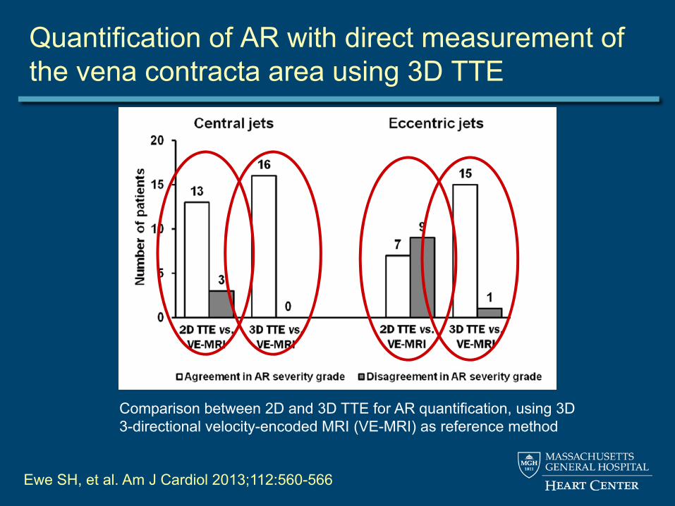
Quantification of AR with direct measurement of the vena contracta area using 3D TTE
Ewe SH, et al. Am J Cardiol 2013;112:560-566
Comparison between 2D and 3D TTE for AR quantification, using 3D 3-directional velocity-encoded MRI (VE-MRI) as reference method

MRI: Advantages
• Unlimited imaging planes• Comprehensive and quantitative • Integrated quantitative flow assessment
– Accurate measure of regurgitant fraction

Survival without surgery for conventional indictions vs. MRI regurgitant fraction ≥ 37%
Myerson SG, et al. J Cardiovasc Magnet Res 2011; 13(Suppl 1):099

Echo remains the modality most often used

Management

Medical Management
• Vasodilators have long been used in cases when AI is moderate or severe – Nifedipine – ACE inhibitors
• 2 key studies, conflicting data– Scognamiglio et al. in 1994 – Evangelista et al. in 2005
Scognamiglio R, et al. N Engl J Med 1994;331:689-69 Evangelista A, et al. N Engl J Med 2005;353:1342-1349

N.B.: there was no placebo control group
Scognamiglio R, et al. N Engl J Med 1994;331:689-69
Probability of AVR in Patients with Severe AI Treated with Nifedipine (20 mg bid) vs. Digoxin
P < 0.001at 6 years

Vasodilator Therapy
• The 1998 ACC/AHA guidelines gave a class I recommendation for the use of long-term vasodilator therapy in patients with chronic, severe AI

Evangelista A et al. N Engl J Med 2005;353:1342-1349
Probability of AVR in Patients with Severe AI Treated with Nifedipine, Enalapril, or Placebo
P = 0.29at 7 years

Efficacy of Vasodilators in Severe AI:A Controlled Animal Model
• Eric Plante and colleagues, Université Laval, Quebec • 60 rats had severe AR (confirmed by echo) induced by
retrograde puncture of the AV leaflets • Randomly divided into 5 groups:
– Normal sham-operated animals– AR untreated– AR treated with nifedipine– AR treated with captopril – AR treated with losartan
• Drug treatments were started 2 weeks after surgical procedure and continued thereafter for 6 months
• Echos at 6 months.
Plant E, et al. Circ Heart Fail 2009; 2: 25-32
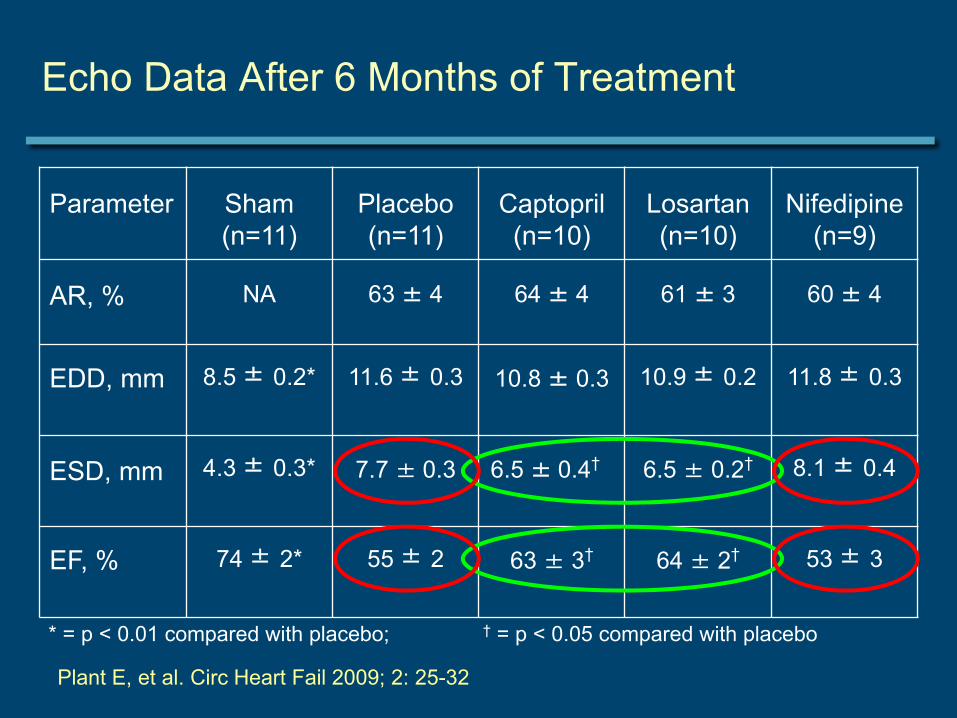
Echo Data After 6 Months of Treatment
Parameter Sham (n=11)
Placebo (n=11)
Captopril (n=10)
Losartan (n=10)
Nifedipine (n=9)
AR, % NA 63 ± 4 64 ± 4 61 ± 3 60 ± 4
EDD, mm 8.5 ± 0.2* 11.6 ± 0.3 10.8 ± 0.3 10.9 ± 0.2 11.8 ± 0.3
ESD, mm 4.3 ± 0.3* 7.7 ± 0.3 6.5 ± 0.4† 6.5 ± 0.2† 8.1 ± 0.4
EF, % 74 ± 2* 55 ± 2 63 ± 3† 64 ± 2† 53 ± 3
Plant E, et al. Circ Heart Fail 2009; 2: 25-32
* = p < 0.01 compared with placebo; † = p < 0.05 compared with placebo

Efficacy of captopril in rat model of pure AI: Showed for the first time a survival benefit
Arsenault M, et al. Circ Heart Fail 2013;6:1021-1028


Retrospective Analysis of 2266 Pts with Mod or Severe AI: Benefits of ACEI/ARB use
Elder DHJ, et al. J Am Coll Cardiol 2011;58:2084–91
Freedom from CV events (CV hospitalization or death)
Users
Non-users

2014 ACC/AHA Guidelines
• Treatment of hypertension (SBP >140 mmHg) is recommended in patients with chronic AR, preferably with dihydropyridine Ca++ channel blockers or ACEIs/ARBs
• I see no good reason to choose nifedipine if patient can tolerate ACEIs or ARBs
Nishimura RA, et al. JACC 2014.02.536

Use of Beta-blockers in Severe AI?

Sampat U, et al. J Am Coll Cardiol 2009;54:452-457
Effect of Beta-Blockers on Survival in Patients with Severe AR: A Cohort of 756

The Timing of Surgery

Surgical Indications Are Based on Symptoms and on LV Size and Function

Indications for Surgery in AI:2014 ACC/AHA guidelines
Class Indication
Nishimura RA, et al. JACC 2014.02.536

Indications for Surgery in AI:2014 ACC/AHA guidelines
Class IndicationI • Any pt with symptomatic severe AI, regardless of LV EF
• Asymptomatic pt with severe AI and LV EF < 50% at rest• Asymptomatic pt with severe AI undergoing cardiac
surgery for other indications
IIa • Asymptomatic pt with severe AI and normal LV function but severe LV dilatation (LVESD > 50 mm or > 25 mm/m2)
IIa • Asymptomatic pt with moderate AI undergoing ascending aortic surgery, mitral valve surgery, or CABG
IIb • Asymptomatic pt with severe AI and normal LV function (LV EF ≥ 50%) but with progressive severe LV dilatation(LVEDD > 65 mm) if surgical risk is low
Nishimura RA, et al. JACC 2014.02.536

Refining Risk Stratification: Biomarkers

BNP to Risk Stratify Patients with Asymptomatic Severe AI and Normal LV Function
Months
Event-free Survival (LV systolic dysfunction, symptoms, or death)
Pizarro R, et al. JACC 2011;58:1705-14

Epidemiology, Pathophysiology, and Management Guidelines of Aortic Insufficiency
Eric M. Isselbacher, MD, MSc
Associate Director, MGH Heart CenterCo-Director, MGH Thoracic Aortic Center Associate Professor of Medicine, Harvard Medical School
1st North American Aortic Valve Repair Symposium, May 14, 2015