Embed Size (px)
Citation preview

Gut, 1967, 8, 8
Eosinophilic granuloma of the gastro-intestinal tractP. R. SALMON' AND J. W. PAULLEY
From Princess Margaret Hospital, Swindon, Wiltshire andIpswich and East Suffolk Hospital, Ipswich, Suffolk
EDITORIAL COMMENT This paper presents a comprehensive review of eosinophilic granuloma of thealimentary tract. The view is put forward that eosinophilic granulomas mainly represent part of asystemic allergic response to one of a number of non-specific allergens. Steroids may be of real valuein treatment.
The first specific reference to an eosinophilic granu-loma of the bowel was that of Mock (1931) who, in adiscussion of infective granuloma, drew attention toa pyloric lesion, possibly eosinophilic. Kaijser (1937)described the first cases, of which one patient, whohad been allergic to onions since youth, developed aneosinophilic granuloma of the pyloric antrum. Theothers, both syphilitics, who were allergic to neo-arsphenamine, developed jejunal lesions. Thesecases assumed greater interest because in the sameyear two papers appeared showing the effect ofallergens on the gastrointestinal tract (Hansen andSimonsen, 1937; Afendoulis and Gulzow, 1937).Later Afendoulis (1943) reported two instances ofdrug allergy associated with gastritis and a furtherexample of allergic gastritis following ingestion ofcheese.Harkavy (1941) in an article entitled 'Pathogenesis
of bronchial asthma with recurrent pulmonaryinfiltration and eosinophilic polyserositis' describedeight cases of bronchial asthma. Six had eosinophilicsterile pleural effusions and three peritoneal involve-ment with eosinophilic ascites. Others had peri-cardial involvement. The peripheral eosinophilcount ranged from 12 to 84%. Angioneuroticoedema occurred twice and polyarteritis nodosaonce. Harkavy concluded that all these patientsshowed an altered vascular response, indicating acommon 'allergenic stimulation'. Most later contribu-tors have mentioned allergy and concluded thathypersensitivity plays an important aetiological role.A disturbance of oestrogen-progesterone metabo-
lism was considered likely by Sison, Dionisio, Silva,and Chavez (1947), whilst Sherman and Moran(1954) felt that gastric granulomata were probablydue to foreign-body reactions, and considered thatthe presence of eosinophils did not justify a diagnosisof eosinophilic granuloma nor an allergic aetiology.Ashby, Appleton, and Dawson (1964) concluded
Present Address: Department of Medicine, Bristol Royal Infirmary,Bristol.
8
that the majority of cases were due to a local gastro-intestinal reaction secondary to repeated infestationby intestinal parasites, especially the 'herring worm'(Eustoma rotundatum) in the bowel. This followedthe observation of Kuipers, van Thiel, and Roskam(1960a) Kuipers, van Thiel, Rodenburg, Wielinga,and Roskam (1960b) who reported 13 examples oflocalized intestinal granulomata associated withEustoma.
It seems to us that previous arguments as towhether eosinophilic granuloma of the bowel isprimarily a local phenomenon or part of a systemicallergic reaction may be irrelevant and dependent onthe extent of the host response and the site and modeof allergic insult. For example, those in the pyloricantrum usually give rise to pyloric stenosis eitherdirectly or by prolapsing into the duodenum, whilstthe primary infiltrative type involving any part of thebowel may be associated with a variety of systemicallergic conditions and high blood eosinophil counts.
In addition eosinophilic infiltration may give riseto a polyserositis (Harkavy, 1941; Salmon andPaulley, 1966) or involve the peritoneum only,causing eosinophilic ascites (Sison, Dionisio, Silva,and Chavez, 1947; Swarts and Young, 1955; Harley,Glushien, and Fisher, 1959), while other viscera maybe involved, as in the case showing involvement ofall the bowel, mesenteric nodes, ovaries, uterus,bladder, and heart (Fossgreen, 1962).
CASE REPORTS
CASE 1 J.E., a farmer, in 1949, when aged 26, wasreferred to one of us (J.W.P.) because of sudden attacks ofindigestion and vomiting occurring regularly every two tothree weeks. When first seen the attacks were usuallypreceded by swelling of the hands and arms, but severalyears previously he had had classical angioneuroticoedema of the face.
Past history included a Caldwell-Luc operation forpersistent rhinorrhoea, and a barium meal and follow-through in 1943 for similar attacks of vomiting and
on 28 January 2019 by guest. Protected by copyright.
http://gut.bmj.com
/G
ut: first published as 10.1136/gut.8.1.8 on 1 February 1967. D
ownloaded from

Eosinophilic granuloma of the gastro-intestinal tract
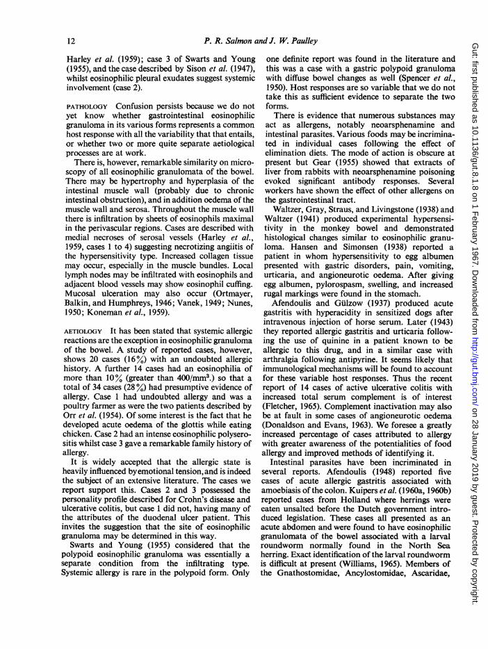
FIG. 1. Diagram (inset) showing filling defect in the pre-pyloric region ('shoulder sign').
indigestion. This showed intermittent pyloric spasm.Tenderness in the right iliac fossa had led to an appen-dicectomy which did not relieve his symptoms. He hadnever passed blood or slime.
There was no family history of allergy, but a sister hadmultiple sclerosis; three brothers were alive and well.The patient was married with one daughter, and was
obsessional about his work to the exclusion of all recrea-tion. For two years he had been worried financially andwas afflicted with insomnia.The patient was a young man of asthenic build. The
caecum was easily palpable and tender, further bariummeal showed clumping of barium in the terminal ileumwith some spasm or narrowing, and a diagnosis of ileo-colitis (Crohn's disease) was considered. He was treatedwith barbiturates and antihistamines. In 1951 he was seenagain because of a further bout of intermittent vomitingand lower abdominal pain. Because of the strong allergichistory elimination diets were tried but without benefit. In1953 he was still complaining of swelling of the lips, eye-lids, and vomiting attacks about once a fortnight. He wasadmitted on 8 June, 1953.Haemoglobin was 89%, W.B.C.s 7,600/mm.3 (eosino-
phils, 2%, 152/mm.3). A barium meal (Fig. 1) reported byDr. K. E. Barlow showed 'some hypotonicity with excessof resting juice. On the greater curve immediately proxi-mal to the pylorus I became suspicious of a smooth roundintramural mass. I consider the appearances sufficientlysuspicious of a so-called innocent tumour to warrantexploration.... ' He was referred for a surgical opinionbut exploratory laparotomy was not performed becausethe patient's symptoms improved and he wished to bedischarged. He did not consult us again but his family saythat his attacks continued at varying intervals.On 27 June 1961, after a fair-sized lunch of roast
chicken he suddenly became cyanosed and dyspnoeic anddied.
Necropsy (Dr. A. B. Lintott) revealed gross oedema ofthe nasopharynx and larynx obstructing the trachea asthe cause of death. The wall of the duodenum and gallbladder were oedematous and the gastric lining showedslight oedema. The pyloric antrum, apart from showingmucosal oedema, did not show an intramural mass asseen on the barium meal in 1953.
CASE 2 G.C., a man aged 36, an only child, had alopeciatotalis since the age of 7. In 1955 (aged 25) he was treatedfor intermittent vomiting which soon cleared up. In 1957,he presented with vomiting and colicky central abdominalpain for three weeks. Vomiting partly relieved the pain.He then developed swelling of the abdomen, and passedfour or five loose motions daily without visible blood ormucus. His appetite was unimpaired. There was a pasthistory of repeated vomiting in the first week of life, andfor his first 18 months he was under medical supervisionbecause of vomiting and failure to thrive. There was noknown family history of allergy or food intolerance, buthe said he was greatly affected by dust; however, he didnot change his job driving an excavator. He seemed youngfor his age, shy, friendless, emotionally detached andsupine. He lived with his mother with whom he had anintense relationship. She did everything for him and wasgenerally protective towards him. He said his hair fellout at age of 7 when he went to school which he hated. Itbecame clear that he was inclined to brood and avoidedany form of rebuff by a submissive smiling and placatoryattitude. His attitude epitomized conformity; he spoke inplatitudes, and he was sensitive to the most subtleexpression of hostility.On admission on 15 July 1957 there was tense ascites
and oedema of the anterior abdominal wall, and bilateralhydrothorax. He had alopecia totalis.Haemoglobin was 106 %, E.S.R. (Westergren) 1 mm. in
the first hour. A chest radiograph showed bilateral basaleffusions, and a film of the abdomen with the patienterect showed a distended small bowel with a few fluidlevels.
Liver function tests were normal. W.B.C.s on 2 Augustwere 7,800/mm.3 (eosinophils 28% or 2,184/mm.3). Abarium meal and follow-through were normal, and atparacentesis abdominis 15 pints of amber fluid wereaspirated and contained 98% eosinophils and 5 g. %protein. A repeat aspiration a few days later contained64% eosinophils.Ten days after admission he developed more oedema of
the anterior abdominal wall, sacrum and legs, and a fewdays later had a bout of diarrhoea and vomiting associ-ated with fever. The gastrointestinal symptoms soonsettled but a low-grade fever persisted. Lower abdominaltenderness with some rebound was found together withresistarce over the right upper quadrant. A presumptivediagnosis of Hodgkin's disease was made. However, hemade a spontaneous recovery and when followed up asan out-patient his only complaint was occasional upperabdominal discomfort after meals with nausea andvomiting.He remained in remission for four years, when, on 3
July 1961, having suppressed anxiety over his mother whorefused to see a doctor for a lump in her breast, hepresented with a recurrence of abdominal distension andloose stools. Examination again revealed ascites andbilateral hydrothorax. Pleural aspiration showed a cellu-lar exudate with 90% eosinophils. W.B.C.s numbered5,000/mm.3 (eosinophils 35%, 1,750/mm.3). A skin andmuscle biopsy was normal.A barium meal and follow-through (Figs. 2-3) showed
'marked evidence of small bowel thickening and narrow-
9
on 28 January 2019 by guest. Protected by copyright.
http://gut.bmj.com
/G
ut: first published as 10.1136/gut.8.1.8 on 1 February 1967. D
ownloaded from

P. R. Salmon and J. W. Paulley
ing with delayed transit suggestive of Crohn's disease'. Atthis stage a diagnosis of eosinophilic granuloma ('eosino-philic Crohn's disease') was made with eosinophilic poly-serositis. He was treated initially with A.C.T.H., 30 unitsdaily, and then changed to a reducing dose of prednisone.He showed dramatic improvement and was dischargedone month later without symptoms and having one bowelaction daily. On his discharge the white cell count was9,000/mm.3 with 2.5 % eosinophils (225/mm.3).He was last seen on 12 December 1964 and said he had
been very well. He is at present taking prednisone, 2-5mg. b.d., and is still working for the same building firm.His social circumstances remain unchanged.
CASE 3 M.S., a male orthodox Jew, in 1964 (July), aged14, presented with a three-week history of diarrhoea,passing blood and mucus, and upper abdominal, prae-cordial and left shoulder tip pain made worse on breathing.On examination he had a pericardial rub and a left
pleural effusion, findings confirmed by electrocardio-graphy and radiography. Sigmoidoscopy showed typicalactive ulcerative colitis, and was confirmed by bariumenema of which the main feature was 'saw-tooth' ulcera-tion of the descending and pelvic colon.The blood count showed eosinophilia. W.B.C.s
numbered 15,800/mm.3 (eosinophils 28%, 4,424/mm.3)and 13,500/mm.3 (eosinophils 18%, 2,430/mm.3).
Stools contained no ova. All these symptoms remittedwhen the patient was given prednisone and terramycin.
After two months' observation as an out-patient thesewere tailed off.He was readmitted in October 1964 febrile and with an
exact recurrence of the previous chest and shoulder painrelieved by sitting forward. Pericarditis and a leftpleural effusion were again present.
W.B.C.s numbered 8,600/mm.3 (eosinophils 11 %,946/mm.3). The E.S.R. (Westergren) was 83 mm. in thefirst hour. Pleural fluid did not show eosinophilia. Faecesagain were negative for ova. A toxocara skin test wasnegative, a Casoni test was negative, and virus studieswere negative. A skin and muscle biopsy was negative.During the period of investigation treatment was withheldapart from analgesics, and fever, pleurisy, and pericarditispersisted.A diagnosis of polyarteritis nodosa concomitant with
ulcerative colitis was suspected and he was given A.C.T.H.with immediate remission of symptoms and signs.The following past and family history seems relevant.
He first passed blood and mucus per rectum in 1950 aged21 months. With substitution of National dried milk forbreast feeding he recovered, but every spring and summeruntil he was 9 years old he had diarrhoea and rectal bleed-ing, a clear-cut diagnosis of ulcerative colitis being madeat the age of 3. Relapses in 1955 and 1956 were especiallysevere, necessitating admission to the paediatric wards forsome weeks when he required blood transfusion for severeanaemia. Because of increasing introspection on the partof the child and tensions with the parents routine hospitaladmissions were stopped. (His mother herself had hadulcerative colitis since the age of 13.) It seemed that forthe following four years relapses were short and mild.The family history which now emerged was strongly
FIG. 2. Five-hour film.
FIG. 3. Seven-hourfilm.
10
on 28 January 2019 by guest. Protected by copyright.
http://gut.bmj.com
/G
ut: first published as 10.1136/gut.8.1.8 on 1 February 1967. D
ownloaded from

Eosinophilic granuloma of the gastro-intestinal tract
allergic on both sides, and we suspect significant in view ofthe age of onset of the patient's colitis and the subsequenteosinophilic reaction, pleurisy, and pericarditis. Thepatient was the eldest of four children. His youngestsister, aged 18 months, was found to be very sensitive tofish. Vomiting and facial oedema occurred after helpingto scale sprats. Now aged 6 she still cannot enter fishshops without a reaction, and a single scale provokesintense oedema.
His paternal grandmother had urticaria requiring ad-mission to hospital, and she was sensitive to white fish. Apaternal uncle had bronchial asthma. His maternal grand-mother and aunt both had urticaria requiring hospitaladmission with no recognized food allergy. A maternalaunt's son (cousin) had bronchial asthma. Neither side ofthe family eat shell fish, which is forbidden to orthodoxJews.
Tragically the patient's colon became available forexamination after his suicide in October 1965, while inhospital receiving exclusion diets in search of an allergicfactor. It showed ulcerative colitis but no eosinophilicinfiltration. However, he had recently had A.C.T.H. for asevere relapse occasioned by the stress of his mother'scolectomy in July 1965. The mother's colon also showedulcerative colitis with a suggestion of early precancerouschange but no evidence of Crohn's disease or eosino-philic granuloma.Enough has been said to indicate that the psychological
ramifications here were relevant and intense, but brevityprohibits fuller treatment of them.
DISCUSSION
CLINICAL PRESENTATION The presentation of gastro-intestinal eosinophilic granuloma is variable anddepends on the site and extent of the lesions, thepresence of submucosal or serosal involvement, andof systemic allergic reactions. Available reportsdemonstrate the great variation in the site and extentof the lesions.Thus gastrointestinal eosinophilic granuloma have
been reported in the pharynx (Brunner, 1951),stomach (Kaijser, 1937; Barrie and Anderson, 1948;Vanek, 1949; Moloney, 1949; Nunes, 1950; Spencer,Comfort, and Dahlin, 1950; Booher and Grant,1951; Doniach and Mckeown 1951; Barnett andKazmann 1952; Fennel, 1952; Ruzic, Dorsey, Huber,and Armstrong, 1952; Helwig and Rainer, 1953;Vrr, Miller, and Russell, 1954; Judd, Civin, andMcllhany, 1955; Swarts and Young, 1955; McCune,1955; Rigler, Black, and Hebbel, 1956; Fischer,1956; Smith, 1956; Toole and Moschopoulos, 1959;Kuipers et al., 1960a, 1960b; Voorhuis and Eijler,1961; Salm, 1965; Russell and Evangeloe, 1965),duodenum (Barrie and Anderson, 1948; Herreraand de la Guardia, 1948; Moloney, 1949; Spenceret al., 1950; Swarts and Young, 1955; Toole andMoschopoulos, 1959; Szechy and Fbldvari, 1962;Ashby et al., 1964; Russell and Evangelou, 1965),
jejunum (Kaijser, 1937; Moloney, 1949; Polayes andKrieger, 1950; Fennel, 1952; Taccani, 1952; Orr etal., 1954; Ferrier and Davis, 1957; Hollmotz andStepan, 1963), ileum (Herrera and de la Guardia,1948; Marek, 1954; Unnewehr and Ohrt, 1954;Virshup and Mandelburg, 1954; Swarts and Young,1955; Pound, 1956; Urban and Lenczyk, 1956;Koneman, Sawyer, and Lubchenko, 1959; Cox,1960; Kuipers et al. 1960a, 1960b; Bauab, 1961;Fossgreen, 1962), appendix (Stemmerman, 1961),caecum (Swarts and Young, 1955), colon (de Santis,1954; Bauab, 1961), and rectum (Steger and Noto,1953).
Distribution within the gastrointestinal tract maybe seen at a glance from Table I. Males havepredominated over females in the proportion of 3:2and the peak incidence is in the sixth decade, the agerange being 14 to 84. Forty per cent of reported caseshave occurred in the stomach, commonly presentingwith epigastric pain and/or vomiting.
TABLE IDISTRIBUTION OF LESIONS IN GASTROINTESTINAL TRACT
Polypoid InfiltrativeGranulomata Granulomata
PharynxStomachBodyAntrum
Small bowelDuodenumJejunumIleum
AppendixCaecumColonRectumGall bladderPeritoneumGeneralized diffuse
Total number of casesTotal number of lesions
314
1
3
35
32122136
630
57
2816
l
155
104
Case 1 eventually showed the classical radiologicalappearance of eosinophilic granuloma of the pyloricantrum although the diagnosis was not made at thetime. Some cases have been discovered accidentally(Rigler et al., 1956). In the small bowel the symptomsare usually those of intermittent subacute obstruc-tion as in case 2, although perforation is described(Russell and Evangelou, 1965). The polypoidgranulomata may present as acute intussusception(Marek, 1954; Koneman et al., 1959; Cox, 1960;von Bogsch and Feher, 1963), granuloma of theappendix with acute abdominal pain, or withoutsymptoms (Stemmerman, 1961) and a distal ileallesion may mimic acute appendicitis (Kuipers et al.,1960b).
Serosal involvement seems to lead to eosinophilicascites as in case 2 and in the cases described by
lt
on 28 January 2019 by guest. Protected by copyright.
http://gut.bmj.com
/G
ut: first published as 10.1136/gut.8.1.8 on 1 February 1967. D
ownloaded from
P. R. Salmon and J. W. Paulley
Harley et al. (1959); case 3 of Swarts and Young(1955), and the case described by Sison et al. (1947),whilst eosinophilic pleural exudates suggest systemicinvolvement (case 2).
PATHOLOGY Confusion persists because we do notyet know whether gastrointestinal eosinophilicgranuloma in its various forms represents a commonhost response with all the variability that that entails,or whether two or more quite separate aetiologicalprocesses are at work.
There is, however, remarkable similarity on micro-scopy of all eosinophilic granulomata of the bowel.There may be hypertrophy and hyperplasia of theintestinal muscle wall (probably due to chronicintestinal obstruction), and in addition oedema of themuscle wall and serosa. Throughout the muscle wallthere is infiltration by sheets of eosinophils maximalin the perivascular regions. Cases are described withmedial necroses of serosal vessels (Harley et al.,1959, cases 1 to 4) suggesting necrotizing angiitis ofthe hypersensitivity type. Increased collagen tissuemay occur, especially in the muscle bundles. Locallymph nodes may be infiltrated with eosinophils andadjacent blood vessels may show eosinophil cuffing.Mucosal ulceration may also occur (Ortmayer,Balkin, and Humphreys, 1946; Vanek, 1949; Nunes,1950; Koneman et al., 1959).
AETIOLOGY It has been stated that systemic allergicreactions are the exception in eosinophilic granulomaof the bowel. A study of reported cases, however,shows 20 cases (16%) with an undoubted allergichistory. A further 14 cases had an eosinophilia ofmore than 10% (greater than 400/mm3.) so that atotal of 34 cases (28 %) had presumptive evidence ofallergy. Case 1 had undoubted allergy and was apoultry farmer as were the two patients described byOrr et al. (1954). Of some interest is the fact that hedeveloped acute oedema of the glottis while eatingchicken. Case 2 had an intense eosinophilic polysero-sitis whilst case 3 gave a remarkable family history ofallergy.
It is widely accepted that the allergic state isheavily influenced byemotional tension, and is indeedthe subject of an extensive literature. The cases wereport support this. Cases 2 and 3 possessed thepersonality profile described for Crohn's disease andulcerative colitis, but case 1 did not, having many ofthe attributes of the duodenal ulcer patient. Thisinvites the suggestion that the site of eosinophilicgranuloma may be determined in this way.
Swarts and Young (1955) considered that thepolypoid eosinophilic granuloma was essentially aseparate condition from the infiltrating type.Systemic allergy is rare in the polypoid form. Only
one definite report was found in the literature andthis was a case with a gastric polypoid granulomawith diffuse bowel changes as well (Spencer et al.,1950). Host responses are so variable that we do nottake this as sufficient evidence to separate the twoforms.There is evidence that numerous substances may
act as allergens, notably neoarsphenamine andintestinal parasites. Various foods may be incrimina-ted in individual cases following the effect ofelimination diets. The mode of action is obscure atpresent but Gear (1955) showed that extracts ofliver from rabbits with neoarsphenamine poisoningevoked significant antibody responses. Severalworkers have shown the effect of other allergens onthe gastrointestinal tract.
Waltzer, Gray, Straus, and Livingstone (1938) andWaltzer (1941) produced experimental hypersensi-tivity in the monkey bowel and demonstratedhistological changes similar to eosinophilic granu-loma. Hansen and Simonsen (1938) reported apatient in whom hypersensitivity to egg albumenpresented with gastric disorders, pain, vomiting,urticaria, and angioneurotic oedema. After givingegg albumen, pylorospasm, swelling, and increasedrugal markings were found in the stomach.
Afendoulis and Gulzow (1937) produced acutegastritis with hyperacidity in sensitized dogs afterintravenous injection of horse serum. Later (1943)they reported allergic gastritis and urticaria follow-ing the use of quinine in a patient known to beallergic to this drug, and in a similar case witharthralgia following antipyrine. It seems likely thatimmunological mechanisms will be found to accountfor these variable host responses. Thus the recentreport of 14 cases of active ulcerative colitis withincreased total serum complement is of interest(Fletcher, 1965). Complement inactivation may alsobe at fault in some cases of angioneurotic oedema(Donaldson and Evans, 1963). We foresee a greatlyincreased percentage of cases attributed to allergywith greater awareness of the potentialities of foodallergy and improved methods of identifying it.
Intestinal parasites have been incriminated inseveral reports. Afendoulis (1948) reported fivecases of acute allergic gastritis associated withamoebiasis of the colon. Kuipers et al. (1960a, 1960b)reported cases from Holland where herrings wereeaten unsalted before the Dutch government intro-duced legislation. These cases all presented as anacute abdomen and were found to have eosinophilicgranulomata of the bowel associated with a larvalroundworm normally found in the North Seaherring. Exact identification of the larval roundwormis difficult at present (Williams, 1965). Members ofthe Gnathostomidae, Ancylostomidae, Ascaridae,
12
on 28 January 2019 by guest. Protected by copyright.
http://gut.bmj.com
/G
ut: first published as 10.1136/gut.8.1.8 on 1 February 1967. D
ownloaded from

Eosinophilic granuloma of the gastro-intestinal tract 13
and Anisakidae can also cause eosinophilic granu-loma (Freeman, 1964). Stemmerman (1961) studied16 cases ofeosinophilic granulomata of the appendix.Six out of 10 patients who had their faeces examinedfor parasites had Strongyloides stercoralis infestation.Thiel (1962) showed that the larvae of Anisakis(normally found in the herring) caused a slight tissuereaction in the bowel of experimentally infestedrabbits when they penetrated the bowel wall. Whenanother larva penetrated near the first site aninflammatory response resembling that seen inhumans was observed.Our view is that eosinophilic granuloma of the
gastrointestinal tract represents part of a systemicallergic response to one of a number of non-specificallergens. The nature of the response is primarilyvascular and represents one end of a spectrum oftissue changes ranging from necrosis and granulomaformation to angiitis. Classical polyarteritis nodosaand the arteritis ofrheumatoid arthritis and dermato-myositis represent the other end of the spectrum witharteritis and minimal granulomatosis. Eosinophilicgranuloma should probably be grouped withWegener's granulomatosis, lethal mid-line granu-loma, and Loeffler's syndrome.
TREATMENT Surgery is indicated for obstructivelesions and in general for cases presenting as an acuteabdomen where the diagnosis is usually in doubt.Ferrier and Davis (1957) used A.C.T.H. for a case ofdiffuse eosinophilic granuloma of the jejunumdiagnosed at laparotomy. A course of 40 units dailyfor two weeks caused immediate and complete reliefof pain and diarrhoea. A recurrence of symptomsone month later was again treated successfully by afurther course. Our case 2 was similarly treated withA.C.T.H. followed by oral steroids. The result wasequally dramatic and after three years there has beenno recurrence of symptoms.
We wish to thank Drs. Trevor Shaw and Basil Morsonfor histological reports and Mr. Maurice Turney for thephotographs.
REFERENCES
Afendoulis, T. C. (1943). O0ber einen Fall von Gastritis allergica.Dtsch. med. Wschr., 69, 398.
-(1948). Observations on acute allergic gastritis. Amer. J. dig. Dis.,15, 90-92.
, and Gulzow, M. (1937). Die allergisch-hyperergische Gastritis.Tierexperimentelle Untersuchungen. Z. ges. exp. Med., 104,167-181.
Alarc6n-Segovia, D., and Brown, A. L., Jr. (1964). Classification andetiologic aspects of necrotizing angiitides. Proc. Mayo Clin., 39,205-222.
Ashby, B. S., Appleton, P. J., and Dawson, I. (1964). Eosinophilicgranuloma of gastro-intestinal tract caused by herring parasiteEustoma rotundatum. Brit. med. J., 1, 1141-1145.
Barnett, L. A., and Kazmann, H. A. (1952). Gastric granuloma witheosinophilic infiltration. Amer. J. Surg., 84, 107-110.
Barrie, H. J., and Anderson, J. C. (1948). Hypertrophy of the pylorus
in an adult with massive eosinophil infiltration and giant-cellreaction. Lancet, 2, 1007-1009.
Bauab, E. G. (1961). Eosinophilic granuloma of the gastrointestinaltract. Massive intestinal haemorrhage caused by multiplelocalization of the caecum, ascendizag colon, transverse colonand the ileum. Bol. Soc. Cirug. B. Aires, 45, 276-290.
Bogsch, A., and Feh6r, E. (1963). Wiederoolt auftretende Dunndar-minvagination bei eosinphoilem Granulom. Z. ges. inn. Med.,18, 1132-1143.
Booher, R. J., and Grant, R. N. (1951). Eos nophilic granuloma of thestomach and small intestine. Surgery, 30, 388-397.
Brunner, H. (1951). Eosinophilic granuloma of mouth, pharynx, andnasal passages. Oral Surg., 4, 623-640.
Cox, J. S. T. (1960). Submucosal ileal granuloma with eosinophilicinfiltration and intussusception. Brit. J. Surg., 48, 149-150.
Donaldson, V. H., and Evans, R. R. (1963). A biochemical ab-normality in hereditary angioneurotic edema. Absence of seruminhibitor of C'l-esterase. Amer. J. Med., 35, 37-44.
Doniach, I., and McKeown, K. C. (1951). A case of eosinophilicgastritis. Brit. J. Surg., 39, 247-250.
Fennel, E. A. (1952). Eosinophilic linitis plastica. Proc. Staff Meet.Straub Clin. (Honolulu), 18, 69-78.
Ferrier, T., and Daves, N. (1957). Eosinophilic infiltration of stomachand small intestine. Med. J. Aust., 1, 789-791.
Fletcher, J. (1965). Serum complement levels in active ulcerative colitis.Gut, 6, 172-175.
Fossgreen, J. (1962). Eosinophile granulomatosis. (In German.) Actapath. microbiol. scand., 56, 143-154.
Fischer, H. G. (1956). Das eosinophile Granulom des Magens.Chirurg, 27, 516-519.
Freeman, R. S. (1964). Studies on responses of intermediate hosts toinfection with Taenia crassiceps (Zeder 1800) (Cestoda).Canad. J. Zool., 42, 367-385.
Gear, J. (1955). Auto-antibodies and the hyper-reactive state in thepathogenesis of disease. Acta med. scand., suppl. 306, 39-55.
Hansen, K., and Simonsen, M. (1937). Rontgenologische Beobachtungund Darstellung der allergischen Gastritis und des allergischenPylorospasmus. Rontgenpraxis, 9, 145-151.
Harkavy, J. (1941). Vascular allergy: pathogenesis of bronchial asthmawith recurrent pulmonary infiltrations and eosinophilic poly-serositis. Arch. intern. Med., 67, 709-734.
Harley, J. B., Glushien, A. S., and Fisher, E. R. (1959). Eosinophilicperitonitis. Ann. intern. med., 51, 301-308.
Helwig, E. B., and Rainer, A. (1953). Inflammatory fibroid polyps ofthe stomach. Surg. Gynec. Obstet., 96, 355-367.
Herrera, J. M., and de la Guardia, J. (1948). Un raro caso de eosino-filia gastro-intestinal motivadora de un cuadro organico deestenosis pilorica. Arch. Hosp. (Santo Tomas), 3, 19-34.
Hollmotz, 0., and Stepan, Z. (1963). Submukozni granulom tenkehoStreva s eozinofilni infiltraci. (Submucous granuloma of thesmall intestine: eosinophil infiltration.) (s. Rentgonol., 17,65-68.
Judd, C. S. Jr., Civin, W. H., and Mcllhany, M. L. (1955). Eosino-philic granuloma of the stomach. Gastroenterology, 28, 453-457.
Kaijser, R. (1937). Zur Kenntnis der allergischen Affektionen desVerdauungskanals vom Standpunkt des chirurgen aus. Langen-becks Arch. klin. Chir., 188, 36-64.
Koneman, E. W., Sawyer, K. C., and Lubchenko, A. E. (1959).Eosinophilic granuloma of the ileum. Arch. Surg., 78, 923-927.
Kuipers, F. C. van Thiel, P. H., and Roskam, E. T. (1960a). Eosin-ofiele flegmone van de dunne darm, veroorzaakt door eenniet aan het lichaam van de mens aangepaste worm. Ned. T.Geneesk., 104, 422-427.
-, van Thiel, P. H., Rodenburg, W., Wielinga, W. J., and Roskam,R. T. (1960b). Eosinophilic phlegmon of the alimentary canalcaused by a worm. Lancet, 2, 1171-1173.
McCune, W. S., Gusack, M., and Newman, W. (1955). Eosinophilicgastroduodenitis with pyloric obstruction. Ann. Surg., 142,510-518.
Marek, S. (1954). Eosinofini granulom tenk6ho stfeva. (Eosinophilicgranuloma of small intestine). Cas. Lek. (es., 93, 484-485.
Mock, H. E. (1931). Infective granuloma. Surg. Gynec. Obstet., 52,672-689.
Moloney, G. E. (1949). Pyloric hypertrophy with eosinophil infiltra-tion. Lancet, 1, 412.
Nunes, M. A. (1950). Granuloma (granuloblastoma) gistrico sub-mucoso com eosin6filos. Gaz. med. port., 3, 751-759.
Orr, I. M., Miller, A. A., and Russell, J. Y. W. (1954). Eosinophilicinfiltration of the stomach and bowel. Postgrad. med. J., 30,485-493.
on 28 January 2019 by guest. Protected by copyright.
http://gut.bmj.com
/G
ut: first published as 10.1136/gut.8.1.8 on 1 February 1967. D
ownloaded from

14 P. R. Salmon and J. W. Paulley
Ortmayer, M., Balkin, R., and Humphreys, E. (1946). Chronic,erosive, granulomatous, atrophic gastritis. Gastroenterology, 6,298-301.
Polayes, S. H., and Krieger, J. L. (1950). Eosinophilic granuloma ofthejejunum. J. Amer. med. Ass., 143, 549-551.
Pound, A. W. (1956). Personal communication. Cited by Ferrier andDavis (1957).
Rigler, L. G., Blank, L., and Hebbel, R. (1956). Granuloma witheosinophils: benign inflammatory polyps of the stomach.Radiology, 66, 169-176.
Russell, J. Y. W., and Evangelou, G. (1965). Eosinophilic infiltrationof the stomach and duodenum complicated by perforation.Postgrad. med. J., 41, 30-33.
Ruzic, J. P., Dorsey, J. M., Huber, H. L., and Armstrong, S. H., Jr.(1952). Gastric lesion of Loeffilers syndrome. J. Amer. med.Ass., 149, 534-537.
Salm, R. (1965). Gastric fibroma with eosinophilic infiltration. Gut, 6,85-9 1.
de Santis, M. (1954). Granuloma eosinofilo del cieco. Policlinoco, Sezprat., 61, 1521-1527.
Sherman, F. E., and Moran, T. J. (1954). Granulomas of stomach. 1.Response to injury of muscle and fibrous tissue of wall ofhuman stomach. Amer. J. clin. Path., 24, 415-421.
Sison, A. B. M., Dioisio, S. A., Silva, J. A., and Chavez, P. C. (1947).Allergic peritonitis: report of a case. J. Amer. med. Ass., 134,1007-1010.
Smith, M. J. (1956). Gastric granuloma with eosinophilic infiltration.Radiology, 66, 177-180.
Spencer, J. R., Comfort, M. W., and Dahlin, D. C. (1950). Eosino-philic infiltration of the stomach and bowel associated withpyloric obstruction and recurrent eosinophilia. Gastroenter-ology, 15, 505-513.
Steger, C., and Noto, L. (1953). Granuloma of the rectum of eosino-philic type. Acta chir. patav., 9, 339-344.
Stemmermann, G. N. (1961). Eosinophilic granuloma of the appendix.
A study of its relation to Strongyloides infestation. Amer.clin. Path., 36, 524-531.
Swarts, J. M., and Young, J. M. (1955). Primary infiltrative eosino-philic gastritis, enteritis, and peritonitis. Gastroenterology, 28,43 1-452.
Szechy, M., and Foldvari, G. (1962). A nyombel eosinophil granulo-maja. (Eosinophilic granuloma of the duodenum.) Orv. Hetil.,103, 501-502.
Taccani, C. (1952). Granuloma eosinofilo dell'ileo. Chirurgia (Milano),7, 454-460.
Thiel, P. H. van. (1961). Reconciliation entre les conceptions concern-ant la biologie de l'Entamoeba histolytica, en rapport avec laperturbation de l'equilibre entre l'homme et le parasite par letraitement medical. Bull. Soc. Path. exot., 54, 824-829.
Toole, H. J., and Moschopoulos, A. N. (1959). Eosinophilic granu-loma of the gastro-intestinal tract: report of two cases. Brit. J.Surg., 46, 445-448.
Unnewehr, F., and Ohrt, H. (1954). Eosinophiles Granulom desDunndarms als Ursache fur einen Ileus. Zbl. Chir., 79, 91-93.
Urban, A. and Lenczyk, M. (1956). W sprawie tzw. granulomaeozynophilicum jelita biodrowego. (So-called eosinophilicgranuloma of the ileum. Pat. pol., 7, 307-311.
Vanek, J. (1949). Gastric submucosal granuloma with eosinophilicinfiltration. Amer. J. Path., 25, 397-411.
Virshup, M., and Mandelberg, A. (1954). Eosinophilic granuloma ofthe gastro-intestinal tract. Ann. Surg., 139, 236-240.
Voorhuis, F. J., and Eijlers, W. (1961). Een ontsekingstumor van demaag, waarschijnlijk veroorzaakt door de haringworm. Ned. T.Geneesk., 105, 2542-2545.
Walzer, M., Gray, I., Straus, H. W., and Livingstone, S. (1938).Studies in experimental hypersensitiveness in the rhesusmonkey: IV. The allergic reaction in passively locally sensitizedabdominal organs. J. Immunol., 34, 91-95.
(1941). Allergy of the abdominal organs. J. Lab. clin. Med., 26,1867-1877.
Williams, H. H. (1965). Roundworms in fishes and so-called 'herring-worm' disease. Brit. med. J., 1, 964-967.
on 28 January 2019 by guest. Protected by copyright.
http://gut.bmj.com
/G
ut: first published as 10.1136/gut.8.1.8 on 1 February 1967. D
ownloaded from


![Eosinophilic granuloma with Splendore-Hoeppli …...agent has been found in eosinophilic gastroenteritis with similar material in ferrets [8], because this is caused by al-lergic reactions,](https://img.dokumen.tips/doc/110x75/5e53e6b07bdaf83730474a9b/eosinophilic-granuloma-with-splendore-hoeppli-agent-has-been-found-in-eosinophilic.jpg)