Embed Size (px)
Citation preview
ENZYMES OF THE LUNG
I. Detection of Esterase with a
New Cytochemical Method
A. E. VATTER, O. K. REISS, JOYCE K. NEWMAN, KARIN
LINDQUIST, and ELLY GROENEBOER
From the Webb-Waring Institute for Medical Research and the Department of Pathology, Tile
University of Colorado School of Medicine, Denver, Colorado 80220
ABSTRACT
The esterases of rabbit lung have been investigated from two viewpoints, the cytochemical
and the biochenijcal. To accomplish this objective, we designed and synthesized a series
of ester substrates which provide both a cytochemical indicator of the location of the enzyme
and a means of following the enzymatic activity in tissue homogenates and subfractions.
The substrates are p-nitrophenylthiol esters which yield, upon hydrolysis, carboxylic acid
and p-nitrothiophenol. The latter can react with aurous ions to give an electron-opaque
deposit; in addition, the strong absorption of p-nitrothiophenol at 410 my permits continu-ous kinetic measurements. Thus, it is possible to correlate the intracellular site of action and
the biochemical behavior of the esterases. The new substrates are the thiol analogues of the
p-nitrophenyl esters frequently employed as esterase substrates. The rates of hydrolysis of
the two series of esters are compared in vitro. During tissue fractionation, most of the ester-
ase activity sediments with a particulate fraction. The effects of a number of commonesterase inhibitors, such as diisopropyl phosphorofluoridate and eserine sulfate, are ex-
amined, and the effects of enzyme concentration and heat inactivation are shown with the
use of the partially purified preparations. The cytochemical work shows that the esteraseactivity is most prominent in the lamellar bodies of the giant alveolar (type II, septal, or
granular pneumatocyte) cells of the lung and to a lesser extent in squamous (type I, or
membranous pneumatocyte) epithelial and endothelial cells. In both the cytochemical andbiochemical studies, the enzymes are inhibited by diisopropyl phosphorofluoridate andphenyl methylsulfonyl fluoride but are insensitive to eserine sulfate.
INTRODUCTION
The enzymatic profiles of lung are quantitativelydifferent from those of liver or kidney (1). Otherinvestigators have observed (2-6), and our studies
confirm, that the lung is rich in hydrolytic ac-tivity; therefore we began our researches in thisarea.
Carboxylic ester hydrolase (3.1.1)1 is defined
We shall refer to this hydrolytic activity as esterasethroughout this report, since we do not know whether
as an enzyme that will catalyze the following
reaction:
0 0
' H0 11 R-C--H + -OHR-C-O-R' + H20 --* R-C-OH + R'-OH
we are dealing with a carboxyl ester hydrolase(3.1.1), a thiol ester hydrolase (3.1.2), or a peptidehydrolase (3.4). See Discussion.
80
Dow
nloaded from http://rupress.org/jcb/article-pdf/38/1/80/1384602/80.pdf by guest on 10 D
ecember 2021
Past studies of esterases have fallen into two
main types, the histochemical work and the bio-
chemical studies of tissue fractions.
Gomori (7) has reviewed the earlier histochem-
ical work. In general, the approach in all these
studies has been to employ a synthetic substratewhich, upon hydrolysis, will yield a product that
can be directly observed at the site of action with
the microscope. Burstone has reviewed the useof many of these substrates (8).
The earliest practical histochemical procedurewas that of Gomori (9). His work led to the use
of ac- and 0f-naphthyl acetates, indoxyl estersand similar compounds as substrates. Uponhydrolysis, the freed, substituted naphthol or
indoxyl is converted into a dye (10, 11). Koelleand Friedenwald (12) introduced a method whichutilized thiocholine. The carboxylic esters of
thiocholine sulfate are hydrolyzed, and the liber-
ated thiocholine sulfate is precipitated in thepresence of copper salts. This reaction forms the
basis of the electron microscopic method, whereby
the "capturing agent," usually copper or lead,
combines with the product to yield an opaque
precipitate (13).Wilson (14) introduced the use of thiolacetic
acid as a substrate for esterase and indicated that
an esterase enzyme had split this molecule into
acetic acid and hydrogen sulfide which could beprecipitated in the presence of lead or copper.Barrnett adapted this technique for electron
microscopy (15). Koelle and Foroglou-Kerameos
(16) used aurous ions as the heavy metal andfound that this procedure yields a finer reactionproduct (17).
The biochemical studies, on the other hand,have concentrated on the esterases in body fluids
and in subcellular fractions of tissues. These in
vitro studies have employed esters of p-nitrophenol(PNP) and similar compounds as substrates be-cause the hydrolytic activity can be followedspectrophotometrically. Rates of hydrolysis, ef-
fects of enzyme and substrate concentrations, andpH effects were thus measurable in vitro. Hugginsand Lapides (18) measured the activity of human
serum enzyme, and other workers have usedsimilar substrates on serum (19), subcellular frac-tions of brain (20, 21), and human gastric juice
(22, 23).For sorting out the different types of esterases
and assigning them places and functions within
the cell, a single substrate which can act both as ahistochemical and as a biochemical detecting
agent would be ideal. We have attempted to
achieve this objective in the present study in
order to correlate structure and function.
MATERIALS AND METHODS
Chemical Syntheses
P-NITROTHIOPHENOL (PNT): p-Nitrothiophe-nol was prepared according to the method of Brownand Rao (24).
6 g of recrystallized bis-(p-nitrothiophenyl) disul-fide (p, 181-182 ° C) (Aldrich Chemical Co.,Milwaukee, Wis.) were reduced by 1 g of sodiumborohydride dissolved in 25 ml of redistilled diglyme(diethylene glycol dimethyl ether; The Ansul Co.,Marinette, Wis.) and 0.9 g of freshly sublimed AIC13suspended in 45 ml of redistilled diglyme. After 1hr, 50 ml of 2 M HCI were added slowly to destroythe excess hydride. The yellow PNT separated aftercooling on an ice bath and was washed with water.The yield was 88% with mp 78.5°-79.5 ° C (TableI). The product showed spontaneous slow oxidationin air to the disulfide.
p-NITROPHENYLTHIOL ACETATE (PNTA):
10 ml of pyridine were treated with a bubblingstream of N2 for 2 or 3 min. 500 mg of PNT weredissolved in the pyridine, and 3 ml of acetyl chloride(Mallinckrodt Chemical Works, St. Louis, Mo.)were added dropwise, with stirring, to the flask whichwas kept in an ice bath. The reaction mixture wasallowed to stand for 5 min, and then 50 ml of colddistilled water were added to it; this dissolved thepyridine hydrochloride and precipitated the PNTA.The mixture was allowed to stand in ice for 1 hr,and the precipitate was recovered by filtration,washed with cold water, and dried; a 44%0 yield ofpale yellow crystals that melted at 78°-79° C wasobtained.
This product is recrystallized from water to givevery fine needles which melt at 81.5°-82° C.
The ester was examined by means of thin-layerchromatography (see below) and gave a single spotwith an Rf value distinctly different from the Rfvalues of the disulfide and the starting material(Table I).
P-NITROPHENYLTHIOL BUTYRATE(PNTB):
4 ml of dry pyridine were added to 100 ml of an-hydrous benzene. 5 g of PNT were added, and then10 ml of freshly distilled butyryl chloride were addedslowly. The mixture was refluxed for 2 hr and wasallowed to stand overnight. The pyridine hydro-chloride was removed by filtration, and the clearsolution was washed successively twice with water,2% NaHCO3, and water again. The solution wasthen dried over Na 2SO 4, and the benzene was evapo-rated from the product under vacuum. The remain-ing mixture of liquid and solid was extracted twice
A. E. VATTER ET AL. Esterase Detection in Lung 81
Dow
nloaded from http://rupress.org/jcb/article-pdf/38/1/80/1384602/80.pdf by guest on 10 D
ecember 2021
with ether; the extracts were pooled, the ether wasevaporated, and the brown oil that remained wasredistilled in a microdistillation apparatus. The esterdistilled at 1150 C, 2 3 mm.
Other homologous esters, such as the propionateand octanoate, were prepared by the same methodas that used for PNTB.
AUROUS P-NITROTHIOLPHENOLATE: Thecompound was formed according to the followingequation:
O2 N SH + Au+
250 mg of AuNa3 (S203)2 .2H 2 0 (Sanocrysin;Ferrosan, Copenhagen, Denmark) were dissolvedin 6.0 ml of 0.175 M sodium maleate buffer pH 7.4and added to 52.2 mg of PNT dissolved in 40 ml ofwater. After it had stood for 60 min, a yellow pre-cipitate formed. The precipitate was washed bycentrifugation twice with cold H2 0, twice with drymethanol, once with ethyl acetate, and once withethyl ether. It was dried 1 hr in vacuo over P20 5.60.6 mg, 173 molese, were recovered for a yield of51%. Analysis was as follows: calculated, Au, 56.09%;C, 20.52%; H, 1.15%; S, 9.13%; found; Au, 55.52%;C, 20.82%; H, 1.28%; S, 9.24%. The mercaptideis insoluble in alcohol, ethyl acetate, and ethyl ether,but is freely soluble in pyridine, like the mercurico-thiophenolate (25).
CHARACTERIZATION OF COMPOUNDS: All
compounds described above were also examined bythin layer chromatography on silica gel HF2 54(Brinkman Laboratories, Inc., Great Neck, N. Y.)plates; a solvent system of hexane:benzene:aceticacid, 50:50:0.5, was used, and the spots were readunder a short wave ultraviolet lamp. The absorptionspectra were determined in the ultraviolet andvisible regions on a Cary 14 recording spectrophoto-meter in several solvents and in the infrared region
in potassium bromide micropellets 1 X 5 mm inthe Beckman IR 7 infrared spectrophotometer.
Preparation of Homogenate and Fractions
Adult rabbits (New Zealand) of either sex andweighing between 1.6 and 2.8 kg were sacrificed byinjecting air into the ear vein. The lungs were re-moved immediately, a portion of a lobe was tiedoff, and the distal section was removed and used forelectron microscopy. The remainder of the lung wasused to prepare a homogenate in 0.25 M sucrose,0.01 M tris-HCI, and 0.001 M potassium EDTA(ethylenediaminetetraacetate). Other details of the
procedure have been described previously (1). Afterthe homogenate had been strained through boltingcloth and centrifuged at 1600 g for 5 min for removalof nuclei, whole cells, and debris, the supernatantfraction was centrifuged at 100,000 g for 30 min,in the Spinco Model L rotor No. 50 so that a pelletand supernatant fraction would be obtained. Thepellet fraction, which under these conditions con-tains the mitochondria and vesiculated membraneswas resuspended in the sucrose-Tris-EDTA solution
0 2N - S-Au + H+
and stored at 2C. All analyses were done within 72hr; however, the enzymatic activity of both frac-tions was stable for at least 1 wk.
Inhibitors
Diisopropyl phosphorofluoridate (DFP) (SigmaChemical Co., St. Louis, Mo.) was prepared as0.1 M solution by dilution with anhydrous propyleneglycol (Distillation Products Industries, Rochester,N. Y.) and stored at 0°C for up to 4 wk from the timeof preparation. After 4 wk of storage, the effective-ness of the stock solution in inhibiting the hydrolysisof PNPA (p-nitrophenyl acetate) by a-chymotrypsinhad decreased less than 10%.
Eserine sulfate (Sigma Chemical Co.) and am-benonium chloride (N,N'-bis-[diethylaminoethyl]oxamide bis-[2-chlorobenzyl chloride]) 2 were pre-pared as 0.1 M stock solution in 0.175 M sodiummaleate and used the same day.
BW 284 (1,5-bis [4-allyl dimethyl ammoniumphenyl] pentan 3-one dibromide) 3 (26) was preparedas 0.1 M stock solution in the maleate buffer.
Phenyl methylsulfonyl fluoride (PMSF, SigmaChemical Co.,) was prepared as 0.1 M stock solutionin the maleate buffer (27).
Analytical Methods
Protein contents of the various fractions weredetermined by a modified Folin procedure (28)
with crystalline bovine plasma albumin (ArmourPharmaceutical Co., Chicago, Illinois) as a standard.Nesslerization after complete digestion was employed
to standardize this procedure (29).
2 We wish to thank the Sterling-Winthrop ResearchInstitute, Rensselaer, N. Y. for a gift of this compound.3 This compound was kindly donated by BurroughsWellcome & Co., Inc., Tuckahoe, N. Y.
82 THE JOURNAL OF CELL BIOLOGY - VOLUME 38, 1968
Dow
nloaded from http://rupress.org/jcb/article-pdf/38/1/80/1384602/80.pdf by guest on 10 D
ecember 2021
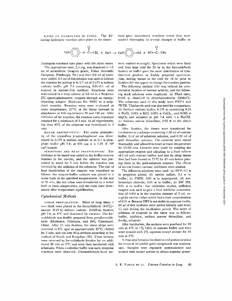
RATES OF HYDROLYSIS IN VITRO: The fol- tions gave inconsistent reactions unless they were
lowing hydrolytic reaction takes place in the tissue:
0 2N Q S--C-CH + H20
Analogous reactions take place with the other esters.The appropriate ester, 2.5 mg, was dissolved in 1.0
ml of acetonitrile (reagent grade, Fisher ScientificCompany, Pittsburgh, Pa.) and then 9.0 ml of waterwere added. 0.3 ml of this solution was used to initiatethe reaction by adding it to 2.7 ml of 0.175 M sodiummaleate buffer pH 7.4 containing 0.01-0.1 ml ofenzyme in sucrose-Tris medium. Reactions weredetermined in a final volume of 3.0 ml in a BeckmanDU spectrophotometer, coupled through an energy-recording adapter (Beckman No. 5800) to a strip-chart recorder. Reaction rates were evaluated atroom temperature, 23°C, as the linear increase inabsorbance at 410 mgu between 30 and 120 sec. Afterinitiation of the reaction, the reaction rates remainedconstant for a minimum of 3 min. In all experiments,less than 40% of the substrate was hydrolyzed in 3min.
MOLAR ABSORPTIVITY: The molar absorptiv-
ity of the crystalline p-nitrothiophenol was deter-mined in 0.175 M sodium maleate or in 0.1 M phos-phate buffer pH 7.4; at 410 m/u it is 1.25 X 104cm 2 /mmole.
INHIBITORS AND HEAT INACTIVATION: Theinhibitor to be tested was added to the buffer-enzymemixture in the cuvette, and the mixture was per-mitted to stand for 5 min before the reaction wasinitiated by the addition of the substrate. The rate ofheat inactivation of the enzyme was examined asfollows: the enzyme-buffer mixture was placed in awater bath at the specified temperatures. At the endof 10 min, the test tubes were transferred to a waterbath at room temperature, and the rates were deter-mined after temperature equilibration.
Cytochemical Methods
TISSUE PREPARATION: Slices of lung tissue 1
mm thick were placed in the formaldehyde (4.0%)-sucrose (0.25 M)-sodium maleate (0.028 M) fixative,pH 7.4, at 4°C and deaerated by vacuum. The for-maldehyde was freshly prepared from paraformalde-hyde (Matheson, Coleman, and Bell, Cincinnati,Ohio). After 15 min fixation, the tissue strips weremounted in 6% agar at approximately 450 C, chilledfor 5 min, and cut into 40-/t sections according to themethod of Smith and Farquhar (30). These sectionswere returned to formaldehyde fixative for an addi-tional 30 min at 4°C and were then incubated withsubstrates. When s-collidine buffer was used, irregularreactions were observed. Glutaraldehyde-fixed sec-
washed thoroughly in several changes of buffer or
O
02N SH + HO-C-CH3
were washed overnight. Specimens which were fixedand then kept cold for 24 hr in the formaldehydefixative or buffer gave the same distribution of cyto-chemical product as freshly prepared specimens.Also, storing tissues in the cold for 18 hr prior tofixation did not appear to change the reaction pattern.
The following method (16) was utilized for cyto-chemical location of esterase activity, and the follow-ing stock solutions were employed. (a) Thiol ester,0.025 M, dissolved in dimethylsulfoxide (DMSO).The substrates used in this study were PNTA andPNTB. Thiolacetic acid was also used for comparison.(b) Sodium maleate buffer, 0.175 M, containing 0.25M NaCI, 0.025 M KCI, 0.025 M CaC12 , and 0.005 MMgCI 2 and adjusted to pH 7.4 with 1 N NaOH.(c) Sodium aurous thiosulfate, 0.02 in the abovebuffer.
After fixation, the tissues were transferred forincubation to a solution containing 1.58 ml of maleatebuffer, 0.12 ml of substrate solution, and 0.30 ml ofgold thiosulfate solution. The contents were mixedthoroughly and allowed to react at room temperaturefor 15-60 min. Controls were made by omitting theappropriate reagent and adjusting to a final volumeof 2 ml with maleate buffer, and also by using tissuesthat had been heated at 750 C for 10 min before plac-ing them in the gold-substrate mixture. The effectsof several known esterase inhibitors were studied.
The following solutions were used: (a) DFP, 0.1 Min propylene glycol; (b) eserine sulfate, 0.1 M inbuffer; (c) PMSF, 0.01 M in isopropanol; (d) am-benonium chloride, 0.01 M in buffer; (e) BW 284,0.01 in buffer. For inhibition studies, sufficientreagent was used to give a final inhibitor concentra-tion of 0.001 M in the reaction mixture of 2 ml, ex-cept for eserine sulfate which had a final concentrationof 0.01 M. Because DFP is not stable in aqueous media,20 1 of this inhibitor were added initially and every15 min during the incubation period. The order ofaddition of reagents to the tissue was as follows:buffer, inhibitor, sodium aurous thiosulfate, andfinally, substrate.
After incubation, the sections were postfixed for 20min at 4C in 1% OsO4 in maleate buffer and thenwere treated with 2% aqueous uranyl acetate for 45min at 4°C.
A rinse step between incubation and postosmicat ionfor removal of soluble gold compounds was unneces-sary. Samples were regularly postosmicated andtreated with uranyl acetate to obtain superior preser-
A. E. VATTEIR ET A.. Esterase etection in Lung 83
Dow
nloaded from http://rupress.org/jcb/article-pdf/38/1/80/1384602/80.pdf by guest on 10 D
ecember 2021
t
E '- -
3 cco o 1t_ o
E : c C br -
o. ° O -O O O u-~ cO
_ - _ _ _
ee cr r r r
co cr ~ ~ .c-
o r- -;m~~~~n, Il- l
ccC) l 0Z
CO ; CNOI
Z c
Z
ec C co -- sc~~~~~~
cc - a, me
I-
06
0
I C 0 =0 u <
z z zC9 4 'ac~d c zc o o o o
a 0o.a -- . ' c o
Z ~ ~ a U) r UZS
~. om <. .~ <z %- "c c b- ~c b -
84 THE JOURNAL OF CELL BIOLOGY · VOLUME 38, 1968
0
~0¢BS
mI
G
67S
ccO
I b.
II
I
0'
Z ac>-0I
A O
G5 j
I0
GO
cc -.5Q)
uu
E
o"4c0)i
I
cc
cc
t.:
I
cc
cc
w
-ci
I9
Q.
cc
ecq
a5-
S
a10)
-a a
0)
E~ -- a
5
a- Obcc~i
cc 5e
0) -5 -0)"
e, cc
cc..,
-00)
50 cc
-1 ) U N
Q&, ' Y-'* +A 055*
Dow
nloaded from http://rupress.org/jcb/article-pdf/38/1/80/1384602/80.pdf by guest on 10 D
ecember 2021
vation and contrast. The use of osmium tetroxide oruranyl acetate did not change the interpretation ofthe gold deposits.
The specimens were dehydrated in hydroxypropylmethacrylate (Rohm & Haas Co., Philadelphia, Pa.)(31) and embedded in Epon. They were sectionedwith a Porter-Blum MT-2 microtome, examined, andphotographed with a Philips EM 200 electron micro-scope. It was not necessary to treat the tissue sectionswith lead stain.
OBSERVATIONS
Chemical
Table I summarizes some of the physical
constants determined. Where possible, these
constants have been compared with values from
the literature. The synthesized compounds mi-
grated as single spots on the thin-layer plates.
After standing, PNT gives an additional spot due
to disulfide formation.The absorption peaks in the infrared region
clearly confirm the presence of the postulated
functional groups. All compounds contain the
characteristic strong absorptions at approximately1340 and 1520 cm-' of the aryl nitro groups.
The carbonyl stretching frequency, strong evi-
dence for the presence of the ester group, is found
at 1718 cm t in the acetate compound and at
1717 cm- 1 in the butyrate compound. No similar
absorption was found in the spectra of the other
nonester compounds examined. The carbon-sulfur
stretch, as described by Nyquist and Potts (32),
was found at 953 cm- 1 for the acetate and at 970
cm-1 for the butyrate. No carbon-sulfur stretch
absorptions are observed for the other compounds
shown, since it is the carbonyl carbon that leads
to this absorption. As expected, no absorption is
found for the disulfide bond, but the weak band
cited by Bellamy (33) as characteristic of the
sulfhydryl group is observed in the p-nitrothio-
phenol at 2550 cm- 1, within the expected region.
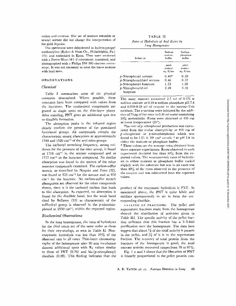
Biochemical Observations
In the lung homogenate, the rates of hydrolyses
for the thiol esters are of the same order as those
for their oxy-analogs, as seen in Table II. Non-
enzymatic hydrolysis was less than 10% of the
observed rate in all cases. Thin-layer chromatog-raphy of the homogenate after 20 min incubation
showed additional spots with Rf values similar
to those of PNT (0.76) and bis-(p-nitrophenyl)disulfide (0.58). This finding indicates that the
TABLE II
Rates of Hydrolysis of Acyl Esters byLung Homogenates
Sodium Sodiummaleate phosphate
Substrate buffer buffer
Imole pmoleproduct/ products
mg N/min mg N/min
p-Nitrophenyl acetate 0.49* 0.59p-Nitrophenylthiol acetate 0.46 0.57p-Nitrophenyl butyrate 1.23 1.93p-Nitrophenylthiol 2.24 3.32
butyrate
The assay mixture contained 2.7 ml of 0.175 Msodium maleate or 0.10 M sodium phosphate pH 7.4and 0.010-0.10 ml of enzyme in the sucrose-Trismedium. The reactions were initiated by the addi-tion of 75 jug of the ester in 0.30 ml water containing10% acetonitrile. Rates were obtained at 410 muat room temperature (23°).
The rate ofp-nitrophenol production was calcu-lated from the molar absorptivity at 410 mk ofp-nitrophenol or p-nitrothiophenol which wasfound to be 1.25 X 104 cm2 mmole-I at pH 7.4 ineither the maleate or phosphate buffer.* These values are the average rates obtained fromthree separate experiments. Rates obtained in eachexperiment deviated less than 10% from the re-ported values. The nonenzymatic rates of hydroly-sis in either maleate or phosphate buffer variedslightly with the substrate but was in all cases lessthan 10% of the rates observed in the presence ofthe enzyme and was subtracted from the reportedvalues.
product of the enzymatic hydrolysis is PNT. As
mentioned above, the PNT is quite labile and
oxidizes spontaneously in air to form the cor-
responding disulfide.
ANALYSIS OF FRACTIONS: The pellet and
supernatant fractions made from the homogenate
showed the distribution of activities given in
Table III. The specific activity of the pellet frac-
tion indicates that this fraction has a 3-5-fold
purification over the homogenate. The data here
suggest that about 12 of the total activity is present
in the pellet, and 12 of it is in the supernatant
fraction. The recovery of total protein from thefractions of the homogenate is good; the total
esterase activity recovered ranges from 78 to 97%.
Fig. I a and shows that the liberation of PNT
is linearly proportional to the pellet protein con-
A. E. VATTER ET AL. Esterase Detection in Lung 85
Dow
nloaded from http://rupress.org/jcb/article-pdf/38/1/80/1384602/80.pdf by guest on 10 D
ecember 2021
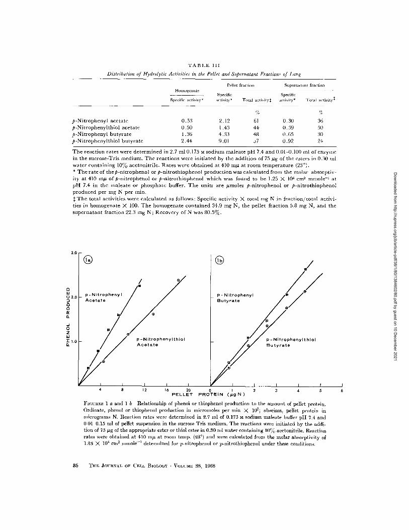
TABLE III
Distribution of Hydrolytic Activities in the Pellet and Supernatant Fractions of Lung
Pellet fraction Supernatant fractionHomogenate
Specific SpecificSpecific activity* activity* Total activity activity* Total activity
% %
p-Nitrophenyl acetate 0.53 2.12 61 0.30 36p-Nitrophenylthiol acetate 0.50 1.43 44 0.39 50p-Nitrophenyl butyrate 1.36 4.33 48 0.65 30p-Nitrophenylthiol butyrate 2.44 9.01 57 0.92 24
The reaction rates were determined in 2.7 ml 0.175 M sodium maleate pH 7.4 and 0.01-0.100 ml of enzymein the sucrose-Tris medium. The reactions were initiated by the addition of 75 ig of the esters in 0.30 mlwater containing 10% acetonitrile. Rates were obtained at 410 mU at room temperature (230).* The rate of thep-nitrophenol or p-nitrothiophenol production was calculated from the molar absorptiv-ity at 410 m of p-nitrophenol or p-nitrothiophenol which was found to be 1.25 X 104 cm
2mmole
-at
pH 7.4 in the maleate or phosphate buffer. The units are moless p-nitrophenol or p-nitrothiophenolproduced per mg N per min.
t The total activities were calculated as follows: Specific activity X total mg N in fraction/total activi-ties in homogenate X 100. The homogenate contained 34.9 mg N, the pellet fraction 5.8 mg N, and thesupernatant fraction 22.3 mg N; Recovery of N was 80.5%.
®
/
p -NitrophenylthiolAcetate
p- NitrophenylButyrate
p - Nitrophenylthiol
Butyrate
4 8 12 16 20 0 1 2 3 4 5PELLET PROTEIN (pg N)
FIGURES I a and 1 b Relationship of phenol or thiophenol production to the amount of pellet protein.Ordinate, phenol or thiophenol production in micromoles per min X 102; abscissa, pellet protein inmicrograms N. Reaction rates were determined in 2.7 ml of 0.175 M sodium maleate buffer pH 7.4 and0.01-0.15 ml of pellet suspension in the sucrose-Tris medium. The reactions were initiated by the addi-tion of 75 g of the appropriate ester or thiol ester in 0.30 ml water containing 10% acetonitrile. Reactionrates were obtained at 410 mu at room temp. (3
°) and were calculated from the molar absorptivity of
1.25 X 104cm
2mmole
- 1determined for p-nitrophenol or p-nitrothiophenol under these conditions.
86 THE JOURNAL OF CELL BIOLOGY · VOLIUME 38, 1968
J.U
UI0 2.0D
0aar
0z
r 1.0n
an
Dow
nloaded from http://rupress.org/jcb/article-pdf/38/1/80/1384602/80.pdf by guest on 10 D
ecember 2021
centration at constant substrate concentrationfor the four substrates tested.
pH EFFECTS: The rates of hydrolysis ofPNPA and PNTA increase with increasing pH. Be-low pH 6.5 the rates of enzymatic hydrolysis arelow, and above pH 8.2 in tris-maleate buffer thespontaneous hydrolysis of the thioesters is rapid.We selected the maleate buffer for all our studiesbecause it appeared to be ideal for the cytochem-ical work. Since maleate loses its buffering capacityat pH values above 7.4, we chose this value.Sodium ethylenediaminetetraacetate (EDTA) orphosphate can substitute for maleate buffer forboth electron microscopic and kinetic work.
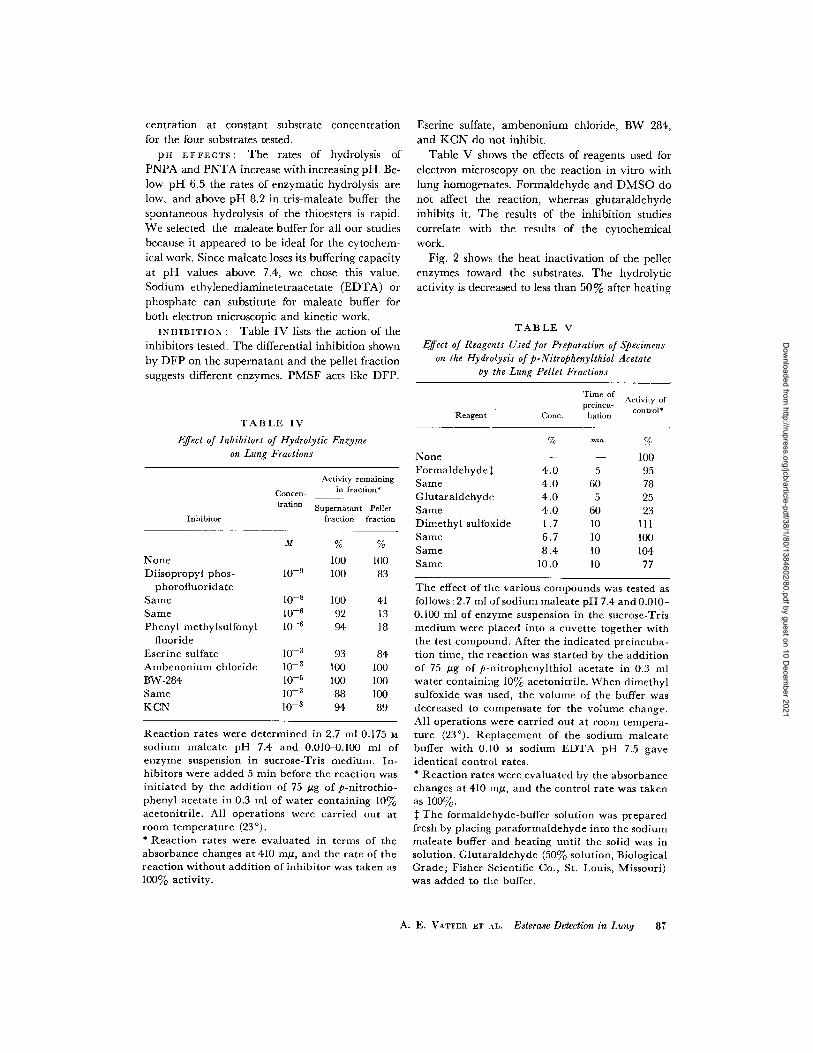
INHIBITION: Table IV lists the action of theinhibitors tested. The differential inhibition shownby DFP on the supernatant and the pellet fractionsuggests different enzymes. PMSF acts like DFP.
TABLE IV
Effect of Inhibitors of Hydrolytic Enzymeon Lung Fractions
Activity remaining
Concern- in fraction
tration Supernatant Pellet
Inhibitor fraction fraction
M % %
None 100 100Diisopropyl phos- 10- 9 100 83
phorofluoridateSame 10-8 100 41Same 10- 6 92 13Phenyl methylsulfonyl 10-6 94 18
fluorideEserine sulfate 10-
3 93 84Ambenonium chloride 10-3 100 100BW-284 10- 5 100 100Same 10-3 88 100KCN 10-
3 94 89
Reaction rates were determined in 2.7 ml 0.175 Msodium maleate pH 7.4 and 0.010-0.100 ml ofenzyme suspension in sucrose-Tris medium. In-hibitors were added 5 min before the reaction wasinitiated by the addition of 75 ug of p-nitrothio-phenyl acetate in 0.3 ml of water containing 10%acetonitrile. All operations were carried out atroom temperature (230).* Reaction rates were evaluated in terms of theabsorbance changes at 410 mg, and the rate of thereaction without addition of inhibitor was taken as100% activity.
Eserine sulfate, ambenonium chloride, BW 284,and KCN do not inhibit.
Table V shows the effects of reagents used forelectron microscopy on the reaction in vitro withlung homogenates. Formaldehyde and DMSO donot affect the reaction, whereas glutaraldehydeinhibits it. The results of the inhibition studiescorrelate with the results of the cytochemicalwork.
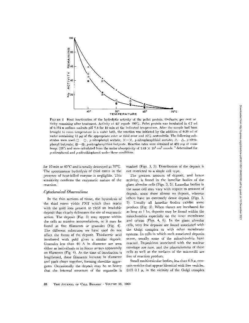
Fig. 2 shows the heat inactivation of the pelletenzymes toward the substrates. The hydrolyticactivity is decreased to less than 50% after heating
TABLE V
Effect of Reagents Used for Preparation of Specimenson the Hydrolysis of p-Nitrophenylthiol Acetate
by the Lung Pellet Fractions
Time ofActivity of
preincu- controlReagent Cone. bation
% min %
None - 100Formaldehyde: 4.0 5 95Same 4.0 60 78Glutaraldehyde 4.0 5 25Same 4.0 60 23Dimethyl sulfoxide 1.7 10 111Same 6.7 10 100Same 8.4 10 104Same 10.0 10 77
The effect of the various compounds was tested asfollows: 2.7 ml of sodium maleate pH 7.4 and 0.010-0.100 ml of enzyme suspension in the sucrose-Trismedium were placed into a cuvette together withthe test compound. After the indicated preincuba-tion time, the reaction was started by the additionof 75 g of p-nitrophenylthiol acetate in 0.3 mlwater containing 10% acetonitrile. When dimethylsulfoxide was used, the volume of the buffer wasdecreased to compensate for the volume change.All operations were carried out at room tempera-ture (23°). Replacement of the sodium maleatebuffer with 0.10 M sodium EDTA pH 7.5 gaveidentical control rates.* Reaction rates were evaluated by the absorbancechanges at 410 In, and the control rate was takenas 100%.: The formaldehyde-buffer solution was preparedfresh by placing paraformaldehyde into the sodiummaleate buffer and heating until the solid was insolution. Glutaraldehyde (50% solution, BiologicalGrade; Fisher Scientific Co., St. Louis, Missouri)was added to the buffer.
A. E. VATTER ET L. Esterase Detection in Lung 87
Dow
nloaded from http://rupress.org/jcb/article-pdf/38/1/80/1384602/80.pdf by guest on 10 D
ecember 2021
100
Z80Z
Lii, 60
> 40
O
20
20
x
40° 50 °60° 70°C
TEMPERATURE
FIGURE 2 Heat inactivation of the hydrolytic activity of the pellet protein. Ordinate, per cent activity remaining after treatment. Activity at 3° equals 100cc. Pellet protein was incubated in .7 nllof 0.175 M sodium maleate pH 7.4 for 10 min at the indicated temperature. After the sample had beenbrought to room temperature in a water bath, the reaction was initiated by the addition of 0.30 nml ofwater containing 75 ug of the appropriate ester or thiol ester and 10% acetonitrile. The following sub-strates were used:0 O , p-nitrophenyl acetate; X-X, p-nitrophenylthiol acetate; A-A, p-nitro-phenyl butyrate; ® ®, p-nitrophenylthiol butyrate. Reaction rates were obtained at 410 mng at roomtemp. (3 °) and were calculated from the molar absorptivity of 1.25 X 104 cm 2 mmolel 1 determined forp-nitrophenol and p-nitrothiophenol under these conditions.
for 10 min at 400C and is totally destroyed at 700C.The spontaneous hydrolysis of thiol esters in thepresence of heat-killed enzyme is negligible. Thissensitivity confirms the enzymatic nature of thereaction.
Cytochemical Observations
In the thin sections of tissue, the hydrolysis ofthe thiol esters yields PNT which then reactswith the gold ions present to yield an insolubledeposit that clearly delineates the site of enzymaticaction. The deposit (Fig. 3) may appear withinthe cells as massive accumulations, or it may befound as fine filaments or granules (Fig. 4).The different substrates we have used do notaffect the forms of the deposit. Thiolacetic acidincubated with gold gives a similar deposit.Granules less than 40 A in diameter are seeneither as individuals or in linear arrays apparentlyon filaments (Fig. 4). As the time of incubation islengthened, these filaments increase in diameterand pack closer together, forming sheetlike aggre-gates. Occasionally the deposit may be so heavythat the internal structure of the organelle is
masked (Figs. 3, 5). Distribution of the deposit is
not restricted to a single cell type.
The greatest amount of deposit, and hence
activity, is found in the lamellar bodies of the
giant alveolar cells (Figs. 3, 5). Lamellar bodies in
the same cell may vary with respect to amount of
deposit; some show almost no deposit, whereas
others have an extremely dense deposit (Figs. 3,
5). Usually all lamellar bodies exhibit some
product (Fig. 3). When tissues are incubated for
as long as 1 hr, deposits may be found within the
mitochondria especially on the inner membrane
and cristae (Figs. 4, 6). In the giant alveolar
cells, very few deposits are found associated with
the Golgi complex or with other membrane
systems. In cells in which such associated depositsoccur, usually some of the mitochondria have
reacted. Depositions associated with the nuclear
envelope are rare, and the plasmalemma of thesecells as well as the surfaces of the microvilli arefree of reaction product.
Small multivesicular bodies, less than 0.5 As, con-
tain vesicles that appear identical with free vesicles,
0.05 0.1 /, in the vicinity of the Golgi complex
88 THE JOURNAL OF CELL BIOLOGY VOLUME 38, 1968
I- -C__
---
Dow
nloaded from http://rupress.org/jcb/article-pdf/38/1/80/1384602/80.pdf by guest on 10 D
ecember 2021
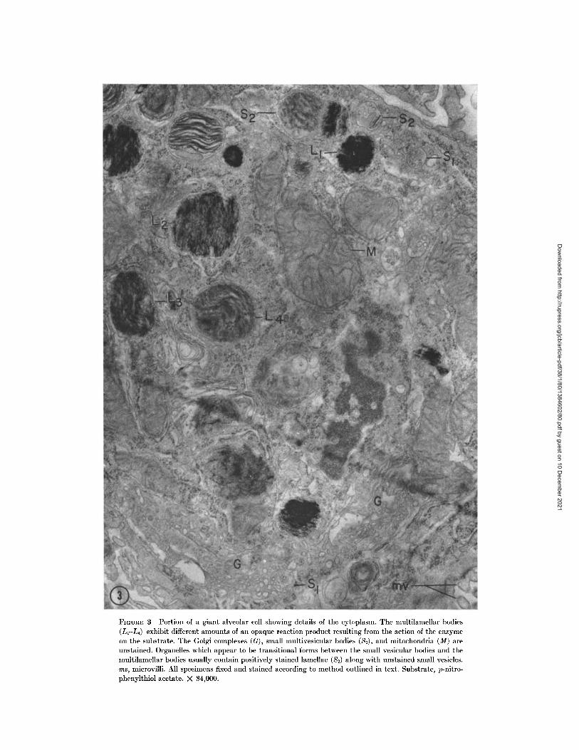
FIGURE 3 Portion of a giant alveolar cell showing details of the cytoplasm. The multilamellar bodies(L1-L4) exhibit different amounts of an opaque reaction product resulting from the action of the enzymeon the substrate. The Golgi complexes (G), small multivesicular bodies (S1), and mitochondria (M) areunstained. Organelles which appear to be transitional forms between the small vesicular bodies and themultilamellar bodies usually contain positively stained lamellae (S2) along with unstained small vesicles.me, microvilli. All specimens fixed and stained according to method outlined in text. Substrate, p-nitro-phenylthiol acetate. X 34,000.
Dow
nloaded from http://rupress.org/jcb/article-pdf/38/1/80/1384602/80.pdf by guest on 10 D
ecember 2021
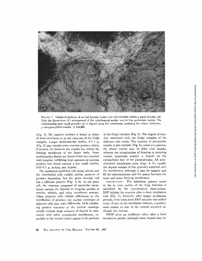
FlGUIE 4 Details of portions of several lamellar bodies and mitochondria within a giant alveolar cell.
Note the filamentous (F) arrangement of the cytochemical marker and its fine particulate nature. Theinitochondria show small granules (g) of deposit along the membranes, including the cristae. Substrate,
p-nitrophenylthiol acetate. X 148,000.
(Fig. 3). No reaction product is found in eitherof these structures or in the cisternae of the Golgicomplex. Larger multivesicular bodies, 0.5-1 ,(Fig. 3) may contain some reaction product which,if present, lies between the vesicles but within thelimiting membrane of the larger body. Somemultilamellar bodies are found which are crowdedwith lamellae exhibiting large amounts of reactionproduct but which contain a few small vesicles,0.05-0.1 , lacking any deposit.
The squamous epithelial cells lining alveoli andthe endothelial cells exhibit similar patterns ofproduct deposition, but the giant alveolar cellhas a different pattern (Figs. 5, 6). In the giantcell, the cisternae composed of agranular mem-brane contain the deposits in irregular profiles ofvesicles, tubules, and other membrane systems.Often adjacent cells exhibit differences in thedistribution of product; the nuclear envelopes ofadjacent cells may stain differently. Cells exhibit-ing positive reactions at the nuclear envelopeusually contain large amounts of deposit in asso-ciation with other cytoplasmic membranes, es-pecially in the vesicles which appear to be portions
of the Golgi complex (Fig. 6). The degree of reac-
tion associated with the Golgi complex of the
different cells varies. The reaction in pinocytotic
vesicles is also variable (Fig. 5); when it is positive,the closed vesicles may be filled with deposit,whereas the invaginations of forming or secretingvesicles sometimes contain a deposit on theextracellular face of the plasmalemma. All mito-chondrial membranes stain (Figs. 4, 6); usuallythe deposit consists of fine granules scattered overthe membranes, although it may be massive andfill the mitochondrion and the spaces between theouter and inner limiting membranes.
INHIBITORS: The inhibition pattern notedin the in vitro studies of the lung fractions isparalleled by the cytochemical observations.DFP inhibits the reaction after a short incubationtime (Fig. 7); however, after longer incubationperiods, even when fresh DFP solution was addedevery 15 min to the incubation mixture, a positivestain similar to that of the control occurred al-though less intense.
PMSF gives an inhibitory effect after a briefincubation period, although some deposit may be
90 THE JOURJNAL OF CELL BIOLOGY VOLUME 38, ,968
Dow
nloaded from http://rupress.org/jcb/article-pdf/38/1/80/1384602/80.pdf by guest on 10 D
ecember 2021
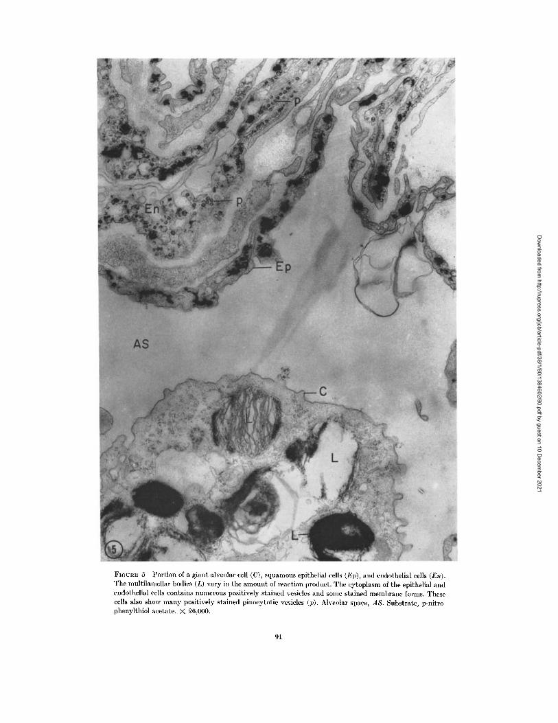
FIGURE 5 Portion of a giant alveolar cell (C), squamous epithelial cells (Ep), and endothelial cells (En).The multilamellar bodies (L) vary in the amount of reaction product. The cytoplasm of the epithelial andendothelial cells contains numerous positively stained vesicles and some stained membrane forms. Thesecells also show many positively stained pinocytotic vesicles (p). Alveolar space, AS. Substrate, p-nitro-phenylthiol acetate. X 26,000.
91
Dow
nloaded from http://rupress.org/jcb/article-pdf/38/1/80/1384602/80.pdf by guest on 10 D
ecember 2021

FrGURE 6 Portion of the cytoplasm of several endothelial cells showing positively stained Golgi coim-plexes (G). Some reaction has taken place on the nuclear envelope (N), mitochondrial membranes (M),pinocytotic vesicles (p), and along portion of the cell membrane (P). Substrate, p-nitrophenylthiol buty-rate. X 38,000.
92
Dow
nloaded from http://rupress.org/jcb/article-pdf/38/1/80/1384602/80.pdf by guest on 10 D
ecember 2021

FIGUREs 7 and 8 Portions of the specimens treated with DFP (Fig. 7) and PMSF (Fig. 8) exhibit fewscattered granules of deposit after short (15 min) incubation time. These results correlate with the invitro experiments. After longer incubation periods (45-60 min) some product does accumulate. L, multi-lamellar bodies. Substrate, p-nitrophenylthiol acetate. Fig. 7, X 25,000; Fig. 8, X 35,000.
93
Dow
nloaded from http://rupress.org/jcb/article-pdf/38/1/80/1384602/80.pdf by guest on 10 D
ecember 2021
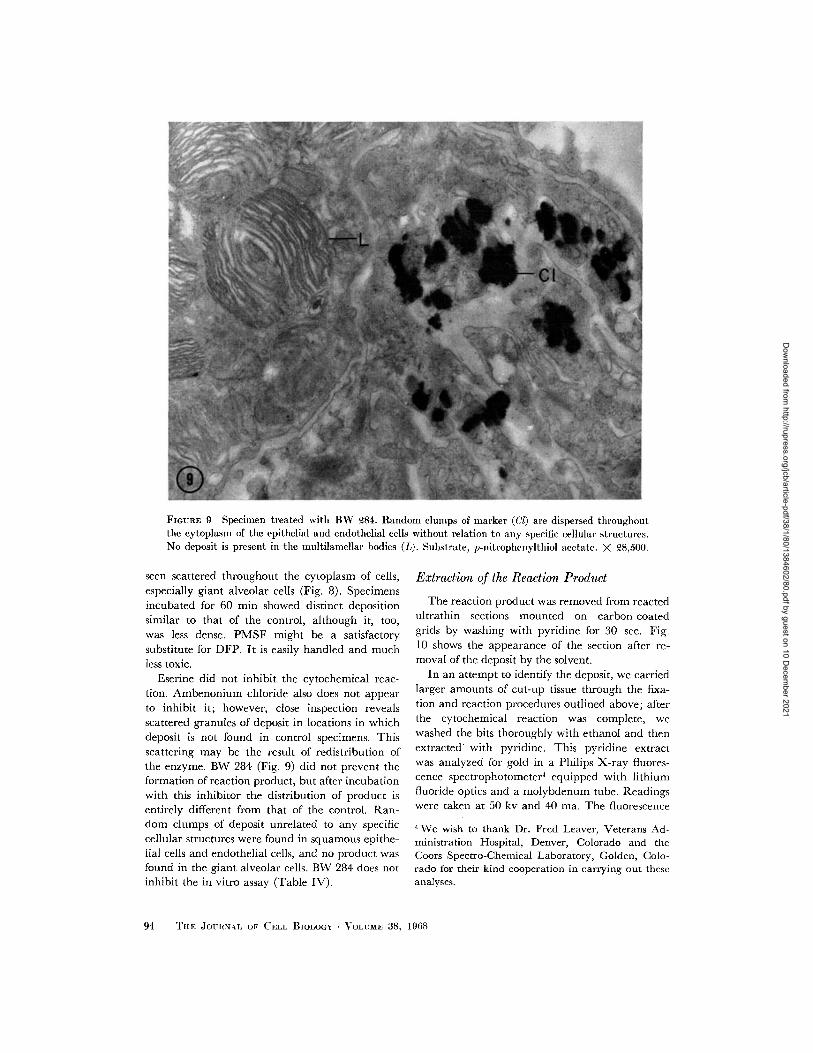
FIGURE 9 Specimen treated with BW 284. Random clumps of marker (Cl) are dispersed throughoutthe cytoplasm of the epithelial and endothelial cells without relation to any specific cellular structures.No deposit is present in the multilamellar bodies (L). Substrate, p-nitrophenylthiol acetate. X 28,500.
seen scattered throughout the cytoplasm of cells,especially giant alveolar cells (Fig. 8). Specimensincubated for 60 min showed distinct depositionsimilar to that of the control, although it, too,was less dense. PMSF might be a satisfactorysubstitute for DFP. It is easily handled and muchless toxic.
Eserine did not inhibit the cytochemical reac-tion. Ambenonium chloride also does not appearto inhibit it; however, close inspection revealsscattered granules of deposit in locations in whichdeposit is not found in control specimens. Thisscattering may be the result of redistribution ofthe enzyme. BW 284 (Fig. 9) did not prevent theformation of reaction product, but after incubationwith this inhibitor the distribution of product isentirely different from that of the control. Ran-dom clumps of deposit unrelated to any specificcellular structures were found in squamous epithe-lial cells and endothelial cells, and no product wasfound in the giant alveolar cells. BW 284 does notinhibit the in vitro assay (Table IV).
Extraction of the Reaction Product
The reaction product was removed from reactedultrathin sections mounted on carbon-coatedgrids by washing with pyridine for 30 sec. Fig.10 shows the appearance of the section after re-moval of the deposit by the solvent.
In an attempt to identify the deposit, we carriedlarger amounts of cut-up tissue through the fixa-tion and reaction procedures outlined above; afterthe cytochemical reaction was complete, wewashed the bits thoroughly with ethanol and thenextracted with pyridine. This pyridine extractwas analyzed for gold in a Philips X-ray fluores-cence spectrophotometer 4 equipped with lithiumfluoride optics and a molybdenum tube. Readingswere taken at 50 kv and 40 ma. The fluorescence
4 We wish to thank Dr. Fred Leaver, Veterans Ad-ministration Hospital, Denver, Colorado and theCoors Spectro-Chemical Laboratory, Golden, Colo-rado for their kind cooperation in carrying out theseanalyses.
94 TIiF JOURtNAI OF CELL BIOLOGY VOLUME 38, 1968
Dow
nloaded from http://rupress.org/jcb/article-pdf/38/1/80/1384602/80.pdf by guest on 10 D
ecember 2021
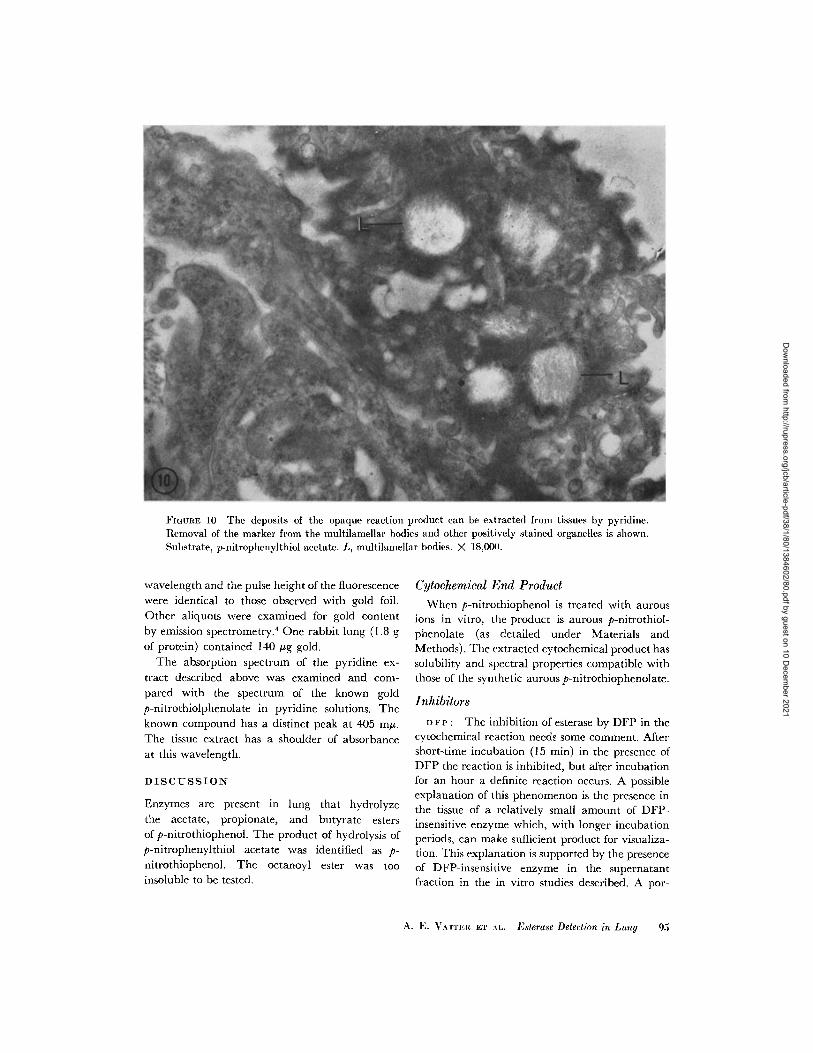
FIGURE 10 The deposits of the opaque reaction product can be extracted from tissues by pyridine.Removal of the marker from the multilamellar bodies and other positively stained organelles is shown.Substrate, p-nitrophenylthiol acetate. L, multilamellar bodies. X 18,00().
wavelength and the pulse height of the fluorescencewere identical to those observed with gold foil.
Other aliquots were examined for gold content
by emission spectrometry.4 One rabbit lung (1.8 g
of protein) contained 140 g gold.
The absorption spectrum of the pyridine ex-tract described above was examined and com-
pared with the spectrum of the known goldp-nitrothiolphenolate in pyridine solutions. Theknown compound has a distinct peak at 405 mu.The tissue extract has a shoulder of absorbance
at this wavelength.
DISCUSSION
Enzymes are present in lung that hydrolyze
the acetate, propionate, and butyrate estersof p-nitrothiophenol. The product of hydrolysis ofp-nitrophenylthiol acetate was identified as p-
nitrothiophenol. The octanoyl ester was tooinsoluble to be tested.
Cytochemical End ProductWhen p-nitrothiophenol is treated with aurous
ions in vitro, the product is aurous p-nitrothiol-phenolate (as detailed under Materials andMethods). The extracted cytochemical product hassolubility and spectral properties compatible withthose of the synthetic aurous p-nitrothiophenolate.
Inhibitors
D FP : The inhibition of esterase by DFP in thecytochemical reaction needs some comment. Aftershort-time incubation (15 min) in the presence ofDFP the reaction is inhibited, but after incubationfor an hour a definite reaction occurs. A possibleexplanation of this phenomenon is the presence inthe tissue of a relatively small amount of DFP-insensitive enzyme which, with longer incubationperiods, can make sufficient product for visualiza-tion. This explanation is supported by the presenceof DFP-insensitive enzyme in the supernatantfraction in the in vitro studies described. A por-
A. E. VATTERI ET \1.. Esterase Detection in Lung 95
Dow
nloaded from http://rupress.org/jcb/article-pdf/38/1/80/1384602/80.pdf by guest on 10 D
ecember 2021
tion of the pellet enzymes is also DFP-insensitive(10-15%).
BW 284, considered a specific inhibitor of
acetyl cholinesterase because of its quaternaryammonium structure, does not inhibit the activityof the homogenate. The cytochemical observa-tions indicate apparent release and dispersionof the enzyme from the lamellar bodies aftertreatment with this inhibitor. The quaternaryammonium group has surface-active proper-
ties which might affect membrane permea-bility and thus account for this observation.
In examining this possibility, we tested the actionof a surface-active agent, Triton WR 1339 whichdoes not have a quaternary ammonium group, on
the cytochemical reaction and found similardispersion of product. This finding indicates that
the lamellar bodies are labile (see below). Tocheck whether some of these dispersions might bethe result of immediate postmortem changes inthe lungs, we prepared some specimens by inject-ing incubation mixture, with and without fixative,intratracheally into the still living lung. These
specimens showed exactly the same distribution ofthe cytochemical product as those tissues preparedin the usual way.
Substrate Specificity
Since the lung homogenate does not hydrolyzeacetyl choline and since the cytochemical reac-tion is not inhibited by eserine, the presence ofacetyl choline hydrolase (3.1.1.7) and acyl-choline acyl-hydrolase (3.1.1.8) can be ruled out.The lung homogenate described in this paperhydrolyzes N-benzoyl-L-tyrosine p-nitroanilide, aspecific substrate for chymotrypsin, a peptidyl
peptide hydrolase (3.4) (34). Chymotrypsin canhydrolyze PNTA (Reiss, O. K. Unpublished ob-servations.) as well as PNPA. The lung also pos-sesses glycerol-ester hydrolase (3.1.1.3) (35), aclass of enzymes which can hydrolyze PNTA(Reiss, O. K. Unpublished observations). Homo-genates of liver can hydrolyze thiol esters (36).
The presence of weak cytochemical reactions onthe mitochondrial and nuclear membranes is notunderstood at this time. The functional signifi-cance of the described enzymatic activity of thelung must await further investigation.
Lamellar Bodies and Pulmonary Surfactant
Most of the esterase activity toward the sub-strates dealt with in this study is found in the
lamellar bodies. Although the pellet fraction ofthe centrifuged homogenate of adult rabbit lungcontains much of this esterase activity, which isDFP-sensitive, our search for the lamellar bodiesin fixed pellet specimens with electron microscopy
has revealed very few of these organelles. Thisobservation agrees with our previous report (1)that the organelles are extremely labile andrupture during homogenization and thus leavesome of the enzymatic protein attached to thesedimented membranes. We have found otherindications that this assumption is correct; the
enzymatic activity of the pellet fraction towardthe thiol esters becomes further solubilized afterit has stood a few days in the cold or after treat-ment with sonic oscillation.
The giant alveolar cell has long been presumedto be secretory, and recent work has suggestedthat it may be a source of the important pulmonarysurfactant (37, 38). This surfactant presumablyfunctions by lowering surface tension and thusstabilizes the alveoli for efficient respiratory gasexchange. It is now believed to be a lipoprotein(39, 40) which contains large amounts of dipalm-itoyl lecithin. Previous work (41) indicates thatsurfactant activity is present in the pellet fractiondescribed in this paper. Partially purified sur-factant fractions obtained from throat washings bythe procedure of Bondurant and Miller (42) canhydrolyze PNTA. This finding suggests that the
esterase and surfactant activities may be inter-related.
The electron micrographs support the idea thatthe lamellar bodies are formed from the multi-vesicular bodies and that they develop their
esterase, and possibly surfactant, activity (43) anddischarge this activity into the alveolar lumen(38, 44). The electron micrographs show a com-plete gradation between small and large multi-vesicular bodies and partially and completelylamellated bodies. Some of the lamellar bodies
stain deeply, some not at all, and some at inter-mediate levels, as would be anticipated in a
sequence of development, activation, and release of
a secretory granule. Similar morphological se-
quences have been shown by Caro and Palade
(45) for the secretory granules of the acinar cell
of the pancreas and by Smith and Farquhar (46)
for granules of the pituitary cells. The lamellar
bodies, like the zymogen and pituitary granules,
lose their structural integrity when released from
the cell.
96 TE JOURNAL OF CELL BIOLOGY VOLUME 38, 1968
Dow
nloaded from http://rupress.org/jcb/article-pdf/38/1/80/1384602/80.pdf by guest on 10 D
ecember 2021

When one considers the nature of the lamellarbodies, the question arises whether they can beclassified as lysosomes. The lamellar bodies give
a weak cytochemical reaction for acid phosphatase,
whereas the lysosomes of the alveolar macrophagesgive an intense reaction. Conversely, the esterase
activity of the lamellar bodies is strong but
that of the macrophage is weak. The enzymes of
the lysosomes are most active in the acid region,
however the esterases described here function in
the neutral or alkaline regions. For these reasons,we favor the interpretation that the lamellar
body is a secretory organelle rather than a lyso-
some.The cytochemical method outlined here may
prove useful in other connections where it is pos-
sible to design synthetic substrates which can
form mercaptides after enzymatic reaction. Theinsolubility and small size of the precipitate of
the mercaptide makes it an ideal cytochemical
product. Furthermore, the removal of these
precipitates by specific solvents permits theirchemical identification.
This study was supported by a grant from the Ameri-can Medical Association Education and ResearchFoundation, a Public Health Service research grantAM-06968 from the National Institutes of Health, anAmerican Cancer Society grant IN 5H No. 11, a VRAgrant RT-10, and a Milheim Foundation grant.
We wish to thank Mr. Vincent Buric, Miss NancyHoover, and Mr. Mark Stephan for their technicalassistance.
Received for publication 4 December 1967, and in revisedform 1 March 1968.
BIBLIOGRAPHY
1. REMss, O. K. 1966. Studies of lung metabolism.I. Isolation and properties of subcellular frac-tions from rabbit lung. J. Cell Biol. 30:45.
2. FRUTON, J. S. 1946. On the proteolytic enzymesof animal tissues. V. Peptidases of skin, lung,and serum. J. Biol. Chem. 166:721.
3. DANNENBERG, A. M., JR., and E. L. SMITH. 1955.Proteolytic enzymes of lung. J. Biol. Chem.215:45.
4. DANNENBERG, A. M., JR., and E. L. SMITH. 1955.Action of proteinase I of bovine lung. Hy-drolysis of the oxidized B chain of insulin;polymer formation from amino acid esters.J. Biol. Chem. 215:55.
5. VON PILZ, W., and I. JOHANN. 1967. Ester
spaltende Fermente der menschlichen Lunge.Hoppe-Seylers Z. Physiol. Chem. 348:73.
6. HOLMES, R. S., and C. J. MASTERS. 1967. Thedevelopmental multiplicity and isoenzyme sta-tus of cavian esterases. Biochim. Biophys. Acta.132:379.
7. GOMORI, G. 1952. Intern. Rev. Cytol. 1:323.8. BURSTONE, M. S. 1962. Chapter VI. In Enzyme
Histochemistry. Academic Press Inc., NewYork. 293.
9. GOMORI, G. 1945. The microtechnical demonstra-tion of sites of lipase activity. Proc. Soc. Exptl.Biol. Med. 58:362.
10. BARRNETT, R. J., and A. M. SELIGMAN. 1951.
Histochemical demonstration of esterases byproduction of indigo. Science. 114:579.
11. HOLT, S. J. 1952. A new principle for the histo-chemical localization of hydrolytic enzymes.Nature. 169:271.
12. KOELLE, G. B., and J. S. FRIEDENWALD. 1949.
A histochemical method for localizing cholin-esterase activity. Proc. Soc. Exptl. Biol. Med.70:617.
13. KOELLE, G. B., and C. G. GROMADZKI. 1966.Comparison of the gold-thiocholine and gold-thiolacetic acid methods for the histochemicallocalization of acetylcholinesterase and cholin-esterases. .1. Histochem. Cytochem. 14:443.
14. WILSON, I. B. 1951. Mechanism of hydrolysis. II.New evidence for an acylated enzyme asintermediate. Biochim. Biophys. Acta. 7:520.
15. BARRNETT, R. J. 1962. The fine structurallocalization of acetylcholinesterase at the myo-neural junction. J. Cell Biol. 12:247.
16. KOELLE, G. B., and C. FOROoLOU-KERAMEOS.
1965. Electron microscopic localization ofcholinesterases in a sympathetic ganglion by agold-thiolacetic acid method. Life Sci. 4:417.
17. DAVIS, R., and G. B. KOELLE. 1965. Electronmicroscopic localization of acetylcholinesteraseat the motor endplate by the gold-thiolaceticacid and gold thiocholine methods. J. Histo-chem. Cytochem. 13:703.
18. HUGGINS, C., and J. LAPIDES. 1947. Chromo-genic substrates: IV. Acyl ester of p-nitrophe-nol as substrates for the colorimetric deter-mination of esterase. J. Biol. Chem. 170:467.
19. NACHLAS, M. M., and A. M. SELIGMAN. 1949.
Evidence for the specificity of esterase andlipase by the use of three chromogenic sub-strates. J. Biol. Chem. 181:343.
20. SELLINGER, O. Z., and V. DE BALBIAN. 1962. An
esterase of rat cerebral cortex acting ono-nitrophenyl acetate: method of assay, proper-
A. E. VATTER ET AL. Esterase Detection in Lung 97
Dow
nloaded from http://rupress.org/jcb/article-pdf/38/1/80/1384602/80.pdf by guest on 10 D
ecember 2021
ties and intracellular distribution. Anal. Bio-chem. 3:479.
21. SELIGMAN, A. M., and M. M. NACHLAS. 1950.The colorimetric determination of lipase andesterase in human serum. J. Clin. Invest.29:31.
22. SZAFRAN, H., Z. SZAFRAN, and T. POPIELA. 1964.
Hydrolysis of aromatic esters by human duo-denal content. Clin. Chim. Acta. 9:190.
23. POPIELA, T., H. SZAFRAN, and Z. SZAFRAN.1965. The activity of human gastric lipasetowards p-nitrophenyl esters. Clin. Chim. Acta.11:283.
24. BROWN, H. C., and B. C. S. RAO. 1956. A newpowerful reducing agent-sodium borohydridein the presence of aluminum chloride and otherpolyvalent metal halides. J. Am. Chem Soc.78:2582.
25. LECHER, H. 1921. Beitraege zum Valenzproblemdes Schwefels. Ber. Deut. Chem. Ges. 53:590.
26. KLAs-BERTIL, A. 1957. In Methods of BiochemicalAnalysis. Interscience Publishers, Inc., NewYork. 5:43.
27. FAHRNEY, D. E., and A. M. GOLD. 1963. Sulfonylfluorides as inhibitors of esterases. I. Rates ofreaction with acetylcholinesterase, oa-chymo-trypsin and trypsin. J. Am. Chem. Soc. 85:997.
28. OYAMA, V. I., and H. EAGLE. 1956. Measurementof cell growth in tissue culture with a phenolreagent (Folin-Ciocalteau). Proc. Soc. Exptl.Biol. Med. 91:305.
29. JOHNSON, M. J. 1941. Isolation and properties ofa pure yeast polypeptidase. J. Biol. Chem.137:575.
30. SMITH, R. E., and M. G. FARQUHAR. 1965.Preparation of nonfrozen sections for electronmicroscope cytochemistry. Sci. Instr. News(RCA). 10:13.
31. VATTER, A. E., and J. ZAMBERNARD. 1966.
Comparative study of water soluble plastic-alcohol dehydrated tissue. J. Appl. Phys.37:3920 (T12).
32. NYQUIST, R. A., and W. J. POTTS. 1959. Charac-teristic infrared absorption frequencies of thiolesters and related compounds. Spectrochim. Acta.15:514.
33. BELLAMY, L. J. 1958. In The Infra-red Spectra of
Complex Molecules. Methuen & Co. Ltd.,London. 2nd Edition. 351.
34. BUNDY, H. F. 1962. A new spectrophotometricmethod for the determination of chymotrypsinactivity. Anal. Biochem. 3:431.
35. ARMSTRONG, H. J., M. C. KUENZIG, and L. F.
PELTIER. 1967. Lung lipase levels in normalrats and rats with experimentally produced fatembolism. Proc. Soc. Exptl. Biol. Med. 124:959.
36. SzuOKI, Z., and T. SuzuOKI. 1953. Enzymatichydrolysis of thioesters, especially in relation too-esterase. J. Biochem. 40:599.
37. BUCKINGHAM, S., and M. E. AVERY. 1962. Timeof appearance of lung surfactant in the foetalmouse. Nature. 193:688.
38. BENSCH, K., K. SCHAEFER, and M. E. AVERY.
1964. Granular pneumocytes: electron micro-scopic evidence of their exocrinic function.Science. 145:1318.
39. KLAUS, M. H., J. A. CLEMENTS, and R. J. HAVEL.1961. Composition of surface-active materialisolated from beef lung. Proc. Natl. Acad. Sci.U. S. 47:1858.
40. PATTLE, R. E., and L. C. THOMAS. 1961. Lipopro-tein composition of the film lining the lung.Nature. 189:844.
41. KLAUS, M., O. K. REISS, W. H. TOOLEY, C.PIEL, and J. A. CLEMENTS. 1962. Alveolar
epithelial cell mitochondria as source of thesurface-active lung lining. Science. 137:750.
42. BONDURANT, S., and D. A. MILLER. 1962. A
method for producing surface active extractsof mammalian lungs. J. Appl. Physiol. 17:167.
43. SOROKIN, S. P. 1967. A morphological and cyto-chemical study on the great alveolar cell. J.Histochem. Cytochem. 14:884.
44. KIscH, B. 1960. Electron microscopy of the lungsin acute pneumonia. Exptl. Med. Surg. 18:273.
45. CARO, L. G., and G. E. PALADE. 1964. Proteinsynthesis, storage, and discharge in the pan-
creatic exocrine cell. An autoradiographic
study. J. Cell Biol. 20:473.46. SMITH, R. E., and M. G. FARQUHAR. 1966.
Lysosome function in the regulation of the
secretory process in cells of the anterior pitui-
tary gland. J. Cell Biol. 31:319.
98 TnI: JOURNAL OF CELL BIOLOGY VOLUME 38, 1968
Dow
nloaded from http://rupress.org/jcb/article-pdf/38/1/80/1384602/80.pdf by guest on 10 D
ecember 2021