Embed Size (px)
Citation preview

Vol. 172, No. 3JOURNAL OF BACTERIOLOGY, Mar. 1990, p. 1380-13840021-9193/90/031380-05$02.00/0Copyright © 1990, American Society for Microbiology
Enzymes of Ammonia Assimilation in Hyphae and Vesicles ofFrankia sp. Strain Cpll
NANCY A. SCHULTZt AND DAVID R. BENSON*
Department of Molecular and Cell Biology, University of Connecticut, Storrs, Connecticut 06269-3044
Received 5 September 1989/Accepted 29 November 1989
Frankia spp. are filamentous actinomycetes that fix N2 in culture and in actinorhizal root nodules. Incombined nitrogen-depleted aerobic environments, nitrogenase is restricted to thick-wailed spherical struc-tures, Frankia vesicles, that are formed on short stalks along the vegetative hyphae. The activities of theNH4'-assimilating enzymes (glutamine synthetase [GS], glutamate synthase, glutamate dehydrogenase, andalanine dehydrogenase) were determined in cells grown on NH4' and N2 and in vesicles and hyphae fromN2-fixing cultures separated on sucrose gradients. The two frankial GSs, GSI and GSII, were present in vesiclesat levels similar to those detected in vegetative hyphae from N2-fixing cultures as shown by enzyme assay andtwo-dimensional polyacrylamide gel electrophoresis. Glutamate synthase, glutamate -dehydrogenase, andalanine dehydrogenase activities were restricted to the vegetative hyphae. Vesicles apparently lack a completepathway for assimilating ammonia beyond the glutamine stage.
Filamentous actinomycetes in the genus Frankia are N2-fixing aerobes that form actinorhizal root nodule symbioseswith certain nonleguminous plants. In aerobically growncultures and in most root nodules, N2 fixation is preceded bythe differentiation of nitrogenase-containing structures, theFrankia vesicles (20, 25, 32). Vesicles develop at the tips ofshort hyphal branches and have a thick, multilayered lipidenvelope that is proposed to protect nitrogenase from inac-tivation by 02 (23, 28, 34, 36). For aerobically culturedFrankia spp., the product of N2 fixation must be transferredin some form to the growing vegetative hyphae. Nitrogenfixation can occur without vesicle formation in microaerobicenvironments in culture (24) and in nodules of certainactinorhizal plants (38).One approach to studying the comparative nitrogen me-
tabolism of the vesicles and hyphae is to assay for thepresence of enzymes involved in ammonia assimilation;ammonia is the first product of N2 fixation. Glutaminesynthetase (GS; EC 6.3.1.2) is the primary NH4+-assimi-lating enzyme in most bacteria during NH4' limitation (37)and in most diazotrophs during N2 fixation (30). GS usesNH4+ plus glutamate to make glutamine. Glutamine can beused for the biosynthesis of proteins or other metabolites, orwith a-ketoglutarate, it can be converted by glutamatesynthase (GOGAT; EC 1.4.7.1) to make two glutamates. Theglutamate formed is used for amino acid biosynthesis and asa substrate for GS (37).Glutamate dehydrogenase (GDH; EC 1.4.1.4) and alanine
dehydrogenase (ADH; EC 1.4.1.1) have also been reportedto participate in ammonia assimilation by the reductiveamination of a-ketoglutarate and pyruvate, respectively.GDH has a characteristically high Km for NH4' and canperform an assimilatory role under conditions of ammoniaexcess (37). It is the predominant NH4+-assimilating enzymein NH4+-grown and N2-fixing cells of certain diazotrophicbacilli (14, 15). ADH can function in assimilation whenNH4+ is present in excess in organisms that apparently lackGDH (1, 4, 13, 16).
* Corresponding author.t Present address: Institute of Water Resources, University of
Connecticut, Storrs, CT 06269.
In previous work on the nitrogen metabolism of Frankiasp. strain CpIl, two GSs were characterized (10, 35). GSI isthe only form of GS detected in NH4+-grown frankialhyphae. Like many other bacterial GSs, GSI is a dodecamerof identical subunits, each of which can be reversibly ade-nylylated (10). Cultures grown with a poor nitrogen sourcelike N2 or glutamate have a highly active, heat-labile GS(GSII) and only low GSI activity (10, 35). GSII is inhibitedby glutamine, other amino acids, and a low energy charge(35); no evidence of regulation by adenylylation has beenfound (10). Frankia GSII is similar to the GSII found inmembers of the family Rhizobiaceae in its pattern of induc-tion and in its sensitivity to inhibition by metabolites (8, 9,11).The restriction of nitrogenase to the vesicles raises ques-
tions about how NH4' is assimilated during N2 fixation inFrankia spp., since N2 fixation is spatially separated fromthe growing hyphal tips of the actinomycete. Addressed inthis paper are the activities and distributions of the twofrankial GSs, GOGAT, GDH, and ADH in vegetative hy-phae and in vesicles of Frankia sp. strain CpIl grown indefined liquid media.
MATERIALS AND METHODSOrganism and culture media. Frankia sp. strain CpIl
(Frankia catalog no. HFP070101), a Comptonia peregrinaroot nodule isolate (7), was maintained in defined liquidmedium with succinate as the carbon source and NH4Cl asthe nitrogen source as previously described (25). To estab-lish N2-grown cultures, hyphae were collected by centrifu-gation and suspended in 500 ml of medium without combinednitrogen (25). Cultures were then incubated in cotton-stop-pered, 1-liter Erlenmeyer flasks for 4 days at 30°C on agyratory shaker (New Brunswick Scientific Co., Inc.) set at150 rpm. NH4+- and N2-grown cells were collected byvacuum filtration onto filter paper and washed with coldbuffer A (10 mM potassium phosphate, pH 6.5) on the filterpaper to remove culture medium. The cells were either usedimmediately for preparing crude extracts or frozen at -80°C.
Vesicle isolation. Vesicles were isolated by a modificationof our published procedure (25). Usually, 2 liters of N2-fixingCpIl culture was collected on filter paper by vacuum filtra-
1380
on May 31, 2018 by guest
http://jb.asm.org/
Dow
nloaded from
NH4+ ASSIMILATION IN FRANKIA HYPHAE AND VESICLES 1381
tion, and the mycelia were removed to ice-cold 50-ml cen-trifuge tubes; cell paste from 1 liter of culture was added toeach tube. Cells were suspended in 20 ml of cold 50% (wt/wt)sucrose in buffer A and homogenized on ice with a Polytrontissue homogenizer until microscopic examination revealedthat most of the vesicles were detached from the hyphae.Chilled 50% sucrose was added to each tube to a finalvolume of 30 ml, and the contents were stirred to obtain auniform suspension of cells in sucrose. After the suspensionwas overlaid with 5 ml of 10% (vol/vol) glycerol in buffer A,the glycerol-sucrose gradients were centrifuged at 35,000 xg for 1 h at 4°C.
During centrifugation, vesicles banded at the glycerol-sucrose interface. The preparation was essentially free ofhyphal contamination, as assessed microscopically. Vesicleswere removed from each tube with a Pasteur pipette andtransferred to a 17-ml ultracentrifuge tube. They were sus-pended in buffer A and washed once by ultracentrifugation at40,000 x g for 15 min at 4°C. Each vesicle pellet wassuspended in 0.5 ml of buffer A, transferred to a microcen-trifuge tube, and repelleted by centrifugation at 10,000 x gfor 10 min at 8°C. This procedure typically yielded 0.5 ml ofpacked vesicles from 2 liters of culture used. Vesicles eitherwere used immediately or were stored frozen at -80°C.The pellets from the glycerol-sucrose gradients were com-
posed of vegetative hyphae and some attached vesicles. Weenriched for vegetative hyphae by gently disrupting thepellets in buffer A with a Dounce-type tissue homogenizer,diluting the suspension with additional buffer A, and centri-fuging at 500 x g for 10 min. This initial centrifugationselectively pelleted the vegetative hyphae, while most of thevesicles and cell debris remained in the supernatant. Theabsolute degree of purity of the hyphal fraction is difficult toassess in the absence of a quantitative marker; however,vesicles are a minority cell type in the original populationand clearly represent less than 1% of the biomass in thehyphal pellet as estimated microscopically. After twowashes at 35,000 x g with buffer A to remove the sucrose,the hyphae were used to make extracts immediately or werestored frozen at -80°C.
Cell extracts. Cell extracts were prepared by sonication at60 W for 20 min with pulsed output on 50% duty cycle in anice bath. For determining GS activity, extracts were made in10 mM imidazole-HCl (pH 6.8) with 2 mM MnCl2 and 1.0%(vol/vol) glycerol. For GOGAT assays, extracts were pre-pared in 10 mM HEPES (N-2-hydroxyethylpiperazine-N'-2-ethanesulfonic acid) buffer (pH 7.5) with 1 mM dithiothre-itol. For ADH and GDH determinations, 10 mM Tris hydro-chloride buffer at pH 8.0 with 1 mM dithiothreitol was usedto make extracts. Each sonic extract was centrifuged at35,000 x g for 20 min at 4°C; the supernatant constituted thecrude cell extract.
Vesicle extracts were concentrated by (NH4)2SO4 precip-itation as follows. While the extract was stirred, solid(NH4)2SO4 was added to 50% saturation; this solution wasstirred in an ice bath at 4°C for 15 min. The extract was thencentrifuged at 35,000 x g for 20 min at 4°C, and the(NH4)2SO4 pellet was dissolved in GOGAT assay buffer.Previous experiments indicated that (NH4)2SO4 had noadverse effect on GOGAT activity.Enzyme assays. GS activities were detected by the y-
glutamyltransferase assay (2) at pH 6.8 for GSI and pH 6.5for GSII as previously described (10). To measure therelative contributions of GSI and GSII to total transferaseactivity, extracts were heated at 70°C for 7 min, cooled atroom temperature for 10 min, and then placed on ice until
assayed for transferase activity. Control samples of eachextract were held at room temperature during the heatingand cooling procedure. Heating eliminates the GSII contri-bution to total transferase activity (10).GOGAT activity was assayed spectrophotometrically by
following the rate of NADH or NADPH oxidation (asspecified in Results) as the decrease in A340 per min at 25°C(5). The 1.0-ml reaction mixtures (final pH, 7.6) contained1.0 mM a-ketoglutarate, 0.1 mM NAD(P)H, 5.0 mM L-glutamine, 200 mM HEPES, and enzyme. The pH of 7.6 wasdetermined to be optimal for Cpll GOGAT in extracts fromNH4+-grown and N2-fixing cells by varying the pH of theassay buffer (200 mM HEPES; data not shown) and measur-ing the pH after mixing the reaction components. Thebackground oxidation of NAD(P)H was measured for 1 min,and then L-glutamine was added to initiate the specificreaction. GOGAT activity was calculated from the differ-ence in the rates of the decrease of absorbance in thepresence and absence of L-glutamine.The reductive amination reactions ofADH and GDH were
measured by monitoring the oxidation of NAD(P)H at 340nm at 25°C (22) and were initiated by the addition of sodiumpyruvate for ADH or sodium a-ketoglutarate for GDH. The1.5-ml reaction mixtures contained 0.1 mM NAD(P)H, 1.0mM sodium pyruvate or sodium a-ketoglutarate, 200 mMNH4Cl, 100 mM Tris hydrochloride (pH 8.6), and enzyme.The difference in the rates of the decrease of absorbance inthe presence and in the absence of pyruvate (for ADH) ora-ketoglutarate (for GDH) was used to determine enzymeactivity. The pH of 8.6 was determined to be at or near theoptimum for both enzymes in preliminary experiments (datanot shown).
Gel electrophoresis. Two-dimensional polyacrylamide gelelectrophoresis was done essentially as described by O'Far-rell (26). For each gel, the volume of cell extract containingabout 10 ,ug of protein was electrophoresed. Isoelectricfocusing gels (pH 5 to 7) were 10 cm in length by 1.5 mm indiameter. The second-dimension sodium dodecyl sulfate-polyacrylamide gels were 0.7 mm thick with a 2.5-cm, 4%acrylamide stacking gel and a 12.5-cm, 7% acrylamideseparating gel with 2.5% bisacrylamide cross-linker. Gelswere stained by the modified silver nitrate method (21).
Protein determination. The method of Bradford (6) forprotein determination was used with bovine serum albuminas the standard.
Materials. Chemicals were obtained from Sigma ChemicalCo., St. Louis, Mo. The Polytron tissue homogenizer wasfrom Brinkmann Instruments, Inc., Westbury, N.Y. Thelow-speed centrifugations were done with a SuperspeedRC2-B automatic refrigerated centrifuge (Ivan Sorvall, Inc.,Norwalk, Conn.). The high-speed centrifugations were donein Ultraclear ultracentrifuge tubes and ultracentrifuge modelLB-SOB (Beckman Instruments, Inc., Palo Alto, Calif.).Sonications were performed with a Sonifier model 200(Branson Sonic Power Co., Danbury, Conn.). Isoelectricfocusing gels contained LKB ampholines (LKB, Paramus,N.J.).
RESULTS
Activities and distributions of GSI and GSII. Table 1 showsthe GS transferase specific activities from a typical heattreatment experiment using extracts from-NH4+-grown hy-phae, N2-grown cells (hyphae with attached vesicles), andthe two sucrose gradient-derived fractions, the isolatedvesicles and the hyphae. The relative contributions of GSI
VOL. 172, 1990
on May 31, 2018 by guest
http://jb.asm.org/
Dow
nloaded from
1382 SCHULTZ AND BENSON
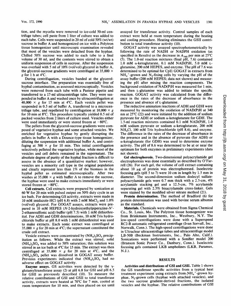
TABLE 1. Total activity and relative contributions of GSIand GSII in Frankia sp. strain Cpll cell types
GS transferase specific % of totalactivitya in: activityb of:
Cell typeControl extracts GSI GSII
NH4' grown 1.2 1.2 100 0N2 grown 2.7 0.03 1 99Gradient pellet 2.2 0.1 5 95Vesicle 2.4 0.1 4 96
a Micromoles of y-glutamyl hydroxamate formed per minute per milligramof protein. All assays were performed in triplicate, and replicates did not varyby more than 5%.
b Calculated as (activity remaining after heat treatment)/(activity in theunheated control) x 100.
and GSII in each extract are reported as percentages of theinitial activity.The GS transferase activity of the NH4+-grown cell ex-
tract remained unchanged after heating; thus, the heat-labileGSII was not detected in these extracts. All cell extractsfrom N2-fixing cultures, including the vesicles, had morethan twice the transferase activity of extracts from NH4+-grown cells. In all N2-grown cell types, GSII accounted for95 to 99%o of the total GS transferase activity.By coelectrophoresis of purified preparations of GSI and
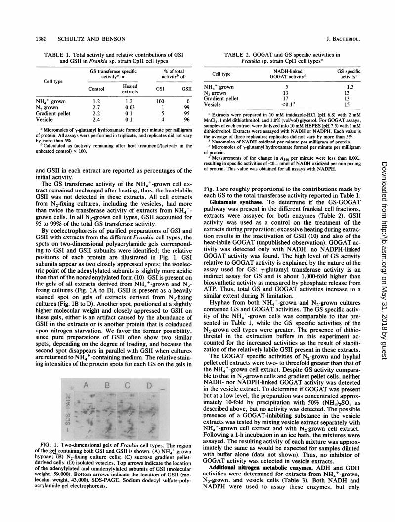
GSII with extracts from the different Frankia cell types, thespots on two-dimensional polyacrylamide gels correspond-ing to GSI and GSII subunits were identified; the relativepositions of each protein are illustrated in Fig. 1. GSIsubunits appear as two closely appressed spots; the isoelec-tric point of the adenylylated subunits is slightly more acidicthan that of the nonadenylylated form (10). GSI is present onthe gels of all extracts derived from NH4+-grown and N2-fixing cultures (Fig. 1A to D). GSII is present as a heavilystained spot on gels of extracts derived from N2-fixingcultures (Fig. 1B to D). Another spot, positioned at a slightlyhigher molecular weight and closely appressed to GSII onthese gels, either is an artifact caused by the abundance ofGSII in the extracts or is another protein that is coinducedupon nitrogen starvation. We favor the former possibility,since pure preparations of GSII often show two similarspots, depending on the degree of loading, and because thesecond spot disappears in parallel with GSII when culturesare returned to NH4+-containing medium. The relative stain-ing intensities of the protein spots for each GS on the gels in
A [3
0-
.f)
_J
FIG. 1. Two-dimensional gels of Frankia cell types. The regionof the gel containing both GSI and GSII is shown. (A) NH4+-grownhyphae; (B) N2-fixing culture cells; (C) sucrose gradient pellet-derived cells; (D) isolated vesicles. Top arrows indicate the locationof the adenylylated and unadenylylated subunits of GSI (molecularweight, 59,000). Bottom arrows indicate the location of GSII (mo-lecular weight, 43,000). SDS-PAGE, Sodium dodecyl sulfate-poly-acrylamide gel electrophoresis.
TABLE 2. GOGAT and GS specific activities inFrankia sp. strain Cpll cell types"
Cell type NADH-linked GS specificCell type ~~GOGAT activityb activityc
NH4' grown 5 1.3N2 grown 13 13Gradient pellet 17 13Vesicle <0.1d 15
aExtracts were prepared in 10 mM imidazole-HCI (pH 6.8) with 2 mMMnCI2, 1 mM dithiothreitol, and 1.0% (vol/vol) glycerol. For GOGAT assays,samples of each extract were dialyzed into 10 mM HEPES (pH 7.5) with 1 mMdithiothreitol. Extracts were assayed with NADH or NADPH. Each value isthe average of three replicates; replicates did not vary by more than 5%.
b Nanomoles of NADH oxidized per minute per milligram of protein.c Micromoles of y-glutamyl hydroxamate formed per minute per milligram
of protein.d Measurements of the change in A340 per minute were less than 0.001,
resulting in specific activities of <0.1 nmol ofNADH oxidized per min per mgof protein. This value was obtained for all assays with NADPH.
Fig. 1 are roughly proportional to the contributions made byeach GS to the total transferase activity reported in Table 1.Glutamate synthase. To determine if the GS-GOGAT
pathway was present in the different frankial cell fractions,extracts were assayed for both enzymes (Table 2). GSIIactivity was used as a control on the treatment of theextracts during preparation; excessive heating during extrac-tion results in the inactivation of GSII (10) and also of theheat-labile GOGAT (unpublished observation). GOGAT ac-tivity was detected only with NADH; no NADPH-linkedGOGAT activity was found. The high level of GS activityrelative to GOGAT activity is explained by the nature of theassay used for GS; -y-glutamyl transferase activity is anindirect assay for GS and is about 1,000-fold higher thanbiosynthetic activity as measured by phosphate release fromATP. Thus, total GS and GOGAT activities increase to asimilar extent during N limitation.Hyphae from both NH4+-grown and N2-grown cultures
contained GS and GOGAT activities. The GS specific activ-ity of the NH4'-grown cells was comparable to that pre-sented in Table 1, while the GS specific activities of theN2-grown cell types were greater. The presence of dithio-threitol in the extraction buffers in this experiment ac-counted for the increased activities as the result of stabili-zation of the relatively labile GSII present in these extracts.The GOGAT specific activities of N2-grown and hyphal
pellet cell extracts were two- to threefold greater than that ofthe NH4+-grown cell extract. Despite GS activity compara-ble to that in N2-grown cells and gradient pellet cells, neitherNADH- nor NADPH-linked GOGAT activity was detectedin the vesicle extract. To determine if GOGAT was presentbut at a low level, the preparation was concentrated approx-imately 10-fold by precipitation with 50% (NH4)2SO4 asdescribed above, but no activity was detected. The possiblepresence of a GOGAT-inhibiting substance in the vesicleextracts was tested by mixing vesicle extract separately withNH4+-grown cell extract and with N2-grown cell extract.Following a 1-h incubation in an ice bath, the mixtures wereassayed. The resulting activity of each mixture was approx-imately the same as would be expected for samples dilutedwith buffer alone (data not shown). Thus, no inhibitor ofGOGAT activity was detected in vesicle extracts.
Additional nitrogen metabolic enzymes. ADH and GDHactivities were determined for extracts from NH4+-grown,N2-grown, and vesicle cells (Table 3). Both NADH andNADPH were used to assay these enzymes, but only
J. BACTERIOL.
!we 0
z 0:.W-,
..- .: A&&. :... J-*l4m 4e -. NW If Av-z4_. 4w -116 - -W'.40 'p 4k 44k.:
.1mA
I # & *.41./P
on May 31, 2018 by guest
http://jb.asm.org/
Dow
nloaded from
VOL. 172, 1990 NH4+ ASSIMILATION IN FRANKIA HYPHAE AND VESICLES 1383
TABLE 3. Specific activities of ADH and GDH inFrankia sp. strain Cpll cell typesa
Cell type NADH-linked NADH-linkedADH activity GDH activity
NH4' grown 3.5 3.0N2 grown 4.0 2.0Vesicles <O.lb <0.1
a Each value (in nanomoles of NADH oxidized per minute per milligram ofprotein) is the average of three replicates; replicates did not vary by more than091%.b Measurements of the change in A340 per minute were less than 0.001,
resulting in a specific activity of <0. 1 nmol ofNADH oxidized per min per mgof protein. This value was obtained for all assays with NADPH.
NADH-linked activity was found. Assays for NADH-linkedADH and GDH in NH4+-grown and N2-fixing culture cellextracts yielded comparable activities. Neither GDH norADH activity was detectable in vesicle cell extracts with theassay conditions used.
DISCUSSION
Frankia actinomycetes produce three cell types: hyphae,vesicles, and spores. Strain CpIl, as grown here, does notsporulate; hence, NH4+-grown CpIl cultures are a relativelyhomogeneous suspension of branching hyphae. All frankialisolates, including Cpll, differentiate to form spherical ni-trogenase-containing vesicles attached to short stalks alongtheir hyphae when cultured aerobically with N2 as the solenitrogen source (33). The purification of vesicles from N2-grown Cpll cultures enabled us to study the NH4' assimi-lation pathways in vesicles versus the vegetative hyphae.The principle conclusions from this work are that vesiclescontain a substantial amount of GSII and a small amount ofGSI but do not contain any detectable GOGAT, ADH, orGDH activity.Frankia sp. strain Cpll makes two GSs. GSI is found in
CpIl cells growing with NH4' and glutamine as sole nitro-gen sources; GSI levels are greatly diminished in cellsgrowing on glutamate or N2 (10, 35). GSII is found at highlevels in Cpll cells growing with glutamate or N2 as nitrogensources but not in cells grown on NH4' or glutamine. GSIIcontributes 95 to 99% of the total GS transferase activity incells from N2-grown cultures, including the vesicles. Thehigh GS activity in vesicles is comparable to that in hyphaefrom N2-fixing cultures. The observation that vesicles havehigh GS activity contradicts our initial finding of low GSactivity in vesicles (25), reported before Frankia sp. strainCpll was known to have a second, heat-labile GS (10). Thelow activity reported for this cell type was most likely thesmall GSI contribution, with GSII having been inactivatedduring vesicle isolation or extract preparation.Hyphae from N2-fixing cultures have two- to threefold-
higher NADH-linked GOGAT activity than do NH4+-grownhyphae. In addition, low, comparable activities of NADH-linked GDH and ADH were found in both NH4+-grown andN2-grown hyphae. Thus, pathways to form both glutamine(GS) and glutamate (GOGAT and GDH) are present in thehyphae of Cpll. ADH most likely functions only to formalanine, with no role obvious role in NH4' assimilation, asthere was no significant difference in the ADH activitiesbetween cells grown with NH4' and those grown with N2.The most intriguing observation is the apparent absence of
a complete NH4+-assimilating cycle in the Frankia vesicle.Despite GS specific activity comparable to that of the N2-
grown hyphae, GOGAT, GDH, and ADH were not detect-able in vesicle extracts, even when such extracts wereconcentrated 10-fold. There are several possible explana-tions for this finding. (i) Enough GOGAT and/or GDH (oreven a different GOGAT) may be present in the vesicles tosupport biosynthetic needs but were not detectable by themethods used; vesicles grow marginally or not at all exceptunder certain defined conditions (29). (ii) Vesicles mayproduce glutamine for transfer to the hyphae and receiveglutamate in return; thus, bulk glutamate formation occurs inthe vegetative hyphae. (iii) NH4' rather than glutamine istransferred from the vesicles to the hyphal cells; if GSII isonly partially active in vesicles because of a low energycharge or lack of substrate (glutamate), the assimilation ofNH4+ would be blocked. NH3 may diffuse from the vesiclesand be taken up via the hyphal NH4' permease (17) forassimilation by GS and GOGAT. The diffusion of NH3 fromactively N2-fixing cyanobacterial heterocysts (18) and fromrhizobial bacteroids (3) occurs in other symbioses during N2fixation.
Free-living, heterocystous cyanobacteria provide an inter-esting parallel with Frankia spp. During N2 fixation, thenitrogenase-containing heterocysts and the vegetative cellsexhibit differential expression of GS and GOGAT; both celltypes contain GS, while GOGAT is restricted to the vegeta-tive cells (19, 27, 31). Thus, glutamine formed by heterocystGS diffuses into the adjacent vegetative cells, where GO-GAT forms two glutamates from glutamine and a-ketogluta-rate. The return of one glutamate to the heterocyst com-pletes the nitrogen metabolic loop (12). Similarly, forFrankia spp., GS activity is detectable in the nitrogenase-containing vesicles and in the vegetative hyphae, whileGOGAT activity is apparently restricted to the vegetativecells.
ACKNOWLEDGMENT
This work was supported by U.S. Department of Agriculturegrant 85-CRCR-1-1657 from the Competitive Research Grants Of-fice.
LITERATURE CITED1. Aharonowitz, Y., and C. G. Friedrich. 1980. Alanine dehydrog-
enase of the P-lactam producer Streptomyces clavuligerus.Arch. Microbiol. 125:137-142.
2. Bender, R. A., K. A. Janssen, A. D. Resnick, M. Blumenburg, F.Foor, and B. Magasanik. 1977. Biochemical parameters ofglutamine synthetase from Klebsiella aerogenes. J. Bacteriol.129:1001-1009.
3. Bergerson, F. J., and G. L. Turner. 1967. Nitrogen fixation bythe bacteroid fraction of breis of soybean root nodules. Bio-chim. Biophys. Acta 141:507-515.
4. Beudeker, R. F., R. Riegmann, and J. G. Kuener. 1982. Regu-lation of nitrogen assimilation by the obligate chemolithotrophThiobacillus neapolitanus. J. Gen. Microbiol. 128:39-47.
5. Boland, M. J., and A. G. Benny. 1977. Enzymes of nitrogenmetabolism in legume nodules: purification and properties ofNADH-dependent glutamate synthase from lupin nodules. Eur.J. Biochem. 79:355-362.
6. Bradford, M. M. 1976. A rapid and sensitive method for thedetermination of microgram quantities of protein utilizing theprinciple of protein-dye binding. Anal. Biochem. 72:248-254.
7. Callaham, D., P. DelTredici, and J. G. Torrey. 1978. Isolationand cultivation in vitro of the actinomycete causing root nodu-lation in Comptonia. Science 199:899-902.
8. Carlson, T. A., G. B. Martin, and B. K. Chelm. 1987. Differen-tial transcription of the two glutamine synthetase genes ofBradyrhizobium japonicum. J. Bacteriol. 169:5861-5866.
9. Darrow, R. A., and R. R. Knotts. 1977. Two forms of glutamine
on May 31, 2018 by guest
http://jb.asm.org/
Dow
nloaded from
1384 SCHULTZ AND BENSON J. BACTERIOL.
synthetase in free-living root-nodule bacteria. Biochem.Biophys. Res. Commun. 78:554-559.
10. Edmands, J., N. A. Noridge, and D. R. Benson. 1987. Theactinorhizal root-nodule symbiont Frankia sp. strain Cpll hastwo glutamine synthetases. Proc. Natl. Acad. Sci. USA 84:6126-6130.
11. Fuchs, R. L., and D. L. Keister. 1980. Comparative properties ofglutamine synthetases I and II in Rhizobium and Agrobacteriumspp. J. Bacteriol. 144:641-648.
12. Haselkorn, R. 1978. Heterocysts. Annu. Rev. Plant Physiol.29:319-344.
13. Johannson, B. C., and H. Gest. 1976. Inorganic nitrogen assim-ilation by the photosynthetic bacterium Rhodopseudomonascapsulata. J. Bacteriol. 128:683-688.
14. Kanamori, K., R. L. Weiss, and J. D. Roberts. 1987. Role ofglutamate dehydrogenase in ammonia assimilation in nitrogen-fixing Bacillus macerans. J. Bacteriol. 169:4692-4695.
15. Kanamori, K., R. L. Weiss, and J. D. Roberts. 1987. Ammoniaassimilation in Bacillus polymyxa: 5N NMR and enzymaticstudies. J. Biol. Chem. 262:11038-11045.
16. Kenealy, W. R., T. E. Thompson, K. R. Schubert, and J. G.Zeikus. 1982. Ammonia assimilation and synthesis of alanine,aspartate, and glutamate in Methanosarcina barkeri and Meth-anobacterium thermoautotrophicum. J. Bacteriol. 150:1357-1365.
17. Mazzucco, C. E., and D. R. Benson. 1984. ['4C]meth-ylammonium transport by Frankia sp. strain Cpll. J. Bacteriol.160:636-641.
18. Meeks, J. C., N. Steinberg, C. M. Joseph, C. S. Enderlin, P. A.Jorgenson, and G. A. Peters. 1985. Assimilation of exogenousand dinitrogen-derived 13NH4' by Anabaena azollae separatedfrom Azolla caroliniana Willd. Arch. Microbiol. 142:229-233.
19. Meeks, J. C., C. P. Wolk, W. Lockau, N. Schilling, P. W.Shaffer, and W.-S. Chien. 1978. Pathways of assimilation of[13N]N2 and 13NH4' by cyanobacteria with and without hetero-cysts. J. Bacteriol. 134:125-130.
20. Meesters, T. M., W. M. Van Vliet, and A. D. L. Akkermans.1987. Nitrogenase is restricted to the vesicles in Frankia strainEAN1P.C. Physiol. Plant. 70:267-271.
21. Merrill, C. R., D. Goldman, S. A. Sedman, and M. H. Ebert.1981. Ultrasensitive stain for proteins in polyacrylamide gelsshows regional variation in cerebrospinal fluid proteins. Science211:1437-1438.
22. Muller, P., and D. Werner. 1982. Alanine dehydrogenase frombacteroids and free-living cells of Rhizobium japonicum. Z.Naturforsch. 37c:927-936.
23. Murry, M. A., M. S. Fontaine, and J. D. Tjepkema. 1984.Oxygen protection of nitrogenase in Frankia sp. HFPArI3.
Arch. Microbiol. 139:162-166.24. Murry, M. A., Z. Zhongze, and J. G. Torrey. 1985. Effect of 02
on vesicle formation, acetylene reduction, and 02-uptake kinet-ics in Frankia sp. HFPCcI3 isolated from Casuarina cunning-hamiana. Can. J. Microbiol. 31:804-809.
25. Noridge, N. A., and D. R. Benson. 1986. Isolation and nitrogen-fixing activity of Frankia sp. strain Cpll vesicles. J. Bacteriol.166:301-305.
26. O'Farrell, P. H. 1975. High resolution two-dimensional electro-phoresis of proteins. J. Biol. Chem. 250:4007-4021.
27. Orr, J., and R. Haselkorn. 1982. Regulation of glutaminesynthetase activity and synthesis in free-living and symbioticAnabaena spp. J. Bacteriol. 152:626-635.
28. Parsons, R., W. B. Silvester, S. Harris, W. T. M. Gruijters, andS. Bullivant. 1987. Frankia vesicles provide inducible and abso-lute oxygen protection for nitrogenase. Plant Physiol. 83:728-731.
29. Schultz, N. A., and D. R. Benson. 1989. Developmental potentialof Frankia vesicles. J. Bacteriol. 171:6873-6877.
30. Scott, D. B. 1978. Ammonia assimilation in N2-fixing systems, p.223-235. In J. Dobereiner, R. H. Burris, and A. Hollaender(ed.), Limitations and potentials for biological nitrogen fixationin the tropics. Plenum Publishing Corp., New York.
31. Thomas, J., J. C. Meeks, C. P. Wolk, P. W. Shaffer, S. M.Austin, and W.-S. Chien. 1977. Formation of glutamine from[S3N]ammonia, ['3N]dinitrogen, and [`4C]glutamate by hetero-cysts isolated from Anabaena cylindrica. J. Bacteriol. 129:1545-1555.
32. Tisa, L. S., and J. C. Ensign. 1987. Isolation and nitrogenaseactivity of vesicles from Frankia sp. strain EAN1peC. J. Bacte-riol. 169:5054-5059.
33. Tjepkema, J. D., C. R. Schwintzer, and D. R. Benson. 1986.Physiology of actinorhizal nodules. Annu. Rev. Plant Physiol.37:209-232.
34. Torrey, J. G., and D. Callaham. 1982. Structural features of thevesicle of Frankia sp. Cpll in culture. Can. J. Microbiol.28:749-757.
35. Tsai, Y.-L., and D. R. Benson. 1989. Physiological characteris-tics of glutamine synthetases I and II of Frankia sp. strain Cpll.Arch. Microbiol. 152:382-386.
36. Tunlid, A., N. A. Schultz, D. R. Benson, D. B. Steele, and D. C.White. 1989. Differences in fatty acid composition betweenvegetative cells and vesicles of Frankia sp. strain Cpll. Proc.Natl. Acad. Sci. USA 86:3399-3403.
37. Tyler, B. 1978. Regulation of the assimilation of nitrogen com-pounds. Annu. Rev. Biochem. 47:1127-1162.
38. Tyson, J. H., and W. S. Silver. 1979. Relationship of ultrastruc-ture to acetylene reduction (nitrogen fixation) in root nodules ofCasuarina. Bot. Gaz. 140(Suppl.):S44-S48.
on May 31, 2018 by guest
http://jb.asm.org/
Dow
nloaded from