Embed Size (px)
Citation preview

Enzyme immunohistochemistry: review of technical aspects and diagnostic applications
Raymond R. Tubbs, D.O.
Department of Pathology
Khalil Sheibani, M.D. Sharad D. Deodhar, M.D., Ph.D.
Department of Immunopathology
William A. Hawk, M.D.
Department of Pathology
The era of immunohistochemistry was intro-duced by Coons et al1 in 1941 when antibodies were successfully labeled with a fluorochromatic com-pound. Shortly thereafter, localization of tissue an-tigens was successfully accomplished with the use of fluorochromatic labels.2 Initially a research tool, immunofluorescence became an essential diagnostic technique for the evaluation of many disease states, particulary autoimmune diseases mediated by im-mune complexes or autoantibody deposition.
It soon became clear that certain limitations such as special instrumentation requirements and lack of permanency were accorded immunofluorescent procedures. Consequently, immunohistochemical systems were developed that permitted the visual localization of a tissue antigen as a permanent preparation with the potential for visualization of adjacent tissue morphology. The successful conju-gation of antibodies with enzymes and unlabeled antibody methods made immunomicroscopy prac-tical. Both enzyme-labeled antibody and unlabeled antibody (antienzyme) methods allowed identifi-cation of tissue antigens by formation of permanent color products in histologic sections with excellent morphologic detail. '4
This paper reviews the rationale underlying en-zymatic immunomicroscopic procedures, tech-
245

246 Cleveland Clinic Quarterly Vol. 48, No. 1
niques currently available, characteris-tics of currently available chromogens, safety for personnel, quality control of immunohistochemical systems, and clinical diagnostic applications of the procedure.
Biochemistry of enzyme immunomi-croscopy
Many different enzymes are potential antibody labels, including acid phos-phatase, ^-glucuronidase, 5'-nucleotid-ase, glucose oxidase, and horseradish peroxidase.5"7 However, horseradish peroxidase has been the enzyme label used most frequently since it is readily available and relatively inexpensive; well-established conjugation methods have been developed for conjugation with antibody.8
The biochemical reaction that occurs at the histochemical level can be sum-marized in the equation below:
H2O2 + H2R Peroxidase (to be (Chromogen *
reduced) donor)
R + 2 H 2 0 (Oxidized-chromogen
donor) The substrate, hydrogen peroxide, is im-portant in the reaction only in that it accepts hydrogen from the chromogen. Ideally, the molecular change in the oxidized chromogen results in a reaction product insoluble in organic solvents which differs in color from its parent compound. The amount of substrate necessary to make the reaction proceed is very small, with usual useful working concentrations of hydrogen peroxide in most systems ranging from 0.0003% to 0.003%.
The proportion of conjugated anti-body to enzyme is evaluated by the ratio of enzyme to antibody protein. This is probably best expressed as a molar ratio implying the number of molecules of
enzyme coupled to one molecule of an-tibody protein. At a ratio of three to four enzyme molecules per protein mol-ecule there is loss of antibody binding, enzyme function, and penetration. For most enzyme-labeled antibody immu-nomicroscopy tests, a molar ratio of 1 enzyme molecule per protein molecule is adequate. This allows high function of both antibody and enzyme and good penetration (peroxidase + antibody = 40,000 + 160,000 = 200,000 molecular weight). For enzyme-linked immuno-sorb assay (ELISA) a molar ratio of between two- and three-enzyme mole-cules per molecule of antibody may be desirable. Currently, numerous com-mercial preparations of high-titer anti-bodies with optimum enzyme-protein conjugation ratios are available. How-ever, lot-to-lot differences and variations between various companies' antisera ex-ist and, therefore, the reactivity and specificity of every commercial reagent must be confirmed.
Tissue processing
The choice of type of tissue processing is largely dependent upon the individ-ual microscopic system. Extracellular immune complexes and autoantibodies may be detected in paraffin-embedded tissue with the use of posttrypsinization techniques. Such preparations do pro-vide superior morphology. However, frozen tissue is preferable for most stud-ies since 10% to 25% of cases positive with cryostat frozen section immuno-fluorescence are negative even if de-waxed paraffin-embedded tissue is pre-treated with trypsin.9'10
The effect of tissue fixation is espe-cially important in the evaluation of lymphoproliferative disorders. It has been shown that fixation using any mor-dant solution markedly alters the im-munoglobulin products associated with non-Hodgkin's and Hodgkin's lym-

Summer 1981 Enzyme immunohistochemistry 247
phoma cells and reactive lympho-cytes.11'12 Although paraffin-embedded tissue sections may be counterstained to give excellent cellular detail in immu-nomicroscopic sections,13 spurious im-munostaining of non-Hodgkin's lym-phomas not infrequently occurs.11'14
Furthermore, immunostained cryostat frozen sections are amenable to counter-staining with hematoxylin and eosin or other counterstains permitting some definition of cellular morphology.15
For paraffin-embedded systems, re-ports vary widely as to the superiority of different fixatives.16-22 It has been suggested that 2% formaldehyde is su-perior to 4% formaldehyde.21 Some in-vestigators have found cacodylate-buffered paraformaldehyde superior to Bouin's solution for cytoplasmic immu-noglobulins and Bouin's best to preserve antigenicity of hormones.22 In our ex-perience, the best approach is to evalu-ate each immunomicroscopic system in-dependently with regard to optimum fixative solutions.
When submitting tissue for paraffin embedding, an important consideration is the thickness of the original tissue
specimen when placed in fixative. To ensure complete tissue penetration, 1- to 2-mm thick sections should be placed in abundant volumes of appropriate fixa-tive.
The alleged problems of immuno-globulin diffusion and spurious staining said to occur with cryostat frozen section immunohistochemistry have not proved serious under close scrutiny.11'13 Study of frozen section material yields repro-ducible results and observations con-sistent with well-established concepts of monoclonality in non-Hodgkin's lym-phomas in most cases.11'12'15,23 Further-more, small amounts of alcohol used in paraffin embedding markedly alter the immunoglobulin phenotypes of prolif-erating lymphoid cells.11'12 Both direct and unlabeled frozen section immuno-histochemistry readily detect appropri-ate immunoglobulin phenotypes.23-25
Chromogens
Table 1 summarizes data currently available for chromogens used in im-munoperoxidase methods. Each chrom-ogen offers certain advantages but has some disadvantages, and many ques-
Table I. Properties of chromogens Carcino- Federal genicity regula-
Solubility in organic (laboratory tions of Immunohis tochemical label solvents Color animals) use
Fluorochromatic Fluoroscein isothiocyanate Not applicable Green ? -
Tetramethyl rhodamine Not applicable Red ? -
Enzymatic Benzidine dihydrochloride - Blue + + DAB (diaminobenzidine) - Brown-black ? * -
T M B (tetramethyl benzidine) - Blue ? t -
H Y R (Hanker-Yates reagent, p-phenylenedi- - Black ? t -
amine & pyrocatechol) AEC (aminoethylcarbazole) + Red-brown + -
* One study has reported that diaminobenzide (3,3',4,4'-Tetraminodiphenylether • 4 HC1 or 4,4'-Oxybis-o-phenediamine) did not act as a carcinogen in experimental animals.31
f Commercial sources of these reagents specify that this chromogen is noncarcinogenic but , to our knowledge, studies of carcinogenesis of these compounds sponsored by the federal government have not been done.

248 Cleveland Clinic Quarterly Vol. 48, No. 1
tions regarding the safety of these com-pounds (and fluorochrome markers) re-main unanswered.26 Benzidine dihydro-chloride gives a stable blue color reac-tion product, which has been associated with greater sensitivity than other avail-able chromogens 27-29 Governmental regulations have made use of this com-pound impractical.30
The most widely used immunohisto-chemical chromogen has probably been 3,3-diaminobenzidine dihydrochloride monohydrate (DAB).13 This reagent yields a brown to black color reaction product, which is not soluble in organic solvents and does not crystallize on the tissue sections. DAB is not currently reg-ulated to our knowledge and in one study did not demonstrate carcinogenesis in experimental animals.31 Tetramethyl-benzidine (TMB) has been advocated as an alternative chromogen that has not been associated with carci-nogenesis in laboratory animals, but crys-tallization on the tissue sections is a problem.28'29'32
The availability of multiple types of peroxidative chromogens yielding dif-ferent color reaction products having different tinctorial properties allows the simultaneous visualization of more than one antigen in the same tissue section.33" 35 These results can be achieved without elaborate double incubation steps.35
Similar double-labeling studies have also been done with the use of a com-bination of enzyme labels such as glu-cose oxidase and horseradish peroxi-dase.36
A m i n o e t h y l c a r b a z o l e (AEC) has also been advocated as a useful chrom-ogen.37,38 However, recent evidence sug-gests that carcinogenic potential in lab-oratory animals does exist, and this re-agent may be regulated by the govern-ment in the future.
Hanker et al40 have developed a chro-mogenic reagent (p-phenylenediamine
and pyrocatechol) that incorporates the better qualities of benzidine derivatives and that has no currently identified car-cinogenic properties.41 The biochemis-try of this chromogen depends upon the peroxidation of aromatic alcohols in the presence of phenolic compounds.40
From the results of a recent study comparing nine methods for immuno-histochemical chromogen systems it was concluded that T M B provided the greatest sensitivity and specificity.32
However, these conclusions have been challenged, and Hanker-Yates reagent (HYR) has been suggested as a superior immunohistochemical chromogen.42
Published reports have described vari-able methodology for H Y R procedures, and the differential sensitivity of T M B and H Y R may be attributable to minor technical variations. H Y R reagent works well when a sequence of fresh substrate-chromogen solutions are used with addition of the substrate just prior to placing the sections into the chrom-ogen solution.41 At present one of the more useful reagents would appear to be H Y R since it has no known carcino-genic potential to our knowledge,42 and has been shown to work well in compar-ative immunomicroscopic systems.41 All ch romogens a n d f luorescent - labeled compounds should be handled as poten-tially hazardous reagents.
Enzyme immunomicroscopic proce-dures
Several enzyme immunohistochemi-cal procedures are available and these are diagrammatically summarized in Figures 1-5. Once the enzyme has been localized at the antigen site by any of these procedures, the techniques for de-velopment of the substrate chromogen reaction product are the same regardless of the immunohistochemical technique chosen.

S u m m e r 1981 Enzyme immunohis tochemist ry 249
HPO C O N J U G A T E D H PO
HPO CONJUGATED GOAT
ANTIRABB IT I gG
Figs. 1-4 . D iagrams of four different immunoperox idase methods to detect IgG in e p i m e m b r a n o u s i m m u n e complexes deposi ted in m e m b r a n o u s glomerulonephri t is . Fig. 1, direct technique; Fig. 2, indirect technique; Fig. 3, unlabeled peroxidase-ant iperoxidase (PAP) technique; a n d Fig. 4, protein A modif icat ion of P A P technique.

250 Cleveland Clinic Quarter ly Vol. 48, No. 1
HPO
PREFORMED A V I D I N (OD) — BIOTINYLATED
PEROX IDASE C O M P L E X
b • p BIOTINYLATED ( (AFFINITY
PURIFIED G O A T ANTI -RABBIT IgG
RABBIT A N T I - H U M A N G L U C A G O N
U C A G O N IN ALPHA CELL OF PANCREATIC ISLET
Fig. 5. Diagram of biotin-avidin " A B C " t echn ique to detect glucagon within pancrea t ic a lpha islet cells. Biotinylated aff ini ty purif ied goat ant i - rabbi t IgG secondary an t ibody links the rabbi t an t ig lucagon p r imary an t ibody to a preformed complex of avidin and biot inylated horseradish peroxidase.
Enzyme-labeled ant ibody methods
Direct method. T h e direct technique is the simplest immunomicroscopic pro-cedure (Fig. I). T h e reagent consists of a specific antibody conjugated with en-zyme. This enzyme-antibody conjugate is overlaid directly on the hydrated tis-sue section. Durat ion of incubation var-ies with the individual immunomicros-copic system. After washing of excess reagent from the tissue surface with an isotonic buffer system, the enzyme-sub-strate color reaction product is devel-oped with one of the chromogens cur-rently available.
T h e direct procedure that uses cryo-stat frozen sections is currently the tech-nique of choice for studying renal tissue. Sensitivity of this procedure, al though
not as high as that for peroxidase-anti-peroxidase (PAP) procedure, is ade-quate for most clinical tissue studies. T h e direct technique also works well for detection of intracellular and surface membrane-associated immunoglobulins in lymphoproliferative disorders.1'®
Indirect method. T h e indirect im-munoperoxidase (IMP) procedure does not differ from its immunofluorescent (IF) counterpart with respect to the basic technique (Fig. 2). T h e primary unconjugated antibody binds specifi-cally to its antigen in the tissue, and after washing off excess primary anti-body from the surface of the tissue, the peroxidase-labeled secondary antibody is applied. Subsequently, the enzyme color reaction product is developed. Since the secondary antibody is labeled

Summer 1981
with the enzyme, a color reaction prod-uct identifies the antigen focus in the tissue.
Unlabeled antibody methods
Triple antibody bridge method. For this method, primary antibody and an-tiperoxidase antibody are raised in the same animal, e.g., rabbit. A bridge an-tibody, e.g., anti-rabbit IgG, is applied in sequence after the primary antibody and before the addition of the antiper-oxidase to the tissue surface. This sec-ondary antibody "bridges" the primary and secondary antibody by virtue of its specificity for the immunoglobulin class in the primary and tertiary reagents. Finally, peroxidase is applied to the tis-sue section and the reaction product developed. Use of this particular reagent has been virtually eliminated by avail-ability of the sensitive PAP unlabeled technique, which uses a preformed sol-uble PAP complex.
Unlabeled PAP method. The unla-beled PAP procedure is illustrated in Figure 3. This particular method differs from the triple antibody method only in that the tertiary reagent consists of a soluble complex of peroxidase and an-tiperoxidase. Excellent commercial sources of PAP are available. This par-ticular procedure is generally more sen-sitive than the other available methods. However, the sensitivity of the labeled (indirect) antibody technique with the use of affinity-purified antibodies is about equal to that of the PAP method.44 In some systems, the sensitiv-ity of the PAP procedure approaches that of radioimmunoassay with useful working dilutions of the primary anti-body approaching 1:100,000.45 Rabbit PAP systems employ in sequence pri-mary rabbit antibody against the tissue antigen in question, a bridge antibody consisting of goat or swine anti-rabbit IgG, and the soluble rabbit PAP com-
Enzyme immunohistochemistry 251
plex. Similarly, the goat PAP system consists of a goat primary antibody, a bridge antibody, e.g., rabbit anti-goat IgG, and a soluble goat PAP complex.
Protein A modification of PAP method. Protein A from Staphylococcus aureus (SPA) has been shown to bind the Fc portion of IgG molecules of several species.4 This particular reagent can be used as a conjugate with peroxidase as a "labeled secondary antibody" as a consequence of its Fc IgG binding.47
Also, SPA can be substituted for bridge antibodies, e.g., goat anti-rabbit IgG or rabbit anti-goat IgG in the unlabeled PAP procedure (Fig. 4)i7~49 However, there are differences in relative avidity of SPA for the PAP complexes of differ-ent animal species; for example, rabbit and guinea pig PAP bind more com-pletely than goat or rat PAP.47"49
Biotin/avidin lectin method
Recent evidence suggests that biot in/ avidin enzyme immunohistochemistry compares favorably with established IMP techniques (Fig. 5).50 The recently developed ABC lectin immunohisto-chemical system has been found to be 8 to 40 times more sensitive than the un-labeled PAP method, yields immuno-stained sections having negligible or no background staining, and is cost effec-tive (about 5% of cost of average PAP procedure).51 The ABC system uses in sequence unconjugated primary anti-body, biotinylated affinity purified sec-ondary antibody, and a preformed com-plex of avidin and biotinylated horse-radish peroxidase as the tertiary re-agent. The extraordinary sensitivity and specificity of this method are due to at least three factors: (1) avidin has high binding affinity for biotin; (2) the avi-din-biotin binding reaction is essentially irreversible; and (3) unlike the second antibody of a PAP system (which must be present in excess since one of its two
252 Cleveland Clinic Quarterly Vol. 48, No. 1
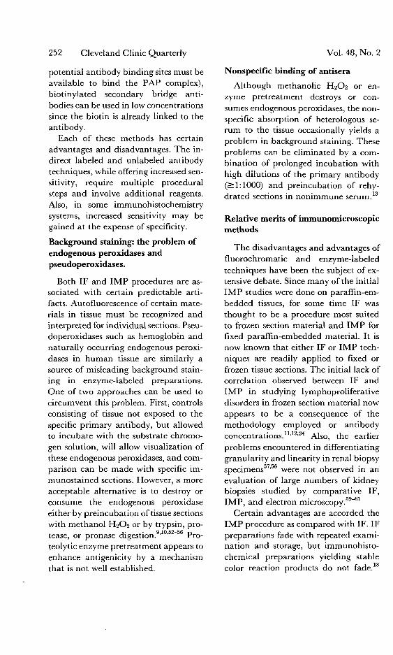
potential antibody binding sites must be available to bind the PAP complex), biotinylated secondary bridge anti-bodies can be used in low concentrations since the biotin is already linked to the antibody.
Each of these methods has certain advantages and disadvantages. The in-direct labeled and unlabeled antibody techniques, while offering increased sen-sitivity, require multiple procedural steps and involve additional reagents. Also, in some immunohistochemistry systems, increased sensitivity may be gained at the expense of specificity.
Background staining: the problem of endogenous peroxidases and pseudoperoxidases.
Both IF and I M P procedures are as-sociated with certain predictable arti-facts. Autofluorescence of certain mate-rials in tissue must be recognized and interpreted for individual sections. Pseu-doperoxidases such as hemoglobin and naturally occurring endogenous peroxi-dases in human tissue are similarly a source of misleading background stain-ing in enzyme-labeled preparations. One of two approaches can be used to circumvent this problem. First, controls consisting of tissue not exposed to the specific primary antibody, but allowed to incubate with the substrate chromo-gen solution, will allow visualization of these endogenous peroxidases, and com-parison can be made with specific im-munostained sections. However, a more acceptable alternative is to destroy or consume the endogenous peroxidase either by preincubation of tissue sections with methanol H2O2 or by trypsin, pro-tease, or pronase digestion.9,10'52"56 Pro-teolytic enzyme pretreatment appears to enhance antigenicity by a mechanism that is not Well established.
Nonspecific binding of antisera
Although methanolic H2O2 or en-zyme pretreatment destroys or con-sumes endogenous peroxidases, the non-specific absorption of heterologous se-rum to the tissue occasionally yields a problem in background staining. These problems can be eliminated by a com-bination of prolonged incubation with high dilutions of the primary antibody (>1:1000) and preincubation of rehy-drated sections in nonimmune serum.13
Relative merits of immunomicroscopic methods
The disadvantages and advantages of fluorochromatic and enzyme-labeled techniques have been the subject of ex-tensive debate. Since many of the initial I M P studies were done on paraffin-em-bedded tissues, for some time IF was thought to be a procedure most suited to frozen section material and I M P for fixed paraffin-embedded material. It is now known that either IF or I M P tech-niques are readily applied to fixed or frozen tissue sections. The initial lack of correlation observed between IF and IMP in studying lymphoproliferative disorders in frozen section material now appears to be a consequence of the methodology employed or antibody concentrations.11,12,24 Also, the earlier problems encountered in differentiating granularity and linearity in renal biopsy specimens ' were not observed in an evaluation of large numbers of kidney biopsies studied by comparative IF, IMP, and electron microscopy.59-61
Certain advantages are accorded the I M P procedure as compared with IF. IF preparations fade with repeated exami-nation and storage, but immunohisto-chemical preparations yielding stable color reaction products do not fade.

Summer 1981
Also, it is not usually possible to visual-ize well simultaneously the imrauno-stained antigen and adjacent tissue mor-phology in IF preparations. Conversely, I M P preparations are readily adaptable to a variety of counterstains enabling the observer to (1) more precisely locate the tissue antigen, and (2) evaluate such additional parameters as inflammatory response to the antigen. An IF micro-scope is necessary for examination of the IF preparations, and photographic doc-umentation is necessary.
Previously, valid disadvantages were also accorded I M P procedures. These included the potential carcinogenic na-ture of the chromogens used with IMP, and the lack of reliable commercial re-agents. Both of these objections are no longer valid, since diaminobenzidine (3,3', 4,4'-Tetraaminodiphenylether • 4 HC1 or 4,4'-oxybis-o-phenylenedia-mine) may not be a carcinogen31 and at least one chromogen, HYR, is now available, which has no currently iden-tified health hazard and yields excellent results. Several manufacturers currently distribute antibody-enzyme conjugates of excellent quality. Objections relating to the more complex nature of I M P procedures are no longer valid, since the direct technique using enzyme conju-gates can be used for most studies that employ immunohistochemistry, i.e., renal diseases and lymphoproliferative disorders. The additional time required to develop the substrate chromogen re-action product is no longer than the additional time required for photogra-phy and cataloging of photographic slides for IF.
It is ideal to have the capability of doing both IF and I M P procedures. This allows the pathologist versatility in the selection of the appropriate proce-dure.
Enzyme immunohistochemistry 253
It seems that the resistance to change from immunofluorescence to immunope-roxidase techniques for the routine ex-amination of renal biopsy material cannot be explained in scientific terms but de-pends largely on emotional ties to a system which has been established for a consid-erable number of years.60
Quality assurance
Quality control of both IF and IMP reagents is an essential and often ne-glected part of fluorescent and enzyme immunohistochemistry. Commercially available antibody should not be as-sumed to be monospecific or of adequate immunoreactivity. The specificity and sensitivity of each reagent purchased should be evaluated when received. Each laboratory should have a protocol for evaluating all new antibodies enter-ing the laboratory. The antisera should be dated when received, and evaluated by Ochterlony immunodiffusion, Im-munoelectrophoresis, competitive bind-ing radioimmunoassay or immunohis-tochemistry with the use of preabsorp-tion and postabsorption with antigen control tissues that have been well char-acterized with respect to the appropriate antigen. Many commercial antisera have package inserts that attest to the reliability of the reagents. However, there may be significant interlot varia-tion and the reagent immunoreactivity may be altered by environmental factors during shipping. Individual techniques should be performed regularly to assure continued competence by technical per-sonnel and adequate performance of re-agents. A detailed record should be kept of all quality assurance tests and docu-mentation of corrective actions taken.
Even when excellent standardization and characterization of antibody have been completed, rigorous in-run con-trols are necessary for valid interpreta-
254 Cleveland Clinic Quarterly Vol. 48, No. 1
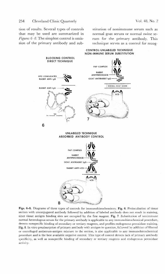
tion of results. Several types of controls that may be used are summarized in Figures 6-8. T h e simplest control is omis-sion of the primary antibody and sub-
stitution of non immune serum such as normal goat serum or normal swine se-rum for the pr imary antibody. This technique serves as a control for recog-
BLOCKING CONTROL DIRECT TECHNIQUE
HPO CONJUGATED
RABBIT ANTI- IgG
RABBIT ANTI- IgG -
CONTROL-UNLABELED TECHNIQUE NON- IMMUNE SERUM SUBSTITUTION
PAP COMPLEX
RABBIT ANTIPEROXIDASE
UNLABELED TECHNIQUE ABSORBED ANTIBODY CONTROL
GOAT ANTIRABBIT IgG
RABBIT A N T I - h C G — - / J
Figs. 6 - 8 . Diagrams of three types of controls for immunohistochemistry. Fig. 6. Preincubation of tissue section with unconjugated antibody followed by addition of labeled antibody does not result in staining, since tissue antigen binding sites are occupied by the first reagent. Fig. 7. Substitution of nonimmune normal heterologous serum for the primary antibody is applicable to any immunohistochemical procedure, detects nonspecific binding of secondary or tertiary reagents, and profiles endogenous peroxidase staining. Fig. 8. In vitro preabsorption of primary antibody with antigen in question, followed by addition of filtered or centrifuged antiserum-antigen mixture to the section, is also applicable to any immunohistochemical procedure and is the best available negative control. This type of control detects lack of primary antibody specificity, as well as nonspecific binding of secondary or tertiary reagents and endogenous peroxidase activity.

Summer 1981 Enzyme immunohistochemistry 255
nition of binding of secondary and ter-tiary antibodies, to the tissue and for endogenous peroxidase in indirect and PAP methods, but cannot adequately assess monospecificity of the primary antibody. The direct I M P procedure can be controlled by preincubation with unlabeled antibody, preferably from the same antisera lots from which the con-jugate was prepared. This type of con-trol effectively blocks the labeled con-jugate from reaching the antigen and although satisfactory for the direct pro-cedure, cannot be applied to indirect or unlabeled modifications of the tech-nique. The most reliable control for all IMP methods is an absorbed antibody control, in which the primary antibody is preincubated with exogenous antigen, thus binding all the available antibody-reacting sites. When the supernatant from the centrifuged mixture of bound antibody-antigen is applied to the tissue section, antibody is not available for the reaction and immunostaining does not occur.
Preabsorption may be necessary to remove nonspecific reactants or reac-tions with related antigens in the tissue. Not uncommonly, secondary anti-bodies, e.g., goat anti-rabbit IgG, or rab-bit anti-goat IgG, will cross react with human immunoglobulins. In such in-stances, absorption with purified human gamma globulin followed by centrifu-gation of the antibody is necessary to assure specificity of the secondary anti-body. For every procedure done on a day-to-day basis and for each tissue an-alyzed, an in-run control should be in-cluded for adequate verification of pos-itive or negative results. Use of affinity purified antibodies may help insure specificity.
Finally, when a new commercial an-tibody is purchased or antibody is made available from other sources, checker-
board titrations with varying combina-tions of antibody dilutions can be used on control tissue sections to evaluate the optimal dilutions of each reagent.
Diagnostic applications
Immunohistochemical procedures have contributed greatly to the under-standing of normal physiology and functional organization of many animal and human biologic systems. This paper will be restricted to reviewing the clini-cal diagnostic applications of I M P methods (Figs. 9-16).
Renal immunohistochemistry
The value of immunomicroscopy in delineating deposition of immunoglob-ulin and complement components or autoantibody in glomeruli of patients with various autoimmune diseases is well established. Once a tool of aca-demic interest, immunomicroscopy is now an essential diagnostic method that must be applied to every renal biopsy specimen. Patterns of IF have been shown to be highly reproducible and predictive histopathologically.62
Comparative studies of IMP and IF of glomerulonephritis were initially fa-vorable.8 Subsequently, studies of kid-ney biopsy specimens with the use of the direct technique and enzyme conjugates were associated with unacceptable back-ground staining, and in some cases a distinction between linear and granular color reaction product was difficult.57,58
Both IF and direct IMP procedures were compared to the unlabeled I M P tech-nique, and the specificity and sensitivity of the unlabeled PAP procedure were demonstrated to be comparable to those of IF.58 However, the length of the pro-cedure and the expense of additional reagents make use of the PAP procedure for frozen renal tissue a poor choice.
In the past few years, improved tech-
256 Cleveland Clinic Quarter ly
• • K M K M i ' V
I Vol. 48, No. 2
f . r
v *
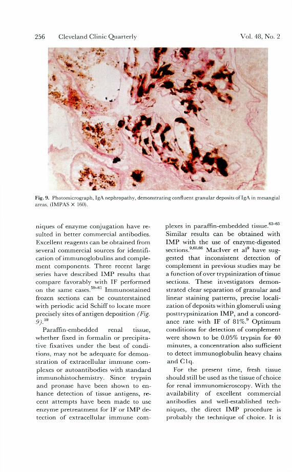
Fig. 9. Photomicrograph, IgA nephropathy, demonstrat ing confluent granular deposits of IgA in mesangial areas, ( IMPAS X 160).
niques of enzyme conjugation have re-sulted in better commercial antibodies. Excellent reagents can be obtained from several commercial sources for identifi-cation of immunoglobulins and comple-ment components. Three recent large series have described I M P results that compare favorably with IF performed on the same cases.59-61 Immunosta ined frozen sections can be counterstained with periodic acid Schiff to locate more precisely sites of antigen deposition (Fig. 9)™
Paraff in-embedded renal tissue, whether fixed in formalin or precipita-tive fixatives under the best of condi-tions, may not be adequate for demon-stration of extracellular immune com-plexes or autoantibodies with s tandard immunohistochemistry. Since trypsin and pronase have been shown to en-hance detection of tissue antigens, re-cent a t tempts have been made to use enzyme pretreatment for IF or I M P de-tection of extracellular immune com-
plexes in paraff in-embedded tissue.63-65
Similar results can be obtained with I M P with the use of enzyme-digested sections.9 '6 ' '66 Maclver et al9 have sug-gested that inconsistent detection of complement in previous studies may be a function of over trypsinization of tissue sections. These investigators demon-strated clear separation of granular and linear staining patterns, precise locali-zation of deposits within glomeruli using posttrypsinization I M P , and a concord-ance rate with IF of 81%. O p t i m u m conditions for detection of complement were shown to be 0.05% trypsin for 40 minutes, a concentration also sufficient to detect immunoglobul in heavy chains and C l q .
For the present time, fresh tissue should still be used as the tissue of choice for renal immunomicroscopy. With the availability of excellent commercial antibodies and well-established tech-niques, the direct I M P procedure is probably the technique of choice. It is

Summer 1981 Enzyme immunohistochemistry 257
Fig. 10. Photomicrograph, B-cell immunoblast ic sarcoma arising in plasmacytoid lymphocytic lymphoma, lymph node. T h e neoplastic cells (brown-black cytoplasm) are immunostained for kappa light chains. A serial section was negative for l ambda light chains. Direct immunoperoxidase technique, counterstained with hematoxylin and eosin, (X 400). Fig. 11. Photomicrograph, beta cell pancreatic apudoma. T h e neoplasm and adjacent normal islets contain immunoreactive insulin. Unlabeled PAP technique was used employing aminoethylcarbozole as the chromogen, (X 64).
not clear at this t ime whether trypsin-ized deparaffinized paraff in-embedded tissue will be acceptable as an immu-nomicroscopic preparat ion for most
forms of glomerular disease, since in the hands of some investigators, enzyme pre t rea tment yields variable tissue digestion and inconsistent immuno-
258 Cleveland Clinic Quarter ly Vol. 48, No. 1
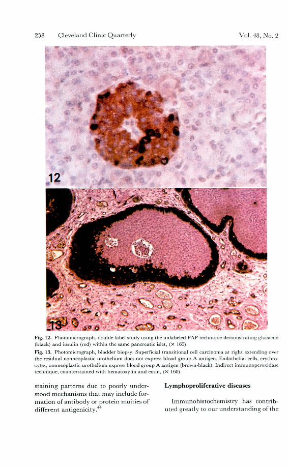
Fig. 12. Photomicrograph, double label study using the unlabeled PAP technique demonstra t ing glucacon (black) and insulin (red) within the same pancreatic islet, (X 160).
Fig. 13. Photomicrograph, bladder biopsy. Superficial transitional cell carcinoma at right extending over the residual nonneoplastic urothelium does not express blood group A antigen. Endothelial cells, erythro-cytes, nonneoplastic urothelium express blood group A antigen (brown-black). Indirect immunoperoxidase technique, counterstained with hematoxylin and eosin, (X 160).
staining patterns due to poorly under- Lymphoprol i fe ra t ive diseases stood mechanisms tha t may include for-mat ion of ant ibody or protein moities of Immunohistochemistry has contrib-different antigenicity.44 uted greatly to our unders tanding of the

Summer 1981
organization of the immune system and to architectural and functional altera-tions in its various components in a variety of disease states. T h e technology of immunohistochemistry has evolved parallel with increased knowledge about lymphoid neoplasia and has contributed significantly to the understanding of the nature of proliferating cells of malignant lymphoma. Immunohistochemistry of malignant lymphomas will eventually play a role similar to that of immuno-microscopy in evaluating renal disease.
Paradoxically, these techniques have
Enzyme immunohistochemistry 259
contr ibuted both to unders tanding and confusion regarding this group of enti-ties. Thus, while documenting the pres-ence of monoclonal cytoplasmic immu-noglobulins in many non-Hodgkin's lymphomas,14 '67"72 these techniques as applied to paraff in-embedded tissues have yielded polyclonal staining of B-cell lymphomas in some reports.14,68
Such observations are not in agreement with the clonal premise upon which most cancer immunology is based.23'73"70
Studies emphasizing the immuno-logic basis for classification of lympho-
S P L E E N I E M O V E D
HYPER1MMUN IZED MICE
I M M U N O B L A S T S IN C E L L S U S P E N S I O N FROM S P L E E N OF HYPER IMMUN IZEO MICE
M O U S E PL A S M A S Y TOM A E STABL I SHED C E L L CULTURE
S U S P E N S I O N OF CULTURED MOUSE M Y E L O M A C E L L S
P O L Y E T H Y L E N E GLYCOL
M O S T C E L L S DIE
A F E W F U S E D C E L L S S U R V I V E 4
S E L E C T I V E CULTURE MED IA
F U S E D C E L L S U B C U L T U R E S
E S T A B L I S H E D HYBR IDOMA C E L L L INES PRODUCING U L T R A S P E C I F I C M O N O C L O N A L A N T I B O D I E S
MONOCLONAL Ab. MONOCLONAL Ac, MONOCLONAL AD,
Fig. 14. Diagram of procedure used to obtain monoclonal hybridoma antibodies. Immunoblasts from hyperimmunized mice are fused with cultured mouse plasmacytoma cells in the presence of polyethylene glycol. Although most of the cells die, a few cells survive which contain the genetic content of both the stimulated immunoblasts and mouse myeloma cells. T h e fused cells are subcultured and cloned, reinjected into mouse peritoneal cavity, and ultraspecific monoclonal antibody harvested as ascitic fluid.

260 Cleveland Clinic Quar te r ly Vol. 48, No. 1
Fig. 15. Photomicrograph, reactive lymphoid hyperplasia, lingual tonsil. Red staining interfollicular T helper / inducer lymphocytes are identified using aminoethylcarbazole as the chromogen. Biotin-avidin " A B C " technique using mouse monoclonal hybridoma primary ant ibody specific for inducer /helper T lymphocytes, with methylene blue counterstain, (X 64). Fig. 16. Photomicrograph, malignant mixed germinal neoplasm of testis. Neoplastic syncytia trophoblasts are immunostaincd for chorionic gonadotropin. Indirect immunoperoxidase technique using mouse mon-oclonal hybridoma primary antibodies specific for beta subunit of human chorionic gonadotropin, peroxidase conjugated affinity purifed goat anti-mouse IgG, and hematoxylin and eosin counterstained, (X 160).

Summer 1981 Enzyme immunohistochemistry 261
mas at tempt to draw parallels between the components of the immune system and the morphologic diversity of non-Hodgkin's lymphomas. Of the several different classifications available for subtyping of non-Hodgkin's lymphoma, only the Lukes-Collins classification,69
which is currently available is directly dependent upon the identification of T-and B-cell marker expression by the neo-plasm. However, immunologic data can be added to the Rappaport morphologic classification.70
Initial functional characterization of lymphomas was done principally by cell suspension (CS) studies.75 With the use of CS techniques, classification as to T, B, or non-T/non-B origin can be ef-fected in a relatively large number of cases.73,74 In recent years, information regarding the reliability of CS studies has accumulated. In a number of B-cell lymphomas marking monoclonal with cryostat frozen section immunohisto-chemistry, polyclonality in CS has been observed.76"78 This apparent discrep-ancy may be due to several factors, most likely selective loss of tumor cells or sampling error resulting in contamina-tion of the suspension with nonneoplas-tic lymphocytes, particularly in nodular lymphomas in which a significant per-centage of the lymphoid parenchyma may be spared by the neoplasm.
When the sensitive PAP immunohis-tochemical technique developed by Sternberger was initially applied to lym-phoproliferative disorders, the use of im-munologic markers to characterize non-Hodgkin's lymphomas was viewed as an academic curiosity rather than a clini-cally useful tool by many pathologists. Recent evidence suggests that when non-Hodgkin's lymphomas are ap-proached from the standpoint of Lukes-Collins classification and interpreted in conjunction with surface marker analy-
sis, data of distinct prognostic signifi-cance are obtained for subsets of lym-phomas.79 However, immunohisto-chemical study of paraffin tissue from patients with multiple myeloma has shown phenotypic expression of both kappa and lambda light chains in cells from patients with well-characterized circulating monoclonal immunoglobu-lins.68 Thus, the initial enthusiasm for immunohistochemistry was tempered by these apparent anomalous staining patterns that violated basic concepts re-garding monoclonality of B-cell neopla-
• 8 0 sia. A similar evolution of understanding
of immunohistochemistry as applied to non-Hodgkin's lymphomas has been ob-served.81"104 The spurious immunostain-ing patterns observed in a significant number of cases evaluated by paraffin-e m b e d d e d immunoh i s tochemis t ry raised serious questions about the valid-ity of results obtained in this man-ner.11'14,105 Initially, it was suggested that such results were a consequence of di-clonal immunoglobulin production by the non-Hodgkin's lymphoma,14 an ex-planation that is not in concurrence with the overwhelming body of evidence for monoclonality in human B-cell lym-phomas.23,106"108
When results of frozen section and paraffin-embedded immunohistochem-istry are compared, it becomes clear that the negative or spurious immunostain-ing patterns associated with paraffin-embedded tissue are probably a conse-quence of processing.11'12,24,28 Currently, cryostat frozen section immunohisto-chemistry (CFSIH) provides the most sensitive and specific procedure for de-tection of monoclonal cell populations, since most non-Hodgkins lymphomas demonstrate monoclonal immunostain-ing with CFSIH.11,15'23,25 However par-affin-embedded techniques provide su-
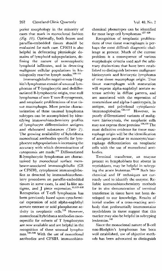
262 Cleveland Clinic Quarterly Vol. 48, No. 1
perior morphology in the minority of cases that mark in monoclonal fashion (Fig. 10). Optimally, both frozen and paraffin-embedded tissues should be evaluated for each case. CFSIH is also helpful in delineating physiologic do-mains of lymphoid subpopulations, de-fining the nature of nonneoplastic lymphoid infiltrates, and in detecting malignant cellular populations in his-tologically reactive lymph nodes.109"111
Immunoglobulin negative non-Hodg-kin's lymphomas consist of unusual lym-phomas of T-lymphocytic and dediffer-entiated B-lymphocytic origin, true null lymphomas of non-T non-B cytogenesis, and neoplastic proliferations of true tis-sue macrophages. More precise charac-terization of these unusual lymphoma subtypes can be accomplished by iden-tifying immunohistochemistry profiles of lymphocyte differentiation antigens and elaborated substances (Table 2). The growing availability of hybridoma monoclonal antibodies specific for lym-phocyte subpopulations is increasing the accuracy with which determinations of cytogenesis are made.112 Differentiated B-lymphocytic lymphomas are charac-terized by monoclonal surface mem-brane-associated immunoglobulin (CS or CFSIH), cytoplasmic immunoglobu-lins as detected by immunohistochem-istry procedures on paraffin-embedded tissues in some cases, la and la-like an-tigens, and J piece expression.11,113"118
Recognition of T-cell lymphomas has been previously based upon cytochemi-cal expression of acid alpha-naphthyl acetate esterase or acid phosphatase ac-tivity in neoplastic cells.119 However, monoclonal hybridoma antibodies mon-ospecific for subsets of T lymphocytes are now available and are helpful in the recognition of these unusual lympho-mas.120"126 With the use of monoclonal antibodies and CFSIH, immunohisto-
chemical phenotypes can be identified for most large cell lymphomas.127"129
Recognition of neoplastic prolifera-tions of true tissue macrophages is per-haps the most difficult diagnostic chal-lenge at present. Much of the current problem is a consequence of various morphologic criteria used and the arbi-trary distinctions that have been estab-lished to distinguish between malignant histiocytosis and histiocytic lymphoma of true tissue macrophage origin. True tissue macrophages with maturation will express alpha-naphthyl acetate es-terase activity in diffuse pattern, and are associated with the expression of muramidase and alpha-1-antitrypsin, la antigen, and polyclonal cytoplasmic immunoglobulins.130"137 However, in poorly differentiated variants of malig-nant histiocytosis, the neoplastic cells may not express these proteins.134,135 The most definitive evidence for tissue mac-rophage origin will be the identification of antigens peculiar to monocy te/mac-rophage differentiation on neoplastic cells with the use of monoclonal anti-bodies.
Terminal transferase, an enzyme present in lymphoblasts but absent in myeloblasts, may be helpful in subtyp-ing the acute leukemas.138,139 Both bio-chemical and IF techniques are cur-rently used to identify the enzyme. Re-liable immunohistochemistry methods for in situ demonstration of terminal transferase in tissue have not been de-veloped to our knowledge. Results of initial studies of a cross-reacting anti-body that preferentially immunostains myeloblasts in tissue suggest that this marker may also be helpful in subtyping leukemias.14
Since the monoclonal nature of B-cell non-Hodgkin's lymphomas has been well established, use of objective meth-ods has been advocated to distinguish

Summer 1981 Enzyme immunohistochemistry 263
Table 2. Immunohistology of lymphoproliferative disorders OKT3 OKTS OKT4
Ig J P Ia Pan Sup Ind
Reactive hyperplasia Follicles
Light zone PC + + O C - O C Dark zone PC + + -
Sinus PC, E - + Paracortex O C , D C - O C • • •
Non-Hodgkin 's lymphomas B cell lymphomas M C + V - - -
With Fc receptors E + V - - -
T cell lymphomas - - V + + * + *
With Fc receptors E - V + + * + * Dedifferentiated B cell
Lymphomas - - + - - -
With Fc receptors E - + - - -
Hodgkin 's disease Plasma cells PC + - - - -
Reactive tissue macrophages PC - + - - -
Neoplastic cells PC - N W E N W E N W E N W E
Abbreviations: Ig = surface a n d / o r cytoplasmic immunoglobulins (best assessed with cryostat frozen section immunohistochemistry). J P = J piece, O K I = monoclonal ant ibody specific for l a antigen, O K T 3 . Pan = monoclonal ant ibody specific for all peripheral blood T lymphocytes, O K T 8 . Sup = monoclonal ant ibody specific for suppressor/cytotoxic T lymphocytes, O K T 4 . I N D = monoclonal ant ibody specific for inducer /he lper T lymphocytes, PC = polyclonal (both k and A light chains present), + = present, — = absent, O C = occasional cells, DC = dendrit ic cells, N W E = not well established, V = variable, M C = monoclonal (only one light chain present, either k or À), Fc = receptors for Fc portion of immunoglobulin) , E = surface-associated immunoglobul in which can be eluted with acidic buffer . * T cell mal ignant lymphoproliferations react with either T Supp. or T inducer /helper monoclonal ant ibody, depending on differentiation of the neoplasm.
reactive lymphoid hyperplasias from non-Hodgkin's lymphomas by charac-terizing surface immunoglobulin phe-notypes. " Since CS may yield spu-rious results in non-Hodgkin's lympho-mas, possibly due to contamination with nonneoplastic populations or selective loss of tumor cells, CSFIH should be used to help determine the biologic po-tential of the lymphoproliferative dis-orders.141"144
The origin of Reed-Sternberg cells in Hodgkin's disease has been the subject of extensive debate. For years the pref-erential involvement of the lymph node sinus and interfollicular zone and de-fects in cellular immunity were inter-preted as evidence for T-cell or tissue
macrophage origin. Immunohistochem-istry studies by Taylor14 and others145" 148 showed that Reed-Sternberg cells contain polyclonal IgG. These observa-tions are consistent with a tissue mac-rophage origin, the cytoplasmic immu-noglobulin probably representing en-gulfed exogenous polyclonal immuno-globulin. Tissue culture cell lines de-rived from Hodgkin's disease display cytochemical and immunologic features of macrophages, and when transplanted into experimental animals produce tu-mors with morphology resembling Hodgkin's disease.149 Furthermore, CS from tissues involved by Hodgkin's dis-ease contain Reed-Sternberg cells that have polyclonal cytoplasmic IgG, and

264 Cleveland Clinic Quarterly
actively bind and internalize labeled ex-ogenous immunoglobulins.150 Although a tissue macrophage origin of Hodgkin's disease appears likely, definitive evi-dence could be obtained with immu-nohistochemistry staining with the use of lymphocyte-monocyte differentiation monoclonal antibodies. To our knowl-edge, such a study has not been done.
Endocrine systems
I M P procedures have contributed greatly to our understanding of the C-cell neoplasms and preneoplastic state of the thyroid gland. Wolfe et al151 have delineated the distribution of C cells in the normal gland and in the thyroid gland of patients at risk for hereditary medullary carcinoma. When evaluated in this fashion, it has been shown that the middle and upper portions of the lateral thyroid lobes show marked in-creases and clustering of calcitonin-con-taining cells in this disease. The immu-nohistochemical demonstration of thy-roglobulin within well-differentiated tu-mors of the thyroid gland of both pap-illary and follicular types has been shown to be helpful in confirmation of thyroidal origin, since tumors of nonthy-roidal histogenesis examined did not show thyroglobulin synthesis.152'153
Immunohistochemistry studies have been helpful in evaluation of both nor-mal and neoplastic pituitary tissue. It has been shown that adenomas of the adenohypophysis, although they may be tinctorially homogeneous, are immuno-histochemically heterogeneous.154' Im-munohistochemistry studies are helpful in delineating the presence of neoplastic cells containing the hormone circulating in the patient. Thus, the documentation of prolactin in resection tissue from the anterior pituitary of a patient with hy-perprolactinemia as assessed by ra-dioimmunoassay is a helpful confirma-
Vol. 48, No. 1
tory study.155'156 Study of the pituitary tissue of acromegalic patients has docu-mented the presence of growth hormone within the neoplastic tissue, an obser-vation corroborating radioimmunoas-say results.157 However, Fukaya et al157
have also demonstrated occasional im-munoreactive cells positive for prolactin and luteinizing hormone in an adenoma removed from a patient with acromeg-aly, raising questions about the signifi-cance of identifying other hormones within a particular tumor. Such immu-noreactive cells may represent residual normal pituitary tissue. Conversely, these observations may suggest that pi-tuitary adenomas are polyclonal neo-plastic proliferations with secretion of one hormone dominating the clinical presentation. Recent cases of pituitary adenomas with ultrastructural and im-munohistochemical evidence for heter-ogeneous cell populations have been de-scribed that were eosinophilic in tincta-torial differentiation but heterogeneous in their elaboration of growth hormone and prolactin.158 '159 It has been sug-gested that at least some of these de-scribed cases may involve technical problems and may not actually repre-sent a stem-cell neoplasm.160 Immuno-histochemistry may prove to be espe-cially helpful in the confirmation of hor-monal homogeneity in small specimens of tissue removed as microadenomas from patients with Cushing's syndrome. Currently, this confirmation is based upon standard histochemical and clini-cal correlations.161 Study of pituitary adenomas occurring in association with M E N I syndrome has confirmed that most of the adenomas are of either pro-
162 163 T
lactin or growth hormone type. ' In the rate occurrence of pituitary carci-noma metastatic to extracranial sites, immunohistochemistry techniques may offer confirmatory evidence for pituitary

Summer 1981
origin of the neoplasm.164 Of greater interest is the immunoreactivity demon-strable in chromophobe adenomas of the adenohypophysis. These tumors are usually characterized by multiple hor-monally positive cells for most of the hormones endogenous to the anterior p i tu i tary . 1 6 5 Immunohis tochemis t ry techniques may also prove helpful in the characterization of "hypoplasias" or preadenomatous states.166 Other appli-cations of immunohistochemistry in the study of pituitary disease include the delineation of decreased hormonal syn-thesis or storage in gonadotrophs in pa-tients with hemochromatosis.167 Confir-mation of elaboration of adenohypo-physeal hormones by ovarian teratomas, oat cell carcinomas, and other extrapi-tuitary tumors may also be done with the use of immunohistochemistry tech-
168-171 niques.
The use of immunohistochemistry in evaluation of the endocrine pancreas has yielded some interesting results (Figs. 11, 12). In experimental animals, the distribution and relative frequency of different immunoreactive cell popu-lations have been documented with I M P and IF.172 Human pancreatic en-docrine tumors are characterized by dis-tinct ultrastructural features that allow subclassification of pancreatic apudo-mas.173 Immunohistochemistry tech-niques have been used to identify the distribution of insulin-positive cells in hyperinsulinemic hypoglycemia of in-fancy,174 to document the presence of hormones such as glandular kallikrein a n d cho lecys tok in in -pancreozymin within islet-cell populations175 '176 and to profile immunohistochemically the hor-monal content of islet-cell tumors177
(Fig. 11). Kurman et al178 180 have used immu-
nohistochemistry to localize elaborated steroid molecules in tumors of the ovary
Enzyme immunohistochemistry 265
and the testis. With these techniques, both testosterone and estradiol were identified in Sertoli-Leydig cells and in primitive spindled cells in these tu-mors.169"172 Estradiol was localized in granulosa cells and in luteinized theca cells, and nonluteinized stromal cells were negative for steroids.178"181 Immu-nohistochemistry techniques may thus prove helpful in the subclassification of ovarian tumors based upon the predom-inant hormone elaborated, and also in the confirmation of gonadal stromal or-igin when such neoplasms occur in an extragonadal location.180'181
When carcinoid tumors are evaluated with immunohistochemistry techniques, positive immunoreactivity for multiple hormones is usually identified.182 How-ever, somatostatin immunoactivity or-dinarily predominates and is usually as-sociated with immunostaining for other hormones such as gastrin or calcitonin.182
Most of the tumors studied in this fashion have been clinically silent with respect to hormonal elaboration.
The emergence of immunohistochem-istry techniques specific for prostatic acid phosphatase have proved to be very helpful in the confirmation of prostatic origin of secondary metastasis.183"187
Nadji et al188 have also recently shown that an antibody can be raised against specific tumor antigens of prostatic car-cinoma rather than the elaborated acid phosphatase product. The antibody thus derived is specific only for carci-noma of prostate histogenesis.
Oncodevelopmental antigens
Oncodevelopmental antigens are a group of substances that are produced early in fetal life, but which disappear with fetal maturation. These substances may reappear in the bloodstream in association with a malignant neoplasm apparently through derepression of the

266 Cleveland Clinic Quarterly Vol. 48, No. 1
genes responsible for production of these markers. It has been shown that the sensitive PAP technique is satisfactory to demonstrate the presence of human chorionic gonadotropin (HCG) in pla-cental syncytial trophoblast and neo-plastic trophoblastic elements.189"191 Ra-dioimmunoassay of beta subunit H C G is helpful in monitoring patients with gestational trophoblastic and gonadal germinal neoplasia.192"196 Ideally, RIA baseline follow-up measurements are used in conjunction with immunohisto-chemical study of initially resected tu-mor tissue. This approach permits a precise morphologic and immunohisto-chemistry characterization of the tumor for definitive subtyping, and suggests which serum markers will be most useful for therapeutic monitoring.197"200 The amount of choriocarcinomatous differ-entiation can be best assessed with im-munohistochemistry for H C G and the degree of endodermal sinus and em-bryonal differentiation best assessed with a-fetoprotein immunohistochemis-try. These techniques may also be help-ful in confirming germinal nature of neoplasms occurring in an extragonadal location such as the intracranial vault.201'202 However, these markers are by no means specific for germinal neo-plasia since they may occur in a variety of nongonadal neoplasm.203"211 Al-though perhaps of limited usefulness in confirming a germinal origin for a par-ticular neoplasm, monitoring of markers
may provide an indication of therapeu-212
tic success. Similar results have been observed for
carcinoembryonic antigen (CEA). The immunohistochemistry method works well for the detection of CEA in tissue.213 However, the elaboration of this oncodevelopmental antigen by a variety of neoplasms makes diagnostic usefulness limited. CEA expression has
been suggested as a useful diagnostic tool in the assessment of lung tumors, since mesotheliomas have been shown with immunofluorescence not to express CEA, whereas bronchogenic carcinomas are associated with CEA elaboration.214
Peeripheral serologic measurements of CEA may be useful in monitoring re-sponse to therapy in patients with breast, stomach, and colorectal can-cer.215-221 A distinction between benign colonic mucosa and dysplastic or frankly carcinomatous changes within the bowel mucosal tissue of patients with ulcerative colitis is theoretically possible with CEA immunostaining, but staining patterns are inconsistent.222'223 It has also been shown that IMP techniques are able to identify certain CEA-positive cervical carcinomas before ovarian CEA concentration is elevated.224 Van Nagell et al225 have shown the usefulness of immunohistochemistry identification of CEA expression by ovarian cystadeno-carcinomas and follow-up serologic measurements to assess therapeutic suc-cess. The presence of CEA seems to correlate best with mucinous rather than serous differentiation of ovarian neoplasms.226-228
An antibody reacting with gp52, a 52,000-dalton glycoprotein of the mouse mammary tumor virus has been shown to immunostain selectively breast carci-noma cells, not reacting with the normal breast tissue or other malignancies. Autoantibodies having specificity for the same virus have been shown to occur in sera of breast cancer patients.230
Blood group antigens
The expression of ABO blood group antigens by different human tissues has been recognized for many years. Normal urothelium, for example, expresses ABO antigen in agreement with the pheno-type of the patient's red blood cells. In

Summer 1981
most urothelial neoplasia, the capacity to express the ABO blood group antigen is lost as the neoplasm becomes more aggressive.231-234 In some superficial transitional cell carcinomas of the blad-der, the natural history of the disease is not characterized by aggressive biologic potential, and thus it would be useful to have a means to identify this particular group of patients who require less ag-gressive therapeutic measures.232 With the mixed red cell agglutination test, as assessed by passive red blood cell im-munoadherence, it is possible to identify neoplasms that retain the ability to ex-press blood group antigens on the tumor cells. The expressin of ABO antigen on neoplastic urothelium seems to indicate a less aggressive biologic potential, i.e., the tumor does not become invasive.231-
234 Passive red cell immunoadherence has also been used to identify secretion of fetal blood group antigens on polyps of the distal colon, and the loss of ABO isoantigen expression in histologically benign lesions and in mammary carci-noma.235'236 ABO antigens in tissue can also be detected by indirect immuno-histochemistry (Fig. 13).
Infectious agents
Immunohistochemistry techniques are readily adaptable to the detection of hepatitis-B surface antigen in fixed and frozen tissue.237"241 These techniques can be used to confirm the presence of sur-face and core hepatitis-B antigen in he-patic cirrhoses or hepatocellular carci-nomas, although the relationship be-tween the presence of the antigen and the pathogenesis of the disease remains problematic.241-244 Recently, it has been shown that hepatitis surface antigen has an affinity for free and bound horserad-ish peroxidase, an observation that man-dates strict use. of controls in the proce-dure.245 With IF, immunohistochemis-
Enzyme immunohistochemistry 267
try techniques have been used to iden-tify the causative agent of non-A, non-B hepatitis in tissue that may also prove useful in IMP studies.246
Antisera specific for certain parasites, polyoma viruses, herpes virus, and var-icella/zoster virus have been effective in the retrospective identification of infec-tious agents in tissue with the use of IF or I M P procedures.247"253'
Other antigens
Other antigens detected in tissue with immunohistochemistry include the dis-tribution of laminin,254 ligandin,255 and Factor VIII coagulation factor.256 Gli-ofibrillary acidic protein can be used to characterize glial cell populations, and other intracranial mass lesions.267"260
However, an inverse relationship be-tween the degree of anaplasia and the intensity of immunostaining with anti-GFA antibody has been observed; thus, in poorly differentiated neoplasms, the technique may have limited use-fulness.258 Neoplastic Paneth cells have been identified with lysozyme antisera in an unusual variant of gastric carci-noma,261 the presence of actin has been documented in meyloepithelial cells in the breast and other tissues of smooth muscle cell origin,262'263 fetal red cells identified in placental intervillous thrombi,264 myoglobin in normal and neoplastic human skeletal muscle,265 ker-atin in a variety of normal human tis-sues,266'267 and basement membrane an-tigen has been demonstrated in Wilm's tumor.268
Immunohistochemistry techniques have also been shown to be useful in serologic studies to detect antinuclear factor in serum,269 cell surface antigens in CS,270 thymus leukemia antigen expression on lymphoid cells,271 and an-tilymphocyte antibodies.272

268 Cleveland Clinic Quarterly
The future of enzyme immunohistochemistry
The future of diagnostic and investi-gational immunohistochemistry will be influenced by several things. The role of this specialized technique in the diag-nostic surgical pathology laboratory will increase substantially as the techniques become more widely accepted and as more systems for application are devel-oped. However, as the techniques be-come more widely used, the necessity for good controls and standardization of reagents and techniques become even
» » 45,273 more important. The federal government will un-
doubtedly play some role in the practi-cal daily use of immunohistochemistry in clinical laboratories. Classification of benzidine as a carcinogen and subse-quent strict regulations governing its use have had considerable bearing on the use of this reagent in cytochemistry. As outlined earlier in this paper, alternative chromogens are available. However, many chromogens currently in use in the United States, other than benzidine and fluorescent compounds have not been fully investigated with regard to their neoplastogenic potential. A frame-work for decision-making by the federal government regarding potential human carcinogens has been recently out-lined.274
The immunohistochemistry detection of specific tumor antigens associated with one particular type of tumor offers great promise. Such antigens have been identified in association with ovarian carcinomas, melanoma, breast as evaluated by an antigen cross-reacting with mouse mammary tumor-related antigens, human cervical squamous cell carcinoma, and mesothelioma.278"279
The role of external photoscanning with radiolabeled antibody tracers will
Vol. 48, No. 1
continue to grow. Immunohistochemis-try may be helpful in the initial immu-nologic characterization of such tumors before noninvasive scanning proce-dures.280'281
Prolactin receptors have been profiled with the use of IMP methodol-
282 283
ogy, ' and the potential exists to de-velop peroxidase-labeled histochemical procedures for the detection of a variety of receptors including estrogen, proges-terone, and testosterone.
Perhaps the most exciting develop-ment in immunohistochemistry relates to hybridoma technology. Since Kohler and Milstein's284,285 reports of the suc-cessful fusion of specific antibody pro-ducing cells in culture with tumor cell lines, the applications of this particular biotechnology have increased greatly.286
Hybridomas are produced by the fusion of a myeloma tumor cell line main-tained in tissue culture with cell lines derived from splenic immunoblasts re-moved from hyperimmunized animals (Fig. 14). Selective tissue cultures yield cloned hybridoma cell lines that elabo-rate monoclonal ultraspecific antibody. As long as the tissue cell lines can be maintained in culture, production of a standardized antibody is guaranteed. The fused cells may be inserted into mouse peritoneum where functioning hybridoma plasmacytomas grow, and ascitic fluid containing the monoclonal antibody can subsequently be har-vested. Monclonal antibodies have been made with specificity for lymphocyte differentiation antigens (Fig. 15), tu-mor specific antigens, «-fetoprotein, CEA, beta subunit of human chorionic gonadotropin (Fig. 16), and many other substances,276"279 and have con-tributed greatly to understanding of lymphocyte maturation and immuno-pathology of neoplasia.287"305

Summer 1981 Enzyme immunohistochemistry 269
Summary
Immunohistochemical procedures have contributed greatly to our under-standing of disease processes and have become a necessary tool in the evalua-tion of many disease states. Initial de-tection systems utilized IF markers. En-zyme immunohistochemical techniques developed during the past decade have circumvented many of the problems in-herent in IF procedures.
This paper outlines the technical aspects and clinical diagnostic applica-tions of enzyme-labeled immunohisto-chemistry. The availability of mono-clonal antibodies and the adaptation of these reagents to immunohistochemistry systems will contribute greatly to further understanding of disease processes and will have continued utility in clinical diagnosis.
References 1. Coons AH, Creech HJ , Jones RN. Immu-
nological properties of an antibody contain-ing a fluorescent group. Proc Soc Exp Biol Med 1941; 47: 200-2.
2. Coons AH, Kaplan M H . Localization of antigens in tissue cells. II. Improvements in a method for the detection of antigen by means of fluorescent antibody. J Exp Med 1950; 91: 1-30.
3. Nakane PK, Pierce GB, J r . Enzyme-labeled antibodies: preparat ion and application for the localization of antigens. J Histochem Cytochem 1966; 14: 929-31.
4. Sternberger LA. Immunocytochemistry. 2nd ed. New York: J o h n Wiley & Sons, 1978.
5. Wisdom GB. Enzyme immunoassay. Clin Chem 1976; 22: 1243-55.
6. Suffin SC, Muck KB, Young J C , Lewin K, Porter DD. Improvement of the glucose ox-idase immunoenzyme technic. Use of a tet-razolium whose formazan is stable without heavy metal chelation. Am J Clin Pathol 1979; 71: 492-6.
7. Boorsma D M , Streefkerk J G . Periodate or glutaraldehyde for preparing peroxidase conjugates? J Immunol Methods 1979; 30: 245-55.
8. M u r p h y W M , Deodhar SD, Cawley LP. Use of horseradish peroxidase in identification of serum antibodies and immune complexes. Clin Chem 1973; 19: 1370-3.
9. Maclver AG, Giddings J , Mepham BL. Demonstration of extracellular immunopro-teins in formalin-fixed renal biopsy speci-mens. Kidney Int 1979; 16: 632-6.
10. H u a n g SN, Minassian H, More J D . Appli-cation of immunofluorescent staining on par-affin sections improved by trypsin digestion. Lab Invest 1976; 35: 383-90.
11. Warnke R, Pederson M, Williams C, Levy R. A study of lymphoproliferative diseases comparing immunofluorescence with im-munohistochemistry Am J Clin Pathol 1978; 70: 867-75.
12. Warnke R. Alteration of immunoglobulin-bearing lymphoma cells by fixation. J His-tochem Cytochem 1979; 27: 1195-6.
13. Taylor CR. Immunoperoxidase techniques. Practical and theoretical aspects. Arch Pa-thol Lab Med 1978; 102: 113-21.
14. Taylor CR. An immunohistological study of follicular lymphoma, reticulum cell sarcoma, and Hodgkin's disease. Eur J Cancer 1976; 12: 61-75.
15. Tubbs R R , Sheibani K, Weiss RA, Sebek BA. Frozen section immunohistochemistry of malignant lymphomas using the direct im-munoperoxidase procedure (abstract). Lab Invest 1980; 42: 156.
16. Burns J , Hambridge M, Taylor CR. Intra-cellular immunoglobulins: A comparat ive study on three s tandard tissue processing methods using horseradish peroxidase and fluorochrome conjugates. J Clin Pathol 1974; 27: 548-57.
17. Arnold W, Kalden J R , VonMayersback H. Influence of different histologic preparat ion methods on preservation of tissue antigens in the immunofluorescent ant ibody technique. Ann NY Acad Sci 1975; 254: 27-34.
18. Miller H R P . Fixation and tissue preserva-tion for antibody studies: A review. Histo-chem J 1972; 4: 305-20.
19. Fel tkamp V T M . Preparation of tissues and cells for immunohistochemical processing. Ann NY Acad Sci 1975; 254: 21-6.
20. Davenport WD, Ball CR . Observation on the results of specific histochemical tech-niques and empirical staining methods on several tissue/organ types using a variety of fixing fluids. Histopathology 1979; 3: 321-7.
21. Taylor CR. Immunohistological observa-tions upon the development of reticulum cell

270 Cleveland Clinic Quarterly Vol. 48, No. 1
sarcoma in the mouse. J Pathol 1976; 118: 201-19.
22. Elias J M , Chandor S, Miller F. Fixative effects on tissue antigenicity. Abstract pre-sented at the spring meet ing of T h e Histo-chemical Society, New Orleans, J u n e 1980.
23. Levy R, Warlike R , Dor fman RF, Haimov-ich J . T h e monoclonali ty of h u m a n B-cell lymphomas. J Exp Med 1977; 145: 1014-28.
24. T u b b s R R , Sheibani K, Sebek BA. Immu-nohistochemistry versus immunofluores-cence for non-Hodgkin 's lymphoma. Am J Clin Pathol 1980; 73: 144-5.
25. T u b b s R R , Sheibani K, Weiss BA, Sebek BA. Immunohistochemistry of fresh frozen lymphoid tissue using the direct immuno-peroxidase technique. Am J Clin Pathol 1981; 75: 172-4.
26. Culling CF, Reid PE, and Sinnott NM. T h e effect of various fixatives and trypsin diges-tion upon the staining of routine parafFm-embedded sections by the peroxidase-anti-peroxidase and immunofluorescent tech-nique. J Histochem 1980; 3: 10-9.
27. Straus W. Factors affecting the cytochemical reaction of peroxidase with benzidine and stability of the blue reaction product . J His-tochem Cytochem 1964; 12 :462-9.
28. Mesulam M M . T h e blue reaction product in horseradish peroxidase neurohistochemistry: Incubation parameters and visibility. J His-tochem Cytochem 1976; 24: 1273-80.
29. Mesulam M M , Rosene DL. Differential sen-sitivity between blue and brown reaction products for H R P neurohistochemistry. Neuroscience Letters 1977; 5: 7-14.
30. Occupat ional Safety and Heal th Adminis-tration Standards. Part 1910-1010. Tit le 29. Code of Federal Regulations, Federal Reg-ister, 1979: 3825-45.
31. Griswold DP, Casey AE, Weisburger EK, Weisburger J H . T h e carcinogenicity of mul-tiple intragastric doses of aromatic hetero-cyclic nitro or amino derivatives in young female Sprague-Dawley rats. Cancer Res 1968; 28: 924-33.
32. Mesulam M M , Rosene DL. Sensitivity in horseradish peroxidase neurohistochemistry: A comparat ive and quant i ta t ive study of nine methods. J Histochem Cytochem 1979; 27: 763-6.
33. Lechago J , Sun NJ, Weinstein W M . Immu-noperoxidase—Immunofluorescence combi-nation for the simultaneous detection of two different antigens in the same tissue section (abstract). Lab Invest 1979; 40: 268.
34. Sternberger LA, Joseph SA. T h e unlabeled ant ibody method. Contrast ing color staining of paired pi tui tary hormones without anti-body removal. J Histochem Cytochem 1979; 27: 1424-9.
35. Joseph SA, Sternberger LA. T h e unlabeled ant ibody method. Contrast ing color staining of /^-lipoprotein and ACTH-associated hy-pothalamic peptides without ant ibody re-moval. J Histochem Cytochem 1979; 27: 1430-7.
36. Lewin KJ , Suffin SC, Porter DD, Muck KB, Young J C . T h e glucose oxidase immunoen-zyme technique (abstract). Lab Invest 1979; 40: 269.
37. Banks PM. Diagnostic applications of an immunoperoxidase method in hematopa-thology. J Histochem Cytochem 1979; 27: 1192-4.
38. T u b b s R R , Velasco M, Benjamin SP. Im-munocytochemical identification of human chorionic gonadotropin. Arch Pathol Lab M e d 1979; 103: 534-6.
39. National Cancer Insti tute Carcinogenesis Technical report series. Bioassay of 3-amino-9-ethylcarbazole hydrochloride for possible carcinogenicity. 1978; No. 93: 1-180.
40. Hanker JS , Yates PE, Metz CB, Rustioni A. A new specific sensitive and non-carcino-genic reagent for the demonstrat ion of horse-radish peroxidase. Histochem J 1977; 9: 789-92.
41. T u b b s R R , Sheibani K. Chromogens for immunohistochemistry (letter). J Histochem Cytochem 1981; 29: 684.
42. Reiner A, Gamin P. On noncarcinogenic chromogens for horseradish peroxidase his-tochemistry. J Histochem Cytochem 1980; 28: 187-8.
43. Sonlag J M . Carcinogenicity of substi tuted benzenediamines (phenylenediamines) in rats and mice. J N C I 1981; 66: 591-602.
44. Heyderman E. Immunoperoxidase tech-nique in histopathology—applications, methods, and controls. J Clin Pathol 1979; 32: 971-8.
45. Petrusz P, DiMeo P, Ord ronneau P, Weaver C, Keefer DA. Improved immunoglobul in-enzyme bridge method for light microscopic demonstrat ion of hormone-containing cells of the rat adenohypophysis. Histochemistry 1975; 46: 9 -26.
46. Vronvall G, Frommel D. Definition of staph-ylococcal protein A reactivity for h u m a n immunoglobul in G fragments. Immuno-chemistry 1970; 7: 124.

Summer 1981
47. Dubois-Dalq M, McFarland H, McFarlin D. Protein-A peroxidase: A valuable tool for the localization of antigens. J Histochem Cyto-chem 1977; 25: 1201-06.
48. Celio M R , Lutz H, Binz H, Fey H. Protein A in immunoperoxidase techniques. J His-tochem Cytochem 1979; 27: 691-3.
49. Notani GW, Parsons JA, Erlandsen SL. Ver-satility of staphylococcus aureus protein A in immunocytochemistry. Use in unlabeled an-tibody enzyme system and fluorescent meth-ods. J Histochem 1974; 27: 1438-44.
50. Guesdon J L , Ternynck T, and Avrameas S. The use of avidin-biotin interaction in im-munoenzymatic techniques. J Histochem Cytochem 1979; 27: 1131-9.
51. Hsu SM, Raine L, Fanger H. A comparat ive study of the PAP method and avidin-biotin-complex method for studying polypeptide hormones with radioimmunoassay anti-bodies. Am J Clin Pathol 1981; 75: 734-8.
52. Fink B, Loepfe E, Wyler R. Demonstration of viral antigen in cryostat sections by a new immunoperoxidase procedure eliminating endogenous peroxidase activity. J Histochem Cytochem 1979; 27: 1299-1301.
53. Straus W. Inhibition of peroxidase by meth-anol and by methanol-nitroferricyanide for use in immunoperoxidase procedures. J His-tochem Cytochem 1971; 19: 682-8.
54. Weir EE, Pretlow TG, Pitts A, Williams EE. Destruction of endogenous peroxidase in or-der to locate cellular antigens by peroxidase labeled antibodies (letter). J Histochem Cy-tochem 1974; 22: 51-4.
55. Afroudakis AP, Liew CT, Peters RL. An immunoperoxidase technic for the demon-stration of the hepatitis B surface antigen in h u m a n livers. Am J Clin Pathol 1976; 65: 533-9.
56. Denk H , Syre G, Weirich E. Immunomor-phologic methods in routine pathology. Ap-plication of immunofluorescence and the un-labeled antibody-enzyme (peroxidase-anti-peroxidase) technique to formalin fixed par-affin embeded kidney biopsies. Beitr Pathol 1977; 160: 187-94.
57. Davey FR, Busch GJ. Immunohistochemis-try of glomerulonephritis using horseradish peroxidase and fluorescein-labeled antibody: A comparison of two technics. Am J Clin Pathol 1970; 53: 531-6.
58. Elias J M , Miller F. A comparison of the unlabeled enzyme method with immuno-fluorescence for the evaluation of human immunologic renal disease. Am J Clin Pathol
Enzyme immunohistochemistry 271
1975; 64: 464-71. 59. Tubbs R R , Gephardt G, Valenzuela R,
Deodhar SD. An approach to immunomi-croscopy of renal disease with immunoperox-idase and periodic-acid-Schiff counterstain ( IMPAS stain). Am J Clin Pathol 1980; 73: 240-4.
60. Turner DR, Wilson D M , Lake A, Heaton J M , Leibowitz S, Cameron JS. An evalua-tion of the immunoperoxidase technique in renal biopsy diagnosis. Clin Nephrol 1979; 11: 13-7.
61. Sheibani K, Tubbs R R , Gephardt GN, M c M a h o n J T , Valenzuela R. Comparison of alternative chromogens for renal immu-nohistochemistry. H u m a n Pathol 1981; 12: 349-54.
62. Valenzuela R, Deodhar S. Atlas on interpre-tation of immunomicroscopic pat tern in renal and skin diseases. Am Soc Clin Pathol. In press.
63. Qua lman SJ, Keren DF. Immunofluores-cence of deparaffinized trypsin-treated renal tissues. Preservation of antigens as an ad-junct to diagnosis of disease. Lab Invest 1979; 41: 483-9.
64. Choi YJ, Reiner L. Immunofluorescence of renal lesions in paraff in-embedded and fresh-frozen sections. Am J Clin Pathol 1980; 73: 116-9.
65. Cur ran RC, Gregory J . T h e unmasking of antigens in paraffin sections of tissue by tryp-sin. Experientia 1977; 33: 1400-1.
66. Turner DR, Wilson D, Cameron JS . Perox-idase-labelled IgG and complement in plas-tic embedded human renal tissue, in First International Symposium on Immunoenzy-matic Techniques, INSERM Symposium No. 2 Feldmann G, Druet P, Bignon J , Avrameas S. eds., Amsterdam, North-Holland Publish-ing Company, 1976: 105-8.
67. Taylor CR, Russell R, Chandor S. An im-munohistologic study or multiple myeloma and related conditions using an immuno-peroxidase method. Am J Clin Pathol 1978; 70: 612-22.
68. Taylor CR, Burns J . T h e demonstration of plasma cells and other immunoglobulin-con-taining cells in formalin-fixed, paraffin-em-bedded tissues using peroxidase labelled an-tibody. J Clin Pathol 1974; 27: 14-20.
69. Lukes RJ . T h e immunologic approach to the pathology of malignant lymphomas. Am J Clin Pathol 1979; 72: 657-69.
70. M a n n RB, Ja f fe ES, Berard CW. Malignant lymphomas—A conceptual understanding

272 Cleveland Clinic Quarterly Vol. 48, No. 1
of morphologic diversity. Am J Pathol 1979; 94: 105-92.
71. Taylor CR. T h e nature of Reed-Sternberg cells and other malignant reticulum cells. Lancet 1974; 2: 802-7.
72. Pinkus GS, Said J . Specific identification of intracellular immunoglobulin in paraff in sections of multiple myeloma and macro-globulinemia using an immunoperoxidase technique. Am J Pathol 1977; 87: 47-58.
73. Aisenberg AC, Wilkes BM, Long J C , Harris NL. Cell surface phenotype in lymphoprolif-erative disease. Am J Med 1980; 68: 206-13.
74. Pinkus GS, Said J W . Characterization of non-Hodgkin's lymphomas using multiple cell markers. Immunologic, morphologic and cytochemical studies of 72 cases. Am J Pathol 1978; 94: 349-80.
75. Green I, Ja f fe ES, Shevach EM, Edelson RL, Frank M M , Berard CW. Determination of origin of malignant reticular cells by the use of surface membrane markers: T h e Reticu-loendothelial system—IAP monograph, No. 16, Rebuck J W and Berard CW, eds. Balti-more: Williams and Wilkins, 1974.
76. Gajl-Peczalski KJ, Kersey J H , BloomfieldC, Frizzera G. T h e value of combined CS and tissue frozen section studies in surface marker evaluation of non-Hodgkin's malignant lym-phomas (abstract). Lab Invest 1979; 40: 254.
77. Harris NL, Poppema S. Detection of surface immunoglobulin in malignant lymphomas with the immunoperoxidase technique (ab-stract). Lab Invest 1981; 44: 2 7A.
78. Tubbs R R , Sheibani K , Weiss R, Sebek BA, Deodhar SD. Tissue immunomicroscopic evaluation of monoclonality of B cell lym-phomas. Comparison with cell suspension studies. Am J Clin Pathol. In Press.
79. Bloomfield CD, Gajl-Peczalska KJ , Frizzera G, Kersey J H , Goldman AI. Clinical utility of lymphocyte surface markers combined with the Lukes-Collins histologic classifica-tion in adult lymphoma. N Engl J Med 1979; 301: 512-18.
80. Mukai K, Rosai J . Application of Immuno-peroxidase Techniques in Surgical Pathol-ogy. Progr Surg Pathol Vol. I, Fenoglio CM, Wolff M, eds. New York: Masson Publishing, 1980.
81. Papadimitr iou CS, Muller-Hermelik U., Lennert K. Histologic and immunohisto-chemical findings in the differential diagno-sis of chronic lymphocytic leukemia of B-cell type and lymphoplasmacytic/ lymphoplas-macytoid lymphoma. Virchows Arch (Pa-thol Anat) 1979; 384: 149-58.
82. Johansen P, Jensen M K . Enzymecytochem-istry and immunohistochemistry in mono-clonal gammopa thy and reactive plasmacy-tosis. Acta Pa th Microbiol Scand 1980; 88: 377-82.
83. Taylor CR. Immunohistological approach to tumor diagnosis. Oncology 1978; 35: 189-97.
84. Mori N, Masafumi ABE, Koj ima M. Study of malignant lymphomas from the aspect of immunoglobulin production. Acta Pathol J p n 1979; 29: 705-22.
85. Halliday D, Davey FR, Marucci AA. Detec-tion of intracellular immunoglobulin in nod-ular lymphomas. Am J Clin Pathol 1978; 69: 587-93.
86. Morgan T W , Banks PM. Large cell neopla-sia: An evaluation of criteria for the distinc-tion of lymphoid from epithelial malignan-cies (abstract). Lab Invest 1979; 40: 273.
87. Li CY, Harrison EG. Histochemical and im-munohistochemical study of diffuse large-cell lymphomas. Am J Clin Pathol 1978; 70: 721-32.
88. Sun NC, Fishkin BG, Nies K M , Glassy EF, Carpentier C. Lymphoplasmacytic mye-loma. An immunological, immunohisto-chemical and electron microscopic study. Cancer 1979; 43: 2268-78.
89. Scott FE, Dupont PA, Webb J W . Plasma-cytoma of the stomach. Diagnosis with the aid of the immunoperoxidase technique. Cancer 1978; 41: 675-81.
90. Mancil la R, Davis GL. Nonsecretory multi-ple myeloma. Immunohistologic and ultra-structural observations on two patients. Am J Med 1977; 63: 1015-22.
91. R a m b a u d J C , Modigliani R, Nguyen Phuoc BK, et al. Non-secretory alpha-chain disease in intestinal lymphoma (letter). N Engl J Med 1980; 303: 53.
92. Levitt LJ, Dawson D M , Rosenthal DS, Mo-loney WC. CNS involvement in the non-Hodgkin's lymphomas. Cancer 1980; 45: 545-52.
93. Clausen PP, Jacobson M, Johansen P, Thommesen N. Immunohistochemical dem-onstration of intracellular immunoglobulin in formalin fixed, paraffin embedded sec-tions, as staining method in diagnostic work. Acta Pathol Microbiol Scand (C), 1979; 87: 308-12.
94. Woda BA, Knowles DM. Nodular lympho-cytic lymphoma eventuating into diffuse his-tiocytic lymphoma. Immunoperoxidase de-tection of monoclonality. Cancer 1979; 43: 303-7.
95. Vernon S, Voet RL, Naeim F, Waisman J .
Summer 1981
Nodular lymphoma with intracellular im-munoglobulin. Cancer 1979; 44: 1273-9.
96. Haghighi P, Tabei Z, Kharazmi A, Gerami 5, Abadi P, Haghsenass M. Immunoperoxi-dase study in a-chain disease. Arch Pathol Lab Med 1978; 102: 555-7.
97. Knowles D M , Shevchuk M. Pleomorphic reticulum cell sarcoma, monoclonal gam-mopathy and amyloidosis. An immuno-peroxidase study. Cancer 1978; 41: 1883-9.
98. Al-Saleem T, Al-Qadiry W, Issa F, King J . T h e immunoselection technic in laboratory diagnosis of alpha heavy-chain disease (let-ter). Am J Clin Pathol 1979; 72: 132-3.
99. Morris JA, Bird CC. Ultrastructural and immunohistological study of immunoblast ic sarcoma developing in child with immuno-blastic lymphadenopathy. Cancer 1979; 44: 171-82.
100. Isaacson P. Middle east lymphoma and al-pha-chain disease. An immunohistochemical study. Am J Surg Pathol 1979; 13: 431-41.
101. Isaacson P, Wright D H , J u d d MA, Jones DB, Payne SV. T h e na ture of the i m m u n o -globulin-containing cells in malignant lym-phoma: An immunoperoxidase study. J His-tochem Cytochem 1980; 28: 761-70.
102. Pangalis GA, Nathwani BN, Rappapor t H . Detection of cytoplasmic immunoglobulin in well-differentiated lymphoproliferative dis-eases by the immunoperoxidase method. Cancer 1980; 45: 1334-9.
103. van den Tweel J G , Taylor C R , Parker J W , Lukes RJ . Immunoglobulin inclusions in non-Hodgkin's lymphomas. Am J Clin Pa-thol 1978; 69: 306-13.
104. Taylor CR. Immunohistologic studies of lymphomas past, present and future. J His-tochem Cytochem 1980; 28: 777-87.
105. Call ihan T R , Braylan RC, Farnham R, J a f f e ES, Soban EJ, Berard CW. Correlation be-tween immunohistochemistry and cell sur-face markers in diffuse large cell (histiocytic) lymphomas (abstract). Lab Invest 1979; 40: 244.
106. Nisonoff A, Hopper J E , Spring SB. T h e ant ibody molecule. New York: Academic Press, Inc., 1975: 542.
107. Warnke R , Levy R. Immunopathology of follicular lymphomas. A model of/3- lympho-cyte homing. N Engl J Med 1978; 298: 481-6.
108. Warnke R, Levy R, Pederson M, Dorfman RF. Tissue section immunofluorescence in the investigation of lymphoproliferative dis-eases (abstract). Lab Invest 1979; 40: 291.
109. Stein H, Bonk A, Tolksdorf G, Lennert K,
Enzyme immunohistochemistry 273
Rodt H, Gerdes J . Immunohistologic analy-sis of the organization of normal lymphoid tissue and non-Hodgkin's lymphomas. J His-tochem Cytochem 1980; 28: 746-60.
110. Tubbs R R , Sheibani K, Weiss RA, Lee V, Sebek BA, Valenzuela R. Immunohisto-chemistry of Warthin 's tumor. Am J Clin Pathol 1980; 74: 795-7.
111. Palutke M, Schnitzer B, Mirchandani I, Ta-baczka P, So K, Garrillo G. Monoclonal lymphoid populations in lymph nodes with reactive hyperplasia (abstract). Lab Invest 1981; 44: 60A.
112. Cooper MD. Immunologic analysis of lymphoid tumors. N Engl J Med 1980; 302: 964-5.
113. Knowles D M , Halper J P . Ia antigen expres-sion by human malignant lymphomas: Cor-relation with conventional lymphoid markers (abstract). Lab Invest 1980; 42: 129.
114. Billing R, Rafizadeh B, Drew I, H a r t m a n G, Gale R, Terasaki P. H u m a n /^-lymphocyte antigens expressed by lymphocytic and mye-locytic leukemia cells. J Exp Med 1976; 144: 167-78.
115. Yamanaka N, Ishii Y, Koshiba H , Mikuni C, Konno M, Kikuchi K. A study of surface markers in acute lymphocytic leukemia by using ant i -T and anti-B lymphocyte sera. Cancer 1978; 42: 2641-7.
116. Halper J P , Knowles D M , YiWang C. Ia antigen expression by h u m a n malignant lymphomas: Correlation with conventional lymphoid markers. Blood 1980; 55: 373-82.
117. Isaacson P. Immunochemical demonstration of J Chain: A marker of B-cell malignancy. J Clin Pathol 1979; 32: 802-7.
118. Mestecky J , Preud 'homme J L , Crago SS, Mihaesco E, Prchal J T , Okos AJ. Presence of J chain in h u m a n lymphoid cells. Clin Exper Immunol 1980; 39: 371-85.
119. Collins RD, Waldron JA , Glick AD. Results of mul t iparameter studies of T-cell lymphoid neoplasms. Am J Clin Pathol 1979; 72: 699-707.
120. Warnke R, Levy R. Detection of T and B cell antigens with hybridoma monoclonal antibodies: A biotin-avidin-horseradish per-oxidase method. J Histochem Cytochem 1980; 2& 771-6.
121. Reinherz EL, Moret ta L, Roper M, et al. H u m a n T-Lymphocyte subpopulations de-fined by Fc receptors and monoclonal anti-bodies: A comparison. J Exp Med 1980; 151: 969-74.
122. Reinherz EL, K u n g PC, Goldstein G, Schlossman SF. A monoclonal ant ibody with

274 Cleveland Clinic Quarterly Vol. 48, No. 1
selective reactivity with functionally mature human thymocytes and all peripheral hu-man T cells. J Immunol 1979; 123: 1312-7.
123. Kung PC, Goldstein G, Reinherz EL, Schlossman SF. Monoclonal antibodies de-fining distinctive h u m a n T-cell surface an-tigens. Science 1979; 206: 347-9.
124. Reinherz EL, Kung PC, Goldstein G, Schlossman SF. A monoclonal ant ibody re-active with the human cytotoxic/suppressor T-cell subset previously defined by a hetero-antiserum termed TH 2 . J Immunol 1980; 124: 1301-7.
125. Reinherz EL, Kung PC, Goldstein G, Schlossman SF. Separation of functional subsets of human T-cells by a monoclonal antibody. Proc Natl Acad Sei USA 1979; 76: 40612-5.
126. Royston I, M a j d a JA, Baird S, Meserve B, Griffiths J . Monoclonal antibody for human T-lymphocytes: Identification of normal and malignant T-cells. Blood 1979; 54(Suppl I): 106a.
127. Warnke R, Miller R, Grogan T, Pederson M, Dilley J , Levy R. Immunologic pheno-types in 30 patients with diffuse large cell lymphoma. N Engl J Med 1980; 303: 293-300.
128. Alsenberg AC, Wilkes BM. Unusual h u m a n lymphoma pheotype defined by monoclonal antibody. J Exp Med 1980; 152: 1126-31.
129. Tubbs R R , Weiss RA, Savage RA, Sebek BA, Weick J K . Determination of immuno-logic phenotypes of large cell lymphoma us-ing monoclonal antibodies (abstract). Lab Invest 1981; 44: 69A.
130. Tubbs R R , Sheibani K, Sebek BA, Savage RA. Malignant histiocytosis. Ultrastructural and immunocytochemical characterization. Arch Pathol Lab Med 1980; 104: 26-9.
131. Isaacson P, Wright DH. Intestinal lym-phoma associated with malabsorption. Lan-cet 1978; 1: 67-70.
132. Buchner SA, Rufli T. Malign histiozytose mit hautmanifestat ionen. Enzymzytochem-ische und immunzytologische. 1980; Nr. II, 14. Marz , 373-7.
133. Tubbs R R , Sheibani K, Savage RA, Sebek BA. Muramidase an immunohistochemical marker of malignant histiocytosis (letter). H u m a n Pathol 1979; 10: 483.
134. Meister P, Na th ra th W. Immunohistochem-ical markers of histiocytic tumors. H u m a n Pathol 1980; 11: 300-1.
135. Mendelsohn G, Eggleston J C , M a n n RB. Relationship of lysozyme (muramidase) to
histiocytic differentiation in malignant his-tiocytosis. An immunohistochemical study. Cancer 1980; 45: 273-9.
136. Meister P, H u h n D, Nathroth W. Malignant histiocytosis. Immunohistochemical charac-terization on paraffin embedded tissue. Vir-chows Arch (Pathol Anat) 1980; 385: 233-46.
137. Ree HJ , Song JY , Leone LA, Crowley J P , Fanger H. Occurrence and patterns of mur-amidase containing cells in Hodgkin's dis-ease, non-Hodgkin's lymphomas and reac-tive hyperplasia. H u m a n Pathol 1981; 12: 49-59.
138. Strass SA, Schumacher H R , Keneklis T P , Bollum FJ. Terminal deoxynucleotidyl transferase immunofluorescence of bone marrow smears. Am J Clin Pathol 1979; 72: 898-903.
139. Janossy G, Hof fbrand AV, Greaves MF, et al. Termina l transferase enzyme assay and immunological membrane markers in the diagnosis of leukemia: A mult iparameter analysis of 300 cases. Br J Haematol 1980; 44: 221-34.
140. Pattengale PK, Taylor CR, Engvall E, Ruos-lahti E. Direct tissue visualization of normal cross-reacting antigen in neoplastic granu-locytes. Am J Clin Pathol 1980; 73: 351-5.
141. Knowles D M , Jakobiec F. Orbital lymphoid neoplasms: A clinicopathologic study of 60 patients (abstract). Lab Invest 1980; 42: 129.
142. Brubaker DB, Whiteside TL. Differentiation between benign and malignant human lymph nodes by means of immunologic markers. Cancer 1979; 43: 1165-76.
143. Astarita R W , Minckler D, Taylor CR, et al. Orbital and adnexal lymphomas. Am J Clin Pathol 1980; 73:615-21.
144. Tubbs R R , Sheibani K, Weiss R, Sebek BA. Reactive lymphoid hyperplasia versus non-Hodgkin's lymphoma: Role of immunohis-tochemistry (abstract). Am J Clin Pathol 1980; 74: 501.
145. Poppema S, Elema J D , Halie M R . The sig-nificance of intracytoplasmic proteins in Reed-Sternberg cells. Cancer 1978; 42: 1793-1803.
146. Garvin AJ, Spicer SS, Parmley RT , Munster AM. Immunohistochemical demonstration of IgG in Reed-Sternberg and other cells in Hodgkin's disease. J Exp Med 1974; 139: 1077-83.
147. Anagnostou D, Parker J W , Taylor C R , et al. Lacunar cells of nodular sclerosing Hodg-kin's disease. An ultrastructural and immu-

Summer 1981
nobistologic study. Cance r 1977; 39: 1032-43.
148. Parmley R T , Spicer SS, M o r g a n SK, Grush O C . Hodgk in ' s disease a n d myelomonocyt ic leukemia. An ul t ras t ructura l a n d i m m u n o -cytochemical s tudy. Cance r 1976; 38: 1188-98.
149. K a p l a n H S , G a r t n e r S. S te rnberg-Reed giant cells of Hodgkin ' s disease: Cul t iva t ion in vitro, he te ro t ransplan ta t ion a n d charac-terization as neoplastic macrophages . Int J Cance r 1977; 19: 511-25.
150. K a d i n M E , Stites DP, Levy P, W a r n k e R . Exogenous immunoglobu l in a n d macro-phage origin of Reed-Sternberg cells in Hodgk in ' s disease. N Engl J M e d 1978; 299: 1208-14.
151. Wolfe H J , Melvin KE, Cervi-Skinner S. C cell hyperplas ia preceding medul la ry thyroid carc inoma. N Engl J M e d 1973; 289: 437-41.
152. Burt A, Goud ie RB. Diagnosis of p r i m a r y thyroid ca rc inoma by immunohis tological demons t ra t ion of thyroglobul in . His topa-thology 1979; 3: 279-86.
153. Bocker W , Dral le H , Husse lmann H , Bay V, Brassow M . Immunohis tochemica l analysis of thyroglobul in synthesis in thyroid carci-nomas . Virchows Arch (Pathol Anat ) 1980; 385: 187-200.
154. H a l m i NS, Duel lo T . Acidophil ic p i tu i ta ry tumors . A reappraisal wi th differential stain-ing a n d immunocytochemica l techniques. Arch Pa tho l L a b M e d 1976; 100: 346-51.
155. Ve ldhu i s J D , Green J E , Kovacs E, Worgu l T J , M u r r a y FT , H a m m o n d J M . Prolact in-secreting p i tu i tary adenomas . Association wi th mul t ip le endocr ine neoplasia, T y p e I. A m J M e d 1979; 67: 830-6.
156. C a r t e r J N , Tyson J E , Tolis G, VanVl ie t S, F a i m a n C, Friesen H G . Prolactin-secret ing tumors a n d hypogonadism in 22 men . N Engl J M e d 1978; 299: 847-52.
157. Fukaya T , K a g e y a m a N, K u w a y a m a A, et al. M o r p h o f u n c t i o n a l s tudy of p i tu i t a ry ad-enomas wi th acromegaly by immunoperox i -dase technique a n d electron microscopy. C a n c e r 1980; 45: 1598-1603.
158. H o r v a t h E, Kovacs K , Singer W , et al. Aci-dophi l s tem cell a d e n o m a of the h u m a n p i tu i t a ry (abstract) . L a b Invest 1980; 42: 122.
159. Duel lo T , H a l m i N. Acidophil s tem cell ad-enomas of the pi tui tary . Arch Pa tho l L a b M e d 1978; 102: 439.
160. H o r v a t h E, Kovacs K, Singer W , Ezrin C,
Enzyme immunohistochemistry 275
Kerenyi NA. Acidophil s tem cell adenoma . Arch Pathol L a b M e d 1977; 101: 594-9.
161. Tyrrel l J B , Brooks R M , Fitzgerald PA, Co-foid PB, Forsham P H . Cushing 's disease. Selective t rans-sphenoidal resection of pitui-tary microadenomas . N Engl J M e d 1978; 298: 753-8.
162. Levine J H , Sagel J , Rosebrock G, et al. Prolactin-secret ing a d e n o m a as par t of the mul t ip le endocr ine neoplas ia-Type I. (Men-I) syndrome. Cancer 1979; 43: 2492-6.
163. H o r v a t h E, Kovacs K. Histologic, immuno-cytologic a n d fine s t ructura l findings in pi-tu i ta ry adenomas associated wi th the mult i-ple endocr ine neoplasia syndrome (MENS) (abstract) . L a b Invest 1979; 40: 261.
164. M u k a i K, Wolseth DG, Bonner R A , O p p e n -heimer J H . Pi tu i ta ry ca rc inoma with liver metastasis: An immunohis tochemica l s tudy (abstract) . L a b Invest 1980; 42: 138.
165. Hassoun J , C h a r p i n C , J a q u e t P, et al. An-alogies immunocy tocehmi ques des adeno-mas hypophysaires de la malad ie de cushing et des adenomes "non fonct ionnels" (adeno-mas chromophobes) de l 'hypophyse. Annals d 'endocrinologie (Paris), 1979; 40: 559-60.
166. Lando t A M . Pi tu i ta ry adenomas . Clinico-morphologic correlations. J His tochem Cy-tochem 1979; 27: 1395-7.
167. Bergeron C, Kovacs K . P i tu i ta ry siderosis. A histologic, immunocytologic a n d ul t rastruc-tura l study. A m J Pa tho l 1978; 93: 295-310.
168. McKee l D W , Askin FB. Ectopic hypophy-seal ho rmona l cells in benign cystic t e r a t o m a of the ovary. Arch Pa tho l L a b M e d 1978; 102: 122-8.
169. Skrabanek P, Powell D. Un i fy ing concept of non-p i tu i ta ry ACTH-sec re t ing tumors . Evi-dence of c o m m o n origin of neural-crest tu-mors, carcinoids, a n d oat-cell carcinomas. Cance r 1978; 42: 1263-9.
170. Spark RF , Connol ly PB, Gluckin DS, W h i t e BA, Sacks B, Landsberg L. A C T H secretion f rom a func t ion ing pheochromocytoma. N Engl J M e d 1979; 301: 416-8 .
171. Cohle SD, Tschen J A , Smi th FE, et al. ACTH-secre t ing ca rc inoma of the breast. Cance r 1979; 43: 2370-6.
172. Fa lkmer S. Immunocy tochemica l studies of the evolution of islet hormones . J His tochem Cytochem 1979; 27: 1281-2.
173. Larsson LI. Endocr ine pancrea t ic tumors . H u m a n Pathol 1978; 9:401-16.
174. J a f f e R, H a s h i d a Y, Yunis EJ. Pancrea t ic pathology in hyper insul inemic hypoglyce-mia of infancy. L a b Invest 1980; 42: 356-66.

276 Cleveland Clinic Quarterly Vol. 48, No. 1
175. Grube D, Maier V, Raptis S, Schlegel W. Immunoreactivity of the endocrine pancreas. Evidence for the presence of cholecystokinin-pancreozymin within the A-cell. Histochem-istry 1978; 56: 13-35.
176. Ole-MoiYoi O , Pinkus GS, Spragg J , Austen KF. Identification of human glandular kal-likrein in the beta cell of the pancreas. N Engl .] Med 1979; 300: 1289-94.
177. Nieuwenhuijzen Kruseman AC, Knijnen-burg G, Erutel de la Riviere G, Bosman FT. Morphology and immunohistochemically-defined endocrine function of pancreatic islet cell tumors. Histopathology 1978; 2: 389-99.
178. K u r m a n RJ , Andrade D, Goebelsmann U , Taylor CR. An immunohistological study of steroid localization in Sertoli-Leydig tumors of the ovary and testis. Cancer 1978; 42: 1772-83.
179. K u r m a n RJ , Goebelsmann U, Taylor CR. Steroid localization in granulosa theca tu-mors of the ovary. Cancer 1979; 43: 2377-84.
180. Taylor CR, Kurman RJ , Warner NE. T h e potential value of immunohistologic tech-niques in the classification of ovarian and testicular tumors. H u m a n Pathol 1978; 9: 417-27.
181. Maurer R, Taylor C R , Schmucki O , Hed-inger CE. Extratesticular gonodal stromal tumor in the pelvis. A case report with im-munoperoxidase findings. Cancer 1980; 45: 985-90.
182. Dayal Y, O'Brian DS, Wolfe HJ , Reichlin S. Carcinoid tumors: A comparison of their immunocytochemical hormonal profile with morphologic and histochemical characteris-tics (abstract). Lab Invest 1980; 42: 111.
183. Leav I, Savage P, Rule A, De Lellis RA, Merk FB. Immunohistochemical detection of human prostatic acid phosphotase (ab-stract). Lab Invest 1979; 40: 267.
184. Jobsis Ac, DeVries GP, Anholt R R , Sanders GT. Demonstration of the prostatic origin of metastases. An immunohistochemical method for formalin-fixed embedded tissue. Cancer 1978; 41: 1788-93.
185. Nadji M, Tabei SZ, Castro A, Chu T M , Morales AR. Prostatic origin of tumors. An immunohistochemical study. Am J Clin Pa-thol 1980; 73: 735-9.
186. Skinner MS, Seckinger D. Evaluation of beta-subunit chorionic gonadotropin as an aid in diagnosis of trophoblastic disease. Ann Clin Lab Sei 1979; 9: 347-52.
187. Manley PN, Mahan DE, Bruce AW, Kipkie
GF, Franchi L. Postradiotherapy biopsy of prostatic adenocarcinoma: A light and im-munohistochemical study (abstract). Lab In-vest 1980; 42: 132.
188. Nadji M, Tabei SZ, Castro A, Ming Chu T, W a n g MC, Morales AR. Prostatic specific antigen. An immunohistologic marker for prostatic neoplasms. Cancer. In Press.
189. Yorde DE, Hussa R O , Garancis J C , Pattillo RA. Immunocytochemical localization of human choriogonadotropin in human malig-nant trophoblast. Model for human chorio-gonadotropin secretion. Lab Invest 1979; 40: 391-8.
190. Nieumenhuijzen-Kruseman AC, VanLent M, Blom AH, Lauw GP. Choriocarcinoma in mother and child, identified by immu-noenzyme histochemistry. Am J Clin Pathol 1977; 67: 679-83.
191. Sheibani K, Tubbs R R , Velasco ME, Ben-j amin S. Immunocytochemical identification of human chronic gonadotropin (hCG) (ab-stract). Lab Invest 1979; 40: 284A.
192. Von Eyben FE. Biochemical markers in ad-vanced testicular tumors. Serum lactate de-hydrogenase, urinary chorionic gonadotro-pin and total urinary estrogens. Cancer 1978; 41: 648-52.
193. Stepanas AV, Samaan NA, Schultz PN, Ho-loye PY. Endocrine studies in testicular tu-mor patients with and without gynecomas-tia. A report of 45 cases. Cancer 1978; 41: 369-76.
194. Schultz H, Sell A, Norgaard-Pedersen, Ar-ends J . Serum alpha-feto-protein and human chorionic gonadotropin as markers for the effect of postoperative radiation therapy a n d / o r chemotherapy in testicular cancer. Cancer 1978; 42: 2182-6.
195. J a v a d p o u r N, Mclnt i re K R , Waldmann TA. H u m a n chorionic gonadotropin (HCG) and alpha-fetoprotein (AFP) in sera and tumor cells of patients with testicular seminoma. A prospective study. Cancer 1978; 42: 2768-72.
196. Javadpour N. Serum and cellular biologic tumor markers in patients with urologic can-cer. H u m a n Pathol 1979; 10: 557-68.
197. Javadpour N. Significance of elevated serum alpohafetoprotein (AFP) in seminoma. Can-cer 1980; 45: 2166-8.
198. K u r m a n RJ , Scardino PT, Mclnt i re K R , Waldmann TA, J avadpour N. Cellular lo-calization of alpha-fetoprotein and human chorionic gonadotropin in germ cell tumors of the testis using an indirect immunoperox-

Summer 1981
idase technique. A new approach to classifi-cation utilizing tumor markers. Cancer 1977; 40: 2136-51.
199. J avadpour N, Mclnt i re KR, Waldmann TA, Bergman SM. T h e role of alpha-fetoprotein and human chorionic gonadotropin in sem-inoma. J Urol 1978; 120: 687-90.
200. Talerman A, Haije WG, Baggerman L. Se-rum alphafetoprotein (AFP) in diagnosis and management of endodermal sinus (yolk sac) tumor and mixed germ cell tumor of the ovary. Cancer 1978; 41: 272-8.
201. Stachura I, Mendelow H. Endodermal sinus tumor originating in the region of the pineal gland. Ultrastructural and immunohisto-chemical study. Cancer 1980; 45: 2131-7.
202. Norgaard-Pedersen B, Lindholm J , Al-brechtsen R, Arends J , Diemer N H , Riishedo J . Alpha-fetoprotein and human chorionic gonadotropin in a patient with a pr imary intracranial germ cell tumor. Cancer 1978; 41: 2315-20.
203. Borkowski A, M u q u a r d t C. H u m a n cho-rionic gonadotropin in the plasma of normal, nonpregnant subjects. N Engl J Med 1979; 301: 298-302.
204. Gerber MA, T h u n g SN, Euzenir S. Emer-gence of heterogeneous cell populations in hepatocellular carcinoma (abstract). Lab In-vest 1979; 40: 256.
205. Odell W D , Wolfsen AR. Hormones from tumors: Are they ubiquitous? Am J Med 1980; 68: 317-8.
206. Franchimont P, Zangerle PF. Present and future clinical relevance of tumour markers. Eur J Cancer 1977; 13: 637-46.
207. Rutanen EM, Seppala M. The HCG-beta subunit radioimmunoassay in nontropho-blastic gynecologic tumors. Cancer 1978; 41: 692-6.
208. Hattor i M, Fukase M, Yoshimi H, Matsu-kura S, Imura H. Ectopic production of hu-man chorionic gonadotropin in malignant tumors. Cancer 1978; 42: 2328-33.
209. Bellet D, Arrang J M , Contesso G, Caillaud J M , Bohuon C. Localization of the /¡-subunit of human chorionic gonadotrophin on var-ious tumors. Eur J Cancer 1980; 16: 433-9.
210. Bender RA, Wein t raub BD, Rosen SW. Pro-spective evaluation of two tumor-associated proteins in pancreatic adenocarcinoma. Cancer 1979; 43: 591-5.
211. Hat tor i M, Imura H, Matsukuba, S, et al. Mult iple-hormone producing lung carci-noma. Cancer 1979; 43: 2429-37.
212. Woo KB, Waalkes T P , Ahmann DL, Tor-
Enzyme immunohistochemistry 277
mey DC, Gehrke CW, Oliverio V T . A quan-titative approach to determining disease re-sponse during therapy using multiple bio-logic markers. Application to carcinoma of the breast. Cancer 1978; 41: 1685-1703.
213. Primus FJ, W a n g R H , Sharkey R M , Gol-denberg DM. Detection of carcinoembryonic antigen in tissue sections by immunoperoxi-dase. J Immunol Methods 1975; 8: 267-76.
214. Wang NS, H u a n g SN, Gold P. Absence of carcinoembryonic antigen-like material in mesothelioma. An immunohistochemical differentiation from other lung cancers. Can-cer 1979; 44: 937-43.
215. Falkson H C , Van der Wat t J J , Portugal MA, Falkson G. Carcinoembryonic antigen in patients with breast cancer. An adjunctive tool to monitor response and therapy. Can-cer 1978; 42: 1308-13.
216. Wahren B, Lidbrink E, Wallgren A, Eneroth P, Jaj icek J . Carcinoembryonic antigen and other tumor markers in tissue and serum or plasma of patients with pr imary mammary carcinoma. Cancer 1978; 42: 1870-8.
217. Ellis DJ, Speirs C, Kingston RD, Brookes VS, Leonard J , Dykes PW. Carcinoem-bryonic antigen levels in advanced gastric carcinoma. Cancer 1978; 42: 623-5.
218. Ejeckan GC, H u a n g SN, McCaughey W T , Gold P. Immunohistopathological study on carcinoembryonic antigen (CEA)-like mate-rial and immunoglobulin A in gastric malig-nancies. Cancer 1979; 44: 1606-14.
219. Evans J T . Carcinoembryonic antigen in prognosis of colorectal cancer (letter). N Engl J Med 1978; 299: 1369.
220. Al-Sarraf M, Baker L, Talley RW, Kithier K, Vaitkevicius VK. T h e value of serial car-cinoembryonic antigen (CEA) in predicting response rate and survival of patients with gastrointestinal cancer treated with chemo-therapy. A Southwest Oncology group study. Cancer 1979; 44: 1222-5.
221. Juber t AV, Talbot t T M , Maycroft T M . Characteristics of adenocarcinomas of the colorectum with low levels of preoperative plasma carcinoembryonic antigen (CEA). Cancer 1978; 42: 635-9.
222. Isaacson P. Tissue demonstration of carci-noembryonic antigen (CEA) in ulcerative colitis. Gut 1976; 17: 561-7.
223. O'Brien M J , Burke B, Zamcheck M, Gott-lieb LS. An assessment of diagnostic value of immunocytochemical demonstration of CEA in benign and malignant colonic mucosa (abstract). Lab Invest 1979; 40: 276.
278 Cleveland Clinic Quarterly Vol. 48, No. 1
224. Ru tanen EM, Lindgren J , Sipponen P, Sten-man U H , Saksela E, Seppala M. Carcinoem-bryonic antigen in malignant and nonmalig-nant gynecologic tumors. Cancer 1978; 42: 581-90.
225. VanNagel l J R , Donaldson ES, Gay EC, Sharkey R M , Rayburn P, Goldenberg DM. Carcinoembryonic antigen in ovarian epi-thelial cystadenocarcinomas. The prognostic value of tumor and serial plasma determi-nations. Cancer 1978; 41: 2335-40.
226. H o m e C H , Towler CM, Milne GD. Detec-tion of pregnancy specific /^-glycoprotein in formalin-fixed tissues. J Clin Pathol 1977; 30: 19-23.
227. Marchand A, Fenoglio C M , Pascal R, Ri-chart R M , Bennett S. Carcinoembryonic an-tigen in human ovarian neoplasms. Cancer Res 1975; 35: 3807-10.
228. Heald J , Buckley C H , and Fox H. An im-munohistochemical study of the distribution of carcinoembryonic antigen in epithelial tu-mours of the ovary. J Clin Pathol 1979; 32: 918-26.
229. Spiegelman S, Keydar I, Mesa-Tejada R, et al. Possible diagnostic implications of a m a m m a r y tumor virus related protein in human breast cancer. Cancer 1980; 46: 879-92.
230. Witkin SS, Sarkar NH, Kinne DW, Good RA, Day NK. Antibodies reactive with the mouse mammary tumor virus in sera of breast cancer patients. Int J Cancer 1980; 25: 721-5.
231. Johnson JD, Lamm DL. Prediction of blad-der tumor invasion with the mixed cell ag-glutination test. J Urol 1980; 123: 25-8.
232. Cummings KB. Carcinoma of the bladder: Predictors. Cancer 1980; 45: 1849-55.
233. Limas C, Lange P, Fraley EE, Vessella RL. A, B, H antigens in transitional cell tumors of the urinary bladder. Correlation with the clinical course. Cancer 1979; 44: 2099-107.
234. Weinstein RS, Alroy J , Farrow G M , Miller AW, Davidsohn I. Blood group isoantigen deletion in carcinoma in situ of the urinary bladder. Cancer 1979; 43: 661-8.
235. Cooper HS, Cox J , Patchefsky AS. Immu-nohistologic study of blood group substances in polyps of the distal colon. Expression of a fetal antigen. Am J Clin Pathol 1980; 73: 345-50.
236. Strauchen JA, Bergman SM, Hanson TA. Expression of A and B tissue isoantigens in benign and malignant lesions of the breast. Cancer 1980; 45: 2149-55.
237. Theodoropoulos G, Nakopoulou L, Repanti M, Papcharalmpous N, Melissinos K. Detec-tion of hepatitis B surface antigen in fixed tissues of patients with cirrhosis and hepa-toma. Virchows Arch (Pathol Anat) 1979; 382: 293-300.
238. Burns J . Immunoperoxidase localisation of hepatitis B antigen (HB) in formalin-paraf-fin processed liver tissue. Histochemistry 1975; 44: 133-5.
239. Wu PC, Lam KC. Cytoplasmic hepatitis B surface antigen and the ground glass ap-pearance in hepatocellular carcinoma. Am J Clin Pathol 1979; 71: 229-34.
240. Sumithran E. Methods for detection of hep-atitis B surface antigen in paraffin sections of liver: A guideline for their use. J Clin Pathol 1976; 30: 460-3.
241. T a p p E, Jones M. HbsAg and HBcAg in the livers of asymptomatic hepatitis B antigen carriers. J Clin Pathol 1977; 30: 671-7.
242. Krugman S, Overby LR, Mushahwar IK, Ling C M , Frosner GG, Deinhardt F. Viral hepatitis, Type B; studies on natural history and prevention re-examination. N Engl J Med 1979; 300: 101-6.
243. Norkrans G, Frosner G, Iwarson S. Deter-mination of HBeAg by radioimmunoassay prognostic implication in hepatitis B. Scand J Gastroenterol 1979; 14: 289-93.
244. Trichopoulos D. Hepatitis B virus and he-patocellular carcinoma (letter). Lancet 1979; 1: 1192.
245. O m a t a M, Liew CT , Ashcavai M, Peters RL. Nonimmunologic binding of horserad-ish peroxidase to hepatitis B surface antigen. A possible source of error in immunohisto-chemistry. Am J Clin Pathol 1980; 73: 626-32.
246. Kabiri M, Tabor E, Gerety RJ. Antigen-ant ibody system associated with non-A, non-B hepatitis detected by indirect immunofluo-rescence. Lancet 1979; 2: 221-4.
247. Willaert E, Stevens AR, Healy GR. Retro-spective identification of Acanthamoeba culbert-som in a case of amoebic meningoencephali-tis. J Clin Pathol 1978; 31: 717-20.
248. Culbertson CG. Soil ameba infection; spe-cific indirect immunoenzymatic (peroxidase) staining of formalin-fixed paraffin sections. Am J Clin Pathol 1975; 63: 475-82.
249. Capron A, Dugimont J C , Fruit J , Bout D. Application of immunoenzyme methods in diagnosis of human parasitic diseases. Ann NY Acad Sci 1975; 254: 331.
250. Benjamin D R , Ray GC. Use of immuno-

Summer 1981
peroxidase on brain tissue for the rapid di-agnosis of herpes encephalitis. Am J Clin Pathol 1975; 64: 472-6.
251. Hansen BL, Hansen GN, Vestergaard BF. Immunoelectron microscopic localization of herpes simplex virus antigens in infected cells using the unlabeled antibody-enzyme method. J Histochem Cytochem 1979; 11: 1455-61.
252. Gerber MA, Shah KV, T h u n g SN, Zu Rhein G. Immunohistochemical demonstration of common antigen of polyomaviruses in rou-tine histologic tissue sections of animals and man. Am J Clin Pathol 1980; 73: 794-7.
253. Drew WL, Mintz L. Rapid diagnosis of var-icella-zoster virus infection by direct immu-nofluorescence. Am J Clin Pathol 1980; 73: 699-701.
254. Foidart J M , Bere EW, Yaar M, et al. Distri-bution and immunoelectron microscopic lo-calization of iaminin, a noncollagenous base-ment membrane glycoprotein. Lab Invest 1980; 42: 336-42.
255. Campbel l JA , Kirsch RE. Immunohistolog-ical localization of l igandin in human tissues. Cancer 1980; 45: 503-10.
256. Nadji M, Gonzalez MS, Castro A, Morales AR. Factor VIH-related antigen: An endo-thelial cell marker (abstract). Lab Invest 1980; 42: 139.
257. Duchesne PY, Gheuens J , Brotchi J , Gerebt-zoff. Normal and reactive astrocytes: A com-parative study by immunohistochemistry and by a classical histological technique. Cell Mol Biol 1979; 24: 237-9.
258. Velasco ME, Dahl D, Roessmann U, Gam-betti P. Immunohistochemical localization of glial fibrillary acidic protein in human glial neoplasms. Cancer 1980; 45: 484-94.
259. Bender BL, Yunis EJ. Ultrastructural histo-chemical and immunohistochemical studies of the central nervous system lesions of tu-berous sclerosis (abstract). Lab Invest. 1980; 42: 169.
260. Ju rco S, Harvey DG. The nature of the stromal cell in hemangioblastomas: Immu-nohistochemical studies (abstract). Lab In-vest 1980; 42: 127.
261. Heitz PV, Wegman W. Identification of neo-plastic paneth cells in an adenocarcinoma of stomach using lysozyme as a marker and electron microscopy. Virchows Arch (Pathol Anat) 1980; 386: 107-16.
262. Bussolati G, Alfani V, Weber K, Osborn M. Immunocytochemical detection of actin in fixed and embedded tissue: Its potential use
Enzyme immunohistochemistry 279
in routine pathology. J Histochem Cytochem 1980; 28: 169-73.
263. Bussolati G. Actin-rich (myoepithelial) cells in lobular carcinoma in situ of the breast. Virchows Arch (Cell Path) 1980; 32: 165-76.
264. Kaplan CG, Blanc WA, Elias J M . Identifi-cation of fetal red cells in placental thrombi by peroxidase-antiperoxidase (PAP) immu-noperoxidase (abstract). Lab Invest. 1980; 42: 173.
265. Mukai K, Rosai J , Halloway BE. Localiza-tion of myoglobin in normal and neoplastic human skeletal muscle cells using an immu-noperoxidase method. Am J Surg Pathol. 1979; 3: 373-6.
266. Schlegel R, Banks-Schlegel S, Pinkus GS. Immunohistochemical localization of keratin in normal human tissues. Lab Invest 1980; 42: 91-6.
267. Battifora H, Sun T T B , Rahu R, Rap S. T h e use of antikeratin antiserum in tumor diag-nosis (abstract). Lab Invest 1980; 42: 100.
268. Franklin W, Ringus J . Basement membrane antigen in Wilms' tumor (abstract). Lab In-vest. 1980; 42: 118.
269. Dorling J , Johnson GD, Webb JA, Smith ME. Use of peroxidase-conjugated antiglob-ulin as an alternative to immunofluorescence for the detection of antinuclear factor in serum. J Clin Pathol 1971; 24: 501-5.
270. Bross KJ , Pangalis GA, Staatz CG, Blume KG. Demonstrat ion of cell surface antigens and their antibodies by the peroxidase-anti-peroxidase method. Transplantat ion 1978; 25: 331-4.
271. J eng M W , Finegold MJ , Basch RS, L a m m ME. Demonstration of thymusleukemia (TL) antigens on mitochondria of lymphoid cells by immunoelectron microscopy. Lab Invest 1978; 38: 41-4.
272. MacPherson BR, Kottmeyer ME. Detection of anti- lymphocyte antibodies using the im-munoperoxidase antiglobulin technic. Am J Clin Pathol 1977; 68: 347-50.
273. De Lellis RA, Sternberger LA, M a n n RB, Banks P M , Nakane PK. Immunoperoxidase technics in diagnostic pathology. Am J Clin Pathol 1979; 71: 483-8.
274. Calkins DR, Dixon RL, Gerber CR, Zarin D, Ommen GS. Identification, characteri-zation, and control of potential human car-cinogens: A framework for Federal decision-making. J N C I 1980; 64: 169-76.
275. Burton R M , McGrew T L , Barrows G H , et al. Occurrence of a thermostable antigen of ovarian carcinoma in normal tissues and

280 Cleveland Clinic Quarterly Vol. 48, No. 1
secretions. Cancer 1979; 43: 2385-91. 276. Stuhlmiller G M , Boylston JA, Seigler HF,
Fetter BF. Immunodiagnosis óf melanoma using chimpanzee an t ihuman melanoma an-tiserum. Am J Clin Pathol 1977; 67: 573-9.
277. Branwood AM, Mesa-Tejada R, Keydar I, et al. Clinical-pathologic correlation in pa-tients with breast carcinoma expressing im-munohistochemically detectable mouse m a m m a r y tumor virus-related antigens (ab-stract). Lab Invest. 1979; 40: 242.
278. Ka to H , Miyaughi F, Morioka H , Fujino T , Torigoe T. T u m o r antigen of human cervical squamous cell carcinoma. Correlation of cir-culating levels with disease progress. Cancer 1979; 43: 585-90.
279. Singh G, Whiteside TL , Dekker A. Immu-nodiagnosis of mesothelioma. Use of anti-mesothelial cell serum in an indirect immu-nofluorescence assay. Cancer 1979; 43: 2288-96.
280. Goldenberg DM, Deland F, Kim E, et al. Use of radiolabeled antibodies to carcinoem-bryonic antigen for the detection and local-ization of diverse cancers by external pho-toscanning. N Engl J Med 1978; 298: 1384-8.
281. Mach J P , Carrell S, Forni M, Ritschard J , Donath A, Alberto P. T u m o r localization for radiolabeled antibodies against carcinoem-bryonic antigen in patients with carcinoma. A critical evaluation. N Engl J Med 1980; 303: 5-10.
282. Partrige RK, Hahnel R. Prolactin receptors in human breast carcinoma. Cancer 1979; 43: 643-6.
283. Witorsch RJ. The application of immuno-peroxidase methodology for the visualization of prolactin binding sites in human prostate tissue. H u m a n Pathol 1979; 10: 521-32.
284. Kohler G, Milstein C. Continuous cultures of fused cells secreting antibody of prede-fined specificity. Nature 1975; 256: 495-7.
285. Kohler G, Milstein C. Derivation of specific antibody-producing tissue culture and tumor lines by cell fusion. Eur J Immunol 1976; 6: 511-9.
286. Wade N. Hybridomas: A potent new bio-technology. Science 1980; 208: 692-3.
287. Tsung YK, Milunsky A, Alpert E. Secretion by a hybr idoma of antibodies against human a-Fetoprotein (letter). N Engl J Med 1980; 302: 180.
288. Accolla RS, Carrel S, Mach J P . Monoclonal antibodies specific for carcinoembryonic an-tigen and produced by two hybrid cell lines. Proc Natl Acad Sci 1980; 77: 563-6.
289. Reinherz EL, K u n g PC, Goldstein G, Levey R H , Schlossman SF. Discrete stages of hu-man intrathymic differentiation: Analysis of normal thymocytes and leukemic lympho-blasts of T-cell lineage. Proc Natl Acad Sci 1980; 77: 1588-92.
290. Thompson J J , Herlyn M, Stephewski Z, Ko-prowski H, Elder DE, Clark W H . First use of monoclonal antibodies to detect mela-noma-related antigens in tissue sections (ab-stract). Lab Invest 1981; 44: 66A.
291. Reinherz EL, Weiner HL, Hauser SL, Cohen JA, Distaso JA, and Schlossman St. Loss of suppressor T cells in active multiple scle-rosis—analysis with monoclonal antibodies. N Engl J Med 1980; 303: 125-9.
292. Hof fman RA, K u n g PC, Hansen W P , Gold-stein G. Simple and rapid measurement of h u m a n T Lymphocytes and their subclasses in peripheral blood. Proc Natl Acad Sci USA 1980; 77: 4914-7.
293. Terhorst C, Van Agthoven A, Reinherz E, Schlossman S. Biochemical analysis of hu-man T lymphocyte differentiation antigens T4 and T5. Science 1980; 209: 520-1.
294. Reinherz EL, K u n g PC, Goldstein G, Schlossman SF. Further characterization of the h u m a n inducer T cell subset defined by monoclonal antibody. J Immunol 1979; 123: 2894-6.
295. Reinherz EL, K u n g PC, Breard J M , Gold-stein G, Schlossman SF. T cell requirements for generation of helper factor(s) in Man : Analysis of the subsets involved. J Immunol 1980; 124: 1883-7.
296. Reinherz EL, Moret ta L, Roper M, Breard J M , Mingari MC, Cooper M D , Schlossman SF. H u m a n T lymphocyte subpopulations defined by Fc receptors and monoclonal antibodies. J Exp Med 1980; 151: 969-74.
297. Reinherz EL, Morimoto C, Penta AC, Schlossman SF. Regulation of B cell immu-noglobulin secretion by functional subsets of T lymphocytes in man. Eur J Immunol 1980; 10: 570-2.
298. Van Wauwe J P , DeMey J R , Goossens JG. O K T 3 : A monoclonal ant i -human T lym-phocyte antibody with potent mitogenic properties. J Immunol 1980; 124: 2708-13.
299. Reinherz EL, K u n g PC, Goldstein G, Schlossman ST. A monoclonal antibody with selective reactivity with functionally mature human thymocytes and all periph-eral h u m a n T cells. J Immunol 1979; 123: 1312-7.
300. Breard J , Reinherz EL, K u n g PC, Goldstein G, Schlossman SF. A monoclonal antibody

Summer 1981 Enzyme immunohistochemistry 281
reactive with h u m a n peripheral blood mon-ocytes. J Immunol 1980; 124: 1943-8.
301. K u n g PC, Goldstein G, Reinherz EL, Schlossman SF. Monoclonal antibodies de-fining distinctive human T cell surface an-tigens. Science 1979; 206: 347-9.
302. Reinherz EL, O'Brien C, Rosenthal P, Schlossman SF. T h e cellular basis for viral-induced immunodeficiency: Analysis of monoclonal antibodies. J Immunol 1980; 125: 1269-74.
303. Reinherz EL, K u n g PC, Pesando J M , Ritz J , Goldstein G, Schlossman SF. Ia determi-nants on h u m a n T cell subsets defined by
monoclonal antibody. J Exp Med 1979; 150: 1472-82.
304. Morimoto C, Reinherz EL, Abe T, H o m m a M, Schlossman SF. Characteristics of ant i -T cell antibodies in systemic lupus erythema-tosus: Evidence for selective reactivity with normal suppressor cells defined by mono-clonal antibodies. Clin Immunol Immuno-pathol 1980; 16: 474-84.
305. Reinherz EL, Schlossman SF. Regulation of the immune response-inducer and suppressor T-lymphocyte subsets in human beings. N Engl J Med 1980; 303: 370-3.


![HBS Advocaten Ooleande[RAAD]](https://img.dokumen.tips/doc/110x75/568caae41a28ab186da3587b/hbs-advocaten-ooleanderaad.jpg)