Embed Size (px)
Citation preview

ENVR 421
Viruses: Overview
Mark Sobsey

Viruses• Not cellular organisms• Small:
– 0.02-0.3 µM diameter
• simple: – nucleic acid– protein coat– (lipoprotein envelope)
• shape: – spherical (icosahedral)– rod-shaped (helical)– complex

Virus Composition
• nucleic acid:– RNA or DNA– double- or single-stranded– one piece or multiple,
genetically distinct pieces • represent separate
genes• some have multiple
copies of same gene– linear, circular or
circular+supercoiled

Virus Composition
• protein coat or capsid:– contains one or more distinct proteins; multiple
copies of each – proteins arranged in a stable array to form capsid– some proteins on virus surface are glycosylated
• envelope:– usually derived from cell membrane
• lipid bilayer with inserted virus-specific proteins (peplomers)
• acquired from cell upon exiting (“budding”)

Virus Replication and Infectivity
• no biological activity outside of host cells/or host organisms
• obligate intracellular parasites; active only in host cell• recruit host cells biosynthetic machinery and building
blocks to make new viruses • typically produce 1000s to 100,000s per infected cell• often destroy (lyse) the host cell as a result of infection
– some viruses: host cell survives to shed viruses over time• productive infection
– other viruses: host cell survives and is transformed by presence of virus genes
• tumor viruses

Taxonomy
• Classify into groups based upon common physical/chemical properties
• Viruses in same group often have similar biological properties– Replication– Disease– Spread

Classification based upon:
• Genome (RNA, DNA, SS, DS etc)• Morphology of virion, envelope• Replication strategy• Serological relationships (Serotypes)
International Committee onTaxonomy of Viruses

Important Definitions ForVirus Replication
• Virion – a virus particle; the virus nucleic acid surrounded by a protein coat and sometimes, a lipoprotein envelope
• Messenger RNA (mRNA) – an RNA molecule transcribed from DNA that contains the genetic material necessary to encode a particular protein
• Plus-strand (+) nucleic acid – an RNA or DNA strand that has the same sense as the mRNA of a virus (can act as mRNA > make viral proteins)
• Minus- strand (-) nucleic acid – an RNA or DNA strand that has the opposite sense (complementary) of the mRNA of a virus

The Central Dogma of Molecular Biology
DNAReplication RNA (mRNA)
Protein
Transcription
Translation
Transcription is carried out by RNA polymeraseTranslation is carried out by ribosomes in the cellReplication is carried out by DNA polymeraseReverse transcriptase copies RNA to DNA


Virus – “Life Cycle”
1. Attachment / Adsorption2. Penetration
1. translocation2. endocytosis3. fusion
3. Uncoating4. Biosynthesis: Replication and Transcription
• segmented: monocystronic mRNA• non-segmented: polycistronic mRNA
5. Synthesis and assembly6. Release7. Maturation
• virus becomes infectious• may be linked to release or may occur after the virus has
been released

Replication of a Nonenveloped RNA Virus

Replication of an Enveloped Virus

Viruses and the Environment• non-enveloped viruses are most persistent in the
environment than enveloped viruses– protein coat confers stability and resistance to stressors
• enteric viruses are important for environmental health – transmitted by direct and indirect contact, fecally
contaminated water, food, fomites and air.– Most enteric viruses are nonenveloped
• respiratory viruses also important – transmitted by direct and indirect contact, air and fomites
(some by water and food, too)– some respiratory viruses are nonenveloped (rhinoviruses and
adenoviruses)– others are enveloped (influenza viruses and coronaviruses)
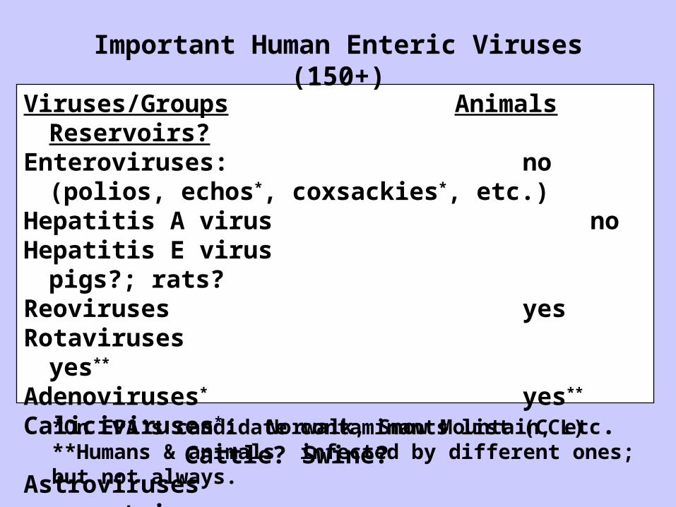
Viruses/Groups Animals Reservoirs?Enteroviruses: no
(polios, echos*, coxsackies*, etc.)Hepatitis A virus noHepatitis E virus pigs?; rats?Reoviruses yesRotaviruses yes**
Adenoviruses* yes**
Caliciviruses*: Norwalk, Snow Mountain, etc. Cattle? Swine?
Astroviruses uncertain*On EPA’s candidate contaminants list (CCL)**Humans & animals infected by different ones; but not always.
Important Human Enteric Viruses (150+)

Enteroviruses• Icosahedral shape• ~27-30 nm diameter• single-stranded +sense RNA
– about 7,500 nucleotides• icosahedral protein coat (capsid)
– 4 capsid proteins: VP1, VP2, VP3, VP4 (all cleaved from VP0)
• >71 antigenically distinct human types– polioviruses (3 types)– coxsackie B viruses (6 types)– coxsackie A viruses (23 types)– echoviruses (31 types)
• distinct animal enteroviruses

Hepatitis A Virus (HAV)
• icosahedral• 27 nM in diameter• non-enveloped capsid• ss(+) RNA genome• At least 3 major structural
polypeptides• Single serotype• Infects humans and non-
human primates• Cell culture reported in
1979 in fetal rhesus monkey kidney cells
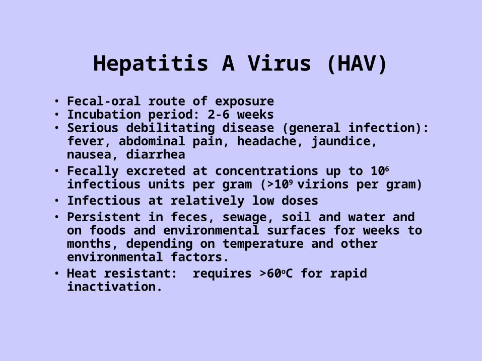
Hepatitis A Virus (HAV)
• Fecal-oral route of exposure• Incubation period: 2-6 weeks• Serious debilitating disease (general infection): fever,
abdominal pain, headache, jaundice, nausea, diarrhea• Fecally excreted at concentrations up to 106 infectious
units per gram (>109 virions per gram)• Infectious at relatively low doses• Persistent in feces, sewage, soil and water and on foods
and environmental surfaces for weeks to months, depending on temperature and other environmental factors.
• Heat resistant: requires >60oC for rapid inactivation.

Hepatitis A Viruscause of infectious or epidemic hepatitis
replicates in liver; viral shedding: 4 weeks

Geographic Distribution of HAV Infection
Geographic Distribution of HAV Infection
Anti-HAV Prevalence
High
Intermediate
Low
Very Low

Hepatitis E Virus
• 32-34 nM in diameter• ss(+) RNA genome• May belong to the caliciviridae
family• Incubation period:
– Average 40 days– Range 15-60 days
• Case-fatality rate:– Overall, 1%-3%
– Pregnant women, 17% - 33%
• Illness severity: increased with age

Geographic Distribution of Hepatitis EGeographic Distribution of Hepatitis E

Reovirus and Rotaviruses• ~spherical; icosahedral• ~75-80 nm diameter• double-layered capsid• nucleic acid:
– double-stranded RNA– 11 segments (rota)– 10 segments (reo)– electropherotypes
• 7 Groups (A-G) by VP6– Subgroups, serotypes
• Group A most important in humans (children)
• Group C causes sporadic illness• Group B has caused large outbreaks
(adults), rare

Rotaviruses

ADENOVIRUSES:• icosahedral• ~80 nm diameter• double-stranded, linear DNA• protein coat; at least 10 proteins
– Hexons, pentons, minor polypeptides– attachment fibers with knobs
• At leat 41 human adenoviruses– types 1-39 mostly respiratory
• but fecally shed– types 40 and 41 are enteric
• Often the most prevalent viruses in treated sewage– resistance to treatment?
• Highly resistant to UV radiation• Distinct animal adenoviruses

•Icosahedral• “structured”; cup-like surface morphology• 30-35 nm diameter• ss(+) RNA, ~7.7 KB•1 major capsid polypeptide, ~60 kD• minor protein, ~30 kD• 4 major HuNOV groups
•(G1 and G2 most prevalent• Sappoviruses; genetically distinct human enteric caliciviruses• NoVs and other caliciviruses are genetically diverse/variable• No culture of human NoVs, (except in humans and maybe chimps)• Distinct animal caliciviruses & noroviruses
•some genetically similar to human NoVs•cross-species transmission?
Noroviruses and Other Caliciviruses

Response of Human Volunteers to Norwalk Virus Infection via the Oral Route
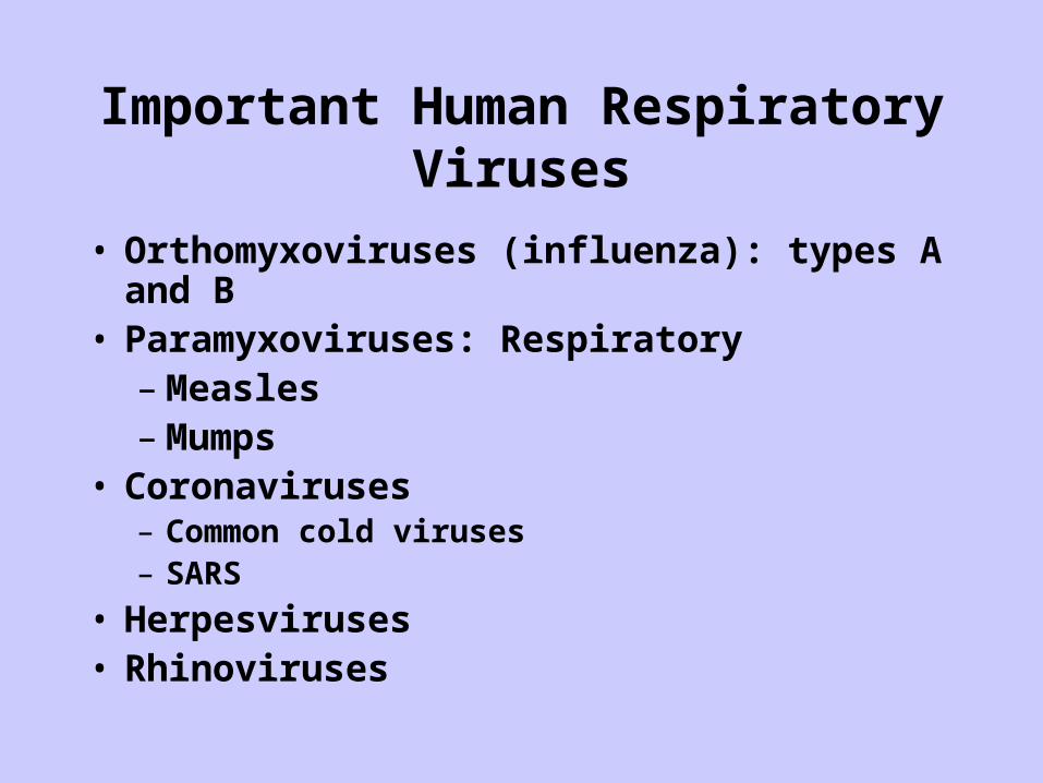
Important Human Respiratory Viruses
• Orthomyxoviruses (influenza): types A and B• Paramyxoviruses: Respiratory
– Measles– Mumps
• Coronaviruses– Common cold viruses– SARS
• Herpesviruses• Rhinoviruses

Influenza Viruses• Pleomorphic, spherical filamentous
forms occur– 50-120 nm diameter, or 20 nm
diameter and 200-300 nm long• Segmented, linear -ssRNA genome
– 7 to 8 segments• Enveloped, filamentous
nucleocapsids– Envelope is lipid bilayer with
~500 spikes (Hs & Ns)• Hemagglutinin and neuraminidase
Causes influenza – “the flu”
Animal reservoirs of influenza viruses that “jump” to humans or co-nfect animals, usually pigs, along with human strains to create new strains that are highly transmissable and virulent

Paramyxoviruses
• Roughly Spherical, Pleomorphic– ~200 nm diameter
• -ssRNA genome, 17-20 kb
• Enveloped, helical nucleocapsid– Envelope is lipid bilayer
with glycoprotein spikes
• Includes Measles, Mumps, and RSV
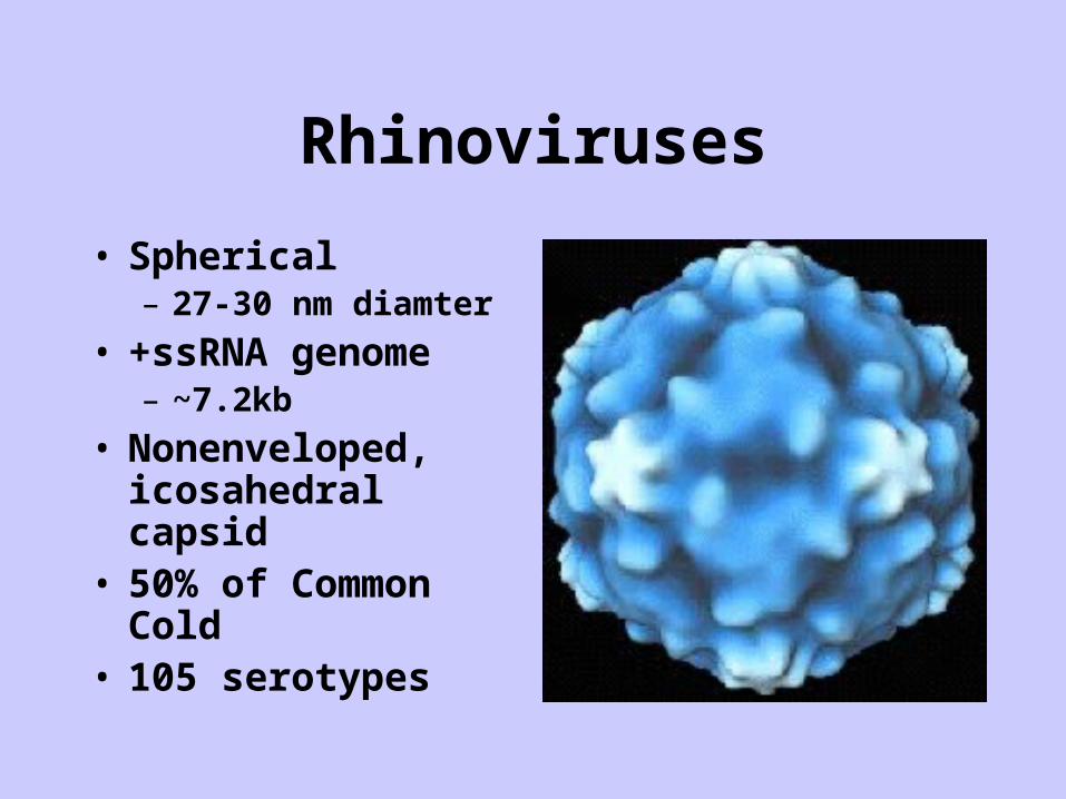
Rhinoviruses
• Spherical– 27-30 nm diamter
• +ssRNA genome– ~7.2kb
• Nonenveloped, icosahedral capsid
• 50% of Common Cold
• 105 serotypes

Coronaviruses
• Irregularly shaped– 60-220 nm diameter
• +ssRNA genome (27-31 kb)
• Enveloped particles with loosely wound nucleocapsid– characteristic “club-shaped”
peplomers
• 10 % of Common cold
• Severe Acute Respiratory Syndrome - SARS

SARS - Coronaviruses• Discovered in March, 2003• pleomorphic, enveloped particles
– 60 and 130 nm
• Short incubation (2 – 7 days)• Complete sequence known
– five major ORFs
• Very distinct group; not related to other HuCo-Vs
• Concensus genotype, but strain variability (epid studies)– High rate of RNA-RNA
recombination
• More environmentally stable than other HuCo-Vs
• Zoonotic pathogen? (civets)

SARS: Clinical Detection
• Up to 109 particles per mL in sputum
• Detected in nasopharyngeal aspirates by RT-PCR in 32% at initial presentation (mean 3.2 days after onset of illness) and in 68% at day 14
• Detected in 97% of patient’s stools and 42% of urine samples two weeks after the onset of illness

SARS
Symptoms: • high fever (>100.4°F; >38.0°C).
Other symptoms: headache, malaise, and body aches.
• Some people also have mild respiratory symptoms at outset.
• 10 percent to 20 percent of patients have diarrhea.
• After 2 to 7 days, SARS patients may develop a dry cough.
• Most patients develop pneumonia.
Source: Initially certain mammals in SE Asia (esp. China): palm civet cat; recent evidence in bats
Spreadclose person-to-person contact.• respiratory droplets (droplet spread)
from coughs or sneezes.– propelled a short distance (generally up
to 3 feet) through the air and deposited on the mucous membranes of the mouth, nose, or eyes of persons who are nearby.
• Also spread by fomites (person touches a surface or object contaminated with infectious droplets and then touches his or her mouth, nose, or eye(s).
• might spread more broadly through the air (airborne spread)

Assay Methods for Viruses• Electron Microscopy (EM) and Immune EM
– Insensitive (>1,000,000 particles/ml)– OK for clinical but not environmental virology
• Animal Infectivity– Slow, cumbersome, expensive, ethical considerations
• Culture or infectivity– Now widely used in environmental virology– Cytopathogenic effects– Growth, but no cytopathogenic effects
• detect viral antigens or nucleic acids• Immunoassays
– insensitive for direct detection– Amplify viruses in cell cultures
• Nucleic acid assays– insensitive for direct detection by hybridization– Amplify in cell culture or in vitro (PCR or RT-PCR)
Virus Infectivity and Infectivity Assays• Viruses differ in their their human infectivity• Enteric viruses and some respiratory viruses are
generally infectious at low doses– As little as one cell culture or animal infectious dose has a high
probability of infecting an exposed human• Many enteric viruses in environmental samples do not
cause cytopathogenic effects (CPE)– will not be detected by microscopic examination– require additional methods to detect their presence
• immunochemical methods– detect antigens in infected cells
• nucleic acid methods– nucleic acid hybridization– nucleic acid amplification

Virus Detection in Cell Culture by Cytopathogenic Effects
Uninfected Cell CultureUninfected Cell Culture Infected Cell Culture with CPEInfected Cell Culture with CPE
From a public health and risk assessment standpoint, microbial assays based on infectivity are the most relevant and easily interpretable ones
Quantifying Human Virus Infectivity is a Challenge
• Some infect only humans• Some infect certain experimental animals, too• Some infect experimental animals and cell cultures
• Different ratios of infectivity to virions (particles)– 1 infectious unit ~ 1 virus particles
• some bacteriophages– 1 infectious unit ~100 virions:
• some cell culture adapted viruses– 1 infectious unit ~10,000-100,000 virions
• many “wild-type or field viruses
• Some viruses (some enteroviruses, adenoviruses, rotaviruses, astroviruses and hepatitis A virus) grow poorly or slowly in cell cultures and produce little or no CPE.
• Greater detection with additional analytical techniques:– Viral antigens
• Immunofluorescence assays, enzyme immunoassays, radioimmunoassays, etc.
– Viral nucleic acid assays: hybridization and/or amplification
• Combined cell culture + RT-PCR demonstrates presence of greater numbers of infectious viruses than CPE alone
– Post-disinfection, more viruses are detected than by CPE
Progress in Virus Detection in Cell Culture

Estimating Viral Dose:Relationship of Infectivity to Virus Particle Count
• As little a one or a few intact, functional virus particles are capable of causing infection in a susceptible host.
• Ratios of virus particles to infectious units are highly variable and are subject to change:– Passage of viruses in susceptible host cells reduces the
ratio of virus particles to infectious units
• rotavirus: – initial ratio: ~50,000 virus particles/infect. unit– after cell culture passage: ~100 particles/infect. Unit
• Norwalk Virus appears to be infectious at doses corresponding to as little as 10-100 virus particles.

Emerging Microbial Indicators of Fecal Contamination for Enteric Viruses
• Somatic and F+ (male-specific) coliphages may be useful indicators of enteric viruses in water, wastewater and other fecally contaminated samples.– Plentiful in raw sewage– Reduced less effectively than are
conventional indicator bacteria by sewage treatment processes.
– Superficially resemble enteric viruses (F+ coliphages)
– Easy, rapid and economical to detect and quantify by reliable methods
somatic
F+

Virus Infections: Some Important Viruses Cause Localized Infections and Others Systemic Infections
Enteric Viruses:• Localized:
– caliciviruses– astroviruses– adenoviruses– rotaviruses
• Generalized:– enteroviruses– hepatitis A and E viruses
Respiratory Viruses:• Localized:
– rhinoviruses– coronaviruses– Orthomyxoviruses(Flu)– paramyxoviruses
• Generalized:– herpesviruses– measles– mumps
Factors Influencing Virus Infection and Illness• The probability of infection varies with:
– Virus factors– Host factors– Route and site of infection and vehicle
• Virus and host availability and encounters• Ingestion, inhalation, eye, skin, etc.• Water, food, droplets, aerosols
• Probablility of illness from infection: high (>50%) for many enteric viruses– Varies with age of host and with type of virus:
• Some: high rates of illness in infants and children• Others: high rates of illness in adults
– Varies with health status: “sensitive populations”• Elderly: high risk of illness with adverse outcomes• Immune compromised: high risk of chronic, lethal illness• Pregnancy: high risk of illness and death (ex: HEV)
– Immune status– Genetics– Nutrition– behavior (personal habits) of host.

Role of Immunity in Virus Infections:Generalized/Systemic/Disseminated Infections
• Immunity against generalized/systemic/disseminated infection is usually lifelong, unless immune system is severely compromised
• Localized (e.g., gastrointestinal) re-infection is possible• Hepatitis A and E and many enteroviruses cause
systemic/generalized/disseminated infections– Typically, immunity against severe illness is long-term and
probably lifelong.• Proof of concept: live, oral poliovirus vaccine and poliomyelitis
eradication; susceptibles are newborns and infants• Antigenic changes in viruses may overcome long-term
immunity and increase risks of re-infection or illness.

Role of Immunity in Virus Infections:Localized Infections:
• Immunity to infection is usually short-term and transient– Gut (secretory or IgA) immunity wanes over time
• Proof of concept: live, oral rotavirus vaccine: – immunity declines over time and reinfection with “wild”
type rotaviruses occurs• Repeated localized (e.g., gastrointestinal) re-infection is
possible• Rotaviruses, caliciviruses, adenoviruses and some
enteroviruses, cause localized infections

Role of Selection of New Viral Strains in Susceptibility to Infection and Illness
• Antigenic changes in viruses overcome immunity, increasing risks of re-infection or illness– Antigenically different strains of viruses appear and are
selected for over time and space– Constant selection of new strains (by antigenic shift and drift)– Partly driven by “herd” immunity and genetic recombination,
reassortment and point mutations• Antigenic Shift:
– Major change in virus genetic composition by gene substitution or replacement (e.g., reassortment)
• Antigenic Drift:– Minor changes in virus genetic composition, often by mutation
involving specific codons in existing genes (point mutations)• A single point mutation can greatly alter virus virulence
– Influena vaccine for 2008 provides protection against only some circulating influenza viruses that emerged in the population; chose vaccine strains different from prevalent ones