Embed Size (px)
Citation preview
International Journal of
Molecular Sciences
Review
Environmental Epigenetics and Genome Flexibility:Focus on 5-Hydroxymethylcytosine
Olga A. Efimova * , Alla S. Koltsova, Mikhail I. Krapivin, Andrei V. Tikhonovand Anna A. Pendina
D. O. Ott Research Institute of Obstetrics, Gynecology and Reproductology, Mendeleevskaya line 3,199034 St. Petersburg, Russia; [email protected] (A.S.K.); [email protected] (M.I.K.);[email protected] (A.V.T.); [email protected] (A.A.P.)* Correspondence: [email protected]; Tel.: +7-812-328-98-09
Received: 5 April 2020; Accepted: 29 April 2020; Published: 2 May 2020�����������������
Abstract: Convincing evidence accumulated over the last decades demonstrates the crucial role ofepigenetic modifications for mammalian genome regulation and its flexibility. DNA methylation anddemethylation is a key mechanism of genome programming and reprogramming. During ontogenesis,the DNA methylome undergoes both programmed changes and those induced by environmental andendogenous factors. The former enable accurate activation of developmental programs; the latterdrive epigenetic responses to factors that directly or indirectly affect epigenetic biochemistry leading toalterations in genome regulation and mediating organism response to environmental transformations.Adverse environmental exposure can induce aberrant DNA methylation changes conducive to geneticdysfunction and, eventually, various pathologies. In recent years, evidence was derived that apartfrom 5-methylcytosine, the DNA methylation/demethylation cycle includes three other oxidativederivatives of cytosine—5-hydroxymethylcytosine (5hmC), 5-formylcytosine, and 5-carboxylcytosine.5hmC is a predominantly stable form and serves as both an intermediate product of active DNAdemethylation and an essential hallmark of epigenetic gene regulation. This makes 5hmC a potentialcontributor to epigenetically mediated responses to environmental factors. In this state-of-the-artreview, we consolidate the latest findings on environmentally induced adverse effects on 5hmCpatterns in mammalian genomes. Types of environmental exposure under consideration includehypnotic drugs and medicines (i.e., phenobarbital, diethylstilbestrol, cocaine, methamphetamine,ethanol, dimethyl sulfoxide), as well as anthropogenic pollutants (i.e., heavy metals, particulateair pollution, bisphenol A, hydroquinone, and pentachlorophenol metabolites). We put a specialfocus on the discussion of molecular mechanisms underlying environmentally induced alterationsin DNA hydroxymethylation patterns and their impact on genetic dysfunction. We conclude thatDNA hydroxymethylation is a sensitive biosensor for many harmful environmental factors each ofwhich specifically targets 5hmC in different organs, cell types, and DNA sequences and induces itschanges through a specific metabolic pathway. The associated transcriptional changes suggest thatenvironmentally induced 5hmC alterations play a role in epigenetically mediated genome flexibility.We believe that knowledge accumulated in this review together with further studies will providea solid basis for new approaches to epigenetic therapy and chemoprevention of environmentallyinduced epigenetic toxicity involving 5hmC patterns.
Keywords: 5-hydroxymethylcytosine; DNA methylation; environmental factors; phenobarbital;narcotics; dimethyl sulfoxide (DMSO); heavy metals; bisphenol A (BPA); particulate air pollution;pentachlorophenol (PCP)
Int. J. Mol. Sci. 2020, 21, 3223; doi:10.3390/ijms21093223 www.mdpi.com/journal/ijms
Int. J. Mol. Sci. 2020, 21, 3223 2 of 22
1. Introduction
In 1972, a group of investigators observed the presence of modified cytosine,5-hydroxymethylcytosine (5hmC), which accounted for about 15% of the total cytosine residuesin rat and mouse brain DNA [1]. Further attempts to reproduce these results failed for a long time,the biological role of 5hmC remaining unknown for roughly the next 40 years. In 2009, however,5hmC was repeatedly detected in mouse brain cells [2] and mouse embryonic stem cells (mESCs) [3].Moreover, 5hmC was identified as the oxidation product of methylated cytosine, 5-methylcytosine(5mC) [3], which is a major player in epigenetic regulation and genome reprogramming in mammaliandevelopment [4–7].
5mC oxidation is mediated by the enzymatic activity of TET (Ten-Eleven-Translocation) familyproteins and consistently yields three oxidative derivatives: 5-hydroxymethylcytosine, 5-formylcytosine(5fC), and 5-carboxylcytosine (5caC). Once targeted by base excision repair enzymes, 5fC and 5caC areexcised from the DNA and replaced with unmodified cytosine [3,8]. TET-mediated 5mC oxidationapparently promotes active (enzymatic) DNA demethylation which is a key event of epigeneticreprogramming in germ cells and mammalian embryos.
Research demonstrated that 5mC oxidation products serve as intermediates in active DNAdemethylation and likewise function in genome regulation. This is particularly true for 5hmC asa predominantly stable oxidation product of 5mC. Thus, 5hmC is specifically recognized by someprotein regulators of cell metabolism, including RPL26, PRP8, MHS6, MeCP2, UHRF, and Thy28 [9–11].Typically, 5hmC would exhibit specific genomic localization—in enhancers, sites flanking promoters(or CpG-islands), and in gene bodies. Furthermore, abundance of 5hmC at enhancers is positivelycorrelated with enhancer activity [12–14]. At CpG-islands, 5hmC is vital to maintain promoters inthe unmethylated state, whereas in intragenic sequences, 5hmC is suggested to have an inhibitoryaction on antisense transcription initiation [15,16]. 5hmC can be thus recognized as a stable cytosinemodification which has its own function [17–21].
On the one hand, in the course of ontogenesis, specific epigenetic profiles that act to initiate geneexpression programs and direct cell differentiation are established, maintained, and altered in a strictlydetermined way [22–24]. Reversible cytosine modification, on the other hand, provides a source ofepigenomic plasticity—the ability of the epigenome to change in response to external factors. Today,there is well-established evidence that aberrant DNA methylation patterns generally associated withsome pathological conditions including genome structural variation and complex rearrangementscan be provoked by endogenous and environmental factors [25–29]. Folic acid deficiency duringpregnancy can induce a deficiency of S-adenosylmethionine (SAM), a methyl group donor, resulting inelevated homocysteine and abnormal DNA methylation. This leads to gene deregulation, includingabnormal biallelic expression of imprinted genes, which are normally characterized by a monoallelicexpression [30]. Metals such as nickel, cadmium, and arsenic perturb DNA methylation patterns anddamage epigenetic regulation of proto-oncogenes and oncosuppressors, thus increasing the risk ofmalignization [31]. Research has shown that disrupted DNA methylation can be induced by syntheticnonsteroidal estrogen—diethylstilbestrol [32–34]. Ample studies suggest evidence of DNA methylationdamage due to exposure to chromium, mercury, trichloroethylene, dichloroacetic and trichloroaceticacid, bisphenol A (BPA), and many other substances [35–42].
While DNA methylation has been extensively investigated, knowledge about how 5hmC patternschange in response to adverse environmental factors is so far scarce [43]. Up-to-date informationis focused on brain-related 5hmC changes upon external exposures [44] or has been reviewed interms of the link between environmental cues and DNA hydroxymethylation, lacking discussionof the underlying molecular mechanisms [45]. The present paper consolidates current evidence on5hmC pattern alterations induced by environmental factors in different organs, tissues, and cell linesand analyzes how they affect genome function. Along with DNA hydroxymethylation changes,the associated alterations of major components for 5hmC production (5mC, TETs, DNA methylases,co-substrates, and co-factors) are reviewed to place special emphasis on the molecular mechanisms
Int. J. Mol. Sci. 2020, 21, 3223 3 of 22
providing epigenetic responses to adverse external effectors. The reviewed data strongly suggest thatenvironmentally induced genome-wide and gene-specific hydroxymethylation alterations are drivenby various metabolic pathways due to effector-specific changes in 5hmC biochemistry. The associatedtranscriptional changes point towards the role of environmentally induced 5hmC alterations inepigenetically mediated genome flexibility.
2. Factors Associated with 5hmC Biochemical Pathways in Mammalian DNA
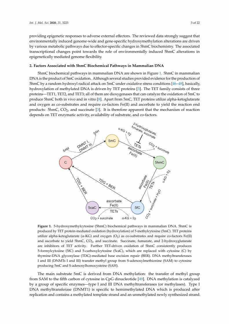
5hmC biochemical pathways in mammalian DNA are shown in Figure 1. 5hmC in mammalianDNA is the product of 5mC oxidation. Although several studies provided evidence for the production of5hmC by a random hydroxyl radical attack on 5mC under oxidative stress conditions [46–48], basically,hydroxylation of methylated DNA is driven by TET proteins [3]. The TET family consists of threeproteins—TET1, TET2, and TET3; all of them are dioxygenases that can catalyze the oxidation of 5mC toproduce 5hmC both in vivo and in vitro [8]. Apart from 5mC, TET proteins utilize alpha-ketoglutarateand oxygen as co-substrates and require co-factors Fe(II) and ascorbate to yield the reaction endproducts: 5hmC, CO2, and succinate [3]. It is therefore apparent that the mechanism of reactiondepends on TET enzymatic activity, availability of substrate, and co-factors.
Int. J. Mol. Sci. 2020, 21, 3223 3 of 22
suggest that environmentally induced genome-wide and gene-specific hydroxymethylation alterations are driven by various metabolic pathways due to effector-specific changes in 5hmC biochemistry. The associated transcriptional changes point towards the role of environmentally induced 5hmC alterations in epigenetically mediated genome flexibility.
2. Factors Associated with 5hmC Biochemical Pathways in Mammalian DNA
5hmC biochemical pathways in mammalian DNA are shown in Figure 1. 5hmC in mammalian DNA is the product of 5mC oxidation. Although several studies provided evidence for the production of 5hmC by a random hydroxyl radical attack on 5mC under oxidative stress conditions [46–48], basically, hydroxylation of methylated DNA is driven by ТЕТ proteins [3]. The TET family consists of three proteins—TET1, TET2, and TET3; all of them are dioxygenases that can catalyze the oxidation of 5mC to produce 5hmC both in vivo and in vitro [8]. Apart from 5mC, TET proteins utilize alpha-ketoglutarate and oxygen as co-substrates and require co-factors Fe(II) and ascorbate to yield the reaction end products: 5hmC, СО2, and succinate [3]. It is therefore apparent that the mechanism of reaction depends on TET enzymatic activity, availability of substrate, and co-factors.
Figure 1. 5-hydroxymethylcytosine (5hmC) biochemical pathways in mammalian DNA. 5hmC is produced by TET protein-mediated oxidation (hydroxylation) of 5-methylcytosine (5mC). TET proteins utilize alpha-ketoglutarate (α-KG) and oxygen (О2) as co-substrates and require co-factors Fe(II) and ascorbate to yield 5hmC, СО2, and succinate. Succinate, fumarate, and 2-hydroxyglutarate are inhibitors of TET activity. Further TET-driven oxidation of 5hmC consistently produces 5-formylcytosine (5fC) and 5-carboxylcytosine (5caC), which are replaced with cytosine (C) by thymine-DNA glycosylase (TDG)-mediated base excision repair (BER). DNA methyltransferases I and III (DNMTs I and III) transfer methyl group from S-adenosylmethionine (SAM) to cytosine producing 5mC and S-adenosylhomocysteine (SAH).
The main substrate 5mC is derived from DNA methylation: the transfer of methyl group from SAM to the fifth carbon of cytosine in CpG dinucleotide [49]. DNA methylation is catalyzed by a group of specific enzymes—type I and III DNA methyltransferases (or methylases). Type I DNA methyltransferase (DNMT1) is specific to hemimethylated DNA which is produced after replication and contains a methylated template strand and an unmethylated newly synthesized strand. DNMT1 methylation of the new strand mediates reproduction of the template strand methylation pattern and thus enables its inheritance by daughter cells during divisions. Conversely, type III DNA
Figure 1. 5-hydroxymethylcytosine (5hmC) biochemical pathways in mammalian DNA. 5hmC isproduced by TET protein-mediated oxidation (hydroxylation) of 5-methylcytosine (5mC). TET proteinsutilize alpha-ketoglutarate (α-KG) and oxygen (O2) as co-substrates and require co-factors Fe(II)and ascorbate to yield 5hmC, CO2, and succinate. Succinate, fumarate, and 2-hydroxyglutarateare inhibitors of TET activity. Further TET-driven oxidation of 5hmC consistently produces5-formylcytosine (5fC) and 5-carboxylcytosine (5caC), which are replaced with cytosine (C) bythymine-DNA glycosylase (TDG)-mediated base excision repair (BER). DNA methyltransferasesI and III (DNMTs I and III) transfer methyl group from S-adenosylmethionine (SAM) to cytosineproducing 5mC and S-adenosylhomocysteine (SAH).
The main substrate 5mC is derived from DNA methylation: the transfer of methyl groupfrom SAM to the fifth carbon of cytosine in CpG dinucleotide [49]. DNA methylation is catalyzedby a group of specific enzymes—type I and III DNA methyltransferases (or methylases). Type IDNA methyltransferase (DNMT1) is specific to hemimethylated DNA which is produced afterreplication and contains a methylated template strand and an unmethylated newly synthesized strand.

Int. J. Mol. Sci. 2020, 21, 3223 4 of 22
DNMT1 methylation of the new strand mediates reproduction of the template strand methylationpattern and thus enables its inheritance by daughter cells during divisions. Conversely, type IIIDNA methyltransferases DNMT3A and DNMT3B are specific to unmethylated DNA and can drivemethylation de novo [50].
Alpha-ketoglutarate is enzymatically produced from isocitrate. The reaction is catalyzed byisocitrate dehydrogenases IDH1, IDH2, and IDH3 [51]. IDH2 dysfunction in gastric cancer cells resultsin 5hmC depletion [52]. In melanoma cells, IDH2 down-regulation is also associated with decreased5hmC levels [53]. The latter is induced by 2-hydroxyglutarate accumulation—an oncometabolitethat competitively inhibits TET enzymes [54]. Fumarate and succinate, generally accumulated bycancer cells that are exposed to fumarate hydratase and succinate dehydrogenase deficiency, alsoshow inhibitory action on TET proteins and prevent their binding with alpha-ketoglutarate [55,56].Conversely, excess of IDH1 and IDH2 facilitates the increase of DNA hydroxymethylation [53,54].Elevated alpha-ketoglutarate in mouse liver cells induces a surge in 5hmC levels [57].
In different cell types, oxygen can affect 5mC hydroxylation in various ways. Throughoutembryogenesis, oxygen gradients differentially regulate TET activity and thus determine cellulardifferentiation [58]. Under hypoxia, human glioblastoma cells exhibit a decrease in 5hmC levelswhich is associated with hypermethylation and loss of TET activity [59]. On the contrary, humanembryonic stem cells (hESCs) show hypoxia-induced TET up-regulation which leads to elevated DNAhydroxymethylation [60]. Similarly, hypoxic neuroblastoma cells demonstrate elevated 5hmC levelsinduced by hypoxia-inducible factor (HIF) activation that in turn enhances TET activity [61].
Ascorbate (l-ascorbic acid or vitamin C) promotes 5hmC increase in the genome [62]. This effectis neither Fe(II)-dependent nor related to changes in Tet or IDH expression and subsequentalpha-ketoglutarate production [63]. Ascorbate directly interacts with the catalytic domain of TETproteins, reducing Fe(III) to Fe(II) and enhancing TET-mediated 5mC oxidation [64]. Considering thatmost, if not all, malignant lesions exhibit decreased 5hmC levels [65], ascorbic acid’s potential as ananti-cancer therapy is currently intensively investigated. Evidence suggests that ascorbate-mediated5hmC increase in melanoma cells suppresses their metastatic capabilities and detains tumor growth [66].
The presence of Fe(II) as another co-factor for 5mC hydroxylation also appears to enhance theactivity of TET to generate 5hmC [67]. Mutation-induced modifications at the Fe(II)-binding domainof TET proteins lead to a decrease/loss of enzymatic activity [3].
Overall, 5hmC production in the mammalian genome predominantly depends on the presenceand level of major components for 5mC oxidation: 5mC itself, TET enzymes, alpha-ketoglutarate,oxygen, Fe(II), and ascorbate.
3. Impact of External Factors on Genomic Hydroxymethylation
3.1. Hypnotics and Medications
3.1.1. Phenobarbital
Phenobarbital is a barbituric acid derivative with antiseizure, hypnotic, and sedative properties.In rodents, chronic exposure to phenobarbital demonstrated hepatocancerogenic action [68]; there is noevidence of hepatocancerogenic hazard of phenobarbital in humans. Mice that had been treated withphenobarbital in drinking water for 28 days showed elevated 5hmC levels in promoters of tumor-relatedgenes of liver tissue. Elevated 5hmC is associated with decreased DNA methylation and up-regulationof these genes, suggesting the initiation of active DNA demethylation [69,70]. Prolonged exposure tophenobarbital of up to 91 days significantly promotes DNA hydroxymethylation [70]. Experimentaldata on phenobarbital-induced hepatocellular adenomas demonstrated changes of hydroxymethylationand gene expression levels, including carcinogenic genes, especially those regulated through theconstitutive androstane receptor (CAR) signaling pathways [71]. This evidence suggests that changesin 5hmC levels related to the initiation of active DNA demethylation indicate hepatic cell response tophenobarbital, associated with carcinogenicity and other effects.

Int. J. Mol. Sci. 2020, 21, 3223 5 of 22
3.1.2. Diethylstilbestrol
Diethylstilbestrol is a synthetic nonsteroidal estrogen that was prescribed to pregnant women until1971 to support pregnancy and prevent miscarriage or other pregnancy complications. The drug wasbanned after the American Cancer Society provided evidence of carcinogenicity [72]. In mice, neonatalexposure to diethylstilbestrol induces alterations in histone modification pattern and a significantreduction in Tet1 expression; this correlates with a decrease in 5hmC levels in adults [73]. Consideringthese results, the authors assumed that it is diethylstilbestrol-induced epigenetic alterations that areresponsible for modifications in female reproductive tract gene expression, infertility, and uterinecancer [73]. First-trimester diethylstilbestrol exposure is associated with an increased risk of benigntumors—uterine leiomyomas [74]. Based on detected 5hmC imbalance in uterine leiomyoma tissue [75]and the dependence of the hydroxymethylation pattern on the hormonal status [76], it can be assumedthat diethylstilbestrol-induced benign tumorigenesis also involves alterations in 5hmC.
3.1.3. Cocaine
Cocaine is a highly addictive alkaloid of the shrub Erythroxylum coca. Mice receiving cocaineintraperitoneal injections for 14 days showed decreased 5hmC levels in liver cells without anyalterations in global DNA methylation; meanwhile in brain cells, hydroxymethylation level remainedunchanged [77]. Another research also reports the absence of alterations in global DNA methylationand hydroxymethylation in mouse nucleus accumbens in response to cocaine administration [78].The authors observed significant down-regulation of Tet1 mRNA and a concomitant decrease in TET1protein [78]. A ~40% decrease in TET1 mRNA was observed in the nucleus accumbens of human cocaineaddicts examined postmortem [78]. This evidence suggests that cocaine can induce locus-specific 5hmCalterations, while a greater abundance of Tet2 and Tet3 mRNAs, characteristic of nucleus accumbens,can presumably compensate for TET1 decrease [78]. Selective chemical labeling for 5hmC followed bydeep sequencing allowed identification of 11511 differentially hydroxymethylated regions, distributedprimarily in gene bodies (~55%) and intergenic regions (~34%) [78]. In rat prefrontal cortex exposed tococaine self-administration, both DNA methylation and hydroxymethylation are decreased within theHomer2 promoter, a glutamate receptor-related scaffolding protein [79]; all Tet genes and Dnmt3b aredown-regulated while Dnmt3a is up-regulated [80]. Combined with the effect on brain structures, thereis indirect evidence of cocaine-induced alterations in DNA hydroxymethylation patterns in mousespermatogenic cells: authors observed decreased Dnmt3b and Tet1 mRNAs and increased Dnmt3a andDNA methylation [81].
3.1.4. Methamphetamine
Methamphetamine is a synthetic neurotoxic psychostimulant. Its intake causes increasein TET1 and TET3 protein levels and changes hydroxymethylation levels in rat nucleusaccumbens [82]. Increased 5hmC concomitant with a decrease in DNA methylation was detectedin corticotropin-releasing hormone (Crh/Crf ) gene promoter and at a CpG-rich region within thearginine vasopressin (Avp) gene body [82]. On the contrary, promoter sequences of GluA1 and GluA2alpha-amino-3-hydroxy-5-methyl-4-isoxazole propionic acid receptor (AMPAR) undergo a decrease in bothstriatal DNA methylation and hydroxymethylation [83]. These changes, combined with histonemodifications [83], seem to suppress striatal glutamate receptor expression, observed in systemicmethamphetamine intake. Other genomic regions are also affected by methamphetamine-drivenchanges in 5hmC. In rats addicted to methamphetamine, nucleus accumbens cells subjected toimmunoprecipitation with polyclonal anti-5hmC antibodies followed by next-generation sequencingshowed numerous differentially hydroxymethylated regions, predominantly in intergenic sites locatedon long and short interspersed elements [84].
The available data provides supporting evidence that cocaine- and methamphetamine-drivenchanges in DNA hydroxymethylation patterns can be a vital factor in promoting drug addiction.

Int. J. Mol. Sci. 2020, 21, 3223 6 of 22
3.1.5. Ethanol
Prenatal exposure to ethanol causes disturbances in normal methylation and hydroxymethylationdynamics of developing mouse hippocampus and cortex [85,86]. Delayed 5mC and 5hmC increaseis observed in the neuroepithelial stem cells. In early maturing neurons, ethanol disrupts thetimely decrease of DNA methylation and increase of hydroxymethylation [85]. Prenatal exposure toethanol has prolonged action: after withdrawal, developing gyrus dentatus undergoes significantchanges in programmed DNA methylation and hydroxymethylation dynamics during the thirdtrimester of pregnancy [85]. After 8 days of ethanol exposure in vitro, mouse neural stem cellsdemonstrate an increased global DNA methylation level, whereas the total 5hmC level remainsunaffected. After withdrawal, however, the DNA hydroxylation level plummets significantly [87].In blood samples of alcohol-dependent humans, hydroxymethylation is significantly lower comparedto controls. During detoxification, a rise of hydroxymethylation is observed [88]. Chronic alcoholconsumption in rats decreases hydroxymethylation levels in liver cells by half [89] and intensifiesapoptosis of hepatocytes [90]. This is accompanied by decreasing TET1 levels, while TET2 and TET3remain unchanged [90]. However, iron supplementation to an alcohol diet prevents changes in DNAhydroxymethylation levels [89].
3.1.6. Dimethyl Sulfoxide
Dimethyl sulfoxide (DMSO) is a bipolar aprotic solvent. DMSO has been known for its enhancedpermeability and capacity to significantly facilitate transdermal permeation of active substances;therefore, DMSO is extensively used in local cosmetic products and medicines for transdermal deliveryof local anti-inflammatory and pain-killing agents. DMSO is also used in cryopreservation of differenttypes of cells. Thaler et al. showed that DMSO exposure induced an increase of both global andgene-specific 5hmC levels in pre-osteoblastic MC3T3-E1 cell line [91]. The authors report that 12 to24 h after DMSO exposure Tet and Gadd45 genes—the key players in DNA hydroxymethylation andnucleotide excision repair—demonstrated increased expression. There was a concurrent decreasedexpression of genes related to DNA methylation: Dnmt1, Dnmt3b, and Hells [91]. The Tet1-dependentpro-apoptotic gene Fas and the early osteoblastic factor Dlx5 demonstrated expression increase.The aforesaid changes in gene expression are associated with a global and gene-specific increasein hydroxymethylation and concomitant gene-specific loss of DNA methylation at Fas and Dlx5promoters [91]. By day 5, the DMSO impact on promoter-specific and global methylation/hydroxylationis reduced or reversed [91]. The 3D microtissues of a maturing cardiac model and a mature hepaticmodel provide indirect evidence of DMSO effect on DNA hydroxymethylation in human cells. AddingDMSO at 0.1% to human 3D cardiac microtissue culture promotes up-regulation of methyltransferasesDNMT1 and DNMT3A and down-regulation of TET1. Transcriptional changes of DNA methylationwriters and erasers are associated with changes of methylation levels in 66,178 regions, where 71%show gain of DNA methylation [92]. In contrast, no deregulation of DNA methylation is observed inDMSO-exposed 3D hepatic microtissue [92].
3.2. Anthropogenic Pollutants
3.2.1. Heavy Metals
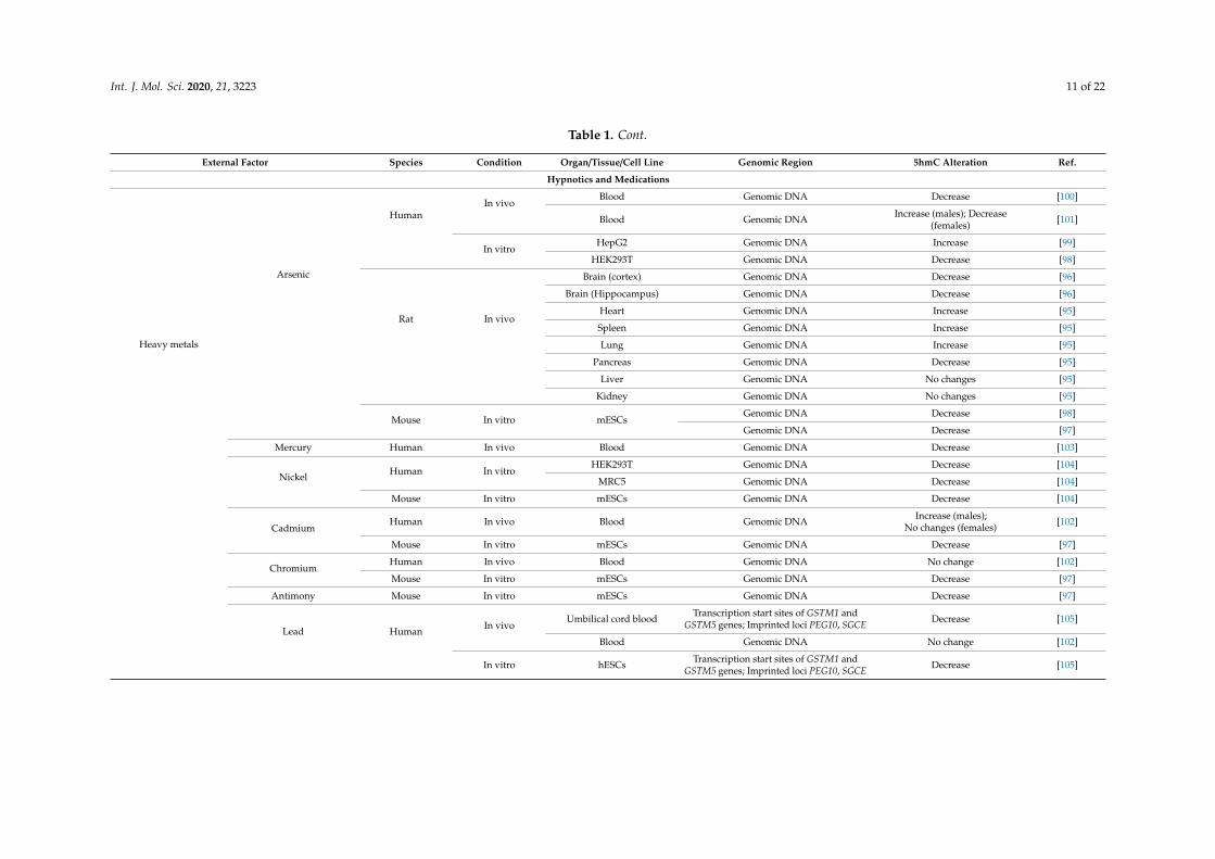
Arsenic is listed among the most dangerous substances in the United Nations EnvironmentProgramme guidelines. The International Agency for Research on Cancer includes arsenic in Group1 ‘Carcinogenic to humans’. Arsenic exposure is associated with cardiovascular diseases, diabetesmellitus, and neurological and reproductive disorders [93]. Once ingested, inorganic arsenic compoundsundergo enzymatic methylation, a pathway of detoxification utilizing SAM as the methyl donor.Significant amounts of arsenic induce SAM depletion, loss in global DNA methylation, and aberrantlocus-specific hypermethylation in multiple regions including p53 and p16 promoters. Disrupted DNA
Int. J. Mol. Sci. 2020, 21, 3223 7 of 22
methylation patterns, particularly in proto-oncogenes and onco-suppressor genes, increase the risk ofmalignization [94].
The effect of arsenic on DNA hydroxymethylation has been recently demonstrated in bothanimal and human models. After 8 weeks of exposure to sodium arsenite dissolved in drinkingwater, male Sprague-Dawley rats showed elevated DNA hydroxymethylation levels in lungs, heart,spleen, and pancreas, while liver and kidney were unaffected. DNA methylation, however, remainedunchanged in these types of organs, except for spleen, where the methylation level was elevated [95].Exposure to arsenic trioxide in drinking water for 6 months induced a decrease in DNA methylationand hydroxymethylation in hippocampus and cortex, presumably promoted by down-regulationof Dnmts and Tets expression [96]. These changes driven by oxidative stress cause deregulationof tricarboxylic acid cycle and alpha-ketoglutarate pathway. No concurrent SAM decrease wasreported [96]. Tricarboxylic acid cycle deregulation and reduced Tet protein activity associated withloss of 5hmC, 5fC, and 5caC are also observed in mESCs under arsenic exposure [97]. Humanembryonic kidney cells (HEK293T) were used to show that arsenite can bind directly to the zinc fingersof Tet proteins, thus causing loss of catalytic activity to catalyze oxidation of 5mC to yield 5hmC, 5fC,and 5caC. A successive decrease in 5hmC and an increase in 5mC depends on arsenite concentration [98].Arsenic-containing hydrocarbons AsHC 332 and AsHC 360 increase global hydroxymethylation andalter expression of a number of genes, including FEN1, XPA, and DNMT3A in culture of human livercells HepG2 [99]. In individuals with high urine concentration of dimethylarsinate over years, DNAmethylation variation in blood correlates positively with changes in 5hmC levels [100]. Remarkably,a different analysis performed on blood samples of arsenic-exposed individuals demonstrated apositive correlation between arsenic exposure and global 5hmC levels in men and a negative correlationin women. Apparently, it is the plasma total homocysteine level that seems to contribute to this sexdifference, as positive correlation is stronger in men with normal plasma total homocysteine, whereasnegative correlation is stronger in hyperhomocysteinemic women [101].
Sporadic studies investigated the effect of other heavy metals on 5hmC patterns. In mESCsculture, cadmium exposure induces a decrease in TET protein activity and a decrease in 5hmC, 5mC,and 5caC levels, while the 5mC level remains unchanged [97]. Exposure to chromium and antimonyhas a similar action on mESCs [97]. In the Central Zhejiang Province of China, children who live in thevicinity of waste incinerators and have elevated blood levels of chromium, cadmium, and lead showlower mean serum levels of 5mC and 5hmC and a higher mean level of percent tail DNA than childrenliving in unpolluted areas. There is a sex difference in correlation with heavy metals in blood andepigenetic changes: in boys, chromium in blood is negatively correlated with 5mC, while cadmiumin blood is positively correlated with 5mC and 5hmC; in girls, however, chromium in blood aloneis negatively correlated with 5mC [102]. Mercury exposure in utero is associated with a decrease in5hmC genomic content and an increase in the 5mC to 5hmC ratio in cord blood at birth and until theage of 5 years [103]. Nickel inhibits TET-mediated 5mC oxidation in human embryonic lung fibroblastscell culture (MRC5) and HEK293T cells as well as in mESCs and significantly reduces the global 5hmClevel [104]. Lead exposure alters hydroxymethylation patterns in CpG islands in hESCs and in the cordblood of newborns [105]. The epigenetic toxicity of other heavy metals is questionable and requiresfuture in-depth research.
3.2.2. Particulate Air Pollution
The relationship between particulate air pollution—a mixture of particles, ranging indiameter—and compromised health has been well documented; this includes a potential progression ofrespiratory and neurodevelopmental disorders and neurodegenerative diseases [106,107]. Mechanismsfor the neuronal pathology of fine particulate matter (PM2.5) involve oxidative stress-mediatedneurocytotoxicity and abnormal DNA hydroxymethylation increase at the genome level and inpromoters of neural genes, including MeCP2, GRIN1, GABRB3, NRXN1, and NLGN3, as shown onSH-SY5Y human neuroblastoma cell line [108]. Mice systematically exposed to concentrated ambient

Int. J. Mol. Sci. 2020, 21, 3223 8 of 22
PM2.5 showed a decrease in global 5hmC levels in lung and liver but not in kidney DNA, while DNAmethylation in these organs remained unchanged [109]. Interestingly, a longitudinal panel studyenrolling 36 healthy college students in Shanghai, China, showed a decrease in methylation of angiotensinconverting enzyme (ACE) and an increase in blood ACE levels concomitant with elevated blood pressureupon short-term exposure to PM2.5 [110]. In mice exposed to PM2.5 through intratracheal instillation,an increase of pulmonary ACE production is accompanied by elevated angiotensin converting enzyme 2(ACE2) level [111]. ACE2 is used for host cell entry by severe acute respiratory syndrome coronaviruses(SARS-CoVs) including severe acute respiratory syndrome coronavirus 2 (SARS-CoV-2) [112,113]detected in Wuhan, China, in December 2019 and causing coronavirus disease 2019 (COVID-19).Recent studies demonstrated that ACE2 is epigenetically regulated and characterized by age andgender difference in DNA methylation in the respiratory system [114]. Aberrant hypomethylation andoverexpression of ACE2 in lupus patients may increase their susceptibility to SARS-CoV-2 infectionand severity of COVID-19 [115]. Although direct evidence of 5hmC involvement into ACE2 regulationby environmental and other factors is to be found, the existing data strongly suggest that DNAmethylation/demethylation control of the ACE2 gene, especially in persons exposed to certain hazards,should be considered as a possible approach for COVID-19 prevention and treatment.
PM10 exposure causes a decrease in 5mC and an increase in 5hmC levels in human peripheralblood mononuclear cells in vitro [116]. In office workers and truck drivers, ambient PM10 exposure ispositively correlated with blood 5hmC, but not 5mC—most probably due to the generation of reactiveoxygen species, which can stimulate oxidation of 5mC into 5hmC [117]. In contrast, buccal cellsdemonstrate a decrease both in 5hmC and 5mC upon exposure to ambient PM2.5 and PM10 levels [118].The aforementioned data indicate that particulate air pollution has significant genome-wide andgene-specific epigenetic effects resulting in altered gene function and thus mediating development ofdifferent disorders.
3.2.3. Bisphenol A
BPA or 4,4′-dihydroxy-2,2-diphenylpropane is one of the highest volume chemicals producedworldwide. BPA is a plastic monomer and plasticizer used in the production of polycarbonateplastics and epoxy resins, which are components of many consumer products including metal jarlids, food-contact surface lacquer coatings for cans, protective coatings and finishes, automobile parts,adhesives, food packaging, and plastic bottles. BPA is ubiquitous in the environment, as it is releasedfrom polycarbonate plastics and epoxy resins during sterilization, autoclaving, and re-heating. BPA isa toxin known to exert low estrogen activity [119]. In mice, high-dose BPA exposure increased fetalloss, whereas low-dose exposure can cause long-term disrupting effects on sexual differentiation, braindevelopment, immune system, and behavior [120,121]. BPA exposure can have a transgenerationaleffect [122,123]. In humans, BPA exposure can induce diabetes mellitus, cardiovascular diseases,obesity, deterioration of sperm quality, and increased risk of reproductive losses [124–127].
A few papers report specific BPA-induced alterations in DNA hydroxymethylation patterns inhuman sperm cells. Workers in factories manufacturing BPA showed increased total 5hmC and LINE-1hydroxymethylation levels in sperm cells [128,129]. Moreover, BPA-exposed individuals containedin sperm DNA 8670 hyper-hydroxymethylated regions and 940 hypo-hydroxymethylated regionsaffecting genes associated with the nervous system, development, cardiovascular diseases and signaltransduction, some maternally expressed imprinted genes, and sperm-expressed genes, includingACHE gene [128,130].
Perinatal BPA exposure alters DNA hydroxymethylation patterns in 5950 regions, including12 regions annotated to imprinted genes (Gnas, Grb10, Plagl1, Klf14, Pde10a, Snrpn, Airn, Cmah, Ppp1r9a,Kcnq1, Phactr2, and Pde4d) in mouse blood; these changes persist throughout adulthood, indicatinglongitudinal effects of BPA on 5hmC [131]. Perinatal BPA exposure increases Kcnq1 expression in thebrains of adult mice, as well as reprograms expression of epigenetic writers Dnmt1 and Tet2 [132].However, 5hmC and 5mC enrichment in Kcnq1 is not affected by BPA, suggesting that alterations in

Int. J. Mol. Sci. 2020, 21, 3223 9 of 22
Kcnq1, Dnmt1, and Tet2 expression are not linked to epigenetic changes in this locus [132]. In humanestrogen-receptor positive breast cancer cells, BPA represses TET2 expression, reduces TET2 proteinproduction and decreases DNA hydroxymethylation, indicating the involvement of the epigeneticpathway in the BPA-mediated tumor cell proliferation [133].
3.2.4. Hydroquinone
Hydroquinone is a metabolite of benzene—an environmental toxicant found in cigarette smokeand petroleum products. In HEK293T cells, hydroquinone exposure promotes the generation ofreactive oxygen species and enhances TET1 activity with a decrease in global 5mC and an increase inglobal 5hmC [134].
3.2.5. Pentachlorophenol metabolites
Pentachlorophenol (PCP) is widely used in wood protection as a bactericide, fungicide,molluscicide, herbicide, algaecide, and insecticide. PCP is resistant to degradation andpersists in soil and water systems for up to several months. The International Agencyfor Research on Cancer classifies PCP as a B2 carcinogen (possibly carcinogenic to humans).Tetrachlorohydroquinone and tetrachloro-1,4-benzoquinone are two reactive metabolites of PCPplaying a central role in its genotoxicity [135]. Both compounds are redox-active quinones thatinduce a 5hmC increase in lung adenocarcinoma (A549), HepG2, MRC5 cells, and mESCs [67,136].The tetrachloro-1,4-benzoquinone-induced increase of hydroxymethylation in MRC5 cells affects 5751genes and alters the expression in 3414 of them, including those related to the apoptosis signalingpathway [67]. The mechanism of action of quinones involves the ability to increase the cellular level ofFe(II) which stimulates the enzymatic activity of TET proteins, thus promoting the oxidation of 5mC to5hmC [67,136].
The data on 5hmC changes upon exposure to the aforementioned hypnotics, medications,and anthropogenic pollutants are summarized in Table 1.

Int. J. Mol. Sci. 2020, 21, 3223 10 of 22
Table 1. 5-hydroxymethylcytosine changes in mammalian genome upon exposure to external factors.
External Factor Species Condition Organ/Tissue/Cell Line Genomic Region 5hmC Alteration Ref.
Hypnotics and Medications
Phenobarbital Mouse In vivo Liver
Upstream, promoter, andgene body regions of multiple genes from
Cyp2b and 2c familiesIncrease [69]
Multiple genes Differential DNAhydroxymethylation [70]
Phenobarbital-inducedhepatocellular adenoma Multiple genes Differential DNA
hydroxymethylation [71]
Diethylstilbestrol Mouse In vivo Uterus Genomic DNA Decrease [73]
Cocaine Mouse In vivo
Liver Genomic DNA Decrease [77]
Brain Genomic DNA No change [77]
Brain (nucleus accumbens) Genomic DNA No change [78]
Multiple genes Differential DNAhydroxymethylation [78]
Rat In vivo Brain (prefrontal cortex) Promoter of Homer2 gene Decrease [79]
Methamphetamine Rat In vivoBrain (striatum) Promoters of GluA1 and GluA2 genes Decrease [83]
Brain (nucleus accumbens)Transcription start site of Crh gene;
intragenic sites of Avp gene Increase [82]
Multiple genes Differential DNAhydroxymethylation [84]
Ethanol
Human In vivoBlood Genomic DNA Decrease during consumption;
Increase after detoxification [88]
Liver Genomic DNA Decrease [90]
Rat In vivo LiverGenomic DNA Decrease [90]
Genomic DNA Decrease [89]
MouseIn vivo
Brain (hippocampus) Genomic DNA Decrease [85]
Brain (cortex: cortical plate) Genomic DNA Increase [86]
Brain (cortex: subplate) Genomic DNA Decrease [86]
Brain (cortex:subventricular
zone/ventricular zone)Genomic DNA Decrease [86]
In vitro Forebrains neural stem cellsPromoters R1, R2, R3, R5 of MeCP2 gene Increase [87]
Genomic DNA No change during exposure,Decrease after withdrawal [87]
Dimethyl sulfoxide Mouse In vitro MC3T3-E1Genomic DNA Short-term increase [91]
Promoters of Fas and Dlx5 genes Short-term increase [91]
Anthropogenic pollutants
Int. J. Mol. Sci. 2020, 21, 3223 11 of 22
Table 1. Cont.
External Factor Species Condition Organ/Tissue/Cell Line Genomic Region 5hmC Alteration Ref.
Hypnotics and Medications
Heavy metals
Arsenic
HumanIn vivo
Blood Genomic DNA Decrease [100]
Blood Genomic DNA Increase (males); Decrease(females) [101]
In vitroHepG2 Genomic DNA Increase [99]
HEK293T Genomic DNA Decrease [98]
Rat In vivo
Brain (cortex) Genomic DNA Decrease [96]
Brain (Hippocampus) Genomic DNA Decrease [96]
Heart Genomic DNA Increase [95]
Spleen Genomic DNA Increase [95]
Lung Genomic DNA Increase [95]
Pancreas Genomic DNA Decrease [95]
Liver Genomic DNA No changes [95]
Kidney Genomic DNA No changes [95]
Mouse In vitro mESCsGenomic DNA Decrease [98]
Genomic DNA Decrease [97]
Mercury Human In vivo Blood Genomic DNA Decrease [103]
Nickel Human In vitroHEK293T Genomic DNA Decrease [104]
MRC5 Genomic DNA Decrease [104]
Mouse In vitro mESCs Genomic DNA Decrease [104]
CadmiumHuman In vivo Blood Genomic DNA Increase (males);
No changes (females) [102]
Mouse In vitro mESCs Genomic DNA Decrease [97]
ChromiumHuman In vivo Blood Genomic DNA No change [102]
Mouse In vitro mESCs Genomic DNA Decrease [97]
Antimony Mouse In vitro mESCs Genomic DNA Decrease [97]
Lead HumanIn vivo
Umbilical cord blood Transcription start sites of GSTM1 andGSTM5 genes; Imprinted loci PEG10, SGCE Decrease [105]
Blood Genomic DNA No change [102]
In vitro hESCs Transcription start sites of GSTM1 andGSTM5 genes; Imprinted loci PEG10, SGCE Decrease [105]

Int. J. Mol. Sci. 2020, 21, 3223 12 of 22
Table 1. Cont.
External Factor Species Condition Organ/Tissue/Cell Line Genomic Region 5hmC Alteration Ref.
Hypnotics and Medications
Particulate air pollution
PM2.5
HumanIn vivo Buccal cells Genomic DNA Decrease [118]
In vitro SH-SY5YGenomic DNA Increase [108]
Promoters of MeCP2, GRIN1, GABRB3,NRXN1, NLGN3 genes Increase [108]
Mouse In vivoLung Genomic DNA Decrease [109]
Liver Genomic DNA Decrease [109]
Kidney Genomic DNA No change [109]
PM10 HumanIn vivo
Blood Genomic DNA Increase [117]
Buccal cells Genomic DNA Decrease [118]
In vitro Blood Genomic DNA Increase [116]
Bisphenol A
HumanIn vivo Sperm
LINE1Increase [129]
Increase [128]
Genomic DNA Increase [128]
ACHE gene Increase [130]
In vitro MCF-7 Genomic DNA Decrease [133]
Mouse In vivo
Brain (cortex) Kcnq1 locus No change [132]
Brain (midbrain) Kcnq1 locus No change [132]
BloodGnas, Grb10, Plagl1, Pde10a, Pde4d genes Increase [131]
Klf14,Airn, Cmah, Snrpn, Ppp1r9a, Kcnq1,Phactr2 genes Decrease [131]
Hydroquinone Human In vitro HEK293Genomic DNA Increase [134]
Open reading frame 2 of LINE1 Decrease [134]
Promoters of GCLC and 14-3-3σ genes Increase [134]
Pentachlorophenolmetabolites
Tetrachloro-1,4-benzoquinone Human In vitroA549 Genomic DNA Increase [67]
HepG2 Genomic DNA Increase [67]
MRC5 Genomic DNA Increase [67]
Mouse In vitro mESCs Genomic DNA Increase [136]
Tetrachloro-1,4-hydroquinone Human In vitroA549 Genomic DNA Increase [67]
HepG2 Genomic DNA Increase [67]
MRC5 Genomic DNA Increase [67]

Int. J. Mol. Sci. 2020, 21, 3223 13 of 22
4. Conclusions and Future Perspectives
Although 5hmC has been known as one of the central components of the epigenetic network for over10 years, our knowledge about molecular mechanisms underlying alterations in hydroxymethylationpatterns and associated changes in mammalian cell gene expression in response to environmentalfactors is still in its early days. Accumulated research findings convincingly demonstrate that 5hmC issensitive to environmental stimuli, and the associated alterations in 5hmC patterns can drive changesin gene expression, resulting in short- or long-term health effects. Most of such studies, however,are purely descriptive and merely establish a causal link between some environmental factor andalterations in DNA hydroxymethylation as a key finding, without shedding light on underlyingmolecular mechanisms. Rigorous studies on biochemical mechanisms of 5hmC modifications causedby environmental factors are scarce. They give trustworthy evidence that environmental factorsin their ability to affect DNA hydroxymethylation directly or indirectly modify key components ofTET-mediated oxidation of 5mC to 5hmC. Thus, different factors would apparently initiate differentmolecular mechanisms of epigenetic changes.
This review provides data showing that modifications in 5hmC patterns induced by environmentalfactors most often involve TET-mediated active DNA demethylation. The latter induces increasedDNA hydroxymethylation and decreased DNA methylation due to reactive oxygen species andiron(II) among other factors. Such a mechanism may be triggered in certain tissues/cell typesby the exposure effect of phenobarbital, DMSO, particulate air pollution, PCP metabolites,and hydroquinone [67,69,70,91,116,134,136]. Decreased 5hmC levels can be associated with both TETdown-regulation [73,92,96,97,104] and preceding reduction in 5mC—a substrate for oxidation [83,96,118].In many cases, however, biochemical pathways of 5hmC alterations remain unknown. An example ofa challenging case in point are elevated 5hmC and unchanged 5mC levels in visceral organs exposed tosodium arsenite [95]. A possible source for 5hmC here can be oxidation of de novo methylated cytosine asis the case for male pronucleus in mouse zygotes [137].
It is still unclear what mechanisms underlie the gene-specific impact of environmental factorson 5hmC. Studies investigating both global and gene-specific effects induced by environmentalfactors rigorously demonstrate that global increase/decrease in hydroxymethylation is accompaniedby gene-specific 5hmC alterations—increased 5hmC in some genes and decreased 5hmC in othergenes [67,78,84,131]—which in turn can differentially impact gene expression. Another curious factto investigate is cell-, organ-, and tissue-specificity of 5hmC alterations upon exposure to the sameenvironmental factor. To some extent, such specificity may come from metabolic properties of affectingenvironmental chemicals. This fact, as well as the observed sex difference in altered patterns ofhydroxymethylation [101,102], demonstrates that changes in 5hmC levels are driven by a large set ofmetabolic pathways.
Another promising and compelling area to study refers to the effect of environmental factors onDNA hydroxymethylation during gametogenesis and embryogenesis, i.e., at stages when epigeneticgenome reprogramming occurs [138–141]. At these particular stages, dynamic changes of 5hmCprofiles determined by the developmental program may enhance susceptibility to environmentaleffects. In turn, environmentally induced epigenetic changes can have transgenerational effects ingametes and a long-term impact in adulthood in preimplantation embryos [142].
To conclude, alterations in DNA hydroxymethylation patterns can be regarded as a sensitiveresponse indicator to many environmental factors. Underlying mechanisms and their impact on genomefunction differ in terms of environmental exposures that specifically target 5hmC in different organs, celltypes, and DNA sequences. The ability of 5hmC patterns to undergo alterations in response to harmfulenvironmental exposure undoubtedly presents a ‘weak link’ within the epigenome. It is epigeneticplasticity, however, which is based on the dynamic interplay between the regulatory effects of histonemodifications and DNA methylation/hydroxymethylation, that apparently ensures genome flexibilityand allows living organisms to adapt to the transforming environment. The sensitivity of DNAhydroxymethylation to environmental factors provides the possibility of purposefully changing 5hmC

Int. J. Mol. Sci. 2020, 21, 3223 14 of 22
patterns by different effectors. Once impaired, a recovery to normal DNA hydroxymethylation could beattempted through modifying the availability of components for DNA methylation/hydroxymethylation.This may imply an appropriate SAM, Fe(II), and ascorbate supplementation as well as antioxidanttherapy to reduce epigenetic consequences of oxidative stress. Such approaches are going to giveopportunities to prevent or ameliorate different pathological conditions that strike residents livingin contaminated areas or those exposed to occupational hazards, anticipate potential epigenetictransgenerational effects, and ensure better safety for future generations. Endeavors to developeffective epigenetic therapies and chemoprevention of environmentally induced epigenetic toxicityinvolving 5hmC patterns require a thorough understanding of molecular mechanisms underlyingalterations in DNA hydroxymethylation to empower further rigorous investigation.
Author Contributions: Conceptualization, O.A.E. and A.A.P.; literature search and analysis, original draftpreparation, review and editing, O.A.E., A.S.K., M.I.K., A.V.T., and A.A.P. All authors have read and agreed to thepublished version of the manuscript.
Funding: This research was funded by the Russian Science Foundation, grant number 18-75-10046 and, in part,by the Ministry of Science and Higher Education of the Russian Federation, Basic Research Program numberAAAA-A19-119021290033-1 (section “Factors associated with 5hmC biochemical pathways in mammalian DNA”).A.S.K. and M.I.K. are grantees of the RF President scholarship.
Conflicts of Interest: The authors declare no conflict of interest.
Abbreviations
5caC 5-carboxylcytosine5fC 5-formylcytosine5hmC 5-hydroxymethylcytosine5mC 5-methylcytosineA549 human Caucasian lung carcinoma cell lineACE angiotensin converting enzymeACE2 angiotensin converting enzyme 2AMPAR alpha-amino-3-hydroxy-5-methyl-4-isoxazole propionic acid receptorAvp arginine vasopressinBPA bisphenol ACAR constitutive androstane receptorCOVID-19 coronavirus disease 2019Crh/Crf corticotropin-releasing hormoneDMSO dimethyl sulfoxideDNMTs DNA methyltransferasesHEK293T human embryonic kidney cell linehESCs human embryonic stem cellsHepG2 human Caucasian hepatocyte carcinoma cell lineHIF hypoxia-inducible factorIDHs isocitrate dehydrogenases humanMC3T3-E1 mouse osteoblastic cell lineMCF-7 human breast cancer cell linemESCs mouse embryonic stem cellsMRC5 human embryonic lung fibroblasts cell culture (medical research council cell strain 5)PCP pentachlorophenolSAM S-adenosylmethionineSARS-CoV severe acute respiratory syndrome coronavirusSARS-CoV-2 severe acute respiratory syndrome coronavirus 2SH-SY5Y human neuroblastoma cell lineTETs Ten-Eleven Translocation enzymes

Int. J. Mol. Sci. 2020, 21, 3223 15 of 22
References
1. Penn, N.W.; Suwalski, R.; O’riley, C.; Bojanowski, K.; Yura, R. The presence of 5-hydroxymethylcytosine inanimal deoxyribonucleic acid. Biochem. J. 1972, 126, 781–790. [CrossRef] [PubMed]
2. Kriaucionis, S.; Heintz, N. The nuclear DNA base 5-hydroxymethylcytosine is present in Purkinje neuronsand the brain. Science 2009, 324, 929–930. [CrossRef] [PubMed]
3. Tahiliani, M.; Koh, K.P.; Shen, Y.; Pastor, W.A.; Bandukwala, H.; Brudno, Y.; Agarwal, S.; Iyer, L.M.; Liu, D.R.;Aravind, L.; et al. Conversion of 5-methylcytosine to 5-hydroxymethylcytosine in mammalian DNA by MLLpartner TET1. Science 2009, 324, 930–935. [CrossRef] [PubMed]
4. Santos, F.; Dean, W. Epigenetic reprogramming during early development in mammals. Reproduction 2004,127, 643–651. [CrossRef]
5. Pendina, A.A.; Efimova, O.A.; Fedorova, I.D.; Leont’eva, O.A.; Shilnikova, E.M.; Lezhnina, J.G.;Kuznetzova, T.V.; Baranov, V.S. DNA methylation patterns of metaphase chromosomes in humanpreimplantation embryos. Cytogenet. Genome Res. 2011, 132, 1–7. [CrossRef]
6. Jones, P.A. Functions of DNA methylation: Islands, start sites, gene bodies and beyond. Nat. Rev. Genet.2012, 13, 484–492. [CrossRef]
7. Vasilyev, S.A.; Tolmacheva, E.N.; Lebedev, I.N. Epigenetic regulation and role of LINE-1 retrotransposon inembryogenesis. Russ. J. Genet. 2016, 52, 1219–1226. [CrossRef]
8. Ito, S.; D’Alessio, A.C.; Taranova, O.V.; Hong, K.; Sowers, L.C.; Zhang, Y. Role of Tet proteins in 5mC to 5hmCconversion, ES-cell self-renewal and inner cell mass specification. Nature 2010, 466, 1129–1133. [CrossRef]
9. Iurlaro, M.; Ficz, G.; Oxley, D.; Raiber, E.A.; Bachman, M.; Booth, M.J.; Andrews, S.; Balasubramanian, S.;Reik, W. A screen for hydroxymethylcytosine and formylcytosine binding proteins suggests functions intranscription and chromatin regulation. Genome Biol. 2013, 14, R119. [CrossRef]
10. Spruijt, C.G.; Gnerlich, F.; Smits, A.H.; Pfaffeneder, T.; Jansen, P.W.; Bauer, C.; Eberl, H.C. Dynamic readersfor 5-(hydroxy) methylcytosine and its oxidized derivatives. Cell 2013, 152, 1146–1159. [CrossRef]
11. Zhou, T.; Xiong, J.; Wang, M.; Yang, N.; Wong, J.; Zhu, B.; Xu, R.M. Structural basis for hydroxymethylcytosinerecognition by the SRA domain of UHRF2. Mol. Cell 2014, 54, 879–886. [CrossRef] [PubMed]
12. Stroud, H.; Feng, S.; Kinney, S.M.; Pradhan, S.; Jacobsen, S.E. 5-Hydroxymethylcytosine is associated withenhancers and gene bodies in human embryonic stem cells. Genome Biol. 2011, 12, R54. [CrossRef]
13. Hon, G.C.; Song, C.X.; Du, T.; Jin, F.; Selvaraj, S.; Lee, A.Y.; Kuan, S. 5mC oxidation by Tet2 modulatesenhancer activity and timing of transcriptome reprogramming during differentiation. Mol. Cell 2014, 56,286–297. [CrossRef]
14. Lu, F.; Liu, Y.; Jiang, L.; Yamaguchi, S.; Zhang, Y. Role of Tet proteins in enhancer activity and telomereelongation. Genes Dev. 2014, 28, 2103–2119. [CrossRef] [PubMed]
15. Williams, K.; Christensen, J.; Helin, K. DNA methylation: TET proteins—Guardians of CpG islands?EMBO Rep. 2012, 13, 28–35. [CrossRef]
16. Song, J.; Pfeifer, G.P. Are there specific readers of oxidized 5-methylcytosine bases? Bioessays 2016, 38,1038–1047. [CrossRef] [PubMed]
17. Branco, M.R.; Ficz, G.; Reik, W. Uncovering the role of 5-hydroxymethylcytosine in the epigenome.Nat. Rev. Genet. 2011, 13, 7–13. [CrossRef] [PubMed]
18. Efimova, O.A.; Pendina, A.A.; Tikhonov, A.V.; Kuznetzova, T.V.; Baranov, V.S. Oxidized form of5-methylcytosine—5-hydroxymethylcytosine: A new insight into the biological significance in the mammaliangenome. Russ. J. Genet. Appl. Res. 2015, 5, 75–81. [CrossRef]
19. Efimova, O.A.; Pendina, A.A.; Tikhonov, A.V.; Baranov, V.S. The evolution of ideas on the biological role of5-methylcytosine oxidative derivatives in the mammalian genome. Russ. J. Genet. Appl. Res. 2018, 8, 11–21.[CrossRef]
20. Kantidze, O.L.; Razin, S.V. 5-hydroxymethylcytosine in DNA repair: A new player or a red herring? Cell Cycle2017, 16, 1499–1501. [CrossRef]
21. Wu, X.; Zhang, Y. TET-mediated active DNA demethylation: Mechanism, function and beyond. Nat. Rev. Genet.2017, 18, 517–534. [CrossRef] [PubMed]
22. Cantone, I.; Fisher, A.G. Epigenetic programming and reprogramming during development. Nat. Struct.Mol. Biol. 2013, 20, 282–289. [CrossRef] [PubMed]

Int. J. Mol. Sci. 2020, 21, 3223 16 of 22
23. Efimova, O.A.; Pendina, A.A.; Krapivin, M.I.; Kopat, V.V.; Tikhonov, A.V.; Petrovskaia-Kaminskaia, A.V.;Navodnikova, P.M.; Talantova, O.E.; Glotov, O.S.; Baranov, V.S. Inter-Cell and Inter-Chromosome Variabilityof 5-Hydroxymethylcytosine Patterns in Noncultured Human Embryonic and Extraembryonic Cells.Cytogenet. Genome Res. 2018, 156, 150–157. [CrossRef] [PubMed]
24. Efimova, O.A.; Pendina, A.A.; Lezhnina, Y.G.; Tikhonov, A.V.; Chiryaeva, O.G.; Petrova, L.I.; Talantova, O.E.;Dudkina, V.S.; Koltsova, A.S.; Krapivin, M.I.; et al. Study of acetylated histone H3K9—An active chromatinmark—In chromosomes from adult and fetal human lymphocytes. Ecol. Genet. 2019, 17, 111–117. [CrossRef]
25. Patkin, E.L.; Sofronov, G.A. Population epigenetics, ecotoxicology, and human diseases. Russ. J. Genet.Appl. Res. 2013, 3, 338–351. [CrossRef]
26. Salemi, R.; Marconi, A.; Di Salvatore, V.; Franco, S.; Rapisarda, V.; Libra, M. Epigenetic alterationsand occupational exposure to benzene, fibers, and heavy metals associated with tumor development.Mol. Med. Rep. 2017, 15, 3366–3371. [CrossRef] [PubMed]
27. Skryabin, N.A.; Vasilyev, S.A.; Lebedev, I.N. Epigenetic silencing of genomic structural variations.Russ. J. Genet. 2017, 53, 1072–1079. [CrossRef]
28. Martin, E.M.; Fry, R.C. Environmental Influences on the Epigenome: Exposure- Associated DNA Methylationin Human Populations. Annu. Rev. Public Health 2018, 39, 309–333. [CrossRef]
29. Koltsova, A.S.; Pendina, A.A.; Efimova, O.A.; Chiryaeva, O.G.; Kuznetzova, T.V.; Baranov, V.S. On theComplexity of Mechanisms and Consequences of Chromothripsis: An Update. Front. Genet. 2019, 10, 393.[CrossRef]
30. Ingrosso, D.; Cimmino, A.; Perna, A.F.; Masella, L.; De Santo, N.G.; De Bonis, M.L.; Vacca, M.; D’Esposito, M.;D’Urso, M.; Galletti, P.; et al. Folate treatment and unbalanced methylation and changes of allelic expressioninduced by hyperhomocysteinaemia in patients with uraemia. Lancet 2003, 361, 1693–1699. [CrossRef]
31. Brocato, J.; Costa, M. Basic mechanics of DNA methylation and the unique landscape of the DNA methylomein metal-induced carcinogenesis. Crit. Rev. Toxicol. 2013, 43, 493–514. [CrossRef] [PubMed]
32. Li, S.; Washburn, K.A.; Moore, R.; Uno, T.; Teng, C.; Newbold, R.R.; McLachlan, J.A.; Negishi, M.Developmental exposure to diethylstilbestrol elicits demethylation of estrogen-responsive lactoferrin gene inmouse uterus. Cancer Res. 1997, 57, 4356–4359. [PubMed]
33. Alworth, L.C.; Howdeshell, K.L.; Ruhlen, R.L.; Day, J.K.; Lubahn, D.B.; Huang, T.H.; Besch-Williford, C.L.;Vom Saal, F.S. Uterine responsiveness to estradiol and DNA methylation are altered by fetal exposure todiethylstilbestrol and methoxychlor in CD-1 mice: Effects of low versus high doses. Toxicol. Appl. Pharmacol.2002, 183, 10–22. [CrossRef]
34. Sato, K.; Fukata, H.; Kogo, Y.; Ohgane, J.; Shiota, K.; Mori, C. Neonatal exposure to diethylstilbestrol altersexpression of DNA methyltransferases and methylation of genomic DNA in the mouse uterus. Endocr. J.2008, 56, 131–139. [CrossRef] [PubMed]
35. Gilbert, K.M.; Blossom, S.J.; Erickson, S.W.; Reisfeld, B.; Zurlinden, T.J.; Broadfoot, B.; West, K.; Bai, S.; Cooney, C.A.Chronic exposure to water pollutant trichloroethylene increased epigenetic drift in CD4(+) T cells. Epigenomics2016, 8, 633–649. [CrossRef]
36. Baccarelli, A.; Bollati, V. Epigenetics and environmental chemicals. Curr. Opin. Pediatr. 2009, 21, 243–251.[CrossRef]
37. Faulk, C.; Kim, J.H.; Anderson, O.S.; Nahar, M.S.; Jones, T.R.; Sartor, M.A.; Dolinoy, D.C. Detection ofdifferential DNA methylation in repetitive DNA of mice and humans perinatally exposed to bisphenol A.Epigenetics 2016, 11, 489–500. [CrossRef] [PubMed]
38. Patkin, E.L.; Grudinina, N.A.; Sasina, L.K.; Noniashvili, E.M.; Pavlinova, L.I.; Suchkova, I.O.; Kustova, M.E.;Kolmakov, N.N.; Van Truong, T.; Sofronov, G.A. Asymmetric DNA methylation between sister chromatidsof metaphase chromosomes in mouse embryos upon bisphenol A action. Reprod. Toxicol. 2017, 74, 1–9.[CrossRef] [PubMed]
39. Noniashvili, E.M.; Grudinina, N.A.; Kustova, M.E.; Suchkova, I.O.; Pavlinova, L.I.; Sasina, L.K.; Patkin, E.L.DNA methylation in early mice embryogenesis under the influence of bisphenol A. Ecol. Genet. 2017, 15,42–53. [CrossRef]
40. Suchkova, I.O.; Sasina, L.K.; Dergacheva, N.I.; Sofronov, G.A.; Patkin, E.L. The influence of low doseBisphenol A on whole genome DNA methylation and chromatin compaction in different human cell lines.Toxicol. Vitr. 2019, 58, 26–34. [CrossRef]

Int. J. Mol. Sci. 2020, 21, 3223 17 of 22
41. Wang, Z.; Yang, C. Metal carcinogen exposure induces cancer stem cell-like property through epigeneticreprograming: A novel mechanism of metal carcinogenesis. Semin. Cancer Biol. 2019, 57, 95–104. [CrossRef][PubMed]
42. Suvorov, A.; Naumov, V.; Shtratnikova, V.; Logacheva, M.; Shershebnev, A.; Wu, H.; Gerasimov, E.;Zheludkevich, A.; Pilsner, J.R.; Sergeyev, O. Rat liver epigenome programing by perinatal exposureto 2,2’,4’4’-tetrabromodiphenyl ether. Epigenomics 2020, 12, 235–249. [CrossRef] [PubMed]
43. Dao, T.; Cheng, R.Y.; Revelo, M.P.; Mitzner, W.; Tang, W. Hydroxymethylation as a Novel EnvironmentalBiosensor. Curr. Environ. Health Rep. 2014, 1, 1–10. [CrossRef] [PubMed]
44. Kochmanski, J.; Bernstein, A.I. The Impact of Environmental Factors on 5-Hydroxymethylcytosine in theBrain. Curr. Environ. Health Rep. 2020, 1–12. [CrossRef]
45. Pulczinski, J.; Yeung, B.H.; Wu, Q.; Cheng, R.Y.; Tang, W.Y. DNA Hydroxymethylation: Implicationsfor Toxicology and Epigenetic Epidemiology. In Toxicoepigenetics: Core Principles and Applications,1st ed.; McCullough, S.D., Dolinoy, D.C., Eds.; Elsevier: Amsterdam, Netherlands, 2018; pp. 191–214.ISBN 978-0-12-812433-8.
46. Castro, G.D.; Díaz Gómez, M.I.; Castro, J.A. 5-Methylcytosine attack by hydroxyl free radicals andduring carbon tetrachloride promoted liver microsomal lipid peroxidation: Structure of reaction products.Chem. Biol. Interact. 1996, 99, 289–299. [CrossRef]
47. Madugundu, G.S.; Cadet, J.; Wagner, J.R. Hydroxyl-radical-induced oxidation of 5-methylcytosine in isolatedand cellular DNA. Nucleic Acids Res. 2014, 42, 7450–7460. [CrossRef]
48. Cadet, J.; Wagner, J.R. Radiation-induced damage to cellular DNA: Chemical nature and mechanisms oflesion formation. Radiat. Phys. Chem. 2016, 128, 54–59. [CrossRef]
49. Cheng, X.; Kumar, S.; Posfai, J.; Pflugrath, J.W.; Roberts, R.J. Crystal structure of the Hhal DNAmethyltransferase complexed with S-adenosyl-L-methionine. Cell 1993, 74, 299–307. [CrossRef]
50. Okano, M.; Xie, S.; Li, E. Cloning and characterization of a family of novel mammalian DNA (cytosine-5)methyltransferases. Nat. Genet. 1998, 19, 219–220. [CrossRef]
51. Krebs, H.A.; Johnson, W.A. The role of citric acid in intermediate metabolism in animal tissues. Enzymologia1937, 4, 148–156.
52. Chou, N.H.; Tsai, C.Y.; Tu, Y.T.; Wang, K.C.; Kang, C.H.; Chang, P.M.; Li, G.C.; Lam, H.C.; Liu, S.I.; Tsai, K.W.Isocitrate Dehydrogenase 2 Dysfunction Contributes to 5-hydroxymethylcytosine Depletion in Gastric CancerCells. Anticancer Res. 2016, 36, 3983–3990. [CrossRef] [PubMed]
53. Lian, C.G.; Xu, Y.; Ceol, C.; Wu, F.; Larson, A.; Dresser, K.; Xu, W.; Tan, L.; Hu, Y.; Zhan, Q.; et al. Loss of5-hydroxymethylcytosine is an epigenetic hallmark of melanoma. Cell 2012, 150, 1135–1146. [CrossRef][PubMed]
54. Xu, W.; Yang, H.; Liu, Y.; Yang, Y.; Wang, P.; Kim, S.H.; Ito, S.; Yang, C.; Wang, P.; Xiao, M.T.; et al.Oncometabolite 2-hydroxyglutarate is a competitive inhibitor of α-ketoglutarate-dependent dioxygenases.Cancer Cell 2011, 19, 17–30. [CrossRef] [PubMed]
55. Xiao, M.; Yang, H.; Xu, W.; Ma, S.; Lin, H.; Zhu, H.; Zhao, S. Inhibition of alpha–KG–dependent histone andDNA demethylases by fumarate and succinate that are accumulated in mutations of FH and SDH tumorsuppressors. Genes Dev. 2012, 26, 1326–1338. [CrossRef]
56. Mason, E.F.; Hornick, J.L. Succinate dehydrogenase deficiency is associated with decreased5-hydroxymethylcytosine production in gastrointestinal stromal tumors: Implications for mechanismsof tumorigenesis. Mod. Pathol. 2013, 26, 1492–1497. [CrossRef]
57. Yang, H.; Lin, H.; Xu, H.; Zhang, L.; Cheng, L.; Wen, B.; Shou, J.; Guan, K.; Xiong, Y.; Ye, D. TET-catalyzed5-methylcytosine hydroxylation is dynamically regulated by metabolites. Cell Res. 2014, 24, 1017–1020.[CrossRef]
58. Burr, S.; Caldwell, A.; Chong, M.; Beretta, M.; Metcalf, S.; Hancock, M.; Arno, M.; Balu, S.; Kropf, V.L.;Mistry, R.K.; et al. Oxygen gradients can determine epigenetic asymmetry and cellular differentiationvia differential regulation of Tet activity in embryonic stem cells. Nucleic Acids Res. 2018, 46, 1210–1226.[CrossRef]
59. Thienpont, B.; Steinbacher, J.; Zhao, H.; D’Anna, F.; Kuchnio, A.; Ploumakis, A.; Hermans, E. Tumour hypoxiacauses DNA hypermethylation by reducing TET activity. Nature 2016, 537, 63–68. [CrossRef]

Int. J. Mol. Sci. 2020, 21, 3223 18 of 22
60. Koutsouraki, E.; Pells, S.; De Sousa, P.A. Sufficiency of hypoxia-inducible 2-oxoglutarate dioxygenases toblock chemical oxidative stress-induced differentiation of human embryonic stem cells. Stem Cell Res. 2019,34, 101358. [CrossRef]
61. Mariani, C.J.; Vasanthakumar, A.; Madzo, J.; Yesilkanal, A.; Bhagat, T.; Yu, Y.; Verma, A. TET1–mediatedhydroxymethylation facilitates hypoxic gene induction in neuroblastoma. Cell Rep. 2014, 7, 1343–1352.[CrossRef]
62. Minor, E.A.; Court, B.L.; Young, J.I.; Wang, G. Ascorbate induces ten-eleven translocation (Tet) methylcytosinedioxygenase-mediated generation of 5-hydroxymethylcytosine. J. Biol. Chem. 2013, 288, 13669–13674.[CrossRef] [PubMed]
63. Dickson, K.M.; Gustafson, C.B.; Young, J.I.; Züchner, S.; Wang, G. Ascorbate-induced generation of5-hydroxymethylcytosine is unaffected by varying levels of iron and 2-oxoglutarate. Biochem. Biophys.Res. Commun. 2013, 439, 522–527. [CrossRef] [PubMed]
64. Yin, R.; Mao, S.Q.; Zhao, B.; Chong, Z.; Yang, Y.; Zhao, C.; Zhang, D.; Huang, H.; Gao, J.; Li, Z.; et al. AscorbicAcid Enhances Tet-Mediated 5-Methylcytosine Oxidation and Promotes DNA Demethylation in Mammals.J. Am. Chem. Soc. 2013, 135, 10396–10403. [CrossRef] [PubMed]
65. Li, W.; Liu, M. Distribution of 5-hydroxymethylcytosine in different human tissues. J. Nucleic. Acids 2011,2011, 870726. [CrossRef] [PubMed]
66. Gustafson, C.B.; Yang, C.; Dickson, K.M.; Shao, H.; Van Booven, D.; Harbour, J.W.; Wang, G. Epigeneticreprogramming of melanoma cells by vitamin C treatment. Clin. Epigenetics 2015, 7, 51. [CrossRef] [PubMed]
67. Zhao, B.; Yang, Y.; Wang, X.; Chong, Z.; Yin, R.; Song, S.H.; Zhao, C.; Li, C.; Huang, H.; Sun, B.F.; et al.Redox-active quinones induces genome-wide DNA methylation changes by an iron-mediated andTet-dependent mechanism. Nucleic Acids Res. 2014, 42, 1593–1605. [CrossRef] [PubMed]
68. Rice, J.M.; Diwan, B.A.; Hu, H.; Ward, J.M.; Nims, R.W.; Lubet, R.A. Enhancement of hepatocarcinogenesis andinduction of specific cytochrome P450-dependent monooxygenase activities by the barbiturates allobarbital,aprobarbital, pentobarbital, secobarbital and 5-phenyl-and 5-ethylbarbituric acids. Carcinogenesis 1994, 15,395–402. [CrossRef]
69. Thomson, J.P.; Lempiainen, H.; Tefferi, A.; Nestor, C.E.; Muller, A.; Bolognani, F.; Oakeley, E.J.; Schubeler, D.;Terranova, R.; Reinhardt, D.; et al. Non-genotoxic carcinogen exposure induces defined changes in the5-hydroxymethylome. Genome Biol. 2012, 13, R93. [CrossRef]
70. Thomson, J.P.; Hunter, J.M.; Lempiainen, H.; Muller, A.; Terranova, R.; Moggs, J.G.; Meehan, R.R.Dynamic changes in 5-hydroxymethylation signatures underpin early and late events in drug exposed liver.Nucleic Acids Res. 2013, 41, 5639–5654. [CrossRef]
71. Ohara, A.; Takahashi, Y.; Kondo, M.; Okuda, Y.; Takeda, S.; Kushida, M.; Kobayashi, K.; Sumida, K.;Yamada, T. Candidate genes responsible for early key events of phenobarbital-promoted mouse hepatocellulartumorigenesis based on differentiation of regulating genes between wild type mice and humanizedchimeric mice. Toxicol. Res. 2017, 6, 795–813. [CrossRef]
72. Herbst, A.L.; Ulfelder, H.; Poskanzer, D.C. Adenocarcinoma of the vagina: Association of maternal stilbestroltherapy with tumor appearance in young women. N. Engl. J. Med. 1971, 284, 878–881. [CrossRef] [PubMed]
73. Jefferson, W.N.; Chevalier, D.M.; Phelps, J.Y.; Cantor, A.M.; Padilla-Banks, E.; Newbold, R.R.; Archer, T.K.;Kinyamu, H.K.; Williams, C.J. Persistently altered epigenetic marks in the mouse uterus after neonatalestrogen exposure. Mol. Endocrinol. 2013, 27, 1666–1677. [CrossRef] [PubMed]
74. Mahalingaiah, S.; Hart, J.E.; Wise, L.A.; Terry, K.L.; Boynton-Jarrett, R.; Missmer, S.A. Prenatal diethylstilbestrolexposure and risk of uterine leiomyomata in the Nurses’ Health Study II. Am. J. Epidemiol. 2014, 179, 186–191.[CrossRef] [PubMed]
75. Navarro, A.; Yin, P.; Ono, M.; Monsivais, D.; Moravek, M.B.; Coon, J.S., 5th; Dyson, M.T.; Wei, J.J.; Bulun, S.E.5-Hydroxymethylcytosine promotes proliferation of human uterine leiomyoma: A biological link to a newepigenetic modification in benign tumors. J. Clin. Endocrinol. Metab. 2014, 99, E2437-45. [CrossRef]
76. Kol’tsova, A.S.; Pendina, A.A.; Efimova, O.A.; Kaminskaya, A.N.; Tikhonov, A.V.; Osinovskaya, N.S.;Sultanov, I.Y.; Shved, N.Y.; Kakhiani, M.I.; Baranov, V.S. Differential DNA Hydroxymethylation in HumanUterine Leiomyoma Cells Depending on the Phase of Menstrual Cycle and Presence of MED12 GeneMutations. Bull. Exp. Biol. Med. 2017, 163, 646–649. [CrossRef]
77. Chao, M.R.; Fragou, D.; Zanos, P.; Hu, C.W.; Bailey, A.; Kouidou, S.; Kovatsi, L. Epigenetically modifiednucleotides in chronic heroin and cocaine treated mice. Toxicol. Lett. 2014, 229, 451–457. [CrossRef]

Int. J. Mol. Sci. 2020, 21, 3223 19 of 22
78. Feng, J.; Shao, N.; Szulwach, K.E.; Vialou, V.; Huynh, J.; Zhong, C.; Le, T.; Ferguson, D.; Cahill, M.E.;Li, Y.; et al. Role of Tet1 and 5 hydroxymethylcytosine in cocaine action. Nat. Neurosci. 2015, 18, 536–544.[CrossRef]
79. Ploense, K.L.; Li, X.; Baker-Andresen, D.; Carr, A.E.; Woodward, N.; Bagley, J.; Szumlinski, K.K.; Bredy, T.W.;Kippin, T.E. Prolonged-access to cocaine induces distinct Homer2 DNA methylation, hydroxymethylation,and transcriptional profiles in the dorsomedial prefrontal cortex of Male Sprague-Dawley rats.Neuropharmacology 2018, 143, 299–305. [CrossRef]
80. Saad, L.; Sartori, M.; Pol Bodetto, S.; Romieu, P.; Kalsbeek, A.; Zwiller, J.; Anglard, P. Regulation of Brain DNAMethylation Factors and of the Orexinergic System by Cocaine and Food Self-Administration. Mol. Neurobiol.2019, 56, 5315–5331. [CrossRef]
81. González, B.; Pantoja, C.R.G.; Sosa, M.H.; Vitullo, A.D.; Bisagno, V.; González, C.R. Cocaine alters themouse testicular epigenome with direct impact on histone acetylation and DNA methylation marks.Reprod. Biomed. Online 2018, 37, 269–278. [CrossRef]
82. Jayanthi, S.; Gonzalez, B.; McCoy, M.T.; Ladenheim, B.; Bisagno, V.; Cadet, J.L. Methamphetamine InducesTET1- and TET3-Dependent DNA Hydroxymethylation of Crh and Avp Genes in the Rat Nucleus Accumbens.Mol. Neurobiol. 2018, 55, 5154–5166. [CrossRef] [PubMed]
83. Jayanthi, S.; McCoy, M.T.; Chen, B.; Britt, J.P.; Kourrich, S.; Yau, H.J.; Ladenheim, B.; Krasnova, I.N.; Bonci, A.;Cadet, J.L. Methamphetamine downregulates striatal glutamate receptors via diverse epigenetic mechanisms.Biol. Psychiatry 2014, 76, 47–56. [CrossRef] [PubMed]
84. Cadet, J.L.; Brannock, C.; Krasnova, I.N.; Jayanthi, S.; Ladenheim, B.; McCoy, M.T.; Walther, D.; Godino, A.;Pirooznia, M.; Lee, R.S. Genome-wide DNA hydroxymethylation identifies potassium channels in thenucleus accumbens as discriminators of methamphetamine addiction and abstinence. Mol. Psychiatry 2017,22, 1196–1204. [CrossRef] [PubMed]
85. Chen, Y.; Ozturk, N.C.; Zhou, F.C. DNA methylation program in developing hippocampus and its alterationby alcohol. PLoS ONE 2013, 8, e60503. [CrossRef] [PubMed]
86. Öztürk, N.C.; Resendiz, M.; Öztürk, H.; Zhou, F.C. DNA Methylation program in normal and alcohol-inducedthinning cortex. Alcohol 2017, 60, 135–147. [CrossRef]
87. Liyanage, V.R.; Zachariah, R.M.; Davie, J.R.; Rastegar, M. Ethanol deregulates Mecp2/MeCP2 in differentiatingneural stem cells via interplay between 5-methylcytosine and 5-hydroxymethylcytosine at the Mecp2regulatory elements. Exp. Neurol. 2015, 265, 102–117. [CrossRef]
88. Koller, G.; Zill, P.; Soyka, M.; Adorjan, K.; Weiss, C.; Kern, A.; Nguyen-Thien, M.L.; Kamp, F.; Proebstl, L.;Krause, D.; et al. Short-term changes in global methylation and hydroxymethylation during alcoholdetoxification. Eur. Neuropsychopharmacol. 2019, 29, 897–903. [CrossRef]
89. Tammen, S.A.; Park, J.E.; Shin, P.K.; Friso, S.; Chung, J.; Choi, S.W. Iron Supplementation Reverses theReduction of Hydroxymethylcytosine in Hepatic DNA Associated with Chronic Alcohol Consumptionin Rats. J. Cancer. Prev. 2016, 21, 264–270. [CrossRef]
90. Ji, C.; Nagaoka, K.; Zou, J.; Casulli, S.; Lu, S.; Cao, K.Y.; Zhang, H.; Iwagami, Y.; Carlson, R.I.; Brooks, K.; et al.Chronic ethanol mediated hepatocyte apoptosis links to decreased TET1 and 5-hydroxymethylcytosineformation. FASEB J. 2019, 33, 1824–1835. [CrossRef]
91. Thaler, R.; Spitzer, S.; Karlic, H.; Klaushofer, K.; Varga, F. DMSO is a strong inducer of DNAhydroxymethylation in pre-osteoblastic MC3T3-E1 cells. Epigenetics 2012, 7, 635–651. [CrossRef]
92. Verheijen, M.; Lienhard, M.; Schrooders, Y.; Clayton, O.; Nudischer, R.; Boerno, S.; Timmermann, B.;Selevsek, N.; Schlapbach, R.; Gmuender, H.; et al. DMSO induces drastic changes in human cellular processesand epigenetic landscape in vitro. Sci. Rep. 2019, 9, 4641. [CrossRef] [PubMed]
93. Hong, Y.S.; Song, K.H.; Chung, J.Y. Health effects of chronic arsenic exposure. J. Prev. Med. Public Health2014, 47, 245. [CrossRef] [PubMed]
94. Ehrlich, M. DNA methylation in cancer: Too much, but also too little. Oncogene 2002, 21, 5400–5413.[CrossRef]
95. Zhang, J.; Mu, X.; Xu, W.; Martin, F.L.; Alamdar, A.; Liu, L.; Tian, M.; Huang, Q.; Shen, H. Exposure to arsenicvia drinking water induces 5-hydroxymethylcytosine alteration in rat. Sci. Total Environ. 2014, 497–498,618–625. [CrossRef] [PubMed]

Int. J. Mol. Sci. 2020, 21, 3223 20 of 22
96. Du, X.; Tian, M.; Wang, X.; Zhang, J.; Huang, Q.; Liu, L.; Shen, H. Cortex and hippocampus DNA epigeneticresponse to a long-term arsenic exposure via drinking water. Environ. Pollut. 2018, 234, 590–600. [CrossRef][PubMed]
97. Xiong, J.; Liu, X.; Cheng, Q.Y.; Xiao, S.; Xia, L.X.; Yuan, B.F.; Feng, Y.Q. Heavy Metals Induce Decline ofDerivatives of 5-Methycytosine in Both DNA and RNA of Stem Cells. ACS Chem. Biol. 2017, 12, 1636–1643.[CrossRef]
98. Liu, S.; Jiang, J.; Li, L.; Amato, N.J.; Wang, Z.; Wang, Y. Arsenite Targets the Zinc Finger Domains ofTet Proteins and Inhibits Tet-Mediated Oxidation of 5-Methylcytosine. Environ. Sci. Technol. 2015, 49,11923–11931. [CrossRef]
99. Müller, S.M.; Finke, H.; Ebert, F.; Kopp, J.F.; Schumacher, F.; Kleuser, B.; Francesconi, K.A.;Raber, G.; Schwerdtle, T. Arsenic-containing hydrocarbons: Effects on gene expression, epigenetics,and biotransformation in HepG2 cells. Arch. Toxicol. 2018, 92, 1751–1765. [CrossRef]
100. Tellez-Plaza, M.; Tang, W.Y.; Shang, Y.; Umans, J.G.; Francesconi, K.A.; Goessler, W.; Ledesma, M.; Leon, M.;Laclaustra, M.; Pollak, J.; et al. Association of global DNA methylation and global DNA hydroxymethylationwith metals and other exposures in human blood DNA samples. Environ. Health Perspect. 2014, 122, 946–954.[CrossRef]
101. Niedzwiecki, M.M.; Liu, X.; Hall, M.N.; Thomas, T.; Slavkovich, V.; Ilievski, V.; Levy, D.; Alam, S.;Siddique, A.B.; Parvez, F.; et al. Sex-specific associations of arsenic exposure with global DNA methylation andhydroxymethylation in leukocytes: Results from two studies in Bangladesh. Cancer Epidemiol. Biomark. Prev.2015, 24, 1748–1757. [CrossRef]
102. Xu, P.; Chen, Z.; Chen, Y.; Feng, L.; Wu, L.; Xu, D.; Wang, X.; Lou, X.; Lou, J. Body burdens of heavymetals associated with epigenetic damage in children living in the vicinity of a municipal waste incinerator.Chemosphere 2019, 229, 160–168. [CrossRef] [PubMed]
103. Cardenas, A.; Rifas-Shiman, S.L.; Godderis, L.; Duca, R.C.; Navas-Acien, A.; Litonjua, A.A.; DeMeo, D.L.;Brennan, K.J.; Amarasiriwardena, C.J.; Hivert, M.F.; et al. Prenatal Exposure to Mercury: Associations withGlobal DNA Methylation and Hydroxymethylation in Cord Blood and in Childhood. Environ. Health Perspect.2017, 125, 087022. [CrossRef] [PubMed]
104. Yin, R.; Mo, J.; Dai, J.; Wang, H. Nickel(ii) inhibits the oxidation of DNA 5-methylcytosine in mammaliansomatic cells and embryonic stem cells. Metallomics 2018, 10, 504–512. [CrossRef] [PubMed]
105. Sen, A.; Cingolani, P.; Senut, M.C.; Land, S.; Mercado-Garcia, A.; Tellez-Rojo, M.M.; Baccarelli, A.A.;Wright, R.O.; Ruden, D.M. Lead exposure induces changes in 5-hydroxymethylcytosine clusters in CpGislands in human embryonic stem cells and umbilical cord blood. Epigenetics 2015, 10, 607–621. [CrossRef]
106. Heusinkveld, H.J.; Wahle, T.; Campbell, A.; Westerink, R.H.S.; Tran, L.; Johnston, H.; Stone, V.; Cassee, F.R.;Schins, R.P.F. Neurodegenerative and neurological disorders by small inhaled particles. Neurotoxicology 2016,56, 94–106. [CrossRef]
107. Calderón-Garcidueñas, L.; Leray, E.; Heydarpour, P.; Torres-Jardón, R.; Reis, J. Air pollution, a risingenvironmental risk factor for cognition, neuroinflammation and neurodegeneration: The clinical impact onchildren and beyond. Rev. Neurol. 2016, 172, 69–80. [CrossRef]
108. Wei, H.; Feng, Y.; Liang, F.; Cheng, W.; Wu, X.; Zhou, R.; Wang, Y. Role of oxidative stress and DNAhydroxymethylation in the neurotoxicity of fine particulate matter. Toxicology 2017, 380, 94–103. [CrossRef]
109. De Oliveira, A.A.F.; De Oliveira, T.F.; Dias, M.F.; Medeiros, M.H.G.; Di Mascio, P.; Veras, M.; Lemos, M.;Marcourakis, T.; Saldiva, P.H.N.; Loureiro, A.P.M. Genotoxic and epigenotoxic effects in mice exposed toconcentrated ambient fine particulate matter (PM(2.5)) from São Paulo city, Brazil. Part. Fibre. Toxicol. 2018,15, 40. [CrossRef]
110. Wang, C.; Chen, R.; Cai, J.; Shi, J.; Yang, C.; Tse, L.A.; Li, H.; Lin, Z.; Meng, X.; Liu, C.; et al. Personal exposureto fine particulate matter and blood pressure: A role of angiotensin converting enzyme and its DNAmethylation. Environ Int. 2016, 94, 661–666. [CrossRef]
111. Lin, C.I.; Tsai, C.H.; Sun, Y.L.; Hsieh, W.Y.; Lin, Y.C.; Chen, C.Y.; Lin, C.S. Instillation of particulate matter 2.5induced acute lung injury and attenuated the injury recovery in ACE2 knockout mice. Int. J. Biol. Sci. 2018,14, 253–265. [CrossRef]
112. Li, W.; Moore, M.J.; Vasilieva, N.; Sui, J.; Wong, S.K.; Berne, M.A.; Somasundaran, M.; Sullivan, J.L.;Luzuriaga, K.; Greenough, T.C.; et al. Angiotensin-converting enzyme 2 is a functional receptor for the SARScoronavirus. Nature 2003, 426, 450–454. [CrossRef] [PubMed]

Int. J. Mol. Sci. 2020, 21, 3223 21 of 22
113. Hoffmann, M.; Kleine-Weber, H.; Schroeder, S.; Krüger, N.; Herrler, T.; Erichsen, S.; Schiergens, T.S.; Herrler, G.;Wu, N.H.; Nitsche, A.; et al. SARS-CoV-2 Cell Entry Depends on ACE2 and TMPRSS2 and Is Blocked bya Clinically Proven Protease Inhibitor. Cell 2020, 181, 271–280. [CrossRef] [PubMed]
114. Corley, M.J.; Ndhlovu, L.C. DNA Methylation Analysis of the COVID-19 Host Cell Receptor, Angiotensin IConverting Enzyme 2 Gene (ACE2) in the Respiratory System Reveal Age and Gender Differences. Preprints2020. [CrossRef]
115. Sawalha, A.H.; Zhao, M.; Coit, P.; Lu, Q. Epigenetic dysregulation of ACE2 and interferon-regulated genesmight suggest increased COVID-19 susceptibility and severity in lupus patients. Clin. Immunol. 2020, 215, 108410.[CrossRef]
116. Faraji, M.; Pourpak, Z.; Naddafi, K.; Nodehi, R.N.; Nicknam, M.H.; Shamsipour, M.; Rezaei, S.; Ghozikali, M.G.;Ghanbarian, M.; Mesdaghinia, A. Effects of airborne particulate matter (PM10) from dust storm and thermalinversion on global DNA methylation in human peripheral blood mononuclear cells (PBMCs) in vitro.Atmos. Environ. 2018, 195, 170–178. [CrossRef]
117. Sanchez-Guerra, M.; Zheng, Y.; Osorio-Yanez, C.; Zhong, J.; Chervona, Y.; Wang, S.; Chang, D.; McCracken, J.P.;Díaz, A.; Bertazzi, P.A.; et al. Effects of particulate matter exposure on blood 5-hydroxymethylation: Resultsfrom the Beijing truck driver air pollution study. Epigenetics 2015, 10, 633–642. [CrossRef]
118. De Nys, S.; Duca, R.C.; Nawrot, T.; Hoet, P.; Van Meerbeek, B.; Van Landuyt, K.L.; Godderis, L. Temporalvariability of global DNA methylation and hydroxymethylation in buccal cells of healthy adults: Associationwith air pollution. Environ. Int. 2018, 111, 301–308. [CrossRef]
119. Laws, S.C.; Carey, S.A.; Ferrell, J.M.; Bodman, G.J.; Cooper, R.L. Estrogenic activity of octylphenol,nonylphenol, bisphenol A and methoxychlor in rats. Toxicol. Sci. 2000, 54, 154–167. [CrossRef]
120. Gioiosa, L.; Fissore, E.; Ghirardelli, G.; Parmigiani, S.; Palanza, P. Developmental exposure to low-doseestrogenic endocrine disruptors alters sex differences in exploration and emotional responses in mice.Horm. Behav. 2007, 52, 307–316. [CrossRef]
121. Koike, E.; Yanagisawa, R.; Win-Shwe, T.T.; Takano, H. Exposure to low-dose bisphenol A during the juvenileperiod of development disrupts the immune system and aggravates allergic airway inflammation in mice.Int. J. Immunopathol. Pharmacol. 2018, 32, 1–14. [CrossRef]
122. Zhang, X.F.; Zhang, L.J.; Feng, Y.N.; Chen, B.; Feng, Y.M.; Liang, G.J.; Shen, W. Bisphenol A exposure modifiesDNA methylation of imprint genes in mouse fetal germ cells. Mol. Biol. Rep. 2012, 39, 8621–8628. [CrossRef][PubMed]
123. Lombó, M.; Fernández-Díez, C.; González-Rojo, S.; Navarro, C.; Robles, V.; Herráez, M.P. Transgenerationalinheritance of heart disorders caused by paternal bisphenol A exposure. Environ. Pollut. 2015, 206, 667–678.[CrossRef] [PubMed]
124. Sugiura-Ogasawara, M.; Ozaki, Y.; Sonta, S.I.; Makino, T.; Suzumori, K. Exposure to bisphenol A is associatedwith recurrent miscarriage. Hum. Reprod. 2005, 20, 2325–2329. [CrossRef] [PubMed]
125. Meeker, J.D.; Ehrlich, S.; Toth, T.L.; Wright, D.L.; Calafat, A.M.; Trisini, A.T.; Hauser, R. Semen quality andsperm DNA damage in relation to urinary bisphenol A among men from an infertility clinic. Reprod. Toxicol.2010, 30, 532–539. [CrossRef]
126. Bae, S.; Kim, J.H.; Lim, Y.H.; Park, H.Y.; Hong, Y.C. Associations of bisphenol A exposure with heart ratevariability and blood pressure. Hypertension 2012, 60, 786–793. [CrossRef]
127. Gong, H.; Zhang, X.; Cheng, B.; Sun, Y.; Li, C.; Li, T.; Huang, K. Bisphenol A accelerates toxic amyloidformation of human islet amyloid polypeptide: A possible link between bisphenol A exposure and type2 diabetes. PLoS ONE 2013, 8, e54198. [CrossRef]
128. Zheng, H.; Zhou, X.; Li, D.K.; Yang, F.; Pan, H.; Li, T.; Miao, M.; Li, R.; Yuan, W. Genome wide alterationin DNA hydroxymethylation in the sperm from bisphenol A exposed men. PLoS ONE. 2017, 12, e178535.[CrossRef]
129. Tian, Y.; Zhou, X.; Miao, M.; Li, D.K.; Wang, Z.; Li, R.; Liang, H.; Yuan, W. Association of Bisphenol A Exposurewith LINE-1 Hydroxymethylation in Human Semen. Int. J. Environ. Res. Public Health 2018, 15, 1770. [CrossRef]
130. Song, X.; Miao, M.; Zhou, X.; Li, D.; Tian, Y.; Liang, H.; Li, R.; Yuan, W. Bisphenol A Exposure and SpermACHE Hydroxymethylation in Men. Int. J. Environ. Res. Public Health 2019, 16, 152. [CrossRef]
131. Kochmanski, J.J.; Marchlewicz, E.H.; Cavalcante, R.G.; Perera, B.P.U.; Sartor, M.A.; Dolinoy, D.C. LongitudinalEffects of Developmental Bisphenol A Exposure on Epigenome Wide DNA Hydroxymethylation at ImprintedLoci in Mouse Blood. Environ. Health Perspect. 2018, 126, 077006. [CrossRef]

Int. J. Mol. Sci. 2020, 21, 3223 22 of 22
132. Malloy, M.A.; Kochmanski, J.J.; Jones, T.R.; Colacino, J.A.; Goodrich, J.M.; Dolinoy, D.C.; Svoboda, L.K.Perinatal Bisphenol A Exposure and Reprogramming of Imprinted Gene Expression in the Adult Mouse Brain.Front. Genet. 2019, 10, 951. [CrossRef] [PubMed]
133. Li, Z.; Lyu, C.; Ren, Y.; Wang, H. Role of TET Dioxygenases and DNA Hydroxymethylation inBisphenols-Stimulated Proliferation of Breast Cancer Cells. Environ. Health Perspect. 2020, 128, 27008.[CrossRef] [PubMed]
134. Coulter, J.B.; O’Driscoll, C.M.; Bressler, J.P. Hydroquinone increases 5-hydroxymethylcytosine formationthrough ten eleven translocation 1 (TET1) 5-methylcytosine dioxygenase. J. Biol. Chem. 2013, 288, 28792–28800.[CrossRef] [PubMed]
135. Schroeder, I.E.; Van Tonder, J.J.; Steenkamp, V. Comparative toxicity of pentachlorophenol with its metabolitestetrachloro-1, 2-hydroquinone and tetrachloro-1, 4-benzoquinone in HepG2 cells. Open Toxicol. J. 2012, 5,11–20.
136. Li, C.; Wang, F.; Wang, H. Tetrachloro-1,4-benzoquinone induces apoptosis of mouse embryonic stem cells.J. Environ. Sci. 2017, 51, 5–12. [CrossRef]
137. Amouroux, R.; Nashun, B.; Shirane, K.; Nakagawa, S.; Hill, P.W.; D’Souza, Z.; Encheva, V. De novo DNAmethylation drives 5hmC accumulation in mouse zygotes. Nat. Cell. Biol. 2016, 18, 225–233. [CrossRef]
138. Yamaguchi, S.; Hong, K.; Liu, R.; Inoue, A.; Shen, L.; Zhang, K. Dynamics of 5-methylcytosine and5-hydroxymethylcytosine during germ cell reprogramming. Cell Res. 2013, 23, 329–339. [CrossRef]
139. Efimova, O.A.; Pendina, A.A.; Tikhonov, A.V.; Fedorova, I.D.; Krapivin, M.I.; Chiryaeva, O.G.;Shilnikova, E.M.; Bogdanova, M.A.; Kogan, I.Y.; Kuznetzova, T.V.; et al. Chromosome hydroxymethylationpatterns in human zygotes and cleavage-stage embryos. Reproduction 2015, 149, 223–233. [CrossRef]
140. Efimova, O.A.; Pendina, A.A.; Tikhonov, A.V.; Parfenyev, S.E.; Mekina, I.D.; Komarova, E.M.; Mazilina, M.A.;Daev, E.V.; Chiryaeva, O.G.; Galembo, I.A.; et al. Genome-wide 5-hydroxymethylcytosine patterns in humanspermatogenesis are associated with semen quality. Oncotarget 2017, 8, 88294–88307. [CrossRef]
141. Pendina, A.A.; Efimova, O.A.; Krapivin, M.I.; Mekina, I.D.; Tikhonov, A.V.; Koltsova, A.S.;Petrovskaia-Kaminskaia, A.V.; Chiryaeva, O.G.; Kogan, I.Y.; Gzgzyan, A.M.; et al. Genomic distribution of5-formylcytosine and 5-carboxylcytosine in human preimplantation embryos. Mol. Reprod. Dev. 2018, 85,893–895. [CrossRef]
142. Tikhodeyev, O.N. Heredity determined by the environment: Lamarckian ideas in modern molecular biology.Sci. Total. Environ. 2020, 710, 135521. [CrossRef] [PubMed]
© 2020 by the authors. Licensee MDPI, Basel, Switzerland. This article is an open accessarticle distributed under the terms and conditions of the Creative Commons Attribution(CC BY) license (http://creativecommons.org/licenses/by/4.0/).