Embed Size (px)
Citation preview

8/7/2019 ENT imp topics by praveen reddy, hyd
http://slidepdf.com/reader/full/ent-imp-topics-by-praveen-reddy-hyd 1/21
TONSILS ADENOIDS
1. Encapsulated 1. Unencapsulated
2. Two in number 2. One
3. Has crypts 3. Has furrows
4. Present in oropharynx 4. Present in nasopharynx
5. Lined by squamous epithelium 5. Lined by ciliated columnar epithelium
6. Has no efferent lymphatics 6. Has both afferent and efferent
lymphatics
Airway foreign bodies
IntroductioN
Foreign bodies involving airways still remains a diagnosticchallenge. It is also difficult to manage these patients.
Foreign bodies inside airway should always be considered as a surgical
emergency.Sudden onset of respiratory symptoms
must alert the clinician to the possibility of airway foreign body.
Clinical presentation:
Airway foreign bodies are common seen in children in the age groupof 1 - 3. It is commonly seen in children of this age group
because of immature dentition and poorly co-ordinated swallowing
mechanism. More over children of this age group tend to be
1
Generated by Foxit PDF Creator © Foxit Softwarehttp://www.foxitsoftware.com For evaluation only.

8/7/2019 ENT imp topics by praveen reddy, hyd
http://slidepdf.com/reader/full/ent-imp-topics-by-praveen-reddy-hyd 2/21
more likely to be active while eating, and hence are prone to
aspiration.
Characteristic features include:
1. Choking / gagging episode as described by mother / caregiver
2. Followed by coughing spells
3. Wheezing
4. Diminished breath sounds
When the foreign body moves distally in the airway, symptoms maybecome less apparent or may even subside. Sometimes
x-ray chest could also be deceptively normal. A high index of
suspicion is a must to diagnose this condition. Children with
doubtful history / symptoms should undergo diagnostic
bronchoscopy.Â
Types of foreign bodies commonly aspirated:
Peanuts, seeds, and beans are the most commonly aspirated foreign
bodies. Peanuts and other organic objects may cause severe
tissue reaction leading to the formation of granulation tissue. The
shape and consistency of the aspirated item can also affect the
clinical
picture. Narrow and oval objects can change position in the airway
and lead to intermittent complete airway obstruction. Grains, beans
and other
vegetable matter may absorb water and may expand causing rapid2
Generated by Foxit PDF Creator © Foxit Softwarehttp://www.foxitsoftware.com For evaluation only.

8/7/2019 ENT imp topics by praveen reddy, hyd
http://slidepdf.com/reader/full/ent-imp-topics-by-praveen-reddy-hyd 3/21
clinical deterioration.
Chest radiographs:
Plain radiograph chest plays an important role in the diagnosis of
airway foreign bodies. Majority of the foreign bodies are
radiolucent, hence
indirect changes should be looked for. The cross sectional area of
the airway increases during inspiration, air can pass beyond the
foreign body. During
expiration, the cross sectional area decreases and the air becomes
trapped. Expiratory films can reveal hyperinflation of ipsilateral
lung.
X-ray chest showing hyperinflated right lung (? due to foreign body)
Atelectasis can be seen when the aspirated object completely
obstructs the distal airway. This is commonly seen in delayed
presentation.
Caution:Â * Radiographs should not be used to rule out the presence
of an airway foreign body. In fact more than 50% of patients with
airway foreign bodies show a negative chest radiograph.*
Diagnostic bronchoscopy should be performed if any of the followingis positive (history, physical examination, chest radiograph) is
considered
positive.
3
Generated by Foxit PDF Creator © Foxit Softwarehttp://www.foxitsoftware.com For evaluation only.

8/7/2019 ENT imp topics by praveen reddy, hyd
http://slidepdf.com/reader/full/ent-imp-topics-by-praveen-reddy-hyd 4/21
Endoscopic removal:
Removal of airway foreign body needs lot of skill to prevent further
trauma to the respiratory epithelium. The skill of the anesthetist is
also vital.
It is better to keep the patient breathing spontaneously because
this avoids significant amounts of positive pressure ventilation which
could
cause distal migration of the foreign body. The patient should be
sprayed with 2% xylocaine topically to the glottis, trachea and
carina.
Post op care:
Chest physiotherapy can be useful in the immediate post operative
period.
Complications of Bronchoscopy:
1. Post op stridor
2. Bronchospasm3. Hypoxia
4. Transient arrhythmias
5. Bradycardia
Synonyms: Antrochoanal polyp, Killian's polyp, Nasal polyp.
Definition: Antrochoanal polyp is a benign solitary polypoidal lesion
4
Generated by Foxit PDF Creator © Foxit Softwarehttp://www.foxitsoftware.com For evaluation only.

8/7/2019 ENT imp topics by praveen reddy, hyd
http://slidepdf.com/reader/full/ent-imp-topics-by-praveen-reddy-hyd 5/21
arising from the maxillary sinus antrum causing opacification and
enlargement of antrum radiologically without any evidence of bone
destruction. It eixts the antrum through the accessory ostium reaches
the nasal cavity, expands posteriorly to exit through the choana into
the post nasal space.
Incidence: It commonly affects children and young adults.
Etiopathogenesis: This disease is commonly seen only in non atopic
persons. Its etiology is still unknown. Infact this disorder is not
associated with nasal allergy.
Proetz theory: Proetz suggested that this disease could be due to
faulty development of the maxillary sinus ostium, since it was alwaysbeen found to be large in these patients. Hypertrophic mucosa of
maxillary antrum sprouts out through this enlarged maxillary sinus
ostium to get into the nasal cavity. The growth of the polyp is due to
impediment to the venous return from the polyp. This impediment
occur at the level of the maxillary sinus ostium. This venous stasis
increases the oedema of the polypoid mucosa thereby increasing its
size.
Bernoulli's phenomenon: Pressure drop next to a constriction causes
a suction effect pulling the sinus mucosa into the nose.
Mucopolysaccharide changes: Jakson postulated that changes in
mucopolysaccharides of the ground substance could cause nasal
polyp.
Infections: Recurrent nasal infections have also been postulated as the
cause for nasal polyp
Vasomotor imbalance theory: This theory attributes polyp formation
due to autonomic imbalance
5
Generated by Foxit PDF Creator © Foxit Softwarehttp://www.foxitsoftware.com For evaluation only.

8/7/2019 ENT imp topics by praveen reddy, hyd
http://slidepdf.com/reader/full/ent-imp-topics-by-praveen-reddy-hyd 6/21
Polypoidal tissue from the maxillary antrum exits out through the
accessory maxillary sinus ostium according to some workers. This
accessory sinus ostium is placed posteriorly, which could be the
reason for the polyp to present posteriorly. The accessory sinus
ostium widens progressively, ultimately at one stage merging with the
natural ostium of the maxillary sinus forming one huge opening into
the maxillary antrum.
Possible reasons for migration of antrochoanal polyp in to the post
nasal space:
1. The accessory ostium through which the polyp gets out of the
maxillary antrum is present posteriorly.
2. The inspiratory air current is more powerful than the expiratory air
current thereby pushes the polyp posteriorly.
3. The natural slope of the nasal cavity is directed posteriorly, hence
the polyp always slips posteriorly.
4. The cilia of the ciliated columnar epithelial cells lining the nasal
cavity always beats anteroposteriorly pushing the polyp behind.
Histology: Shows respiratory epithelium over normal basement
membrane. The interstitial layer is grossly oedematous, with no
eosinophils. The interstial layer contains other inflammatory cells.
Clinical features: Since the disorder is unilateral (commonly) the
patient always present with
1. Unilateral nasal obstruction
2. Unilateral nasal discharge
3. Headache (mostly unilateral)
4. Epistaxis
6
Generated by Foxit PDF Creator © Foxit Softwarehttp://www.foxitsoftware.com For evaluation only.

8/7/2019 ENT imp topics by praveen reddy, hyd
http://slidepdf.com/reader/full/ent-imp-topics-by-praveen-reddy-hyd 7/21
5. Sleep apnoea
6. Rhinolalia clausa due to presence of polyp in the post nasal space
7. Difficulty in swallowing if the polyp extends into the oropharynx
Anterior rhinoscopy may show the polyp as glistening polypoidal
structures. They will be insensitive to touch. this feature helps to
differentiate it from a hypertrophied nasal turbinate.
Postnasal examination will show the polyp if extending posteriorly at
the level of choana. If it fills up the nasopharynx it will be visible
there.
Xray paranasal sinuses will show a hazy mazillary antrum.
CT scan of paranasal sinuses is diagnostic. It will show the polyp filling
the maxillary antrum and exiting out through the accessory ostium
into the nasal cavity.
he antrochoanal polyp is dumb bell shaped with three components i.e.
antral, nasal and nasopharyngeal.
Treatment:
This is a surgical problem. Formerly it was treated by avulsion of the
polyp transnasally. This method led to recurrences. A caldwel luc
approach was preferred in patients with recurrences. In caldwel luc
procedure in addition to the polypectomy, the maxillary antrum is
entered via the canine fossa and the antral component is completely
excised.
Endoscopic approach: With the advent of nasal endoscope this
approach is the preferred one. Using an endoscope it is always easy
7
Generated by Foxit PDF Creator © Foxit Softwarehttp://www.foxitsoftware.com For evaluation only.

8/7/2019 ENT imp topics by praveen reddy, hyd
http://slidepdf.com/reader/full/ent-imp-topics-by-praveen-reddy-hyd 8/21
to completely remove the polypoid tissue. The uncinate process must
also be completely excised. Endoscopic approach has the advantage
of a complete surgical excision with negligible recurrance rates.
DEVIATED NASAL SEPTUM
Septal deviations are pretty common occurrence. Infact a dot central
nasal septum is a clinical curiosity. Eventhough septal deviations arecommon they are usually not severe enough to cause symptoms.
Aetiology:
Direct trauma - Many septal deviations are a result of direct trauma
and this is frequently associated with damage to other parts of the
nose such as fractures of nasal bone.
Birth moulding theory - Many patients with septal deviation do not givehistory of trauma. Birth moulding theory was propounded by Gray.
According to him abonromal intrauterine posture may result in
compression forces acting on the nose and upper jaws. Displacement
of septum can occur in these patients due to torsion forces that occur
during parturition. Dislocations are more common in primipara and
when the second stage of labour lasted for more than 15 minutes.
Dislocations are generally to the right in the case of left
occipitoanterior presentations and to the left with rightoccipitoanterior presentations. Subsequent growth of nose
accentuates these asymmetries.
Differential growth between nasal septum and palate - This is the most
acceptable theory today. When the nasal septum grows faster in8
Generated by Foxit PDF Creator © Foxit Softwarehttp://www.foxitsoftware.com For evaluation only.

8/7/2019 ENT imp topics by praveen reddy, hyd
http://slidepdf.com/reader/full/ent-imp-topics-by-praveen-reddy-hyd 9/21
certain individuals than the palate then the nasal septum starts to
buckle under pressure.
Pathophysiology:
Deformity of nasal septum may be classified into:
1. Spurs
2. Deviations
3. Dislocations
Spurs - These are sharp angulations seen in the nasal septum occuringat the junction of the vomer below, with the septal cartilage and / or
ethmoid bone above. This type of deformity is the result of vertical
compression forces. Fractures that occur through nasal septum during
injury to the nose may also produce sharp angulations . These
fractures heal by fibrosis that extend to the adjacent
mucoperichondrium. This increases the difficulty of flap elevation in
this area.
Deviations - May be C shaped or S shaped. These can occur in either
vertical or horizontal plane. It may also involve both cartilage and
bone.
Dislocations - In this the lower border of the septal cartilageis
displaced from its medial position and projects into one of the
nostrils.
In patients with septal deviation a compensatory hypertrophy of the
turbinates and bulla may occur on the side opposite to the deviation.
If compression forces are involved the septal deviations are often
asymmetrical and may also involve the maxilla, producing flattenting9
Generated by Foxit PDF Creator © Foxit Softwarehttp://www.foxitsoftware.com For evaluation only.

8/7/2019 ENT imp topics by praveen reddy, hyd
http://slidepdf.com/reader/full/ent-imp-topics-by-praveen-reddy-hyd 10/21
of the cheek, elevation of the floor of the affected nasal cavity,
distortion of the palate and associated orthodontic abnormalities. The
maxillary sinus is usually slightly smaller on the affected side.
Anterior septal deviations are often associated with deviations in the
external nasal pyramid. Deviations may affect any of the three vertical
components of the nose causing:
1. Cartilagenous deviation
2. The C deviation
3. The S deviation.
Cartilagenous deviations:
In these patients the upper bony septum and the bony pyramid are
central, but there is a dislocation / deviation of the cartilagenous
septum and vault.
The C deviation:
Here there is displacement of the upper bony septum and the pyramidto one side and the whole of the cartilagenous septum and vault to
the opposite side.
The S deviation:
Here the deviation of the middle third (the upper cartilagenous vault
and associated septum) is opposite to that of the upper and lower
thirds. With deviations of the nose, the dominant factor is the
position of the nasal septum, hence the adage 'as the septum goes, so
goes the nose'. The first step, therefore in treating the twisted nose is
to straighten the septum, and if this objective is not achieved, there is
no hope of successfully straightening the external pyramid.
10
Generated by Foxit PDF Creator © Foxit Softwarehttp://www.foxitsoftware.com For evaluation only.
8/7/2019 ENT imp topics by praveen reddy, hyd
http://slidepdf.com/reader/full/ent-imp-topics-by-praveen-reddy-hyd 11/21
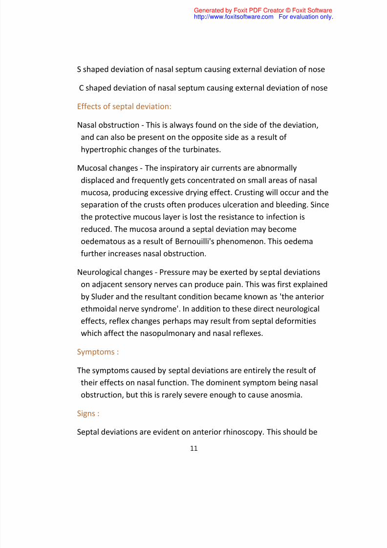
S shaped deviation of nasal septum causing external deviation of nose
C shaped deviation of nasal septum causing external deviation of nose
Effects of septal deviation:
Nasal obstruction - This is always found on the side of the deviation,
and can also be present on the opposite side as a result of
hypertrophic changes of the turbinates.
Mucosal changes - The inspiratory air currents are abnormally
displaced and frequently gets concentrated on small areas of nasal
mucosa, producing excessive drying effect. Crusting will occur and theseparation of the crusts often produces ulceration and bleeding. Since
the protective mucous layer is lost the resistance to infection is
reduced. The mucosa around a septal deviation may become
oedematous as a result of Bernouilli's phenomenon. This oedema
further increases nasal obstruction.
Neurological changes - Pressure may be exerted by septal deviations
on adjacent sensory nerves can produce pain. This was first explainedby Sluder and the resultant condition became known as 'the anterior
ethmoidal nerve syndrome'. In addition to these direct neurological
effects, reflex changes perhaps may result from septal deformities
which affect the nasopulmonary and nasal reflexes.
Symptoms :
The symptoms caused by septal deviations are entirely the result of
their effects on nasal function. The dominent symptom being nasalobstruction, but this is rarely severe enough to cause anosmia.
Signs :
Septal deviations are evident on anterior rhinoscopy. This should be
11
Generated by Foxit PDF Creator © Foxit Softwarehttp://www.foxitsoftware.com For evaluation only.

8/7/2019 ENT imp topics by praveen reddy, hyd
http://slidepdf.com/reader/full/ent-imp-topics-by-praveen-reddy-hyd 12/21
done without the use of nasal speculum because the insertion of
speculum is sufficient to straighten the nasal septum. When the tip of
the nose is lifted septal deviation become evident. Nasal obstruction
may also be present on the opposite side (paradoxical nasal
obstruction). This is due to the presence of hypertrophied turbinates.
If the hypertrophy is limited to turbinate mucosa alone then it will
shrink when decongestant drugs are used in the nasal cavity. If the
hypertrophy is bony then deconstant drops is useless.
Septal deviations in the region of the nasal valve area cause the
greatest obstruction, since this is the narrowest part of the nasal
cavity. This can be identified by the cottle test. A positive cottle test
will confirm the fact that narrowing is present in the nasal valve area.
This is done by asking the patient to pull the cheek outwards and this
manuver is supposed to open up the area thus reducing the block.
The septum should not be considered in isolation and it is necessary
to do a careful examination of the lateral wall of the nasal cavity.
When ever sinus complications like sinusitis is suspected due to
obstruction to the drainage channel of the sinuses by the deviation
xray sinus must be taken.
Septal deviation in new born is associated with asymmetry of the
nostrils, an oblique columella and tip which points in the direction
which is opposite to the deviation. Most of these patients are
diagnosed by the use of Gray's struts. These struts are 4mm wide and
2mm thick and after lubrication, are inserted into the nostrils andthen gently pushed backwards along the floor of the nasal cavity,
hugging the nasal septum. Normally these struts can be introduced
for a distance of 4 - 5 cms, but in cases of septal deviation a frank
obstruction is encountered, usually 1 - 2 cms from the nostril.
12
Generated by Foxit PDF Creator © Foxit Softwarehttp://www.foxitsoftware.com For evaluation only.

8/7/2019 ENT imp topics by praveen reddy, hyd
http://slidepdf.com/reader/full/ent-imp-topics-by-praveen-reddy-hyd 13/21
Cottle has classified septal deviations into three types :
Simple deviations: Here there is mild deviation of nasal septum, there
is no nasal obstruction. This is the commonest condition encountered.
It needs no treatment.
Obstruction: There is more severe deviation of the nasal septum,
which may touch the lateral wall of the nose, but on vasoconstriction
the turbinates shrink away from the septum. Hence surgery is not
indicated even in these cases.
Impaction: There is marked angulation of the septum with a spur which
lies in contact with lateral nasal wall. The space is not increased even
on vasoconstriction. Surgery is indicated in these patients.
Indications for submucous resection of nasal septum:
1. Marked septal deviation occurring behind the vertical line passing
between the nasal processes of the frontal and maxillary bones. This
deviation must be the cause for the patient's symptoms.
2. Closure of septal perforations
3. Source of grafting material
4. To obtain surgical access in hypophysectomy, and vidian
neurecTOMY
Surgically the septum is divided into anterior and posterior segments
by a
vertical line passing between the nasal processes of frontal and
maxillay bones
Procedure:
Submucosal resection of nasal septum is ideally performed under local13
Generated by Foxit PDF Creator © Foxit Softwarehttp://www.foxitsoftware.com For evaluation only.
8/7/2019 ENT imp topics by praveen reddy, hyd
http://slidepdf.com/reader/full/ent-imp-topics-by-praveen-reddy-hyd 14/21
anaesthesia. 4% xylocaine is used as topical anesthetic agent by nasal
packing. 2% xylocaine is used as infiltrative anesthetic agent. It is
mixed with 1 in 1 lakh adrenaline. Infiltration is done at the
mucocutaneous junction on both sides just behind the columella. The
floor of the nasal cavity is also infiltrated on the concave side. Killian's
incision is preferred for SMR operations. Killian's incision is the
commonly used incision. It is an oblique incision given about 5mm
above the caudal border of the septal cartilage
The cartilagenous and bony nasal septum is exposed by elevation of
mucoperichondrial and mucoperiosteal flaps on both sides. This is
done by slicing the septal cartilage just above the columella to access
the opposite side. Flaps are elevated on both sides of the nasalseptum. the cartialge is fully exposed from both sides and is remove
using a Luc's forceps or a Ballanger's swiwel knife. The flaps are
allowed to fall back in place and wound is closed with catgut. Bony
deviations along the floor of the nose if any are also chissled out
before wound closure.
SMR should not be performed in children because it may affect
growth.
Complications of SMR:
1. Septal hematoma
2. Septal abscess
3. Septal perforation
4. Nasal deformities due to excessive removal of dorsal strut of the
septum
5. Removal of the columella cartilage will cause pig snout deformity
Septoplasty:
14
Generated by Foxit PDF Creator © Foxit Softwarehttp://www.foxitsoftware.com For evaluation only.

8/7/2019 ENT imp topics by praveen reddy, hyd
http://slidepdf.com/reader/full/ent-imp-topics-by-praveen-reddy-hyd 15/21
This is a more conservative procedure. The anesthesia is the same as
described for SMR operation. The incision is always sited on the
concave side of the septum. Freer's hemitransfixation incision is
preferred. This is made at the lower border of the septal cartilage. A
unilateral Freer's incision is sufficient for septoplasty. Three tunnels
are created as shown in the figure.
Exposure: The cartilagenous and bony septum are exposed by a
complete elevation of a mucosal flap on one side only. Since flap is
retained on the opposite side the vascularity of the septum is not
compromised.
Mobilisation and straightening: The septal cartilage is freed from all its
attachments apart from the mucosal flap on the convex side. Most of
the deviations are maintained by extrinsic factors such as caudal
dislocation of cartilage from the vomerine groove. Mobilisation alone
will correct this problem. When deviations are due to intrinsic causes
like the presence of healed fracture line then it must be excised along
with a strip of cartilage. Bony deviations are treated either by fracture
and repositioning or by resection of the fragment itself.
Fixation:
The septum is maintained in its new position by sutures and splints.
Advantages of Freer's incision:
1. The incision is cited over thick skin making elevation of flap easy.
2. There is minimal risk of tearing the flap
3. The whole of the nasal septum is exposed.
4. If need arises Rhinoplasty can be done by extending the same
incision to a full transfixation one.
Advantages of Septoplasty:
15
Generated by Foxit PDF Creator © Foxit Softwarehttp://www.foxitsoftware.com For evaluation only.

8/7/2019 ENT imp topics by praveen reddy, hyd
http://slidepdf.com/reader/full/ent-imp-topics-by-praveen-reddy-hyd 16/21
1. More conservative procedure
2. Performed even in children
3. Less risk of septal perforation
4. Less risk of septal hematoma
Killian's incision is preferred for SMR operations.It is an oblique incision
given about 5mm above the caudal border of the septal cartilage
septoplasty
incision always sited on the concave side of the septum. Freer's
hemitransfixation incision is preferred. This is made at the lower
border of the septal cartilage. A unilateral Freer's incision is sufficient
for septoplasty. Three tunnels are created
complications of c.sinusitis
Anatomically the paranasal sinuses are closely adjacent to vital
structures like orbit, brain etc. Complications attributed to sinus
infections are caused by spread of infection from the paranasal
sinuses to these adjacent areas.
Routes of spread:
1. Bacterial infections from the sinuses can spread through natural
dehiscences and weakness of the bony barriers. In chronic infections
the surrounding bone undergoes sclerosis, while in acute sinusitis
massive osteolysis is commonly seen.
16
Generated by Foxit PDF Creator © Foxit Softwarehttp://www.foxitsoftware.com For evaluation only.

8/7/2019 ENT imp topics by praveen reddy, hyd
http://slidepdf.com/reader/full/ent-imp-topics-by-praveen-reddy-hyd 17/21
2. Lamina papyracea is a paper thin bone separating the orbit from the
ethmoidal sinuses. Congenital dehiscences of this bone is commonly
seen through which spread of infection can occur from the ethmoids
into the orbit. In childhood the frontal sinuses are underdeveloped
and orbital complications are caused commonly by acute ethmoiditis.
3. Floor of the frontal sinuses form the roof of the orbit. In older
children and in adults frontal sinus infections can spread into the orbit
causing orbital complications.
4. Infraorbital canal in the floor of the orbit is a weak area through
which infections from orbit may enter into the maxillary sinus.
5. Spread of infection can also occur via diploic veins present in thefrontal bone. These veins are known as the veins of Breschet. This is
preceded by thrombophlebitis.
6. Venous connections between the sinuses and the orbit donot have
any valves facilitating spread of infection from the sinuses to the
orbit.
7. The roots of the second premolar and the first upper molar are
intimately related to the floor of the maxillary sinus. This facilitates atwo way spread of infection. In cases of isolated maxillary sinusitis
dental causes must be suspected.
Predisposing factors responsible for complications:
1. Immunocompromised patient (e.g. HIV)
2. Diabetes mellitus
3. Irregular treatment for sinus infections
4. Inappropriate / Inadequate antibiotic therapy
17
Generated by Foxit PDF Creator © Foxit Softwarehttp://www.foxitsoftware.com For evaluation only.

8/7/2019 ENT imp topics by praveen reddy, hyd
http://slidepdf.com/reader/full/ent-imp-topics-by-praveen-reddy-hyd 18/21
Orbital complications of sinusitis:
Commonly caused in young individuals. These are frequently due to
ethmoiditis. This is the common cause for orbital complication in
children. In adults involvement of frontal sinuses could commonlycause orbital complications. In adults acute sphenoiditis can cause
involvement of optic nerve leading on to blindness.
Hubert's classification of orbital complications of sinusitis:
Hubert classified orbital complications arising from sinusitis into five
groups:
Group I: Inflammatory oedema of eyelids with or without oedema of
orbital contents.
Group II: Subperiosteal abscess with oedema of lids or spread of pus to
the lids.
Group III: Abscess of orbital tissues
Group IV: Mild to severe orbital cellulitis with phlebitis of ophthamicveins
Group V: Cavernous sinus thrombosis
Smith & Spencer classification of orbital complications:
Group I: Preseptal cellulitis - Characterised by oedema of eyelids
without tenderness, visual loss or limitation of ocular mobility.
Group II: Orbital cellulitis without abscess formation - characterised by
diffuse oedema of adipose tissues of orbit.
Group III: Orbital cellulitis with subperiosteal abscess formation with
18
Generated by Foxit PDF Creator © Foxit Softwarehttp://www.foxitsoftware.com For evaluation only.

8/7/2019 ENT imp topics by praveen reddy, hyd
http://slidepdf.com/reader/full/ent-imp-topics-by-praveen-reddy-hyd 19/21
displacement of the globe. May or may not be associated with visual
loss. Ocular mobility is restricted.
Group IV: Orbital cellulitis with intraperiosteal abscess. Here the
displacement of globe is severe with restriction of ocular mobility.
Group V: Cavernous sinus thrombosis.
All these ocular complications are associated with pain, tenderness,
and displacement of the globe. These symptoms are not seen in
preseptal cellulitis. Presence of preseptal cellulitis should be viewedwith caution and treated aggressively as it may lead to further sinister
complications. If the infection affects the posterior ethmoids and
onodi cells optic nerve could be affected. In these patients color
vision gets disturbed first. They will have difficulty distinguishing
between red from brown and blue from black.
If thrombophlebitis extends posteriorly from the orbit it will involve thecavernous sinus. Opposite side is also affected in this condition. This is
one of the common complication of sphenoiditis. These patients will
complain of headache, and paraesthesia along the distribution of
trigeminal nerve.
All patients with impending complications should undergo a CT scan.
This will clearly indicate the site of abscess. Visual acuity tests must be
performed. Changes in visual acuity must be distinguished from
diplopia due to restricted ocular mobility. Proptosis is always axial in
patients with orbital oedema. Severe proptosis will cause chemosis.
Sudden increase in proptosis should be relieved within 2 hours if 19
Generated by Foxit PDF Creator © Foxit Softwarehttp://www.foxitsoftware.com For evaluation only.

8/7/2019 ENT imp topics by praveen reddy, hyd
http://slidepdf.com/reader/full/ent-imp-topics-by-praveen-reddy-hyd 20/21
permanent damage to vision is to be prevented. Globe will tolerate
even 2cm of displacement if proptosis is chronic as in mucoceles
These patients must receive broad spectrum antibiotics. These
antibiotics must be administered parenterally. If there is any sign of
visual disturbance surgical decompression should be immediatly
resorted to. Sinus infections must be treated surgically after the acute
crisis is over.
Long term sequelae of orbital complications:
1. Vision loss
2. Ophthalmoplegia
3. Exposure keratitis due to corneal anesthesia
4. Uveitis
5. Glaucoma
Intracranial complications:
Intracranial complications are less common than orbital complications
of sinusitis. These complications can coexist. Intracranial
complications are common in adolescents and young adults because
the diploic system of veins reaches its peak during adolescence.
Males are more commonly affected than females.
Intracrainal complications can be divided into:
20
Generated by Foxit PDF Creator © Foxit Softwarehttp://www.foxitsoftware.com For evaluation only.

8/7/2019 ENT imp topics by praveen reddy, hyd
http://slidepdf.com/reader/full/ent-imp-topics-by-praveen-reddy-hyd 21/21
1. Abscesses - extradural, subdural or intracerebral
2. Meningitis
3. Encephalitis
4. Cavernous sinus thrombosis, superior sagittal sinus thrombosis
Intracranial abscesses are commonly found subdurally. Second
common abscess is frontal lobe abscess. Intracerebral abscess begin
as a focus of cerebritis, which gets localised with necrosis and pus
formation. Multiple intracranial abscess formation indicates septic
thrombophlebitis. These abscesses can be acute or chronic. Abscesses
involving the frontal lobe cause very few discomfort or symptoms till
a large size is reached.
CT scan is diagnostic. Antibiotics must be administered in large doses
parenterally to enable adequate concentrations of the drug reaching
the brain. Intracranial abscess needs to be drained surgically.
Pott's puffy tumor: is one of the bony complications of frontal sinusitis.
This condition was first described by Sir Percival Pott. The frontalbone is a diploic bone with intervening bone marrow. Osteomyelitis of
frontal bone leads to this condition. In this condition both the anterior
and posterior walls of frontal sinus is involved. Infection may spread
anteriorly on to the forehead of posteriorly to form subdural abscess.
Debridement of the bone must be performed under cover of
intravenous antibiotics.
21
Generated by Foxit PDF Creator © Foxit Softwarehttp://www.foxitsoftware.com For evaluation only.


![[XLS]hmda.gov.inhmda.gov.in/LRSStatus/Manchirevula.xls · Web viewV.Neeraja Reddy V.Neeraja Reddy, P.No.403, Shilpi Homes, DD Colony, Bagh Amberpet, Hyd DD No. 001153, 31/12/08, HDFC,](https://img.dokumen.tips/doc/110x75/5ab4b92a7f8b9a0f058c2bc4/xlshmdagov-viewvneeraja-reddy-vneeraja-reddy-pno403-shilpi-homes-dd-colony.jpg)