Embed Size (px)
Citation preview
Research ArticleEnhancement Effect of Microbubble-Enhanced Ultrasound inMicrowave Ablation in Rabbit VX2 Liver Tumors
Shuyi Xiao,1,2,3 Zhiwen Hu ,2,3 Yan He,2,3 Hai Jin,2,3 Yuwen Yang,2,3 Liping Chen,2,3
Qiaoli Chen,2,3 Qiong Luo,3 and Jianhua Liu 2,3
1 e First Affiliated Hospital, Jinan University, Guangzhou, China2Department of Medical Ultrasound, Guangzhou First People’s Hospital, Guangzhou Medical University, Guangzhou, China3 e Second Affiliated Hospital of South China University of Technology, Guangzhou, China
Correspondence should be addressed to Jianhua Liu; [email protected]
Received 24 August 2019; Accepted 30 December 2019; Published 23 January 2020
Academic Editor: Tobias De Zordo
Copyright © 2020 Shuyi Xiao et al. ,is is an open access article distributed under the Creative Commons Attribution License,which permits unrestricted use, distribution, and reproduction in any medium, provided the original work is properly cited.
Objectives. One reason for the high recurrence and metastatic rates of tumors such as hepatocellular carcinoma (HCC) treated bymicrowave ablation (MWA) is the presence of residual foci in the tumor due to heat sink effect. Microbubble-enhanced ul-trasound (MEUS) can noninvasively disrupt and block the tumor blood perfusion and has the potential to overcome the heat sinkeffect and enhance the therapeutic effect of MWA.,e study aimed at evaluating the potential additional benefit of microbubble-enhanced ultrasound (MEUS) in hepatocellular carcinoma (HCC) treated by microwave ablation (MWA).Methods. In this study,a new strategy of combining MWA with MEUS for treating HCC was proposed. Twenty-four rabbits with VX2 tumors in liverswere randomly divided into MEUS+MWA, MEUS alone, MWA alone, and blank control groups, respectively (n� 6). In theMEUS group, the tumors were directly exposed to therapeutic ultrasound for 5min with a concurrent intravenous injection ofmicrobubbles (0.1ml/kg diluted into 5ml saline). In the MWA group, the tumors were treated by MWA for 1min. In theMEUS+MWA group, tumors were ablated by MWA for 1min after ultrasound cavitation enhanced by microbubbles as in theMEUS group. In the blank control group, the tumors received probe sham and intravenous saline. Contrast-enhanced ultrasound(CEUS) was performed before treatment and immediately after treatment to display the size, shape, and contour of the tumors.,roughout the treatment process, the local temperature of the treatment area was detected by a temperature needle puncturedinto the tumor. ,e blood samples of animals were obtained after treatment for evaluating the liver function. Tumor cell necrosisand apoptotic rates were observed after treatment by histological examination. Results. CEUS showed that although perfusiondefects appeared in all the treatment groups, especially in the MEUS+MWA group, there was no significant difference betweenthe two groups on the volumes of perfusion defects, which were 1.78± 0.31 (cm3) in the MWA group and 1.84± 0.20 (cm3) in thecombined group (P< 0.01).,e time to reach the peak temperature of the treatment area was 21.7± 5.0 (s) in theMWA group and10.3± 5.0 (s) in the MEUS+MWA group (P< 0.01). ,e peak temperature (PT) of the two groups were 100.9± 5.0 (°C) and134.1± 6.0 (°C), respectively (P< 0.01). ,is showed that the local temperature of the treatment area was sharply increased to ahigher PT using MEUS+MWA. Histological examination results showed that the apoptosis rate and necrosis rate in theMEUS+MWA group were 23.6± 4.6% and 60.5± 9.7%, respectively, which are significantly higher than those in the MWA group(17.9± 4.5% and 37.6± 3.4%) and those in the MEUS group (18.2± 1.0% and 37.6± 3.4%). ,ey are all higher than those in thecontrol group (3.85± 1.72% and 5.3± 1.1%). Hematological examination showed no significant differences between treatmentgroups on liver function. Conclusions. ,ese results suggested MEUS treatment alone may significantly reduce tumor bloodperfusion and led to a sharp rise in the local temperature of the treatment area to a higher PT using MEUS+MWA with higherrates of necrosis and apoptosis of cancer cells without severe liver function damage, which might be a safe strategy fortreating HCC.
HindawiBioMed Research InternationalVolume 2020, Article ID 3050148, 10 pageshttps://doi.org/10.1155/2020/3050148

1. Introduction
Hepatocellular carcinoma (HCC) is one of the leadingcauses of cancer-related deaths [1]. Current treatmentstrategies for HCC include surgical treatment, thermalablation, and localized embolization chemotherapy aloneor in combination [2]. Among these, surgical treatment isthe most important and effective treatment for HCC atpresent, which includes surgical resection and livertransplantation [3]. ,e 5-year survival rate of patientsundergoing surgical resection is as high as 70%, while thetreatment is limited to HCC patients without hepatocir-rhosis, which comprises about 20–30% of patients withHCC [4]. Despite a 4-year overall survival rate of 85% and arecurrence-free survival rate of 92%, liver transplantation isstill limited due to strict criteria, surgical candidacy, tumorburden, and the availability of donors [5].
,ermal ablations such as microwave ablation (MWA),radiofrequency ablation (RFA), and high-intensity focusedultrasound are important complements of surgical treat-ment for HCC. ,ermal ablation kills the tumors by in-creasing the temperature of solid tumors through heataccumulation [6]. ,is method has obvious advantageswith regard to safety (less invasive), good tolerance, re-peatability, and efficiency. HCC nodules are considered asthe most common targets of thermal ablation clinically[7, 8]. Microwave ablation MWA causes irreversiblethermal necrosis of the tissue through the delivery ofmicrowave energy. Previous studies have reported thatMWA can treat HCC nodules which are larger than 3 cm,resulting in a complete ablation rate of 92.6%, local re-currence rate of 22%, and 3-year survival rate of 30.9%[9, 10]. According to previous studies, “heat sedimentationeffect” is one of the major factors that influence the ablationsize and shape, leading to the local residual focus of thetumors. Blood flow through tumors or major peripheralblood vessels promoted heat loss and prevented heat de-position by removing the heat [11], causing a slow or in-sufficient temperature rise in the treatment area. Due tothis, the tumor cells cannot be completely ablated aftertreatment and the residual foci may lead to recurrence.How to acquire a sufficient ablation area for HCC treat-ment has become a major issue in the use of MWAtechnique [12].
One of the strategies to achieve a more thorough thermalablation area is to block the blood flow of tissues beforeablation. If the blood supply of HCC and surrounding livertissues is reduced and the “heat sedimentation effect” isreduced, the efficiency of heat ablation will be improved[13, 14]. Transarterial embolization or chemoembolization(TAE/TACE) can reduce blood perfusion by slowing downblood flow, causing local ischemia and increasing heat re-tention [15, 16]. ,is has been performed in combinationwith RFA and MWA, resulting in an improved completeablation response and long-term survival rate [17, 18].Several studies have reported the use of microbubble-en-hanced ultrasound (MEUS) in the disruption of tumormicrovasculature [19–21]. ,e inertial cavitation induced byhigh-amplitude, low-intensity ultrasound and microbubbles
severely damages the small vessels and vasculature, resultingin the cessation of circulation in relevant tissues [22].According to a previous study, MEUS was applied to disrupttumor microvasculature and arrested tumor perfusion forup to 24 h [20].,e combination ofMEUS and percutaneousethanol ablation (PEA) increased the necrosis rate of tumorin rats significantly from 81.0% to 97.5% [23]. In normalrabbit liver, MEUS blocked the circulation for 15–60minand enlarged the PEA ablation volume up to 10-fold [24]. So,MEUS combined with PEA can obviously enlarge the ab-lation area [25].
Hence, this study aimed at investigating the possibility,safety, and effectiveness of MEUS-induced perfusionblockage to enhance MWA of HCC in vivo.
2. Materials and Methods
2.1. Experimental Design. Twenty-four New Zealand rabbitsweighing 2.6 kg to 3.0 kg, regardless of gender, were includedin this study.,ey were purchased from the Medical AnimalExperimental Center of Guangdong (MAECG) and were fedwith standard laboratory diet and tap water ad libitum. ,eexperiments on laboratory animals were performed with theapproval of the Institutional Animal Care and Use Com-mittee (IACUC) of the MAECG, Guangdong, China.Fourteen days after tumor implantation, 24 rabbits withpalpable tumors (approximately 1.5–2.0 cm) were randomlydivided into four groups, with 6 rabbits in each group: (i)blank control group; (ii) MEUS treatment group; (iii) MWAtreatment group; and (iv) MEUS+MWA treatment group.,e animals were anesthetized by using 0.3mL/kg of 2%pentobarbital before surgery and then were placed in supineposition for removing the upper abdominal hair. After amiddle surgical incision of the abdominal wall, the liverlobes with tumor nodules were exposed and fixed ex vivo insitu. In the MEUS group, the liver tumors were treated withultrasound cavitation therapy for 5 minutes combined withan intravenous injection of diluted SonoVue saline micro-bubble suspension at a dose of 0.1mL/kg. In the MWAgroup, the liver tumors were treated withMWA for 1min. Inthe MEUS+MWA group, the liver tumors were treated byultrasound cavitation therapy firstly followed by MWA onthe same target region. In the blank control group, rabbitswere injected with the same amount of saline and receivedno ultrasound or MWA treatment. CEUS was performedbefore treatment and immediately after treatment to displaythe size, shape, and contour of the tumors and the size,shape, and contour of the effective treatment areas.,roughout the treatment process, the local temperature ofthe treatment area was detected by a temperature needlepunctured into the tumor. ,e blood samples of animalswere obtained before and immediately after the treatment toevaluate liver function. Tumor cell necrosis and apoptoticrate were observed after treatments by histologicalexamination.
2.2. Rabbit VX2 Liver Tumor Model. ,e rabbit VX2 tumorliver model was established by an interventional method as
2 BioMed Research International

described previously [26]. Firstly, we removed the VX2tumor tissue from the thigh of a tumor-bearing rabbit andcut into 3-4mm3 cubes under sterile conditions. ,en thetumor was placed in normal saline and sliced into 1 to 2mm3
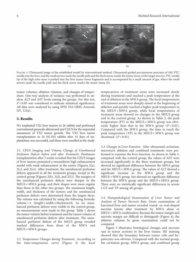
fragments. Next, the 26 animal recipients were anesthetizedwith an intramuscular injection of 2% pentobarbital at a doseof 0.2mL/kg, and their upper abdomens were cleanly shavedfor undergoing a preliminary ultrasound (Mindray M7,Myry Biomedical Electronics Co., Ltd., Shenzhen, China)examination using a linear array transducer to determine thetarget implantation site within the liver. We used an 18GPTC puncture needle (Hakko, 1490 O-aza, Japan) to per-cutaneously puncture the liver tissue (Figure 1(a)), whichincluded a hollow core and a sharp, blunt inner stylet. ,esharp inner stylet was removed, and a small tumor fragment(1-2mm3) was pushed through the core into the liver. Ahyperechogenic area representing the tumor fragment wasvisible with ultrasound imaging (Figure 1(b)). After im-plantation, CEUS was performed to monitor the growth ofthe tumor and to measure the tumor size.
2.3. Ultrasound Cavitation erapy. We used an ultrasoundcavitation device with an ultrasound therapy apparatus (FullDigital Ultrasound Microbubble Cavitation ,erapy In-strument, Shenzhen WELL. D Medical Electronics Co., Ltd.,Shenzhen, China). A probe with a 1MHz central frequencywas used to transmit 400-cycle pulses at a 10Hz pulserepetition frequency for 5min. ,e transducer works in anintermittent mode of 5 s on and 5 s off. ,e actual workingduty cycle was approximately 0.20%. ,e transducer gen-erated a peak positive pressure of 1MPa. All rabbits wereanesthetized with an intravenous 2% pentobarbital dose of0.2mL/kg injected into the ear vein with a 21-gauge needle.,e cavitation probe was then placed on the exposed surfaceof the liver tumor. In the MEUS group, the cavitationtherapy was combined with a continuous intravenous in-jection of SonoVue saline solution (0.1mL/kg) at the sametime through the ear vein.
2.4. MWA Ablation. MWA was performed by using themicrowave therapeutic instrument (EC0-100C, Microwave,erapeutic Instrument, Yigao Medical Equipment Co.,Ltd., Nanjing, China) that is equipped with an ablationneedle (ECO-100AI3, Microwave ,erapeutic Instrument,Yigao Medical Equipment Co., Ltd., Nanjing, China) whosediameter is 1.6mm. ,e treatment needle was inserted intothe centre of the tumor nodule and its tip is ready topuncture into the deeper margins of the tumor. MWA wasperformed with an output power of 20W for 5 s, and then,the needle was withdrawn.
2.5. CEUS and Size Measurements. CEUS imaging of therabbit liver was performed before and after therapy. ,eSonoVue microbubble suspension (0.05mL/kg) was dis-solved in 5mL saline and injected as a bolus dose into the earvein, followed by 2mL saline. A commercially availableultrasound imaging system (GE Vivid E9, GE Medical
Health Co. Ltd, USA) that was equipped with an L9 high-frequency linear array probe was used for CEUS. Contrastmodality was conducted by using a low mechanical index(MI� 0.05), and the frequency was 5–9MHz. ,e ultra-sound probe was placed on the surface of the liver. ,eCEUS images were digitally stored for up to 5min to per-form a quantitative analysis off-line. Grayscale ultrasoundwas used to determine the location, echogenicity, and vol-ume of the tumor. CEUS images were used to determine thelocation, echogenicity, and volume of the effective treatmentarea. ,e maximum length, width, and thickness of thetumors and the unenhanced perfusion defects of CEUSimages were determined from the largest slices. ,e volumewas calculated by using the following formula: volume� π(length×width× thickness)/6 [27].
2.6. Temperature Monitoring. During the treatment, oneneedle thermometer (SENDAE-115, Guangzhou Sun-GunCorp., Guangzhou, China) was inserted into the liver tu-mors, just near to the tip of the ablation needle or in thecentre of ultrasound irradiation area. ,e thermometerautomatically recorded the local temperature changes dur-ing the treatment.
2.7. Blood Sample Collection. Blood samples of animals ineach group were obtained through the auricular artery orthe external jugular vein immediately after treatment. ,emain liver function indicators such as alanine amino-transferase (ALT), aspartate aminotransferase (AST),R-glutamyl transferase (r-GT), and total protein (TP) weremeasured.
2.8. Histological Examination. ,e rabbits were sacrificedimmediately after treatment. Liver lobes with tumor noduleswere harvested, fixed in formalin, and then embedded inparaffin. Samples were cut into 4mm cryosections, stainedwith hematoxylin and eosin (H&E), and then subjected toterminal deoxynucleotidyl transferase dUTP nick-end la-beling (TUNEL) for apoptosis analysis.
H&E staining was completed by using a standard pro-tocol for gross histological assessment of cellular density,necrosis, and fibrosis. Images of tumor sections stained withH&E were acquired with a Mirax Scanner (Carl Zeiss,Oberkochen, Germany) by using a 20x objective. ,e per-centage of necrotic area was assessed under a 1000x ob-jective. ,e degree of apoptosis and tumor necrosis wereassessed with TUNEL staining (Roche Diagnostics)according to the manufacturer’s instructions. Tumor ne-crosis rate was measured by ImageJ software (NationalInstitutes of Health). ,e necrosis rate (%) was calculated bythe following formula: necrosis rate (%)� (overall area-− survival area)/overall area 100%.
2.9. Statistical Analysis. All data were expressed as mean-± standard deviation. An independent sample t-test wasused to compare the two independent samples, and analysisof variance was used among the three or four groups of the
BioMed Research International 3
tumor volumes, ablation volumes, and changes of temper-ature. One-way analysis of variance was performed to an-alyze ALT and AST levels among the groups. For this test,P< 0.05 was considered to indicate statistical significance.All data were analyzed by using SPSS 19.0 (IBM, Armonk,NY, USA).
3. Results
We implanted VX2 liver tumors in 26 rabbits and performedconventional grayscale ultrasound and CEUS for the sequentialassessment of VX2 tumor growth. ,e VX2 liver tumortransplantation in 24 (92.3%) rabbits after 14 days of im-plantation was successful, and these were enrolled in the study.
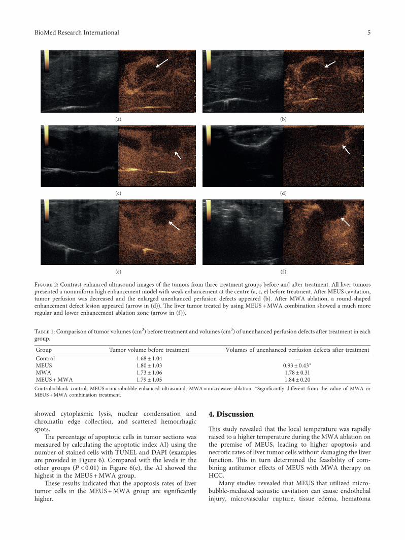
3.1. CEUS Imaging and Volume Change of UnenhancedPerfusion Defects before and after Treatment. Posttumortransplantation after 2 weeks revealed that the CEUS imagesof liver tumors presented a nonuniform high enhancementmodel with weak enhancement at the centre (Figures 2(a),2(c), and 2(e)). After treatment, the unenhanced perfusiondefects appeared in all the treatment groups, except in thecontrol group (Figures 2(b), 2(d), and 2(f)). ,e margins ofthe unenhanced perfusion defects were sharper in theMEUS+MWA group, and their shapes were more regularthan those in the other two groups. ,e maximum length,width, and thickness of the tumors and the unenhancedperfusion defects were determined from the largest slices.,e volume was calculated by using the following formula:volume� π (length×width× thickness)/6. As no unen-hanced perfusion defects were visible in the control group,no measurements were made in this group. Table 1 showsthe tumor volume before treatment and the tumor volume ofunenhanced perfusion defects after treatment. ,e unen-hanced perfusion defects of the MEUS group revealedmarked differences from those of the MWA andMEUS+MWA groups.
3.2. Temperature Changes during Treatment. According tothe time-temperature curve (Figure 3) the local
temperatures of treatment areas were increased slowlyduring treatments and reached a peak temperature at theend of ablation in the MWA group. ,e local temperaturesof treatment areas were sharply raised at the beginning ofablation and quickly reached a higher peak temperature inthe MEUS +MWA group, while local temperatures oftreatment areas showed no changes in the MEUS groupand in the control group. As shown in Table 2, the peaktemperature (PT) in the MEUS +MWA group was obvi-ously higher than that in the MWA group (P< 0.01).Compared with the MWA group, the time to reach thepeak temperature (TP) in the MEUS +MWA group wasshortened (P< 0.01).
3.3. Changes in Liver Function. After ultrasound cavitation,microwave ablation and combined treatments were per-formed to examine the liver function. As shown in Table 3,compared with the control group, the values of AST wereincreased significantly in the three treatment groups, butshowed no significant difference between the MWA groupand the MEUS+MWA group. ,e values of ALT showed asignificant increase in the MWA group and theMEUS+MWA group, but showed no significant differencebetween the MWA group and the MEUS+MWA group.,ere were no statistically significant differences in serumr-GT and TP among all groups.
3.4. Histopathological Examination of Liver Tumor andAnalysis of Tumor Necrosis Rate. Gross examination ofharvested liver and tumor revealed round- or oval-shapednecrotic lesions after treatment by MWA alone orMEUS+MWA combination. Because the tumor margin andnecrotic margin are difficult to distinguish (Figure 4), theablation volumes by gross examination could not becompared.
Figure 5 illustrates histological changes and necrosisrate in tumor sections in the liver tissues. HE stainingshowed that the boundary between tumor cells and he-patocytes was obvious. Compared with the normal group,the cavitation group, MWA group, and combined group
(a) (b)
Figure 1: Ultrasound image of the established model by interventional method. Ultrasound-guided percutaneous puncture of 18G PTCneedle into the liver, and the small arrows mark the needle path and the thick arrowmarks the tumor tissue of the target area (a). PTC needletip of the high echo mass is pushed into the liver tumor tissue fragments and is accompanied by a small amount of gas, where the smallarrows mark the needle path and the thick arrow marks the tumor tissue (b).
4 BioMed Research International
showed cytoplasmic lysis, nuclear condensation andchromatin edge collection, and scattered hemorrhagicspots.
,e percentage of apoptotic cells in tumor sections wasmeasured by calculating the apoptotic index AI) using thenumber of stained cells with TUNEL and DAPI (examplesare provided in Figure 6). Compared with the levels in theother groups (P< 0.01) in Figure 6(e), the AI showed thehighest in the MEUS+MWA group.
,ese results indicated that the apoptosis rates of livertumor cells in the MEUS+MWA group are significantlyhigher.
4. Discussion
,is study revealed that the local temperature was rapidlyraised to a higher temperature during the MWA ablation onthe premise of MEUS, leading to higher apoptosis andnecrotic rates of liver tumor cells without damaging the liverfunction. ,is in turn determined the feasibility of com-bining antitumor effects of MEUS with MWA therapy onHCC.
Many studies revealed that MEUS that utilized micro-bubble-mediated acoustic cavitation can cause endothelialinjury, microvascular rupture, tissue edema, hematoma
Table 1: Comparison of tumor volumes (cm3) before treatment and volumes (cm3) of unenhanced perfusion defects after treatment in eachgroup.
Group Tumor volume before treatment Volumes of unenhanced perfusion defects after treatmentControl 1.68± 1.04 —MEUS 1.80± 1.03 0.93± 0.43∗MWA 1.73± 1.06 1.78± 0.31MEUS+MWA 1.79± 1.05 1.84± 0.20Control� blank control; MEUS�microbubble-enhanced ultrasound; MWA�microwave ablation. ∗Significantly different from the value of MWA orMEUS+MWA combination treatment.
(a) (b)
(c) (d)
(e) (f )
Figure 2: Contrast-enhanced ultrasound images of the tumors from three treatment groups before and after treatment. All liver tumorspresented a nonuniform high enhancement model with weak enhancement at the centre (a, c, e) before treatment. After MEUS cavitation,tumor perfusion was decreased and the enlarged unenhanced perfusion defects appeared (b). After MWA ablation, a round-shapedenhancement defect lesion appeared (arrow in (d)). ,e liver tumor treated by using MEUS+MWA combination showed a much moreregular and lower enhancement ablation zone (arrow in (f)).
BioMed Research International 5

Table 2: Temperature changes during treatments.
Group TP (s) PT (°C)Control — 34.5± 2.0MEUS — 34.7± 2.0MWA 21.7± 5.0 100.9± 5.0MEUS+MWA 10.3± 2.6∗ 134.1± 6.0∗
Control� blank control; MEUS�microbubble-enhanced ultrasound;MWA�microwave ablation; T� time to the peak; PT�peak temperature.∗Significantly different from the other groups.
Table 3: Changes of liver function after treatments.
Group ALT AST r-GT TPControl 35.7± 6.5∗∗ 43.3± 12.1∗ 1.7± 0.6∗ 67.0± 5.8MEUS 37.7± 13.4 90.5± 20.1 5.0± 1.9 69.8± 3.0MWA 99.7± 25.3 132.2± 44.1 5.8± 1.5 59.7± 9.2MEUS+MWA 97.0± 8.4 135.3± 16.5 6.8± 3.3 59.6± 7.8∗Significantly different from the value of other groups. ∗∗Significantlydifferent from the value of MWA or MEUS+MWA combinationtreatment.
21:14:17 21:14:56 21:15:36 21:16:16 21:16:56 21:17:36ROM
104
89
74
59
44
29
ROM
(a)
21:44:10 21:45:04 21:45:59 21:46:54 21:47:49 21:48:44ROM
149
124
99
74
49
24
ROM
(b)
ROM19:53:18 19:55:22 19:57:27 19:59:32 20:01:37
50
45
40
35
30
25
ROM
(c)
Figure 3: ,e temperature curves of treatment groups during treatment. ,e temperature curve during treatment in the MWA group (a) isincreased slowly and reached the peak temperature at the end of ablation. ,e temperature curve during MWA treatment in theMEUA+MWA group (b) is sharply raised at the beginning of ablation, and the peak temperature is obviously higher than that in the MWAgroup. ,e temperature curve during treatment in the MEUA+MWA group (b) showed fluctuation during MEUS therapy (c), and there isno significant difference in the temperature fluctuation.
6 BioMed Research International

formation, and thrombosis in many tissues and tumors[18, 28]. Our results were consistent with the findings ofthese studies. In the MEUS alone group, the ultrasoundcavitation therapy alone indeed reduced the tumor bloodperfusion without accompanying a rise in the temperature.,e tumor circulation was stopped (Figure 1(b)) with ir-regularly unenhanced perfusion defects. ,e mechanicaleffects of MEUS produced sonoporation, microvascularrupture, hemorrhage, and microvascular endothelial injuryin a variety of tumors [28, 29]. In our study, histopatho-logical examination revealed mild cytoplasmic lysis, nuclearcondensation and chromatin edge collection, and scatteredhemorrhagic spots. ,ese vascular effects provide an op-portunity to overcome the heat sink effect.
It makes sense that MEUS+MWA combination therapyresulted in a rapid rise in the local temperature to a higherpeak (Figure 3). ,is is a very intuitive indication that ul-trasound cavitation could enhance the effect of MWA onliver tumors by overcoming the heat sink effect by blockingthe blood supply of tumors and promoting the dissolution oftumors. ,is was also supported by the results of histo-pathological examination. Previous study [25] showed thatthe ablation liver volume induced by MEUS+RFA com-bination therapy was 2.8 times larger than that induced byRFA alone in simple liver tissue, while in the present study, itwas difficult to identify the margins of the tumors and theablation areas, and so the volumes of unenhanced per-fusion defects in different groups were compared, whichare considered as effective ablation areas. Although therewas no statistical difference between the volumes ofunenhanced perfusion defects between the MEUS +MWAgroup and the MWA group, the shape of the ablation areaappeared more regular and round and the margin of theablation area was sharper in the MEUS +MWA group.
,ese phenomena are consistent with the results of his-topathological examinations. Pathological staining anal-ysis showed that the necrosis area of VX2 liver tumors inthe MWA group was larger than that in the MEUS group,and the necrosis area of the combination therapy groupshowed the largest, further proving that the combinationtherapy can rapidly warm up the local temperature to ahigher temperature in a short time, and promoting thecoagulation as well as the necrosis of tumor tissues. Ul-trasound cavitation and MWA can promote the apoptosisand necrosis of tumor tissues. However, no significantdifferences were observed in the volumes of contrast-en-hanced defects before and after MWA in the MWA ab-lation group and in the MEUS +MWA group. ,is mightbe due to the fact that the tumors enrolled in this study andthe power and time of microwave treatment were all small.So, it cannot fully reflect the effect of MEUS or MWA.
In this study, the effect on liver function after treatmentremains a concern. It was found that MWA andMEUS +MWA can lead to increased ALT and AST levelswhen compared with the blank control group, which wasthe manifestation of liver function damage, but there wereno significant changes between these two groups bythemselves. ,is suggested that both ultrasound cavitationtherapy and MWA therapy and their combination cancause some damages to the liver function, but the damagein the combined group was not obviously higher thanthose in the alone treatment group. ,ese results dem-onstrated the safety of combined therapy for treating HCC.Furthermore, previous study [25] found that ALTand ASTwere peaked at about 24–48 h in the MEUS + RFA and RFAgroups, but were almost recovered after 8 d. However, ourstudy was carried out immediately after treatment andcannot reflect the long-term changes of liver function. It is
(a) (b)
(c) (d)
Figure 4: Macroscopic view of liver tumors after treatments. (a) ,e control group; (b) microbubble-enhanced ultrasound alone; (c) MWAablation alone; (d) MEUS+MWA ablation. Tumor margins and necrotic margins are difficult to distinguish.
BioMed Research International 7

one of the limitations of the present study that we did notcontinuously observe the liver function after treatment tofind out whether the liver function might be restoredwithin a few days.
Regarding the mechanism of the effect of ultrasoundcavitation combined with MWA on liver tumors, a seriesof continuous changes initiated by acoustic cavitation wereobserved. Firstly, acoustic cavitation enhanced bymicrobubbles generated mechanical effects, includinghigh-pressure shock waves and microjets [28], leading totransient porosity in the cellular membranes (sonopora-tion) and damage to the vascular wall. ,e endothelial celldamage combined with basement membrane exposure isfollowed by platelet activation, which further promotes theformation of thrombus and increases circulation resis-tance, and finally reducing or blocking tumor bloodperfusion [29]. Finally, the effect of reducing heat sink was
achieved. In this study, CEUS showed that blood perfusionin the MEUS +MWA group and the single treatmentgroup was blocked and the blocking effect in theMEUS +MWA group was better when compared to thealone treatment group. Meanwhile, higher PT and shortertime to reach the PT in the MEUS +MWA group showedthat ultrasound cavitation could effectively block the bloodsupply of tumors and surrounding liver tissues, promotingthe injury of neovascularization of tumors and signifi-cantly reducing the impact of “heat sedimentation effect.”,is confirmed that the temperature promotes coagulationand necrosis of tumor tissues.
5. Conclusions
Overall, we demonstrated that MEUS can enhance the ab-lation effect of MWA in rabbit liver tumors without
100μm×63
20μm×400
(a)
100μm×63
20μm×400
(b)
100μm×63
20μm×400
(c)
100μm×63
20μm×400
(d)
∗∗∗
Control
∗∗∗
MEUS MWA MEUS + MWA
∗∗∗&#
0
20
40
60
80
Nec
rosis
rate
(%)
(e)
Figure 5: Photographs of hematoxylin and eosin staining.,is showed that the boundary between tumor cells and hepatocytes was obvious.Compared with the control group (a), the MEUS group (b), the MWA group (c), and the MEUS+MWA group (d) had cytoplasmic lysis,nuclear condensation and chromatin edge collection, and scattered hemorrhagic spots. ,e necrosis rate in the MEUS+MWA group wassignificantly different from the control group (∗∗∗P< 0.05). ,ere was a significant difference between the necrosis rates of theMEUS+MWA group and the other two treatment groups, respectively (#P< 0.05 and &P< 0.05).
8 BioMed Research International

significant damage to the liver function and lead to morecomplete necrosis and apoptosis of cancer cells. ,is mightbe a new approach for the enhancement of MWA that hasthe potential to achieve more complete ablation of tumors.Further studies to observe the long-term effects of thismethod such as growth trend, recurrence rate of tumors, andsurvival rate should be conducted.
Data Availability
,e data used to support the findings of this study areavailable from the corresponding author upon request.
Conflicts of Interest
,e authors declare that they have no conflicts of interest.
Authors’ Contributions
Shuyi Xiao and Zhiwen Hu contributed equally to this work.
Acknowledgments
,is work was supported by the Natural Science Foundationof Guangdong Province (Grant no. 2016A030313461), China.
References
[1] H. R. Rosen, M. G. Ghany, R. T. Chung, and A. S. F. Lok,“NAM 2017 report: a national plan to eliminate hepatitis Band C in the United States by 2030 and the AASLD’s re-sponse,” Hepatology, vol. 66, no. 4, pp. 1020–1022, 2017.
[2] J. Bruix, M. Reig, and M. Sherman, “Evidence-based diagnosis,staging, and treatment of patients with hepatocellular carci-noma,” Gastroenterology, vol. 150, no. 4, pp. 835–853, 2016.
[3] S. Roayaie, G. Jibara, P. Tabrizian et al., “,e role of hepaticresection in the treatment of hepatocellular cancer,” Hep-atology, vol. 62, no. 2, pp. 440–451, 2015.
[4] S. Berhane, H. Toyoda, T. Tada et al., “Role of the GALAD andBALAD-2 serologic models in diagnosis of hepatocellularcarcinoma and prediction of survival in patients,” ClinicalGastroenterology and Hepatology, vol. 14, no. 6, pp. 875.e6–886.e6, 2016.
×15
(a)
×15
(b)
×15
(c)
×15
(d)
∗∗∗
∗∗∗
∗∗∗ # &
Control MEUS MWA MEUS + MWA0
10
20
30
Apo
ptos
is ra
te (%
)
(e)
Figure 6: Identification and quantification of TUNEL (terminal deoxynucleotidyl transferase dUTP nick-end labeling) positive VX2 livertumor cells. (a–d) Representative images from the control group, theMEUS group, theMWA group, and theMEUS+MWAgroup.,eVX2liver tumor cells are stained with TUNEL (green) and DAPI (4′,6-diamidino-2-phenylindole, dihydrochloride) (blue), indicating a higherlevel of apoptosis in the treated tumors relative to the control tumors. ,e apoptotic rate in the MEUS+MWA group was significantlydifferent from the control group (∗∗∗P< 0.05). ,ere was a significant difference between the apoptotic rates of the MEUS+MWA groupand the other two treatment groups, respectively (#P< 0.05 and &P< 0.05).
BioMed Research International 9

[5] B. Charriere, C. Maulat, B. Suc, and F. Muscari, “Contributionof alpha-fetoprotein in liver transplantation for hepatocellularcarcinoma,” World Journal of Hepatology, vol. 8, no. 21,pp. 881–890, 2016.
[6] A. Forner, J. M. Llovet, and J. Bruix, “Hepatocellular carci-noma,” e Lancet, vol. 79, no. 9822, pp. 1245–1255, 2012.
[7] A. Forner, M. E. Reig, C. Rodriguez de Lope, and J. Bruix,“Current strategy for staging and treatment: the BCLC updateand future prospects,” Seminars in Liver Disease, vol. 30, no. 1,pp. 61–74, 2010.
[8] S. Shiina, R. Tateishi, T. Arano et al., “Radiofrequency ablationfor hepatocellular carcinoma: 10-year outcome and prog-nostic factors,”American Journal of Gastroenterology, vol. 107,no. 4, pp. 569–577, 2012.
[9] M.-D. Lu, H.-X. Xu, X.-Y. Xie et al., “Percutaneous microwaveand radiofrequency ablation for hepatocellular carcinoma: aretrospective comparative study,” Journal of Gastroenterology,vol. 40, no. 11, pp. 1054–1060, 2005.
[10] A.-X. Sun, Z. L. Cheng, P. P. Wu et al., “Clinical outcome ofmedium-sized hepatocellular carcinoma treated with mi-crowave ablation,”World Journal of Gastroenterology, vol. 21,no. 10, pp. 2997–3004, 2015.
[11] F. G. M. Poch, C. Rieder, H. Ballhausen et al., “,e vascularcooling effect in hepatic multipolar radiofrequency ablationleads to incomplete ablationex vivo,” International Journal ofHyperthermia, vol. 32, no. 7, pp. 749–756, 2016.
[12] W. J. Heerink, A. M. Solouki, R. Vliegenthart et al., “,erelationship between applied energy and ablation zone vol-ume in patients with hepatocellular carcinoma and colorectalliver metastasis,” European Radiology, vol. 28, no. 8,pp. 3228–3236, 2018.
[13] Q. Yang, P. Tang, G. He, S. Ge, L. Liu, and X. Zhou,“Hemocoagulase combined with microbubble-enhanced ul-trasound cavitation for augmented ablation of microvascu-lature in rabbit VX2 liver tumors,” Ultrasound in Medicine &Biology, vol. 43, no. 8, pp. 1658–1670, 2017.
[14] Z. Liu, F. Gao, G. Yang et al., “Combination of radiofrequencyablation with transarterial chemoembolization for hepato-cellular carcinoma: an up-to-date meta-analysis,” Tumor Bi-ology, vol. 35, no. 8, pp. 7407–7413, 2014.
[15] K. Wang, W. X. Guo, M. S. Chen et al., “Multimodalitytreatment for hepatocellular carcinoma with portal vein tu-mor thrombus: a large-scale, multicenter, propensitymathching score analysis,” Medicine, vol. 95, no. 11, p. e3015,2016.
[16] J. Kang, Q. Nie, R. Du et al., “Stereotactic body radiotherapycombined with transarterial chemoembolization for hepato-cellular carcinoma with portal vein tumor thrombosis,”Molecular and Clinical Oncology, vol. 2, no. 1, pp. 43–50, 2014.
[17] X. Duan, G. Zhou, X. Han et al., “Radiofrequency ablationcombined with transcatheter therapy in rabbit VX2 livertumours: effects and histopathological characteristics,” ActaRadiologica, vol. 56, no. 1, pp. 87–96, 2015.
[18] C. Liu, P. Liang, F. Liu et al., “MWA combined with TACE as acombined therapy for unresectable large-sized hepotocellularcarcinoma,” International Journal of Hyperthermia, vol. 27,no. 7, pp. 654–662, 2011.
[19] C. W. Burke, A. L. Klibanov, J. P. Sheehan, and R. J. Price,“Inhibition of gliomagrowth by microbubble activation in asubcutaneous model using low duty cycle ultrasound withoutsignificant heating,” Journal of Neurosurgery, vol. 114, no. 6,pp. 1654–1661, 2011.
[20] Z. Liu, S. Gao, Y. Zhao et al., “Disruption of tumor neo-vasculature by microbubble enhanced ultrasound: a potential
new physical therapy of anti-angiogenesis,” Ultrasound inMedicine & Biology, vol. 38, no. 2, pp. 253–261, 2012.
[21] A. K. W. Wood and C. M. Sehgal, “A review of low-intensityultrasound for cancer therapy,” Ultrasound in Medicine &Biology, vol. 41, no. 4, pp. 905–928, 2015.
[22] X. Hu, A. Kheirolomoom, L. M Mahakian et al., “Insonationof targeted microbubbles produces regions of reduced bloodflow within tumor vasculature,” Investigative Radiology,vol. 47, no. 7, pp. 398–405, 2012.
[23] W. Gao, L. Qiao, Y Gao et al., “Effect of microbubble-en-hanced ultrasound on percutaneous ethanol ablation of ratwalker-256 tumour,” European Radiology, vol. 26, no. 9,pp. 3017–3025, 2016.
[24] Q. Liu, H. Zhao, S. Wu et al., “Impact of microbubble-en-hanced ultrasound on liver ethanol ablation,” Ultrasound inMedicine & Biology, vol. 39, no. 6, pp. 1039–1046, 2013.
[25] Z. Chen, H. Zhao, X. Qiao et al., “Effect of microbubble-enhanced ultrasound on radiofrequency ablation of rabbitliver,” Ultrasound in Medicine & Biology, vol. 44, no. 7,pp. 1451–1459, 2018.
[26] W. Luo, X. Zhou, M. Yu et al., “Ablation of high-intensityfocused ultrasound assisted with SonoVue on Rabbit VX2liver tumors: sequential findings with histopathology, im-munohistochemistry, and enzyme histochemistry,” Annals ofSurgical Oncology, vol. 16, no. 8, pp. 2359–2368, 2009.
[27] D. Danno, M. Kanno, S. Fujimoto, L. B. Feril, T. Kondo, andS. Nakamura, “Effects of ultrasound on apoptosis induced byanti-CD20 antibody in CD20-positive B lymphoma cells,”Ultrasonics Sonochemistry, vol. 15, no. 4, pp. 463–471, 2008.
[28] P. T. Prentice, A. L. Cuschieri, E. Krupinski, M. Prausnitz, andP. Campbell, “Membrane disruption by optically controlledmicrobubble cavitation,” Nature Physics, vol. 1, no. 2,pp. 107–110, 2005.
[29] J. H. Hwang, J. Tu, A. A. Brayman, T. J. Matula, andL. A. Crum, “Correlation between inertial cavitation dose andendothelial cell damage in vivo,” Ultrasound in Medicine &Biology, vol. 32, no. 10, pp. 1611–1619, 2006.
10 BioMed Research International
![DBD plasma microbubble reactor for pre-treatment of … · DBD plasma microbubble reactor for pre-treatment of lignocellulosic biomass [poster] ... DBD plasma microbubble reactor](https://img.dokumen.tips/doc/110x75/5e4523a0e85b14090f08d100/dbd-plasma-microbubble-reactor-for-pre-treatment-of-dbd-plasma-microbubble-reactor.jpg)