Embed Size (px)
Citation preview

Antibody in Combination with an EP4 AntagonistPD-L1−Enhanced Immunotherapeutic Efficacy of Anti
Maekawa, Sotaro Fujisawa, Yukinari Kato, Yasuhiko Suzuki, Shiro Murata and Kazuhiko OhashiYamato Sajiki, Satoru Konnai, Zimeng Cai, Kensuke Takada, Tomohiro Okagawa, Naoya
http://www.immunohorizons.org/content/4/12/837https://doi.org/10.4049/immunohorizons.2000089doi:
2020, 4 (12) 837-850ImmunoHorizons
This information is current as of November 18, 2021.
MaterialSupplementary
plementalhttp://www.immunohorizons.org/content/suppl/2020/12/19/4.12.837.DCSup
Referenceshttp://www.immunohorizons.org/content/4/12/837.full#ref-list-1
, 12 of which you can access for free at: cites 53 articlesThis article
Email Alertshttp://www.immunohorizons.org/alertsReceive free email-alerts when new articles cite this article. Sign up at:
ISSN 2573-7732.All rights reserved.1451 Rockville Pike, Suite 650, Rockville, MD 20852The American Association of Immunologists, Inc.,
is an open access journal published byImmunoHorizons
by guest on Novem
ber 18, 2021http://w
ww
.imm
unohorizons.org/D
ownloaded from
by guest on N
ovember 18, 2021
http://ww
w.im
munohorizons.org/
Dow
nloaded from

Enhanced Immunotherapeutic Efficacy of Anti–PD-L1Antibody in Combination with an EP4 Antagonist
Yamato Sajiki,* Satoru Konnai,*,† Zimeng Cai,‡ Kensuke Takada,‡ Tomohiro Okagawa,† Naoya Maekawa,† Sotaro Fujisawa,*Yukinari Kato,§,{ Yasuhiko Suzuki,†,k Shiro Murata,*,† and Kazuhiko Ohashi*,†
*Department of Disease Control, Faculty of Veterinary Medicine, Hokkaido University, Sapporo 060-0818, Japan; †Department of Advanced
Pharmaceutics, Faculty of Veterinary Medicine, Hokkaido University, Sapporo 060-0818, Japan; ‡Department of Applied Veterinary Sciences,
Faculty of Veterinary Medicine, Hokkaido University, Sapporo 060-0818, Japan; §Department of Antibody Drug Development, Tohoku University
Graduate School of Medicine, Sendai 980-8575, Japan; {New Industry Creation Hatchery Center, Tohoku University, Sendai 980-8575, Japan; andkDivision of Bioresources, Research Center for Zoonosis Control, Hokkaido University, Sapporo 001-0019, Japan
ABSTRACT
Combination treatment approaches are increasingly considered to overcome resistance to immunotherapy targeting
immunoinhibitory molecules such as programmed death (PD)–1 and PD-ligand 1 (PD-L1). Previous studies have demonstrated that
the therapeutic efficacy of anti–PD-L1 Abs is enhanced by combination treatment with cyclooxygenase-2 inhibitors, through
downregulation of the immunosuppressive eicosanoid PGE2, although the underlying mechanism remains unclear. In this study, we
show that serum PGE2 levels are upregulated after anti–PD-L1 Ab administration in a bovine model of immunotherapy and that PGE2directly inhibits T cell activation via its receptor E prostanoid (EP) 4. Additionally, anti–PD-L1 Ab induces TNF-a production and TNF-a
blockade reduces PGE2 production in the presence of anti–PD-L1 Ab, suggesting that anti–PD-L1 Ab–induced TNF-a impairs T cell
activation by PGE2 upregulation. Our studies examining the therapeutic potential of the dual blockade of PD-L1 and EP4 in bovine
and murine immune cells reveal that the dual blockade of PD-L1 and EP4 significantly enhances Th1 cytokine production in vitro.
Finally, we show that the dual blockade decreases tumor volume and prolongs survival in mice inoculated with the murine lymphoma
cell line EG7. Altogether, these results suggest that TNF-a induced by anti–PD-L1 Ab treatment is associated with T cell dysfunction
via PGE2/EP4 pathway and that the dual blockade of PD-L1 and EP4 should be considered as a novel immunotherapy for cancer.
ImmunoHorizons, 2020, 4: 837–850.
Received for publication October 18, 2020. Accepted for publication November 10, 2020.
Address correspondence and reprint requests to: Dr. Satoru Konnai, Department of Disease Control, Faculty of Veterinary Medicine, Hokkaido University, Kita-18Nishi-9, Kita-ku, Sapporo 060-0818, Japan. E-mail address: [email protected]
ORCIDs: 0000-0003-1237-4686 (Z.C.); 0000-0002-1391-9732 (T.O.); 0000-0001-5385-8201 (Y.K.); 0000-0002-1313-1494 (Y. Suzuki).
The microarray data presented in this article have been submitted to ArrayExpress (https://www.ebi.ac.uk/arrayexpress/) under accession number E-MTAB-9576.
This work was supported by Grants-in-Aid for Scientific Research and a Japan Society for the Promotion of Science (JSPS) Research Fellowship, Research Project forImproving Animal Disease Prevention Technologies to Combat Antimicrobial Resistance 2017–2021 FY of the Ministry of Agriculture, and grants from the Project of theNARO, Bio-oriented Technology Research Advancement Institution (Research Program on Development of Innovative Technology 26058 BC [to S.K.] and SpecialScheme Project on Regional Developing Strategy Grant 16817557 [to S.K.]). The funders had no role in study design, data collection and analysis, decision to publish, orpreparation of the manuscript.
Y. Sajiki performed most of the experiments, analyzed the data, prepared figures, and wrote the manuscript. S.K. conceived and designed experiments, analyzed the data,and revised the manuscript. Z.C. and K.T. assisted in the experimental design and the experiments using the murine tumor model and revised the manuscript. T.O. andN.M. helped with immunological analyses using bovine immune cells, analyzed the data, and assisted in manuscript writing. S.F., Y.K., and Y. Suzuki provided laboratorymaterials and reagents. S.M. and K.O. assisted in the experimental design and revised the manuscript. All authors reviewed and approved the final manuscript.
Abbreviations used in this article: BLV, bovine leukemia virus; COX, cyclooxygenase; EP, E prostanoid; FLK, fetal lamb kidney; PD, programmed death; PD-L1, PD-ligand 1;qPCR, quantitative real-time PCR; TNFRII, TNF receptor type II.
The online version of this article contains supplemental material.
This article is distributed under the terms of the CC BY-NC-ND 4.0 Unported license.
Copyright © 2020 The Authors
https://doi.org/10.4049/immunohorizons.2000089 837
RESEARCH ARTICLE
Clinical and Translational Immunology
ImmunoHorizons is published by The American Association of Immunologists, Inc.
by guest on Novem
ber 18, 2021http://w
ww
.imm
unohorizons.org/D
ownloaded from
INTRODUCTION
Programmeddeath (PD)–1 is an immunecheckpointmolecule thatnegatively regulates T cell function via the interaction with itsligands, PD-ligand 1 (PD-L1) and PD-L2 (1). Upregulation of PD-1plays a key role in T cell exhaustion, and PD-1/PD-L1 pathway isinvolved in the progression of a variety of tumors and chronicinfections (2, 3). In contrast, previous reports have demonstratedthat the inhibition of the PD-1/PD-L1 pathway using specific Absrestores the effector functions of exhausted T cells and enhancesantitumor immune responses (4–6). Therefore, the immunother-apy targeting the PD-1/PD-L1 pathway has become a promisingtherapeutic strategy for the treatment of patients with tumors (7,8). Recently, studies have also demonstrated the efficacy of PD-1/PD-L1 blockade in thefield of veterinarymedicine (9–13).Wehavepreviously shown the therapeutic potential of anti–PD-1/PD-L1Abs for the treatment of bovine leukemia virus (BLV)–infectedcattle (9, 10, 12). BLV, an oncogenic deltaretrovirus of cattle that isclosely related tohumanTcell leukemiavirus type 1, infectsBcells,and 1–5% of BLV-infected cattle develop fatal lymphoma orlymphosarcoma after a long latent period (14, 15). After theadministration of anti–PD-1/PD-L1 Abs, BLV proviral loads weresignificantly reduced in peripheral blood (9, 10, 12), suggesting thatthis strategy might contribute to a reduced risk of tumorigenesisassociated with BLV infection.
Although the immunotherapy targeting the PD-1/PD-L1pathway have been approved for cancer treatment in humans, asignificant proportion of the patients remains less responsive (16).A potential strategy to overcome this issue is combining anti–PD-1/PD-L1 Abs with other therapies. PGE2 is one of the candidatetargets for combination treatment with anti–PD-1/PD-L1 Abs.PGE2 is known as an inflammatory mediator derived from arachi-donic acid by cyclooxygenase (COX)-1, COX-2, andPGE synthases(17). COX-1 is a constitutive enzymeandwidely expressed inmanytissues, whereas COX-2 is an inducible enzyme whose expressionis regulated by the activation of NF-kB by inflammatory cytokinesand growth factors (18, 19). There are four PGE2 receptors,E prostanoid (EP) 1, EP2, EP3, and EP4 (20). PGE2 inhibitsthe activity of immune cells, such as T cells, dendritic cells,and NK cells, via EP2 and EP4 receptors (21). In addition,numerous studies have demonstrated the role of COX2/PGE2
in tumor microenvironments (22). The increased expressionof COX-2 in breast and colorectal cancers is associated withpoor prognosis (23, 24). In the tumor microenvironment,many cell types, including tumor cells and endothelial cells,produce PGE2 via COX-2 activation, and PGE2 enhancestumor cell progression via several pathways such as angio-genesis (25–27). In addition, PGE2 affects the immune cells intumor microenvironments. Specifically, PGE2 regulates theactivity of Th1 cells, whereas it enhances the function ofimmunosuppressive cells, such as regulatory T cells (21, 28).Interestingly, several studies have reported the role of PGE2 asan inducer of PD-L1 expression. PGE2 upregulates theexpression of PD-L1 in murine and bovine models (29–31),and treatment with a COX-2 inhibitor reduces PD-L1
expression in an in vitro murine model (29). Furthermore,combination treatment with aspirin, a COX inhibitor, andanti–PD-1 Ab has been shown to suppress tumor growth inseveral murine tumor models (32). We have previously shownthat combination treatment with anti–PD-L1 Ab and a COX-2inhibitor has a significantly enhanced therapeutic efficacy inBLV-infected cattle (12), although the underlying mecha-nisms have not been fully elucidated.
In the current study, we focused on the function of PGE2,which is upregulated after the administrationof anti–PD-L1Abs inBLV-infected cattle. Our analyses using bovine immune cellsrevealed that PGE2 directly suppressed the activity of T cells viaEP4. In addition, TNF-a induced by anti–PD-L1 Ab treatmentupregulated PGE2 production from PBMCs. These findingsrevealed that the PGE2/EP4 axis might be a mechanism un-derlying resistance to anti–PD-L1 Ab treatment. Furthermore, ourinvestigation in a murine lymphoma model revealed the thera-peutic potential of combination treatmentwith anti–PD-L1Abandan EP4 antagonist based on the inhibition of tumor growth andprolonged survival in tumor-bearing mice, suggesting that thecombination treatment should be considered as a novel immuno-therapeutic approach in cancer.
MATERIALS AND METHODS
CellsBlood samples derived from BLV-infected and -uninfected cattlewere collected at several farms in Hokkaido, Japan, and BLVinfection was diagnosed as described previously (33). Informedconsent was obtained from all owners of cattle sampled in thecurrent study. PBMCs were separated from the blood samples bydensity gradient centrifugation on Percoll (GE Healthcare, LittleChalfont,U.K.). For isolationofCD3+ andCD4+ cells, PBMCs fromBLV-uninfected cattle were incubated with anti-bovine CD3mAb(MM1A;Washington State University mAb Center, Pullman,WA)or anti-bovine CD4 mAb (CC8; Bio-Rad Laboratories, Hercules,CA) at 4°C for 30 min, followed by incubation with anti-mouseIgG1 MicroBeads (Miltenyi Biotec, Bergisch Gladbach, Germany)at 4°C for 15 min. CD3+ and CD4+ cells were sorted from thePBMCs using AutoMACS Pro (Miltenyi Biotec), according to themanufacturer’s protocol. The purity of cells, confirmed usingFACS Verse (BD Biosciences, San Jose, CA), was routinely.90%.PBMCs and isolated CD3+ and CD4+ cells were cultured in 200mlRPMI 1640medium(Sigma-Aldrich, St. Louis,MO) supplementedwith 10% heat-inactivated FCS (Thermo Fisher Scientific,Waltham, MA), 100 U/ml penicillin (Thermo Fisher Scientific),100 mg/ml streptomycin (Thermo Fisher Scientific), and 2 mML-glutamine (Thermo Fisher Scientific). All bovine cell cultureswere grown in 96-well plates (Corning, Corning, NY) at 37°Cunder 5% CO2 atmosphere.
Eight-week-old female BALB/c mice were purchased fromSankyoLaboServiceCorporation (Tokyo, Japan) and sacrificedbyisoflurane inhalation and cervical dislocation. The spleens werecollected, minced with scissors, digested in RPMI 1640 medium
https://doi.org/10.4049/immunohorizons.2000089
838 T CELL SUPPRESSION BY PD-L1 BLOCKADE–INDUCED PGE2 ImmunoHorizons
by guest on Novem
ber 18, 2021http://w
ww
.imm
unohorizons.org/D
ownloaded from
containing 0.2 mg/ml DNase I (Sigma-Aldrich) and 0.67 U/mlresearch-grade Liberase DL (Sigma-Aldrich) for 30 min at 37°C,and passed through a 100-mmcell strainer (BDBiosciences). Next,thecellswerewashed twicewithPBSandpassed througha40-mmcell strainer (BD Biosciences). The isolated splenocytes were thencultured in culture medium as described above.
EG7 murine T cell lymphoma cell line (34) was obtained fromAmerican Type Culture Collection (Manassas, VA) and main-tained in RPMI 1640 medium supplemented with 10% heat-inactivated FBS (Sigma-Aldrich), 1% penicillin-streptomycin
(FUJIFILM Wako Pure Chemical Corporation, Osaka, Japan),and 0.4 mg/ml G418 (FUJIFILM Wako Pure Chemical Corpora-tion) at 37°C under 5% CO2 atmosphere.
Serum samplesFour BLV-infected cattle (animals 1–4) were i.v. administered1 mg/kg anti-bovine PD-L1 Ab (Boch4G12, a rat-bovine chimericAb) (9). Serum samples of the cattle obtained on days 0, 1, 3, 7, 14,28, and 56 were stored at280°C until use in the experiments. Allexperimental procedures using cattle were conducted following
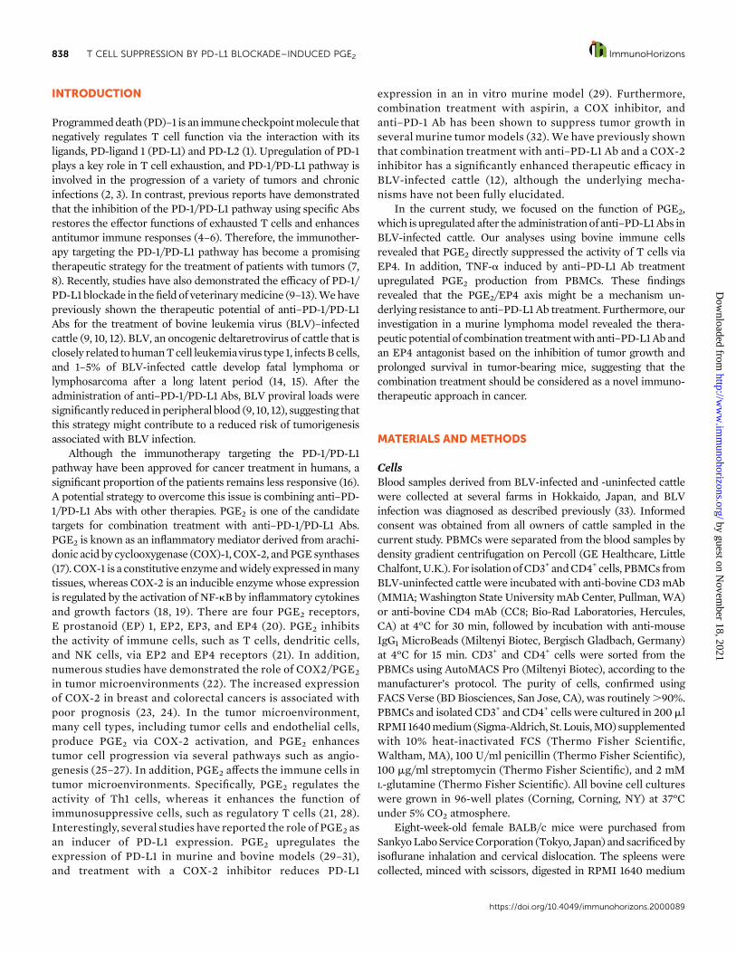
FIGURE 1. Changes in serum PGE2 concentrations after anti–PD-L1 Ab inoculation.
(A and B) BLV-infected cattle (animals 1–4) were administered 1 mg/kg anti–PD-L1 Ab (Boch4G12), and serum samples were collected on
days 0, 1, 3, 7, 14, 28, and 56. Serum PGE2 concentrations were measured by ELISA. (B) Statistical significance was determined by the
Mann–Whitney U test.
https://doi.org/10.4049/immunohorizons.2000089
ImmunoHorizons T CELL SUPPRESSION BY PD-L1 BLOCKADE–INDUCED PGE2 839
by guest on Novem
ber 18, 2021http://w
ww
.imm
unohorizons.org/D
ownloaded from

FIGURE 2. Functional analysis of EP signaling in PBMCs.
(A) Following an hour of incubation with indicated EP antagonists, PBMCs from BLV-uninfected cattle (n = 6) were incubated with PGE2 in the
presence of anti-CD3 mAb and anti-CD28 mAb. After incubation, IFN-g concentrations in culture supernatants were determined by ELISA. (B)
PBMCs from BLV-uninfected cattle (n = 6) were cultured with the EP4 agonist, and the expression of mRNA-encoding IFN-g was quantitated by
qPCR. DMSO was used as a vehicle control. (C) PBMCs from BLV-uninfected cattle (n = 7) were incubated with the EP4 agonist in the presence of
anti-CD3 mAb and anti-CD28 mAb. After incubation, IFN-g concentrations in culture supernatants were determined by ELISA. (Continued)(Continued)
https://doi.org/10.4049/immunohorizons.2000089
840 T CELL SUPPRESSION BY PD-L1 BLOCKADE–INDUCED PGE2 ImmunoHorizons
by guest on Novem
ber 18, 2021http://w
ww
.imm
unohorizons.org/D
ownloaded from
approval from the local committee for animal studies at HokkaidoUniversity (approval number 17-0024).
Quantitative real-time PCRQuantitative real-time PCR (qPCR) was performed as describedpreviously (30). Briefly, total RNA was extracted from culturedcells using TRI Reagent (Molecular Research Center, Cincinnati,OH) and cDNA was synthesized from the total RNA by Prime-Script Reverse Transcriptase (TaKaRa Bio, Otsu, Japan) followingthe manufacturers’ protocols. Next, qPCR was performed usinga LightCycler 480 System II (Roche Diagnostics, Mannheim,Germany) with SYBR Premix DimerEraser (TaKaRa Bio), follow-ing the manufacturers’ protocols. b-actin (ACTB) was used asa reference gene, and relative expression levels were calculatedusing the DD cycle threshold method. The primers were 59-ATAACCAGGTCATTCAAAGG-39 and 59-ATTCTGACTTCTCTTCCG CT-39 for bovine IFN-g, 59-ACG TTT TCT CGT GAA GCCCT-39 and 59-TCT ACC AGA AGG GCG GGA TA-39 for bovineCOX2, and 59-TCT TCC AGC CTT CCT TCC TG-39 and 59-ACCGTG TTG GCG TAG AGG TC-39 for bovine ACTB.
ELISAPGE2 concentrations in sera and culture supernatants weremeasured by PG E2 Express ELISA Kit (Cayman Chemical, AnnArbor, MI), following the manufacturer’s instructions. BovineIFN-g and mouse IL-2 concentrations in culture supernatantswere measured by Bovine IFN-g ELISA Development Kit(Mabtech, Nacka Strand, Sweden) and Mouse IL-2 Matched AbPair Kit (Abcam, Cambridge, U.K.), respectively, according to themanufacturers’ protocols.
Flow cytometryFor CD69 expression levels, collected cells were incubated in PBScontaining 10%goat serum(ThermoFisherScientific) for 15minat25°C to prevent nonspecific reactions. Next, the cells were stainedfor 20min at 25°C using the following Abs: FITC-conjugated anti-bovine CD4 mAb (CC8), PE-conjugated anti-bovine CD8 mAb(CC63; Bio-Rad Laboratories), and Alexa Fluor 647–labeled anti-bovine CD69 mAb (KTSN7A; Kingfisher Biotech, St. Paul, MN).KTSN7AwasprelabeledwithaZenonAlexaFluor647Mouse IgG1
Labeling Kit (Thermo Fisher Scientific). The stained cells werewashed twice and analyzed immediately by FACS Verse.
For the intracellular staining of IFN-g and TNF-a, thecollected cells were incubated in PBS containing 10% goat serumas described above, followed by staining with FITC-conjugatedanti-bovine CD4 mAb (CC8), PerCP/Cy5.5–conjugated anti-bovine CD8 mAb (CC63), and PE-labeled anti-bovine IgM mAb(IL-A30; Bio-Rad Laboratories) for 20 min at 25°C. IL-A30 wasprelabeled with a Zenon R-PEMouse IgG1 Labeling Kit (ThermoFisher Scientific). CC63 was conjugated with PerCP/Cy5.5 byusing a Lightning-Link Ab Labeling Kit (Innova Biosciences,Cambridge, U.K.). After surface staining, the cells were fixed andpermeabilized using FOXP3 Fix/PermKit (BioLegend, San Diego,CA). Next, the cells were stained with biotinylated anti-bovineIFN-gmAb (MT307;Mabtech) or biotinylated anti-bovineTNF-amAb (CC328; Bio-Rad Laboratories) for 20min at 25°C. The cellswere then incubated with allophycocyanin-conjugated streptavi-din (BioLegend) for 20 min at 25°C. After the final staining, thecellswerewashed twiceandanalyzed immediatelybyFACSVerse.
PBMC cultureTo examine the individual effects of EP antagonists, PBMCs fromBLV-uninfected cattle were incubated for 1 hwith 1mg/ml of eachof the following EP antagonists from Cayman Chemical: EP1 (SC-19220), EP2 (AH6809), EP3 (L-798,106), and EP4 (ONO-AE3-208), and then 250 nM PGE2 (Cayman Chemical) was added toeach culture. The PBMCswere stimulated by adding 1mg/ml anti-bovine CD3 mAb (MM1A) and 1 mg/ml anti-bovine CD28 mAb(CC220;Bio-RadLaboratories) to eachwell. After 24h, the culturesupernatants were collected to measure IFN-g concentrations byELISA.
To examine the effect of the EP4 agonist, PBMCs from BLV-uninfected cattle were incubatedwith 1mg/ml Rivenprost, an EP4agonist (CaymanChemical). The expression of IFN-g after 24 h ofincubation with no additional stimulation was determined byqPCR as described above, and IFN-g concentrations in culturesupernatants after 24 h of incubation with 1 mg/ml anti-bovineCD3 mAb (MM1A) and 1 mg/ml anti-bovine CD28 mAb (CC220)were measured by ELISA as described above. Similarly, IFN-gproduction in bovine lymphocyte subsets was measured by flow
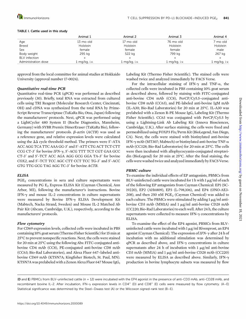
TABLE I. Cattle used in this study
Animal 1 Animal 2 Animal 3 Animal 4
Age 13 mo old 17 mo old 76 mo old 7 mo oldBreed Holstein Holstein Holstein HolsteinSex female female female maleBody weight 295 kg 522 kg 799 kg 267 kgBLV infection + + + +Administration dose 1 mg/kg, i.v. 1 mg/kg, i.v. 1 mg/kg, i.v. 1 mg/kg, i.v.
(D and E) PBMCs from BLV-uninfected cattle (n = 12) were incubated with the EP4 agonist in the presence of anti-CD3 mAb, anti-CD28 mAb, and
recombinant bovine IL-2. After incubation, IFN-g expression levels in CD4+ (D) and CD8+ (E) cells were measured by flow cytometry. (A–E)
Statistical significance was determined by the Steel–Dwass test (A) or the Wilcoxon signed-rank test (B–E).
https://doi.org/10.4049/immunohorizons.2000089
ImmunoHorizons T CELL SUPPRESSION BY PD-L1 BLOCKADE–INDUCED PGE2 841
by guest on Novem
ber 18, 2021http://w
ww
.imm
unohorizons.org/D
ownloaded from

cytometry by stimulating the cells with 2 mg/ml anti-bovine CD3mAb (MM1A), 2 mg/ml anti-bovine CD28 mAb (CC220), and10ng/ml recombinant bovine IL-2 (KingfisherBiotech). Following19hof incubation, the cellswere incubatedwith 10mg/mlbrefeldin
A (Sigma-Aldrich) for additional 5 h, and analyzed as describeabove.Toexamine theeffect of anti–PD-L1AbandTNF-a onPGE2
production, PBMCs from BLV-uninfected cattle were incubatedwith 10 mg/ml anti-bovine PD-L1 Ab (Boch4G12) or 10 ng/ml
FIGURE 3. Functional analysis of EP4 signaling in CD3+ cells.
(A–C) CD3+ cells isolated from PBMCs of BLV-uninfected cattle (n = 8) were cultured with the EP4 agonist in the presence of anti-CD3 mAb and
anti-CD28 mAb. After incubation, CD69 expression levels in CD4+ (A) and CD8+ (B) cells and IFN-g concentrations in culture supernatants (C) were
measured by flow cytometry and ELISA, respectively. (D) The heat-map for the changes in gene expression levels in CD4+ cells by the EP4 agonist.
(A–C) Statistical significance was determined by the Wilcoxon signed-rank test. MFI, Mean fluorescence intensity.
https://doi.org/10.4049/immunohorizons.2000089
842 T CELL SUPPRESSION BY PD-L1 BLOCKADE–INDUCED PGE2 ImmunoHorizons
by guest on Novem
ber 18, 2021http://w
ww
.imm
unohorizons.org/D
ownloaded from

bovine rTNF-a (Kingfisher Biotech) for 72 or 24 h, respectively.Bovine IgG (Sigma-Aldrich) was used as a negative control for theanti–PD-L1 Ab Boch4G12, and PBS was used as a vehicle controlfor bovine rTNF-a. After incubation, the culture supernatantswere collected to measure PGE2 concentrations by ELISA, andthe cells were collected for the quantification of COX2 expressionby qPCR. Additionally, to investigate whether treatment withanti–PD-L1 Ab induces TNF-a production, PBMCs from BLV-uninfected cattlewere incubatedwith 10mg/ml anti-bovinePD-L1Ab (Boch4G12) in the presence of 2 mg/ml anti-bovine CD3 mAb(MM1A), 2 mg/ml anti-bovine CD28 mAb (CC220), and 10 ng/mlrecombinant bovine IL-2. Following 19 h of incubation, thecultures were incubated with 10 mg/ml brefeldin A for 5 h, afterwhich the cultured PBMCs were harvested, and TNF-a expres-sion levels were measured by flow cytometry. Furthermore, toexamine whether the blockade of TNF-a reduces PGE2 pro-duction in the presence of anti–PD-L1 Ab, PBMCs from BLV-uninfected cattle were incubated with 172 nM bovine TNFreceptor type II (TNFRII)–Ig, a decoy receptor for bovine TNF-a(35), in the presence of 10 mg/ml anti-bovine PD-L1 Ab(Boch4G12). Control Ig, which comprised the signal peptide ofbovineTNFRII and the Fc domainof bovine IgG1 (35), was used asa negative control for TNFRII-Ig. Cultures were stimulated byadding 1 mg/ml anti-bovine CD3 mAb (MM1A) and 1 mg/ml anti-bovine CD28 mAb (CC220) to each well. After 72 h, the culturesupernatants were collected to measure PGE2 concentrations byELISA.
To examine the effect of the dual blockade of PD-L1 andEPs incattle, PBMCs from BLV-uninfected or BLV-infected cattle werecultured with 10 mg/ml anti-bovine PD-L1 Ab (Boch4G12) and1 mg/ml each EP antagonist. PBMCs from BLV-uninfected cattlewere cultured in the presence of 1 mg/ml anti-bovine CD3 mAb(MM1A) and 1 mg/ml anti-bovine CD28 mAb (CC220) for 72 h,whereas PBMCs from BLV-infected cattle were cultured in the
presence of a BLV Ag, fetal lamb kidney (FLK)–BLV (2% heat-inactivated culture supernatant of FLK-BLV cells), for 144 h. Afterincubation, the culture supernatants were collected, and IFN-gconcentrations were determined by ELISA.
CD3+ cell cultureIsolated CD3+ cells were cultured with 1 mg/ml the EP4 agonistin the presence of 1 mg/ml anti-bovine CD3 mAb (MM1A) and1mg/ml anti-bovineCD28mAb (CC220) for 72h.After incubation,CD69 expression levels and IFN-g concentrations in culturesupernatants were measured by flow cytometry and ELISA,respectively.
MicroarrayIsolatedCD4+ cellswere culturedwith 0.5mg/ml anti-bovineCD3mAb (MM1A) and 0.5 mg/ml anti-bovine CD28 mAb (CC220).Following 18 h of incubation, the cultures were incubated with1mg/ml theEP4 agonist orDMSO for 4h.Microarray analysiswasperformed using Agilent Bos taurus (Bovine) Oligo Microarray v2(Design ID: 023647; Agilent Technologies, Santa Clara, CA). Aftercell collection, total RNA was extracted using RNeasy Mini Kit(Qiagen, Hilden, Germany) according to the manufacturer’sinstructions. Synthesis and labeling of cRNA were performedusing Low Input Quick Amp Labeling Kit (Agilent Technologies)according to the manufacturer’s instructions. The Cy3-labeledcRNA was purified using RNeasy Mini Kit (Qiagen), andhybridization was performed using Gene Expression Hybridiza-tion Kit (Agilent Technologies), according to the manufacturers’instructions. Next, scanning of the hybridized microarray anddata analysis were performed using Agilent DNA MicroarrayScanner (Agilent Technologies), Feature Extraction software(Agilent Technologies), and GeneSpring (Agilent Technolo-gies), according to the manufacturers’ instructions. The micro-array data were deposited in ArrayExpress (E-MTAB-9576,
TABLE II. The change of gene expression in microarray analysis
Gene Symbol Gene Name Fold Change p Value
IL2 IL-2 22.1255753 0.008147CCL3 Chemokine (CC motif) ligand 3 22.0820506 0.004651TNF TNF 22.0492654 0.001460CSF2 CSF-2 22.0447707 0.005276IL12A IL-12A 21.9293255 0.012709CXCL10 CXC motif chemokine ligand 10 21.8574089 0.019228CCL20 CC motif chemokine ligand 20 21.8484110 0.010516LTA Lymphotoxin-a 21.8189231 0.019710CCL4 Chemokine (CC motif) ligand 4 21.7715497 0.000768FGB Fibrinogen b-chain 21.7014802 0.026826IFNB3 IFN-b 3 21.5604529 0.043236CXCL9 CXC motif chemokine ligand 9 21.4785548 0.000302IFNG IFN-g 21.4232383 0.054541CREM cAMP-responsive element modulator 2.4109561 0.001597NR4A3 Nuclear receptor subfamily 4 group A member 3 1.9490188 0.029402NR4A2 Nuclear receptor subfamily 4 group A member 2 1.8775752 0.001489PTGS2 PG-endoperoxide synthase 2 1.7771128 0.042600NFKB1 NF-kB subunit 1 1.5010872 0.001316CTLA4 CTL–associated protein 4 1.1734980 0.011518PDCD1 Programmed cell death 1 1.0657109 0.355153
https://doi.org/10.4049/immunohorizons.2000089
ImmunoHorizons T CELL SUPPRESSION BY PD-L1 BLOCKADE–INDUCED PGE2 843
by guest on Novem
ber 18, 2021http://w
ww
.imm
unohorizons.org/D
ownloaded from

https://www.ebi.ac.uk/arrayexpress/). The microarray proce-dures from RNA extraction to data analysis were conducted atHokkaido System Science (Sapporo, Japan).
Splenocyte cultureTo examine the effects of anti–PD-L1 Ab treatment on PGE2
production in mice, splenocytes were cultured with 10 mg/ml
anti-mouse PD-L1 Ab (10F.9G2; BioXCell, West Lebanon, NH) or10 mg/ml rat IgG2b isotype control (LTF-2; BioXCell). PGE2
concentrations in supernatants of culture incubated with orwithout 10 mg/ml Con A (Sigma-Aldrich) for 72 h were measuredby ELISA as described above. To examine the effect of the dualblockade of PD-L1 and EP4 in mice, splenocytes were culturedwith 10 mg/ml anti-mouse PD-L1 Ab (10F.9G2) and 1 mg/ml each
FIGURE 4. TNF-a induced by PD-L1 blockade upregulates PGE2 production.
(A and B) PBMCs from BLV-uninfected cattle were cultured with anti–PD-L1 Ab (Boch4G12), and COX2 expression levels (A, n = 8) and PGE2concentrations in culture supernatants (B, n = 6) were measured by qPCR and ELISA, respectively. Bovine IgG was used as a negative control of
anti–PD-L1 Ab (Boch4G12). (C and D) PBMCs from BLV-uninfected cattle (n = 11) were incubated with anti–PD-L1 Ab (Boch4G12) in the presence of
anti-CD3 mAb, anti-CD28 mAb, and recombinant bovine IL-2. After incubation, TNF-a expression levels in CD4+ (C) and CD8+ (D) cells were
measured by flow cytometry. (E and F) PBMCs from BLV-uninfected cattle were cultured with recombinant bovine TNF-a, and COX2 expression
levels (E, n = 10) and PGE2 concentrations in culture supernatants (F, n = 7) were measured by qPCR and ELISA, respectively. PBS was used as a
vehicle control. (G) PBMCs from BLV-uninfected cattle (n = 6) were cultured with TNFRII-Ig in the presence of anti–PD-L1 Ab (Boch4G12). Cultures
were stimulated by adding anti-CD3 mAb and anti-CD28 mAb. Control Ig was used as a negative control for TNFRII-Ig. PGE2 concentrations in
culture supernatants were determined by ELISA. (A–G) Statistical significance was determined by the Wilcoxon signed-rank test.
https://doi.org/10.4049/immunohorizons.2000089
844 T CELL SUPPRESSION BY PD-L1 BLOCKADE–INDUCED PGE2 ImmunoHorizons
by guest on Novem
ber 18, 2021http://w
ww
.imm
unohorizons.org/D
ownloaded from

EP antagonist. Cultures were stimulated by adding 1 mg/ml anti-mouse CD3e mAb (145-2C11; Thermo Fisher Scientific) to eachwell. After 72 h, the culture supernatants were collected, and IL-2concentrations were measured by ELISA.
Tumor grafting and tumor growth measurementSix-week-old male C57BL/6 mice (Japan SLC, Hamamatsu,Japan) were s.c. inoculated with EG7 (5 3 106 cells per mouse).The day of EG7 injection was defined as day 0. For anti–PD-L1 Abtreatment, mice were i.p. injected with anti-mouse PD-L1 mAb(10F.9G2) (10 mg/kg, once a day) on days 7, 10, and 14. For EP4antagonist treatment, mice were orally administered with ONO-AE3-208 (10 mg/kg/d) added to drinking water from day 7 to day23. Tumor size was monitored at least every other day, starting onday 5, using a caliper until the length or width exceeded 20 mm.Tumor volumewas calculated according to the following formula:tumor volume (mm3) = (length 3 width2)/2. The animal exper-iments were performed with the approval of the InstitutionalAnimal Care and Use Committee of the Graduate School ofVeterinary Medicine at Hokkaido University (approval number:16-0131). The animals were handled in accordance with the Guidefor the Care and Use of Laboratory Animals, Graduate School ofVeterinary Medicine, Hokkaido University (approved by theAssociation for Assessment and Accreditation of LaboratoryAnimal Care International).
StatisticsIn Fig. 1, differences were assessed using the Mann–Whitney Utest. In Figs. 2–5 and 6A–C, differences were assessed using theWilcoxon signed-rank test for two-group comparisons and theSteel–Dwass test for multiple-group comparisons. In Fig. 6E and
6F, differences were assessed using the Tukey test and the log-rank test, respectively. In microarray analysis (Table II), differ-ences were assessed using the paired t test. A p value ,0.05was considered to indicate statistical significance.
RESULTS
Serum PGE2 concentration is increased withanti–PD-L1 immunotherapyWefirst analyzed the serumsamples fromBLV-infected cattle thatwere administered the anti–PD-L1 blocking Ab (Table I) andfound that the serum PGE2 concentrations were increased afterthe anti–PD-L1 Ab treatment (Fig. 1). Therefore, we specificallyexamined the role of PGE2 in the blockade of PD-1/PD-L1interaction in the current study.
T cell activation is suppressed via PGE2/EP4 pathwayWe have previously shown that PGE2 suppresses Th1 responses,such as Th1 cytokine production and T cell proliferation, in cattle(30). In the current study, we further aimed to identify specificPGE2 receptors involved in PGE2-mediated immune dysfunctionusing EP antagonists and agonists. Bovine PBMCs were preincu-bated with individual EP antagonists, followed by culturing thecells with PGE2 in the presence of anti-CD3 and anti-CD28mAbs.Pretreatment with the EP4 antagonist inhibited the suppressiveeffect of PGE2, whereas IFN-g production was suppressed byPGE2 in PBMCs pretreated with the antagonists of other EPs(EP1–EP3) (Fig. 2A). Additionally, treatment with the EP4 agonistsignificantly inhibited IFN-g mRNA and its protein expression inPBMCs (Fig. 2B, 2C). Flow cytometric analysis revealed that the
FIGURE 5. Functional analysis of the dual blockade of PD-L1 and EP4 in cattle.
(A and B) PBMCs from BLV-uninfected (n = 8) and BLV-infected (n = 7) cattle were cultured with anti–PD-L1 Ab (Boch4G12) and indicated EP
antagonists. PBMCs from BLV-uninfected cattle were stimulated by anti-CD3 mAb and anti-CD28 mAb. PBMCs from BLV-infected cattle were
stimulated by FLK-BLV, a BLV Ag. After incubation, IFN-g concentrations in culture supernatants were measured by ELISA. Statistical significance
was determined by the Steel–Dwass test.
https://doi.org/10.4049/immunohorizons.2000089
ImmunoHorizons T CELL SUPPRESSION BY PD-L1 BLOCKADE–INDUCED PGE2 845
by guest on Novem
ber 18, 2021http://w
ww
.imm
unohorizons.org/D
ownloaded from

FIGURE 6. Functional analysis of the dual blockade of PD-L1 and EP4 in mice.
(A and B) Murine splenocytes (A, n = 6; B, n = 8) were cultured with anti–PD-L1 Ab (10F.9G2). Cultures were stimulated with or without Con A, and
PGE2 concentrations in culture supernatants were measured by ELISA. Statistical significance was determined by the Wilcoxon signed-rank test. (C)
Murine splenocytes (n = 6) were cultured with anti–PD-L1 Ab (10F.9G2) and indicated EP antagonists in the presence of anti-mouse CD3e mAb. IL-2
concentrations in culture supernatants were determined by ELISA. Statistical significance was determined by the Steel–Dwass test. (D–F) Evaluation
of the antitumor effects of dual blockade in the EG7 mouse model. (D) Experimental design. (E) Tumor growth in each group. (Continued)(Continued)
https://doi.org/10.4049/immunohorizons.2000089
846 T CELL SUPPRESSION BY PD-L1 BLOCKADE–INDUCED PGE2 ImmunoHorizons
by guest on Novem
ber 18, 2021http://w
ww
.imm
unohorizons.org/D
ownloaded from

EP4 agonist decreased the percentage of IFN-g+ cells in both theCD4+andCD8+cell populations (Fig. 2D,2E,SupplementalFig. 1A,1B). Furthermore, to examine whether PGE2 directly suppressesthe activity of bovine T cells, isolated CD3+ T cells were culturedwith the EP4 agonist, and the expression levels of CD69, anactivation marker, in these cells and IFN-g production in culturesupernatants were assayed by flow cytometry and ELISA,respectively.Treatmentwith theEP4agonist significantly reducedthe CD69 expression levels in CD4+ and CD8+ cells and IFN-gproduction (Fig. 3A–C, Supplemental Fig. 1C, 1D). Microarrayanalysis revealed that the EP4 agonist treatment downregulatedthe expression of Th1-related cytokine genes, such as IL-2, IFN-g,TNF-a, and IL-12, inCD4+ cells (Fig. 3D,Table II). Taken together,these data suggest that PGE2 induced by PD-L1 blockade directlyinhibits T cell activation via the EP4 signaling.
PD-L1 blockade–mediated induction of TNF-a upregulatesPGE2 productionAs shown in Fig. 1, the serumPGE2 concentrationswere increasedafter anti–PD-L1 Ab treatment. To examine whether treatmentwith the anti–PD-L1 Ab induces PGE2 production in vitro, bovinePBMCs were cultured with anti–PD-L1 Ab (Boch4G12), whichsignificantly induced the COX2 expression and PGE2 productionin vitro (Fig. 4A, 4B). Blockade of the PD-1/PD-L1 pathwayusing specific Abs reactivates exhausted T cells, leading to theenhancement ofTh1 cytokine production fromTcells (5, 6). In thecurrent study, flow cytometric analysis revealed that anti–PD-L1Ab (Boch4G12) significantly increased the TNF-a expressionlevels in both CD4+ andCD8+ cells (Fig. 4C, 4D, Supplemental Fig.1A, 1C). Previous reports have clearly demonstrated that TNF-ainduces NF-kB activation, which is essential for COX-2 upregu-lation (36–38). Therefore, we examined whether anti–PD-L1Ab–induced TNF-a is involved in the observed PGE2 upregula-tion. Treatment with bovine rTNF-a significantly induced boththeCOX2 expression and PGE2 production in bovine PBMCs (Fig.4E, 4F). Interestingly, the blockade of TNF-a using the decoyreceptor TNFRII-Ig (35) reduced PGE2 production in the pres-ence of anti–PD-L1 Ab (Fig. 4G). Collectively, these results suggestthat TNF-a induced by PD-L1 blockade upregulates PGE2
production, contributing to the impaired efficacy of anti–PD-L1Ab treatment via the PGE2/EP4 signaling.
Th1 cytokine production is enhanced by the dual blockade ofPD-L1 and EP4To examine whether the inhibition of EP4 enhances the efficacyof anti–PD-L1 Ab in vitro, bovine PBMCs were cultured withindividual EP antagonists in the presence of anti–PD-L1 Ab(Boch4G12). As shown in Fig. 5A, the dual blockade of PD-L1 andEP4 increased IFN-g production compared with other treatment
groups. Additionally, the dual blockade of PD-L1 and EP4significantly enhanced the BLV-specific IFN-g production fromPBMCs of BLV-infected cattle (Fig. 5B). Taken together, theseresults suggest that combinationwithanEP4antagonistmightbe anovel strategy to enhance the efficacy of anti–PD-L1 Ab treatmentin cattle.
Antitumor effects are enhanced by the dual blockade ofPD-L1 and EP4Our studies in bovine immune cells revealed the novelmechanismof anti–PD-L1 Ab resistance and the potential of enhancing Th1cytokine production by the dual blockade of PD-L1 and EP4. Wethen used murine splenocytes to examine whether the dualblockadeenhancesTh1 immune responses inother animalmodels.As shown in Fig. 6A and 6B, anti–PD-L1 Ab induced PGE2
production from murine splenocytes stimulated with or withoutCon A (Fig. 6A, 6B). Additionally, treatment with the EP4antagonist increased IL-2 production from murine splenocytesin the presence of anti–PD-L1 Ab (10F.9G2) (Fig. 6C), suggestingthat the dual blockade enhanced Th1 responses not only in cattlebut also in mice. Finally, based on these results, we used a mouselymphomamodel to evaluate the potential antitumor effects of thedual blockade as a potent immunotherapy in cancers resistant toanti–PD-1/PD-L1 Ab alone. C57BL/6 mice were inoculated with alymphoma cell line, EG7, and the EG7-bearing mice wereadministered anti–PD-L1 Ab (10F.9G2) i.p. and theEP4 antagonistorally (Fig. 6D). Compared with the animals treated with the EP4antagonist or the anti–PD-L1Abalone, the growthofEG7cellswasinhibited in those administered the combination treatment (Fig.6E). Additionally, the survival of the combination treatment groupwas significantly prolonged compared with that of the untreatedgroup (Fig. 6F). Taken together, these data suggest that the dualblockade of PD-L1 and EP4 is a promising strategy as a novelimmunotherapy.
DISCUSSION
Numerous studies have recently elucidated the mechanisms ofresistance to cancer immunotherapy (39–41). For instance,Koyama et al. (39) demonstrated that therapeutic PD-1 blockadeupregulated the expression of alternative immune checkpointmolecules, which caused resistance to the PD-1/PD-L1 blockade.Treatments including anti–PD-1/PD-L1 Abs in combination withother medicines to overcome resistance are garnering increasingattention. Previous studies have shown that combination treat-ment with anti–PD-1/PD-L1 Abs with COX inhibitors enhancesthe therapeutic efficacy in murine and bovine models (12, 32).However, the mechanisms underlying the observed therapeutic
Data are presented as means, and the error bars indicate SEs. Statistical significance was determined by the Tukey test. (F) The Kaplan–Meier Curve
for survival in all groups. Statistical significance was determined by the log-rank test. (D–F) Data are representative of two independent experiments,
each performed with five to eight mice per group.
https://doi.org/10.4049/immunohorizons.2000089
ImmunoHorizons T CELL SUPPRESSION BY PD-L1 BLOCKADE–INDUCED PGE2 847
by guest on Novem
ber 18, 2021http://w
ww
.imm
unohorizons.org/D
ownloaded from

effect of these combination approaches remain unclear. In thecurrent study, we identify a novel mechanism of resistance relatedto PGE2 using a bovine model (Supplemental Fig. 2). It wasrevealed that the anti–PD-L1Ab treatment induced theproductionof Th1 cytokines, such as TNF-a, and that TNF-a–induced PGE2
suppressed the activation of T cells via EP4. This might partiallyexplain a reason that the combined treatment enhances theefficacy of anti–PD-1/PD-L1 Abs. In addition, our study clearlyshowed the therapeutic potential of combination treatment withanti–PD-L1Abs andEP4antagonists in bovine andmurinemodels.To the best of our knowledge, this is the first study to demonstratethe therapeutic efficacy of a dual blockade strategy using ananti–PD-L1 Ab and an EP4 antagonist in an in vivo model. Futurestudies in other murine tumor models are warranted to furtherinvestigate the efficacy of the dual blockade.
Among the four PGE2 receptors EP1–4 (20), EP2 and EP4 areinvolved in PGE2-associated immune dysfunction (21). In thecurrent study, the blockade of EP4, but not EP2, inhibited thesuppression of IFN-g production by PGE2. EP4 is a high-affinityreceptor for PGE2, whereas EP2 requires significantly higherPGE2 concentrations for effective signaling (21). Thus, theobserved differences in the results following the blockade of EP2and EP4 might be due to the difference in the affinity of eachreceptor. The contribution of EP2 to immunosuppression shouldcarefully be investigated in other preclinical models in whichhigher PGE2 levels are expected during disease progression.
Anti–PD-1/PD-L1 Abs reactivate exhausted T cells, leading tothe production of Th1 cytokines, such as IFN-g and TNF-a (5, 6).TNF-a not only plays a critical role in cellular immunity againstcancer, but also has a direct cytotoxic effect on tumor cells byinducing apoptosis (42, 43). Although known as an antitumorcytokine, TNF-a paradoxically promotes tumor progression insome circumstances (44–46). For example, serum TNF-a con-centration is correlated with the progression of several cancertypes, such as renal cell carcinoma and prostate cancer (47, 48).Additionally, the blockade of TNF-a using Abs inhibits tumorgrowth (49). Furthermore, recent studies have shown that theblockade ofTNF-a improves the efficacy of PD-1blockade (50, 51);however, the underlying detailed mechanism has not been fullyelucidated. In the current study,we demonstrated that TNF-awasinvolved in PGE2 upregulation under anti–PD-L1 Ab treatment,and that the dual blockade of PD-1/PD-L1 and EP4 enhanced theefficacy of immunotherapy. Our strategy might be more effectivethan the dual blockade of PD-1/PD-L1 and TNF-a because theantitumor effects of TNF-a are not inhibited. Further studies arenecessary to compare the efficacy between the two strategies.
PGE2/EP4 signaling increases cAMP production (52). Onestudy has previously shown that the PGE2/EP4/cAMP upregu-lates the expression of T cell Ig and mucin domain-3 (TIM-3), animmunoinhibitory molecule, in a human T cell line (53). Addi-tionally, several reports have investigated that TIM-3expression isinduced after the PD-1/PD-L1 blockade, leading to the resistanceto PD-1/PD-L1 blockade (39, 50). Therefore, PGE2 upregulationafter the PD-1/PD-L1 blockademight also contribute to resistancevia the upregulation of other immunoinhibitory molecules.
PD-L1blockade–TNF-a–PGE2–EP4axis is anovelmechanismof resistance to anti–PD-1/PD-L1 immunotherapy. A novelcombined approach targeting PD-L1 and EP4 as immunotherapymay overcome resistance to the anti–PD-1/PD-L1 Ab therapy.Further studies using different murine tumor models as well asother animalmodelswill heraldnewavenues forcancer treatment.
DISCLOSURES
The authors have no financial conflicts of interest.
ACKNOWLEDGMENTS
We thank Dr. Hideyuki Takahashi, Dr. Yasuyuki Mori, and Dr. Tomio Ibayashifor valuable advice and discussions. We thank Enago (www.enago.jp) for theEnglish language review.
REFERENCES
1. Okazaki, T., and T. Honjo. 2007. PD-1 and PD-1 ligands: from dis-covery to clinical application. Int. Immunol. 19: 813–824.
2. Blank, C., T. F. Gajewski, and A. Mackensen. 2005. Interaction of PD-L1 on tumor cells with PD-1 on tumor-specific T cells as a mechanismof immune evasion: implications for tumor immunotherapy. CancerImmunol. Immunother. 54: 307–314.
3. Blank, C., and A. Mackensen. 2007. Contribution of the PD-L1/PD-1pathway to T-cell exhaustion: an update on implications for chronicinfections and tumor evasion. Cancer Immunol. Immunother. 56:739–745.
4. Iwai, Y., M. Ishida, Y. Tanaka, T. Okazaki, T. Honjo, and N. Minato.2002. Involvement of PD-L1 on tumor cells in the escape from hostimmune system and tumor immunotherapy by PD-L1 blockade. Proc.Natl. Acad. Sci. USA 99: 12293–12297.
5. Keir, M. E., M. J. Butte, G. J. Freeman, and A. H. Sharpe. 2008. PD-1and its ligands in tolerance and immunity. Annu. Rev. Immunol. 26:677–704.
6. Dulos, J., G. J. Carven, S. J. van Boxtel, S. Evers, L. J. Driessen-Engels,W. Hobo, M. A. Gorecka, A. F. de Haan, P. Mulders, C. J. Punt, et al.2012. PD-1 blockade augments Th1 and Th17 and suppresses Th2responses in peripheral blood from patients with prostate and ad-vanced melanoma cancer. J. Immunother. 35: 169–178.
7. Iwai, Y., J. Hamanishi, K. Chamoto, and T. Honjo. 2017. Cancer im-munotherapies targeting the PD-1 signaling pathway. J. Biomed. Sci.24: 26.
8. Ribas, A., and J. D. Wolchok. 2018. Cancer immunotherapy usingcheckpoint blockade. Science 359: 1350–1355.
9. Nishimori, A., S. Konnai, T. Okagawa, N. Maekawa, R. Ikebuchi,S. Goto, Y. Sajiki, Y. Suzuki, J. Kohara, S. Ogasawara, et al. 2017. Invitro and in vivo antivirus activity of an anti-programmed death-ligand 1 (PD-L1) rat-bovine chimeric antibody against bovine leuke-mia virus infection. PLoS One 12: e0174916.
10. Okagawa, T., S. Konnai, A. Nishimori, N. Maekawa, R. Ikebuchi,S. Goto, C. Nakajima, J. Kohara, S. Ogasawara, Y. Kato, et al. 2017.Anti-bovine programmed death-1 rat-bovine chimeric antibody forimmunotherapy of bovine leukemia virus infection in cattle. Front.Immunol. 8: 650.
11. Maekawa, N., S. Konnai, S. Takagi, Y. Kagawa, T. Okagawa,A. Nishimori, R. Ikebuchi, Y. Izumi, T. Deguchi, C. Nakajima, et al.2017. A canine chimeric monoclonal antibody targeting PD-L1 andits clinical efficacy in canine oral malignant melanoma or undiffer-entiated sarcoma. Sci. Rep. 7: 8951.
https://doi.org/10.4049/immunohorizons.2000089
848 T CELL SUPPRESSION BY PD-L1 BLOCKADE–INDUCED PGE2 ImmunoHorizons
by guest on Novem
ber 18, 2021http://w
ww
.imm
unohorizons.org/D
ownloaded from

12. Sajiki, Y., S. Konnai, T. Okagawa, A. Nishimori, N. Maekawa, S. Goto,K. Watari, E. Minato, A. Kobayashi, J. Kohara, et al. 2019. Prosta-glandin E2-induced immune exhaustion and enhancement of antiviraleffects by anti-PD-L1 antibody combined with COX-2 inhibitor inbovine leukemia virus infection. J. Immunol. 203: 1313–1324.
13. Goto, S., S. Konnai, Y. Hirano, J. Kohara, T. Okagawa, N. Maekawa,Y. Sajiki, K. Watari, E. Minato, A. Kobayashi, et al. 2020. Clinicalefficacy of the combined treatment of anti-PD-L1 rat-bovine chimericantibody with a COX-2 inhibitor in calves infected with Mycoplasmabovis. Jpn. J. Vet. Res. 68: 77–90.
14. Sagata, N., T. Yasunaga, J. Tsuzuku-Kawamura, K. Ohishi, Y. Ogawa,and Y. Ikawa. 1985. Complete nucleotide sequence of the genome ofbovine leukemia virus: its evolutionary relationship to other retro-viruses. Proc. Natl. Acad. Sci. USA 82: 677–681.
15. Schwartz, I., and D. Lévy. 1994. Pathobiology of bovine leukemia vi-rus. Vet. Res. 25: 521–536.
16. Gong, J., A. Chehrazi-Raffle, S. Reddi, and R. Salgia. 2018. Develop-ment of PD-1 and PD-L1 inhibitors as a form of cancer immuno-therapy: a comprehensive review of registration trials and futureconsiderations. J. Immunother. Cancer 6: 8.
17. Phipps, R. P., S. H. Stein, and R. L. Roper. 1991. A new view ofprostaglandin E regulation of the immune response. Immunol. Today12: 349–352.
18. Subbaramaiah, K., N. Telang, J. T. Ramonetti, R. Araki, B. DeVito,B. B. Weksler, and A. J. Dannenberg. 1996. Transcription ofcyclooxygenase-2 is enhanced in transformed mammary epithelialcells. Cancer Res. 56: 4424–4429.
19. Morita, I. 2002. Distinct functions of COX-1 and COX-2. Prosta-glandins Other Lipid Mediat. 68–69: 165–175.
20. Sugimoto, Y., and S. Narumiya. 2007. Prostaglandin E receptors.J. Biol. Chem. 282: 11613–11617.
21. Kalinski, P. 2012. Regulation of immune responses by prostaglandinE2. J. Immunol. 188: 21–28.
22. Kobayashi, K., K. Omori, and T. Murata. 2018. Role of prostaglandinsin tumor microenvironment. Cancer Metastasis Rev. 37: 347–354.
23. Tomozawa, S., N. H. Tsuno, E. Sunami, K. Hatano, J. Kitayama,T. Osada, S. Saito, T. Tsuruo, Y. Shibata, and H. Nagawa. 2000.Cyclooxygenase-2 overexpression correlates with tumour recurrence,especially haematogenous metastasis, of colorectal cancer. Br. J.Cancer 83: 324–328.
24. Denkert, C., K. J. Winzer, B. M. Muller, W. Weichert, S. Pest,M. Kobel, G. Kristiansen, A. Reles, A. Siegert, H. Guski, and S. Hauptmann.2003. Elevated expression of cyclooxygenase-2 is a negative prognosticfactor for disease free survival and overall survival in patients with breastcarcinoma. Cancer 97: 2978–2987.
25. Rigas, B., I. S. Goldman, and L. Levine. 1993. Altered eicosanoid levelsin human colon cancer. J. Lab. Clin. Med. 122: 518–523.
26. Wang, D., H. Wang, Q. Shi, S. Katkuri, W. Walhi, B. Desvergne, S. K.Das, S. K. Dey, and R. N. DuBois. 2004. Prostaglandin E(2) promotescolorectal adenoma growth via transactivation of the nuclear perox-isome proliferator-activated receptor delta. Cancer Cell 6: 285–295.
27. Wang, D., and R. N. Dubois. 2010. Eicosanoids and cancer. Nat. Rev.Cancer 10: 181–193.
28. Li, H., M. L. Edin, A. Gruzdev, J. Cheng, J. A. Bradbury, J. P. Graves,L. M. DeGraff, and D. C. Zeldin. 2013. Regulation of T helper cellsubsets by cyclooxygenases and their metabolites. ProstaglandinsOther Lipid Mediat. 104–105: 74–83.
29. Prima, V., L. N. Kaliberova, S. Kaliberov, D. T. Curiel, and S. Kusmartsev.2017. COX2/mPGES1/PGE2 pathway regulates PD-L1 expression intumor-associated macrophages and myeloid-derived suppressor cells.Proc. Natl. Acad. Sci. USA 114: 1117–1122.
30. Sajiki, Y., S. Konnai, T. Okagawa, A. Nishimori, N. Maekawa, S. Goto,R. Ikebuchi, R. Nagata, S. Kawaji, Y. Kagawa, et al. 2018. Prosta-glandin E2 induction suppresses the Th1 immune responses in cattlewith Johne’s disease. Infect. Immun. 86: e00910–e00917.
31. Goto, S., S. Konnai, Y. Hirano, J. Kohara, T. Okagawa, N. Maekawa,Y. Sajiki, K. Watari, E. Minato, A. Kobayashi, et al. 2020. Upregulationof PD-L1 expression by prostaglandin E2 and the enhancement ofIFN-g by anti-PD-L1 antibody combined with a COX-2 inhibitor inMycoplasma bovis infection. Front. Vet. Sci. 7: 12.
32. Zelenay, S., A. G. van der Veen, J. P. Bottcher, K. J. Snelgrove,N. Rogers, S. E. Acton, P. Chakravarty, M. R. Girotti, R. Marais, S. A.Quezada, et al. 2015. Cyclooxygenase-dependent tumor growththrough evasion of immunity. Cell 162: 1257–1270.
33. Sajiki, Y., S. Konnai, A. Nishimori, T. Okagawa, N. Maekawa, S. Goto,M. Nagano, J. Kohara, N. Kitano, T. Takahashi, et al. 2017. In-trauterine infection with bovine leukemia virus in pregnant dam withhigh viral load. J. Vet. Med. Sci. 79: 2036–2039.
34. Moore, M. W., F. R. Carbone, and M. J. Bevan. 1988. Introduction ofsoluble protein into the class I pathway of antigen processing andpresentation. Cell 54: 777–785.
35. Fujisawa, S., S. Konnai, T. Okagawa, N. Maekawa, A. Tanaka,Y. Suzuki, S. Murata, and K. Ohashi. 2019. Effects of bovine tumornecrosis factor alpha decoy receptors on cell death and inflammatorycytokine kinetics: potential for bovine inflammation therapy. BMCVet. Res. 15: 68.
36. Jobin, C., O. Morteau, D. S. Han, and R. Balfour Sartor. 1998. SpecificNF-kappaB blockade selectively inhibits tumour necrosis factor-alpha-induced COX-2 but not constitutive COX-1 gene expression inHT-29 cells. Immunology 95: 537–543.
37. Ghosh, S., and M. Karin. 2002. Missing pieces in the NF-kappaBpuzzle. Cell 109(Suppl): S81–S96.
38. Bouwmeester, T., A. Bauch, H. Ruffner, P. O. Angrand, G. Bergamini,K. Croughton, C. Cruciat, D. Eberhard, J. Gagneur, S. Ghidelli, et al.2004. A physical and functional map of the human TNF-alpha/NF-kappa B signal transduction pathway. [Published erratum appears in2004 Nat. Cell Biol. 6: 465.] Nat. Cell Biol. 6: 97–105.
39. Koyama, S., E. A. Akbay, Y. Y. Li, G. S. Herter-Sprie, K. A. Buczkowski,W. G. Richards, L. Gandhi, A. J. Redig, S. J. Rodig, H. Asahina, et al.2016. Adaptive resistance to therapeutic PD-1 blockade is associatedwith upregulation of alternative immune checkpoints. Nat. Commun.7: 10501.
40. Sharma, P., S. Hu-Lieskovan, J. A. Wargo, and A. Ribas. 2017. Primary,adaptive, and acquired resistance to cancer immunotherapy. Cell 168:707–723.
41. Nowicki, T. S., S. Hu-Lieskovan, and A. Ribas. 2018. Mechanisms ofresistance to PD-1 and PD-L1 blockade. Cancer J. 24: 47–53.
42. Goodsell, D. S. 2006. The molecular perspective: tumor necrosisfactor. Oncologist 11: 83–84.
43. Tamada, K., and L. Chen. 2006. Renewed interest in cancer immu-notherapy with the tumor necrosis factor superfamily molecules.Cancer Immunol. Immunother. 55: 355–362.
44. Moore, R. J., D. M. Owens, G. Stamp, C. Arnott, F. Burke, N. East,H. Holdsworth, L. Turner, B. Rollins, M. Pasparakis, et al. 1999. Micedeficient in tumor necrosis factor-alpha are resistant to skin carci-nogenesis. Nat. Med. 5: 828–831.
45. Balkwill, F. 2006. TNF-a in promotion and progression of cancer.Cancer Metastasis Rev. 25: 409–416.
46. Szlosarek, P., K. A. Charles, and F. R. Balkwill. 2006. Tumour necrosisfactor-alpha as a tumour promoter. Eur. J. Cancer 42: 745–750.
47. Yoshida, N., S. Ikemoto, K. Narita, K. Sugimura, S. Wada, R. Yasumoto,T. Kishimoto, and T. Nakatani. 2002. Interleukin-6, tumour necrosisfactor alpha and interleukin-1beta in patients with renal cell carcinoma.Br. J. Cancer 86: 1396–1400.
48. Michalaki, V., K. Syrigos, P. Charles, and J. Waxman. 2004. Serumlevels of IL-6 and TNF-alpha correlate with clinicopathological fea-tures and patient survival in patients with prostate cancer. [Publishederratum appears in 2004 Br. J. Cancer 91: 1227.] Br. J. Cancer 90:2312–2316.
https://doi.org/10.4049/immunohorizons.2000089
ImmunoHorizons T CELL SUPPRESSION BY PD-L1 BLOCKADE–INDUCED PGE2 849
by guest on Novem
ber 18, 2021http://w
ww
.imm
unohorizons.org/D
ownloaded from

49. Scott, K. A., R. J. Moore, C. H. Arnott, N. East, R. G. Thompson, B. J.Scallon, D. J. Shealy, and F. R. Balkwill. 2003. An anti-tumor necrosisfactor-alpha antibody inhibits the development of experimental skintumors. Mol. Cancer Ther. 2: 445–451.
50. Bertrand, F., A. Montfort, E. Marcheteau, C. Imbert, J. Gilhodes,T. Filleron, P. Rochaix, N. Andrieu-Abadie, T. Levade, N. Meyer, et al.2017. TNFa blockade overcomes resistance to anti-PD-1 in experi-mental melanoma. Nat. Commun. 8: 2256.
51. Perez-Ruiz, E., L. Minute, I. Otano, M. Alvarez, M. C. Ochoa,V. Belsue, C. de Andrea, M. E. Rodriguez-Ruiz, J. L. Perez-Gracia,
I. Marquez-Rodas, et al. 2019. Prophylactic TNF blockade uncouplesefficacy and toxicity in dual CTLA-4 and PD-1 immunotherapy. Na-ture 569: 428–432.
52. Yokoyama, U., K. Iwatsubo, M. Umemura, T. Fujita, and Y. Ishikawa.2013. The prostanoid EP4 receptor and its signaling pathway. Phar-macol. Rev. 65: 1010–1052.
53. Yun, S. J., B. Lee, K. Komori, M. J. Lee, B. G. Lee, K. Kim, and S. Park.2019. Regulation of TIM-3 expression in a human T cell line bytumor-conditioned media and cyclic AMP-dependent signaling. Mol.Immunol. 105: 224–232.
https://doi.org/10.4049/immunohorizons.2000089
850 T CELL SUPPRESSION BY PD-L1 BLOCKADE–INDUCED PGE2 ImmunoHorizons
by guest on Novem
ber 18, 2021http://w
ww
.imm
unohorizons.org/D
ownloaded from