Embed Size (px)
Citation preview

Copyright © 2015 Tech Science Press MCB, vol.12, no.4, pp.249-263, 2015
Enhanced External Counterpulsation Treatment MayIntervene The Advanced Atherosclerotic Plaque
Progression by Inducing The Variations of MechanicalFactors: A 3D FSI Study Based on in vivo Animal
Experiment
Jianhang Du1,2,3, Liang Wang4
Abstract: Growing evidences suggest that long-term enhanced external counter-pulsation (EECP) treatment can inhibit the initiation of atherosclerotic lesion byimproving the hemodynamic environment in aortas. However, whether this kindprocedure will intervene the progression of advanced atherosclerotic plaque re-mains elusive and causes great concern in its clinical application presently. In thecurrent paper, a pilot study combining animal experiment and numerical simulationwas conducted to investigate the acute mechanical stress variations during EECPintervention, and then to assess the possible chronic effects.An experimentally induced hypercholesterolemic porcine model was developedand the basic hemodynamic measurement was performed in vivo before and dur-ing EECP treatment. Meanwhile, A 3D fluid-structure interaction (FSI) model ofblood vessel with symmetric local stenosis was developed for the numerical cal-culation of some important mechanical factors. The results show that EECP aug-mented 12.21% of the plaque wall stress (PWS), 57.72% of the time average wallshear stress (AWSS) and 43.67% of the non-dimensional wall shear stress gradient(WSSGnd) at throat site of the stenosis. We suggest that long-term EECP treat-ment may intervene the advanced plaque progression by inducing the significantvariations of some important mechanical factors, but its proper effects will need afurther research combined follow-up observation in clinic.
Keywords: Enhanced external couterpulsation, advanced plaque progression, fluid-
1 School of Engineering, Guangdong Ocean University, Zhanjiang 524088, China2 Key laboratory on assisted circulation, Ministry of Health, Guangzhou 500089, China3 Corresponding Author: Jianhang Du, School of Engineering, Guangdong Ocean University, Zhan-
jiang 524088, China4 Mathematical Sciences Department, Worcester Polytechnics Institute, 100 Institute Road, Worces-
ter, MA 01609, USA
250 Copyright © 2015 Tech Science Press MCB, vol.12, no.4, pp.249-263, 2015
structure interaction, plaque wall stress, flow shear stress.
1 Introduction
As a kind of non-invasive and atraumatic assisted circulation procedure, Enhancedexternal counterpulsation (EECP) has exhibited itself to be an effective, safe andeconomical therapy in clinic for the management of ischemic cardiovascular andcerebrovascular diseases in the recent decades [Manchanda and Soran (2007); Braith,Conti, Nichols, Choi, Khuddus, Beck and Casey (2010); Lin, Xiong, Han, Leung,Soo, Chen and Wong (2012); Liu, Xu, Wong, Wang and Wang (2014)], and hasbeen introduced into the AHA/ACC Guideline of Coronary Artery Disease since2002. The technique of EECP involves the use of the EECP device to inflate anddeflate a seriesof compression cuffs wrapped around the patient’s calves, lowerthighs, and upper thighs, as showed as figure 1(a). As the result, the enhanced flowperfusion is derived from the device’s propelling blood from veins of lower bodyto arteries of upper body and increases the blood supply for the important organsand brain.
Besides its advantage of improving the acute perfusion of vital organs through dias-tolic augmentation [Lin, Xiong, Han, Leung, Soo, Chen and Wong (2012)], EECPprocedure has been demonstrated in former studies to be able to inhibit intimal hy-perplasia and development of atherosclerosis by effectively improving the hemo-dynamic environment in aortas [Zhang, He, Chen, Ma, Liu, Luo, Du, Jin, Xiong,He et al. (2007); Yang and Wu (2013); Beck, Martin, Casey, Avery, Sardina andBraith (2014); DU, Wu, Zheng, Dai and feng (2014)]. However, considering thatEECP treatment significantly induces elevated diastolic pressure in aortas, whichwill be even higher than systolic pressure, whether it will increase the potentialrisks of some acute and chronic hypertension-related events causes great concernin its clinical application [Lin, Xiong, Han, Leung, Soo, Chen and Wong (2012)].But the relevant studies, especially the exact effects of long-term EECP treatmenton the progression of advanced atherosclerotic plaque and its subsequent stability,are almost blank even to this day.
The instability and final rupture of atherosclerotic vulnerable plaque is responsi-ble for most acute cardiac syndromes, including heart attack, myocardial infarction,and cerebral stroke. Although there is no general agreement has been found yet[Assemat, Armitage, Siu, Contreras, Dart, Chin-Dusting and Hourigan (2014)], themechanical stresses in plaques are thought playing an important role in advancedplaque progression and the final rupture [Gijsen and Migliavacca (2014); Tang,Kamm, Yang, Zheng, Canton, Bach, Huang, Hatsukami, Zhu, Ma et al. (2014)],and can potentially be used for assessment of plaque vulnerability [Gijsen andMigliavacca (2014)]. Wall shear stress (WSS) exerted by blood flow and plaque

Size-Dependent Diffusion of Dextrans in Excised Porcine Corneal Stroma 251
wall stress (PWS) induced by blood pressure are two mechanical factors that arethought correlating closely with plaque progression. The low and oscillatory WSShypothesis for plaque formation and early progression has been proposed by nu-merous groups [Peiffer, Sherwin and Weinberg (2013); Chatzizisis, Baker, Sukho-va, Koskinas, Papafaklis, Beigel, Jonas, Coskun, Stone, Maynard et al. (2011)].Meanwhile, several studies suggested that the advanced plaque progression and fi-nal rupture might be associated with high WSS distribution [Groen, Gijsen, van derLugt, Ferguson, Hatsukami, van der Steen, Yuan and Wentzel (2007); Yang, Can-ton, Yuan, Ferguson, Hatsukami and Tang (2010); Gijsen, van der Giessen, van derSteen and Wentzel (2013)]. However, considering the massive difference of the s-cale, structural stresses, such as PWS, are thought playing a more important role inadvanced plaque process [Tang, Yang, Mondal, Liu, Canton, Hatsukami and Yuan(2008); Tang, Teng, Canton, Yang, Ferguson, Huang, Zheng, Woodard and Yuan(2009); Sadat, Teng and Gillard (2010)]. PWS shows a negative correlation withadvanced plaque progression [Yang, Canton, Yuan, Ferguson, Hatsukami and Tang(2010); Gijsen, van der Giessen, van der Steen and Wentzel (2013); Tang, Yang,Mondal, Liu, Canton, Hatsukami and Yuan (2008)], and is suggested to be a betterpredictor of carotid plaque rupture sites than WSS [Tang, Teng, Canton, Yang, Fer-guson, Huang, Zheng, Woodard and Yuan (2009); Sadat, Teng and Gillard (2010);Teng, Canton, Yuan, Ferguson, Yang, Huang, Zheng, Woodard and Tang (2010)].A plaque is suggested to be unstable if its plaque cap stress (PCS) is in excess ofa threshold value of 300 kPa, and cap thickness < 60 µm lead to PCS > 300 kPa[Williamson, Lam, Younis, Huang, Patel, Kaazempur-Mofrad and Kamm (2003)].
EECP treatment significantly induces the augmentation of aortic perfusion andblood pressure in cardiac cycles. We hypothesizes that it will affect the acutemechanical environment of advanced plaques, and in turn will chronically affectthe subsequent progression. To understand the impact of biomechanical stimuli onadvanced plaque during EECP intervention, a 3D fluid–structure interaction (FSI)model of blood vessel with symmetric stenosis was developed in the current pa-per, based on in vivo hemodynamic measurement performed in an experimentallyinduced hypercholesterolemic animal model.
2 Animal model and in vivo hemodynamic measurement
An experimentally induced hypercholesterolemic porcine model was developed inthe current study. A male porket of 3-week-old, with the weight of 7.3kg, wasfed with a high-cholesterol atherogenic diet containing 4% cholesterol, 10% yolkpowder, 8% lard and 1.2% salts over a course of 15weeks, with the final weight of45 kg, to initiate and accelerate the process of atherosclerotic lesion. At the endof the course, the in vivo hemodynamic measurement was performed before and
252 Copyright © 2015 Tech Science Press MCB, vol.12, no.4, pp.249-263, 2015
(a) (b)
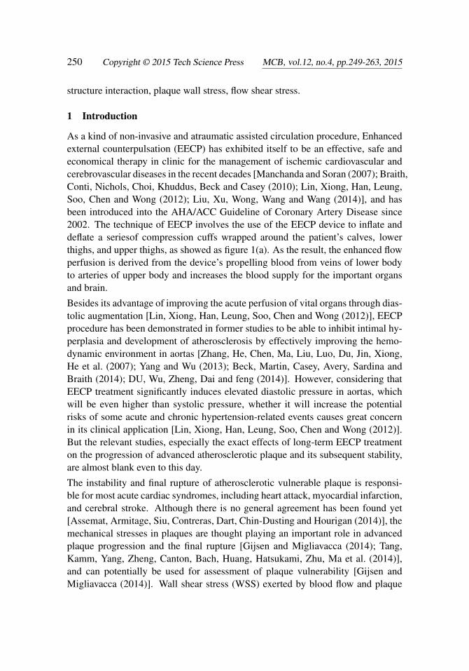
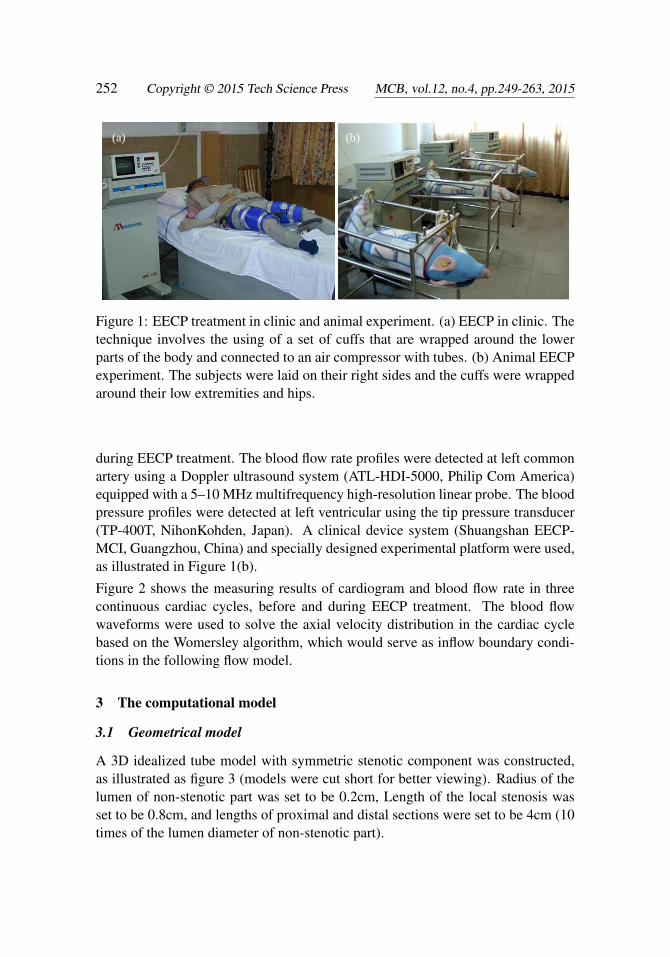
Figure 1: EECP treatment in clinic and animal experiment. (a) EECP in clinic. Thetechnique involves the using of a set of cuffs that are wrapped around the lowerparts of the body and connected to an air compressor with tubes. (b) Animal EECPexperiment. The subjects were laid on their right sides and the cuffs were wrappedaround their low extremities and hips.
during EECP treatment. The blood flow rate profiles were detected at left commonartery using a Doppler ultrasound system (ATL-HDI-5000, Philip Com America)equipped with a 5–10 MHz multifrequency high-resolution linear probe. The bloodpressure profiles were detected at left ventricular using the tip pressure transducer(TP-400T, NihonKohden, Japan). A clinical device system (Shuangshan EECP-MCI, Guangzhou, China) and specially designed experimental platform were used,as illustrated in Figure 1(b).
Figure 2 shows the measuring results of cardiogram and blood flow rate in threecontinuous cardiac cycles, before and during EECP treatment. The blood flowwaveforms were used to solve the axial velocity distribution in the cardiac cyclebased on the Womersley algorithm, which would serve as inflow boundary condi-tions in the following flow model.
3 The computational model
3.1 Geometrical model
A 3D idealized tube model with symmetric stenotic component was constructed,as illustrated as figure 3 (models were cut short for better viewing). Radius of thelumen of non-stenotic part was set to be 0.2cm, Length of the local stenosis wasset to be 0.8cm, and lengths of proximal and distal sections were set to be 4cm (10times of the lumen diameter of non-stenotic part).

Size-Dependent Diffusion of Dextrans in Excised Porcine Corneal Stroma 253
a b
Figure 2: In vivo hemodynamic measurement. (a) Pre-EECP. (b) During EECP.Note that EECP intervention significantly changed the patterns of blood flow incardiac cycles, and augmented the level of perfusion.
Define the stenosis severity normally as the following equation.
S0 =R0−Rmin
R0×100%, (1)
Then, the radius of the lumen at rest is specified as the following equation.
H0(z) = R0−
12
S0R0[1− cos(2π(z− z1)/(z2− z1))], z1 ≤ z≤ z2
0, otherwise(2)
Where R0 is the radius of the lumen of non-steotic part; z1 and z2 are the beginningand ending positions of the stenotic part;
Figure 3: 3D Geometrical model of tube with local symmetric stenosis, S0 = 70%.(a) Flow lumen; (b) Wall structure.

254 Copyright © 2015 Tech Science Press MCB, vol.12, no.4, pp.249-263, 2015
3.2 Governing equations and boundary conditions
3.2.1 The flow model
Assuming that the blood flow was laminar, Newtonian, viscous, and incompress-ible. The incompressible Navier-Stokes equations with arbitrary Lagrangian-Eulerin(ALE) formulation were used as the governing equations, and are given as the fol-lowing:
ρ(∂u/∂ t +((u−ug) ·∇)u) =−∇p+µ∇2u, (3)
∇ ·u = 0, (4)
u|Γ= (0,0,0), (5)
Where u is flow velocity, ug is mesh velocity, p is pressure, ρ is fluid density andµ isthe kinematical viscosity. Γ is the inner boundary of the lumen.
3.2.2 The wall model
Assuming that material for arterial wall is hyperelastic, isotropic, incompressibleand homogeneous. The following equilibrium equations and boundary conditionswere used for the wall model [Tang, Yang, Mondal, Liu, Canton, Hatsukami andYuan (2008)].
σSi j, j = 0, (6)
dS∣∣innerwall = d f
∣∣innerwall , (7)
σSi j, j ·n j
∣∣innerwall
= σf
i j, j ·n j
∣∣∣innerwall
, (8)
σSi j, j ·n j
∣∣outerwall
= 0, (9)
Where dS, d f , σSi j, j and σ
fi j, j are displacement and stress tensors for solid and fluid
respectively. Meanwhile, as the constraint conditions, the inlet and outlet face werefixed in axial (longitudinal) direction, but allowed to expand/contract with flowotherwise [Tang, Yang, Mondal, Liu, Canton, Hatsukami and Yuan (2008)].
3.2.3 Boundary conditions
The velocity and pressure boundary conditions were given to solve the FSI model.The Womersley method for pulsatile flow along a long and rigid circular vessel,as described as below, was introduced to solve the inlet axial velocity profiles ina cardiac cycle based on the in vivo measured blood flow rate waveform. TheWomersley velocity profiles were used as the inlet boundary conditions for thesolving of flow shear stress (FSS) and pressure drop of the model, before and duringEECP treatment.

Size-Dependent Diffusion of Dextrans in Excised Porcine Corneal Stroma 255
Performing a Fourier Transform to the blood flow rate waveform of a cardiac cycle:
Q(t)≈N
∑n=0
Bne jnωt (10)
And then, the Womersley velocity for the axial component of velocity, w(r, t) isgiven by
w(r, t) =2B0
πR20[1− (
rR0
)2]
+N
∑n=1
Bn
πR20[(1−
J0(αnr
R0j3/2)
J0(αn j3/2))/(1− 2J1(αn j3/2)
αn j3/2J0(αn j3/2))]e jnωt (11)
Here, k is the number of the sequential flow rate discrete points in a cardiac cycle,Q(t) is the flow rate, N is the order of the Fourier Transfer, j =
√−1, ω = 2π/T is
the angular frequency, T is the cardiac cycle, Bn is the Fourier coefficients, R0 is theradius of the artery, αn is known as the Womersley number, and αn = R0
√nω/ν ,
ν is the kinematic viscosity of blood, J0 . . . and J1 are Bessel function of the firstkind of order 0 and 1.
a b
Figure 4: Womersley velocity profiles at different time points of a cardiac cycle.(a) Pre-EECP; (b) during EECP.
The pressure boundary conditions were given at inlet and outlet faces, as illustratedas figure 5, for the solving of structural stress and deformation of the wall. Theinlet blood pressure profiles were measured in vivo. The outlet pressure profileswere calculated based on the inlet pressure and the pressure drops solved above.

256 Copyright © 2015 Tech Science Press MCB, vol.12, no.4, pp.249-263, 2015
a b
Figure 5: Pressure conditions specified at the inlet and outlet faces. (a) Pre-EECP;(b) During EECP.
3.3 Tissue properties
For the structural model, assuming that vessel wall is hyperelastic, isotropic, in-compressible, and homogeneous. Then, the tissue propertie can be described byusing the nonlinear modified Mooney-Rivlin model [Yang, Canton, Yuan, Fergu-son, Hatsukami and Tang (2010); Tang, Yang, Mondal, Liu, Canton, Hatsukamiand Yuan (2008)]. The strain energy function is given as following.
W = c1(I1−3)+ c2(I2−3)+D1[exp(D2(I1−3))−1] (12)
I1 = ∑Cii, I2 =12[I2
1 −Ci jCi j] (13)
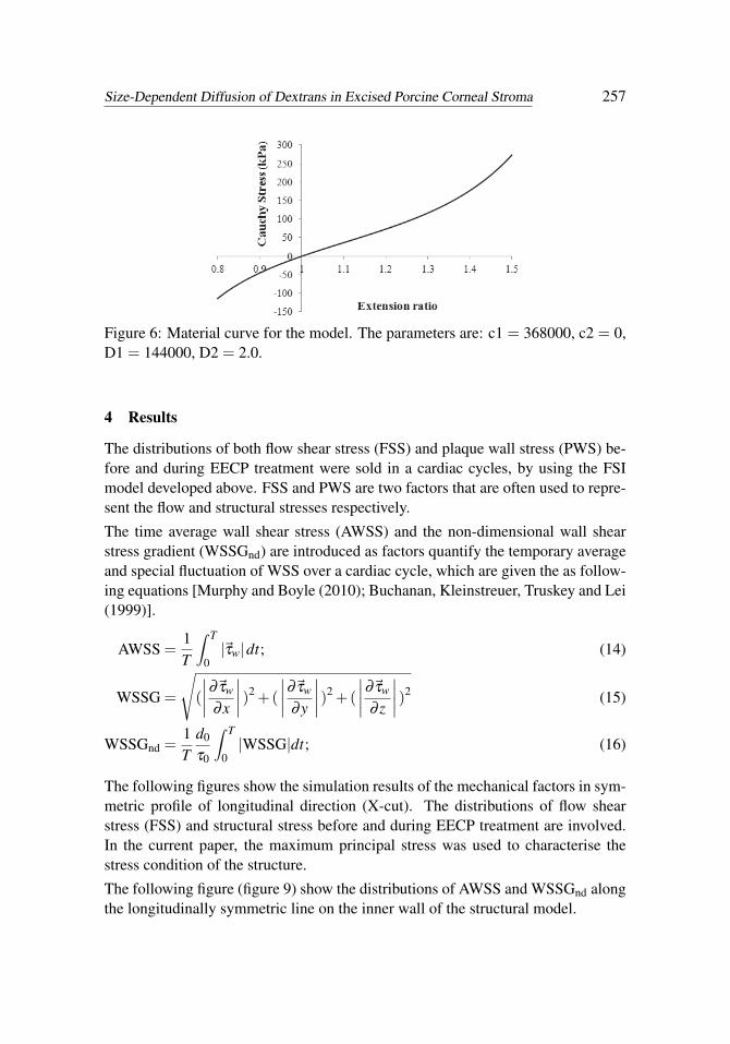
Where I1 and I2 are the first and second strain invariants, C = [Ci j] = XTX is theright Cauchy-Green deformation tensor, X = [Xi j] = [∂xi/∂a j], (xi) is current posi-tion, (ai) is original position. ci and Di are material parameters. The stress-stretchcurves derived from the modified Mooney-Rivlin model are illustrated as figure6, where the material parameters are chosen based on Tang, Yang, Mondal, Liu,Canton, Hatsukami and Yuan (2008).
3.4 Computational solving strategy
The FSI models were solved by ADINA. ADINA uses unstructured finite elementmethods for both fluid and solid models. Nonlinear incremental iterative proce-dures are used to handle FSI. The governing finite element equations for both solidand fluid models were solved by Newton-Raphson iteration method [Tang, Yang,Mondal, Liu, Canton, Hatsukami and Yuan (2008); Tang, Teng, Canton, Yang, Fer-guson, Huang, Zheng, Woodard and Yuan (2009)].
Size-Dependent Diffusion of Dextrans in Excised Porcine Corneal Stroma 257
Figure 6: Material curve for the model. The parameters are: c1 = 368000, c2 = 0,D1 = 144000, D2 = 2.0.
4 Results
The distributions of both flow shear stress (FSS) and plaque wall stress (PWS) be-fore and during EECP treatment were sold in a cardiac cycles, by using the FSImodel developed above. FSS and PWS are two factors that are often used to repre-sent the flow and structural stresses respectively.
The time average wall shear stress (AWSS) and the non-dimensional wall shearstress gradient (WSSGnd) are introduced as factors quantify the temporary averageand special fluctuation of WSS over a cardiac cycle, which are given the as follow-ing equations [Murphy and Boyle (2010); Buchanan, Kleinstreuer, Truskey and Lei(1999)].
AWSS =1T
∫ T
0|~τw|dt; (14)
WSSG =
√(
∣∣∣∣∂~τw
∂x
∣∣∣∣)2 +(
∣∣∣∣∂~τw
∂y
∣∣∣∣)2 +(
∣∣∣∣∂~τw
∂ z
∣∣∣∣)2 (15)
WSSGnd =1T
d0
τ0
∫ T
0|WSSG|dt; (16)
The following figures show the simulation results of the mechanical factors in sym-metric profile of longitudinal direction (X-cut). The distributions of flow shearstress (FSS) and structural stress before and during EECP treatment are involved.In the current paper, the maximum principal stress was used to characterise thestress condition of the structure.
The following figure (figure 9) show the distributions of AWSS and WSSGnd alongthe longitudinally symmetric line on the inner wall of the structural model.

258 Copyright © 2015 Tech Science Press MCB, vol.12, no.4, pp.249-263, 2015
Figure 7: Flow shear stress (FSS) distributions, x-cut profile. (a) Pre-EECP, t =35T/130; (b) During EECP, t = 80T/132.
Figure 8: Structural stress distributions, x-cut profile. (a) Pre-EECP, t = 35T/130;(b) During EECP, t = 80T/132.
a b
Figure 9: Wall shear stress and its spacial oscillation over a cardiac cycle. (a)AWSS before and during EECP; (b) WSSGnd before and during EECP.
According to the simulation results, the peak structural stresses appeared at up-stream site of the local stenosis in both cases of before and during EECP, and thelocal stenotic site showed lower level of structural stress distribution. EECP inter-

Size-Dependent Diffusion of Dextrans in Excised Porcine Corneal Stroma 259
vention significantly changed the distribution characteristics of the structural stressin cardiac cycle and augmented its level. During EECP treatment, the peak val-ue of the structural stress (1223446 dyn/cm2) happened in diastole (t = 80T /132).Correspondingly, the structural stress at the middle of the stenosis (throat site) was487000 dyn/cm2 at that time point during EECP. Whereas, the peak value of thestructural stress before EECP treatment (1051716 dyn/cm2) happened in systole(t = 35T /130). And at that time point, the structural stress at the middle of thestenosis (throat site) was 432000 dyn/cm2. EECP treatment increased 12.21% ofthe structural stress at the middle of the stenosis, compared with the normal physi-ological status (before EECP).
Meanwhile, the peak FSS values appeared at the throat of the stenotic site in bothcases of before and during EECP, and the non-stenotic sites were where lower FSSlevel distributed. During EECP treatment, the peak FSS value (1232.0 dyn/cm2)happened in diastole (t = 80T /132), compared with that of 607.7 dyn/cm2 beforeEECP which happened in systole (t = 35T /130). The peak FSS value during EECPaugmented 102.73% compared to that of before EECP.
Finally, both AWSS and WSSGnd showed higher-level distributions in local stenot-ic site. The peak values of AWSS over a cardiac cycle, which appeared at thethroat site of the steosis, were 315.74 dyn/cm2 (during EECP) and 200.19 dyn/cm2
(before EECP) respectively. The peak values of WSSGnd over a cardiac cycle,which appeared at the throat site of the steosis, were 1298.05 (during EECP) and903.47(before EECP) respectively. EECP intervention induced an augmentation of57.72% of AWSS level and an augmentation of 43.67% of WSSGnd level comparedto that of before EECP.
5 Conclusions
The pilot study of the present paper aimed to explore the influence of EECP treat-ment, which has been introduced into the AHA/ACC Guideline of Coronary ArteryDisease for over ten years, on flow and structural stress conditions of blood vesselwith advanced plaque. The study was conducted based on a combination of in vivoanimal experiment and 3D numerical simulation.
The stress conditions are thought playing important roles in the progression of theplaque and its final rupture. Many studies suggested that PWS showed a neg-ative correlation with plaque progression [Yang, Canton, Yuan, Ferguson, Hat-sukami and Tang (2010); Tang, Yang, Mondal, Liu, Canton, Hatsukami and Yuan(2008); Tang, Teng, Canton, Yang, Ferguson, Huang, Zheng, Woodard and Yuan(2009); Sadat, Teng and Gillard (2010)]. Meanwhile, some studies suggested thatFSS/WSS might play a positive correlation with advanced plaque progression [Yang,
260 Copyright © 2015 Tech Science Press MCB, vol.12, no.4, pp.249-263, 2015
Canton, Yuan, Ferguson, Hatsukami and Tang (2010); Gijsen, van der Giessen,van der Steen and Wentzel (2013)], yet in others, lower FSS/WSS was thoughtcontributing to continued plaque progression [Tang, Yang, Mondal, Liu, Canton,Hatsukami and Yuan (2008)]. Considering the massive difference of the scale,structural stress was thought to be a more critical stress factor that affected ad-vanced plaque progression than FSS/WSS [Sadat, Teng and Gillard (2010)]. Theresults obtained in the present paper show that EECP treatment induces significantaugmentations of the instant level of both FSS/WSS and structural stress (PWS) atthe local stenotic site, as well as their characteristics of distributions in cardiac cy-cles. We thus suggest that long-term EECP treatment may intervene the advancedatherosclerotic plaque progression by inducing the variations of mechanical fac-tors. However, either EECP treatment may play a positive effect on inhibiting thecontinued plaque progression by increasing the PWS level to some extent, or a neg-ative effect by increasing the WSS level and its spatial oscillation, which will needfurther studies combined with clinical observations.
6 Limitations
In the present pilot study, the conclusions were obtained based on idealized bloodvessel model with symmetric stenosis of plaque, and the components of the plaquewere not taken into account. The geometries and the plaque components, especiallythe lipid pool and fibrous cap, are thought having influences on the critical FSS andstructural stress [Tang, Yang, Mondal, Liu, Canton, Hatsukami and Yuan (2008);Tang, Teng, Canton, Yang, Ferguson, Huang, Zheng, Woodard and Yuan (2009);Teng, Canton, Yuan, Ferguson, Yang, Huang, Zheng, Woodard and Tang (2010);Yuan, Teng, Feng, Zhang, Brown, Gillard, Jing and Lu (2015)]. Attempt will bemade in our future work to improve the model.
Acknowledgement: The present research is supported by the Key Clinical Projectfrom the Ministry of Health (25400) and the National Natural Science Foundationof China (81170272).
References
Assemat, P.; Armitage, J. A.; Siu, K. K.; Contreras, K. G.; Dart, A. M.; Chin-Dusting, J. P.; Hourigan, K. (2014): Three-dimensional numerical simulation ofblood flow in mouse aortic arch around atherosclerotic plaques. Applied Mathe-matical Modelling, vol. 38, no. 17, pp. 4175–4185.
Beck, D. T.; Martin, J. S.; Casey, D. P.; Avery, J. C.; Sardina, P. D.; Braith,R. W. (2014): Enhanced external counterpulsation improves endothelial function

Size-Dependent Diffusion of Dextrans in Excised Porcine Corneal Stroma 261
and exercise capacity in patients with ischaemic left ventricular dysfunction. Clin-ical and Experimental Pharmacology and Physiology, vol. 41, no. 9, pp. 628–636.
Braith, R. W.; Conti, C. R.; Nichols, W. W.; Choi, C. Y.; Khuddus, M. A.;Beck, D. T.; Casey, D. P. (2010): Enhanced external counterpulsation improvesperipheral artery flow-mediated dilation in patients with chronic angina a random-ized sham-controlled study. Circulation, vol. 122, no. 16, pp. 1612–1620.
Buchanan, J. R.; Kleinstreuer, C.; Truskey, G. A.; Lei, M. (1999): Relationbetween non-uniform hemodynamics and sites of altered permeability and lesiongrowth at the rabbit aorto-celiac junction. Atherosclerosis, vol. 143, no. 1, pp.27–40.
Chatzizisis, Y. S.; Baker, A. B.; Sukhova, G. K.; Koskinas, K. C.; Papafaklis,M. I.; Beigel, R.; Jonas, M.; Coskun, A. U.; Stone, B. V.; Maynard, C. et al.(2011): Augmented expression and activity of extracellular matrix-degradingenzymes in regions of low endothelial shear stress colocalize with coronary athero-mata with thin fibrous caps in pigs. Circulation, vol. 123, no. 6, pp. 621–630.
DU, J.-h.; Wu, G.-f.; Zheng, Z.-s.; Dai, G.; feng, M.-z. (2014): Enhancedexternal counterpulsation inducing arterial hemodynamic variations and its chroniceffect on endothelial function. Chinese Journal of Biomedical Engineering, vol. 3,pp. 006.
Gijsen, F.; Migliavacca, F. (2014): Plaque mechanics. Journal of biomechanics,vol. 47, no. 4, pp. 763.
Gijsen, F.; van der Giessen, A.; van der Steen, A.; Wentzel, J. (2013): S-hear stress and advanced atherosclerosis in human coronary arteries. Journal ofbiomechanics, vol. 46, no. 2, pp. 240–247.
Groen, H. C.; Gijsen, F. J.; van der Lugt, A.; Ferguson, M. S.; Hatsukami,T. S.; van der Steen, A. F.; Yuan, C.; Wentzel, J. J. (2007): Plaque rupture inthe carotid artery is localized at the high shear stress region a case report. Stroke,vol. 38, no. 8, pp. 2379–2381.
Lin, W.; Xiong, L.; Han, J.; Leung, T. W. H.; Soo, Y. O. Y.; Chen, X.; Wong,K. S. L. (2012): External counterpulsation augments blood pressure and cerebralflow velocities in ischemic stroke patients with cerebral intracranial large arteryocclusive disease. Stroke, vol. 43, no. 11, pp. 3007–3011.
Liu, L.-P.; Xu, A.-D.; Wong, L. K.; Wang, D. Z.; Wang, Y.-J. (2014): Chineseconsensus statement on the evaluation and intervention of collateral circulation forischemic stroke. CNS neuroscience & therapeutics, vol. 20, no. 3, pp. 202–208.
Manchanda, A.; Soran, O. (2007): Enhanced external counterpulsation andfuture directions: step beyond medical management for patients with angina and

262 Copyright © 2015 Tech Science Press MCB, vol.12, no.4, pp.249-263, 2015
heart failure. Journal of the American College of Cardiology, vol. 50, no. 16, pp.1523–1531.
Murphy, J.; Boyle, F. (2010): Predicting neointimal hyperplasia in stented arter-ies using time-dependant computational fluid dynamics: a review. Computers inbiology and medicine, vol. 40, no. 4, pp. 408–418.
Peiffer, V.; Sherwin, S. J.; Weinberg, P. D. (2013): Does low and oscillatorywall shear stress correlate spatially with early atherosclerosis? a systematic review.Cardiovascular research, vol. 99, pp. 242–250.
Sadat, U.; Teng, Z.; Gillard, J. H. (2010): Biomechanical structural stresses ofatherosclerotic plaques. Expert review of cardiovascular therapy, vol. 8, no. 10,pp. 1469–1481.
Tang, D.; Kamm, R. D.; Yang, C.; Zheng, J.; Canton, G.; Bach, R.; Huang,X.; Hatsukami, T. S.; Zhu, J.; Ma, G. et al. (2014): Image-based modelingfor better understanding and assessment of atherosclerotic plaque progression andvulnerability: Data, modeling, validation, uncertainty and predictions. Journal ofbiomechanics, vol. 47, no. 4, pp. 834–846.
Tang, D.; Teng, Z.; Canton, G.; Yang, C.; Ferguson, M.; Huang, X.; Zheng,J.; Woodard, P. K.; Yuan, C. (2009): Sites of rupture in human atheroscleroticcarotid plaques are associated with high structural stresses an in vivo mri-based 3dfluid-structure interaction study. Stroke, vol. 40, no. 10, pp. 3258–3263.
Tang, D.; Yang, C.; Mondal, S.; Liu, F.; Canton, G.; Hatsukami, T. S.; Yuan,C. (2008): A negative correlation between human carotid atherosclerotic plaqueprogression and plaque wall stress: in vivo mri-based 2d/3d fsi models. Journal ofbiomechanics, vol. 41, no. 4, pp. 727–736.
Teng, Z.; Canton, G.; Yuan, C.; Ferguson, M.; Yang, C.; Huang, X.; Zheng,J.; Woodard, P. K.; Tang, D. (2010): 3d critical plaque wall stress is a betterpredictor of carotid plaque rupture sites than flow shear stress: an in vivo mri-based3d fsi study. Journal of biomechanical engineering, vol. 132, no. 3, pp. 031007.
Williamson, S.; Lam, Y.; Younis, H.; Huang, H.; Patel, S.; Kaazempur-Mofrad, M.; Kamm, R. (2003): On the sensitivity of wall stresses in diseasedarteries to variable material properties. Journal of biomechanical engineering, vol.125, no. 1, pp. 147–155.
Yang, C.; Canton, G.; Yuan, C.; Ferguson, M.; Hatsukami, T. S.; Tang, D.(2010): Advanced human carotid plaque progression correlates positively withflow shear stress using follow-up scan data: an in vivo mri multi-patient 3d fsistudy. Journal of biomechanics, vol. 43, no. 13, pp. 2530–2538.

Size-Dependent Diffusion of Dextrans in Excised Porcine Corneal Stroma 263
Yang, D.-y.; Wu, G.-f. (2013): Vasculoprotective properties of enhanced externalcounterpulsation for coronary artery disease: beyond the hemodynamics. Interna-tional journal of cardiology, vol. 166, no. 1, pp. 38–43.
Yuan, J.; Teng, Z.; Feng, J.; Zhang, Y.; Brown, A. J.; Gillard, J. H.; Jing,Z.; Lu, Q. (2015): Influence of material property variability on the mechanicalbehaviour of carotid atherosclerotic plaques: A 3d fluid-structure interaction anal-ysis. International journal for numerical methods in biomedical engineering, vol.31, no. 8.
Zhang, Y.; He, X.; Chen, X.; Ma, H.; Liu, D.; Luo, J.; Du, Z.; Jin, Y.; X-iong, Y.; He, J. et al. (2007): Enhanced external counterpulsation inhibits intimalhyperplasia by modifying shear stress–responsive gene expression in hypercholes-terolemic pigs. Circulation, vol. 116, no. 5, pp. 526–534.


![Intra-aortic balloon pump counterpulsation [IABP] for ...Intervention Review] Intra-aortic balloon pump counterpulsation (IABP) for myocardial infarction complicated by cardiogenic](https://img.dokumen.tips/doc/110x75/5b0c16167f8b9a8b038b916c/intra-aortic-balloon-pump-counterpulsation-iabp-for-intervention-review-intra-aortic.jpg)