Embed Size (px)
Citation preview

1
Introduction
In case of lipophilic drugs which belong to BCS type-II with a high permeability through biomembranes, enhancement of the in vitro dissolution rate may lead directly to increased oral bioavailability1,2. Nowadays, the most common strategies used to enhance the dissolution rate are particle size reduction3, or transformation of the crystalline form to the amorphous state4. However, amor-phous solids are generally less physically and chemically stable than corresponding crystals due to their higher energy, entropy and free energy5. At present, five drug nanocrystal products are on the market compared with the large number of conventional dosage forms prepared using coarse crystalline drug substances6.
According to the Noyes–Whitney equation, a marked reduction in particle size down to the submicron level would dramatically increase the effective particle surface area available for dissolution7,8. Recently, there have been significant advances in reducing the size of solid particles down to the sub-micron region. Nanoparticle precipita-tion by the anti-solvent method is a direct and simple procedure for the preparation of drug nanocrystals9–13. The process of anti-solvent precipitation is carried out by mixing organic drug solution with an aqueous anti-solvent solution in the presence of stabilizing surfactants or polymers to form submicron particles. However, it is difficult to control the particle size of some drugs (such as simvastatin) due to particle growth and agglomeration.
RESEARCH ARTICLE
Enhanced dissolution rate and oral bioavailability of simvastatin nanocrystal prepared by sonoprecipitation
Tongying Jiang, Ning Han, Buwen Zhao, Yuling Xie and Siling Wang
Department of Pharmaceutics, School of Pharmacy, Shenyang Pharmaceutical University, Shenyang, PR China
AbstractBackground: Simvastatin is classified as a Biopharmaceutics Classification System (BCS) Class-II compound with a poor aqueous solubility and an acceptable permeability through biomembranes. The strategy of increasing the in vitro dissolution has the potential to enhance the oral bioavailability when using nanosized crystalline drugs.
Objective: The aim of this article was to prepare simvastatin nanocrystals to enhance its dissolution rate and bioavailability by exploiting sonoprecipitation.
Methods: Injecting 0.50% (w/v) methanol solution of simvastatin into 0.20% (w/v) water solution of F68 under sonication amplitude of 400 W and processing temperature of 3°C.
Results: Simvastatin nanocrystal with average diameter of 360 ± 9 nm could be obtained. X-ray powder diffraction (XRPD) and differential scanning calorimetry (DSC) confirmed the decreased crystallinity of nanoparticles stabilized by F68. The results of in vitro study demonstrated that the saturation solubility and dissolution rate of simvastatin nanocrystals were enhanced by 1 fold and 4 fold respectively, compared with crude simvastatin and the dissolution rate improved with the decrease in particle size. The C
max and AUC
(0–24 h) values of simvastatin nanocrystal group were
approximately 1.50-fold and 1.44-fold greater than that of simvastatin nanocrystal group, respectively. Additionally, the T
max of simvastatin nanocrystal group was 1.99 h, comparing to 2.88 h of reference group.
Conclusion: Sonoprecipitation method can produce small and uniform simvastatin nanocrystals with an improved saturation solubility, dissolution rate and oral bioavailability.
Keywords: Nanocrystal, Simvastatin, sonoprecipitation, dissolution rate, bioavailability, anti-solvent precipitation, stabilizer
Address for Correspondence: Siling Wang, Department of Pharmaceutics, School of Pharmacy, Shenyang Pharmaceutical University, Shenyang 110016, PR China. Tel: +86 24 23986348. Fax: +86 24 23986348. E-mail: [email protected]
(Received 16 May 2011; revised 24 November 2011; accepted 28 November 2011)
Drug Development and Industrial Pharmacy, 2012, 1–10, Early Online© 2012 Informa Healthcare USA, Inc.ISSN 0363-9045 print/ISSN 1520-5762 onlineDOI: 10.3109/03639045.2011.645830
Drug Development and Industrial Pharmacy
2012
00
00
000
000
16 May 2011
24 November 2011
28 November 2011
0363-9045
1520-5762
© 2012 Informa Healthcare USA, Inc.
10.3109/03639045.2011.645830
LDDI
645830
Dru
g D
evel
opm
ent a
nd I
ndus
tria
l Pha
rmac
y D
ownl
oade
d fr
om in
form
ahea
lthca
re.c
om b
y Sh
enya
ng P
harm
aceu
tical
Uni
vers
ity o
n 07
/11/
12Fo
r pe
rson
al u
se o
nly.

2 T. Jiang et al.
Drug Development and Industrial Pharmacy
To overcome this problem of physical instability, the process of anti-solvent precipitation under sonoprecipi-tation may be employed. For ultrasound can intensify the mass transfer to avoid inefficient mixing, atomize the drug solution into fine droplets which attributes to the formation of smaller particles, reduce the particle size of the newly formed particles and suppress the agglomera-tion of fine particles by cavitation14–17.
In the nanometer range, particles possess additional Gibbs energy resulting in particles aggregating to reduce the particle surface area. It is essential to provide barri-ers between particles by adding stabilizers6 which can be adsorbed on the particle surface to generate repul-sive forces, including steric stabilization or electrostatic stabilization to control particle size and morphology. Polymers such as HMPC, HPC, PVP, F68, PVA and sur-factants including SDS, DOSS, and Tween 80 are widely used as nanocrystals stabilizers alone or in combination. The appropriate stabilizers were screened for smaller particle size by trial and error from PVA, PVP
K30, HPMC
E5,
HPC, F68, F127, SDS and Tween 80.Simvastatin acts by inhibiting HMG-CoA reductase
and is used for the treatment of hypercholesterolemia. It is a class-II drug according to the BCS18 and, therefore, has a dissolution rate-limited absorption in vivo and, hence, suboptimal oral bioavailability. It has one stable, com-mon crystal form and no known hydrates. Several drug delivery systems for improving simvastatin solubility and dissolution rate have been reported in the literature19,20. However, no study in which applying sonoprecipitation method to prepare simvastatin nanoparticles has yet been published. The aim of this study was to prepare sim-vastatin nanocrystal by sonoprecipitation method and assess the dissolution rate and bioavailability. The par-ticle size, morphology, solid state, behavior of the solid transformations and chemical structure of surface were determined by dynamic light scattering, SEM, XRPD, DSC and FT-IR, respectively. The in vitro dissolution and in vivo bioavailability were investigated to evaluate sim-vastatin nanocrystal.
Materials and methods
MaterialsSimvastatin was used as a model drug and was a gener-ous gift from Zhejiang Haizheng Pharmaceutical Co. Ltd. (China) and the mean particle size of crude crystals was 14 μm. F68 (BASF), PVP
K30 (ISP) and HPMC
E5 (DOW)
were obtained from the companies shown. Methanol, ethyl acetate and acetone of chromatographic grade were purchased from Concord Chemical Agent (China). All other chemicals and solvents were of reagent grade.
MethodsPreparation of nanocrystalsSonoprecipitation was used to produce the simvasta-tin solid nanoparticles. An apparatus (JY-92-II sonifier, Ningbo Scientz Biotechnology Co. Ltd., China) consisting
of a probe and sonifier was employed to provide the source of the ultrasound and a power of 400 W was applied in the precipitation process. A glass vessel holding the sta-bilizer solution was fixed under the probe (a tip of 8 mm in diameter) which was immersed 10 mm in the liquid. Methanol and water were used as solvent and anti-sol-vent, respectively. And the volume ratio of solvent and anti-solvent was 1:20 (v:v). Briefly, 0.5 g simvastatin was dissolved in 5 ml methanol to obtain an organic solution and 500 μl of the solution was injected into 10 ml 0.2% stabilizer solution maintained at 3°C through a syringe under ultrasonic conditions at 400 W. The concentra-tion ratio of drug to stabilizer was 5:2 (w:w). This process resulted in the formation of milky simvastatin nanodis-persions. Then an intermittent sonication was applied to the nanodispersions by sonication for 5 seconds at 5 seconds intervals for a total sonication time of 10 min. The dispersions were kept under vacuum at room tem-perature for 1 h to remove the methanol21. Filter-drying was used for solidification of simvastatin nanosuspen-sions to study saturation solubility and dissolution rate. Simvastatin suspension was added to the Bush funnel housed with 0.1 μm microporous membrane, the liquid was removed from the system through force of vacuum pump. Then washed twice with double distilled water, and put the resulting cake in a vacuum desiccator for 24 h, and dry powers of simvastatin can be obtained.
Particle size measurementParticle size analysis of the simvastatin nanosuspen-sions was performed by dynamic light scattering using a zpw388-NICOMP instrument (Nicomp Particle Sizing Systems, Santa Barbara, CA, USA). Particle-size distri-bution which typically includes d(10), d(50) and d(90) representing the percentage of particles below a given size. The nanodispersions were diluted with water for the measurement to give an intensity of 300 Flux as recom-mended by the manufacturer. The mean intensity par-ticle size was obtained. Each sample was measured in triplicate.
Scanning electron microscopyThe shape and morphology was examined using Scanning electron microscopy (SEM) (JEOL JSM-7001F). Freshly prepared simvastatin nanodispersion after dilution and unprocessed crude samples were deposited on a glass slide, and then the glass slide was kept under vacuum at 50°C for 1 h. The samples were coated with a thin gold-palladium layer using a sputter coater unit. The scanning electron microscope was operated at an acceleration voltage of 15 kV.
X-ray powder diffractionThe X-ray powder diffraction (XRPD) patterns were recorded using a powder X-ray diffractometer (Rigaku Geigerflex XRD, Co., Japan) with the Cu-Ka line as the source of radiation and the instrument was operated at a voltage of 40 kV and a current of 45 mA. All samples were
Dru
g D
evel
opm
ent a
nd I
ndus
tria
l Pha
rmac
y D
ownl
oade
d fr
om in
form
ahea
lthca
re.c
om b
y Sh
enya
ng P
harm
aceu
tical
Uni
vers
ity o
n 07
/11/
12Fo
r pe
rson
al u
se o
nly.

Simvastatin nanocrystal 3
© 2012 Informa Healthcare USA, Inc.
measured in the 2θ angle range between 5° and 40° with a scan rate of 5°/min and a step size of 0.01°.
Differential scanning calorimetryDifferential scanning calorimetry (DSC) analyses were carried out on a differential scanning calorimeter DSC-60 (Shimadzu, Japan). Indium/Zinc standards were used to calibrate the DSC temperature and enthalpy scale. Samples were accurately weighed (2–3 mg), sealed in aluminum pans and heated over a range of 50–200°C under a nitrogen purge. A nitrogen flow rate of 20 ml/min was used for DSC runs.
Fourier transform infrared spectroscopyFourier transform infrared spectroscopy (FT-IR) spectra of samples were recorded with an FT-IR spectrometer (Bruker IFS 55, Switzerland). Samples were diluted with dry potassium bromide and scanned from 4000–400 cm−1 to evaluate the molecular state of crude simvastatin and simvastatin nanocrystal.
Contact angle measurementContact angle measurement has been conducted to evaluate the wettability of different samples. The mea-surement was performed on Dynamic Absorption Tester (JY-82, Chengde Dingsheng Tester Equipment Co., Ltd, China). Powder of Simvastatin nanocrystal and crude simvastatin were compressed into tablets (powder of 200 mg, pressure as 6 ± 0.4 kg) prior to measurement. Water was dripped on the plane surface of tablet through a microinjector and the image was immediately cap-tured by a CCD camera. The values of contact angle were obtained by analysis of software. Each sample was mea-sured triplicate to ensure accuracy.
Saturation solubilityThe saturation solubility of crude simvastatin and pre-cipitated drug nanocrystals of simvastatin was deter-mined by the Higuchi method. An excess amount of each sample was added to 10 ml of the phosphate buf-fer solution (pH 6.8) in test-tubes sealed with stoppers. They were kept in a constant temperature shaking bath maintained at 37 ± 0.5°C until equilibrium was reached (for 48 h). A portion of the solution was then taken out and passed through a syringe filter and detected at 238 nm in a UV-spectrophotometer (UNICO UV-2000, Shanghai, China). The experiments were conducted in triplicate.
In vitro dissolution testThe dissolution studies were performed according to the USP apparatus 2 (paddle) method (RC-8D, Tianjin Guoming Medical Equipment Co. Ltd.). Samples equiva-lent of 10 mg drug were placed in a dissolution vessel containing 900 ml pH6.8 buffer solution, maintained at 37 ± 0.5°C and stirred at 100 r/m. Samples were removed periodically and replaced with fresh dissolution medium. After filtration by 0.22 μm microporous membrane, the
concentration of simvastatin was determined spectro-photometrically at 238 nm.
Bioavailability studyExperimental procedure Wistar rats weighing between 230–279 g were chosen as the experimental animals in this study. All experiments were conducted according to the “Guidelines for the Care and Use of Laboratory Animals” and the protocol was approved by Animal Ethics Committee of Shenyang Pharmaceutical University. Wistar rats (n = 10) were fasted 24 h before dosing. The rats were randomly divided into 2 groups. Rats in group A were given simvastatin nanocrystal in aqueous suspen-sion form through intragastric administration at a dose of 10 mg/kg. Rats in Group B were given crude simvastatin in aqueous suspension form (freshly prepared in 0.1% w/v carboxymethyl cellulose just before dosing) through intragastric administration at a dose of 10 mg/kg as con-trol. After anesthetization of animals, a series of blood samples (0.5 ml each) were collected into heparinized tubes at designed time intervals (0.25, 0.5, 0.75, 1.0, 1.5, 2.0, 2.5, 3.0, 4.0, 6.0, 8.0, 12.0, 24.0 h) using retro orbital puncture technique. Plasma was separated by centrifu-gation at 8000 r/m for 5 min and then stored at −20°C for subsequent analyzing by HPLC.
Assay of plasma concentration Frozen plasma samples were thawed at room temperature. A 200 μl aliquot of plasma was transferred into capped test tube, 20 μl internal standard solution (lovastatin 10 μg/ml in mobile phase) and 800 μl extract consisting of ethyl acetate: acetone (1:4, v:v) were added respectively. The sample was fully mixed by vortexing for 5 min, and cen-trifuged at 10000 rpm for 10 min. Then the organic phase was transferred into a clean test tube and evaporated to dryness by centrifugal at 40°C (Labconco Centrivap, USA). The residues were redissolved in 50 μl of mobile phase, vortexed for 5 min and centrifuged at 12000 rpm for 10 min, then 20 μl of the final clear supernatant was injected into HLPC system.
The concentration of simvastatin acid was assessed by a HLPC system Hitachi L-7000 series including a Hitachi 7100 pump, a Hitachi L-7400 UV detector (both from Hitachi technologies corporation, Japan) equipped with a Diamonsil C18 column (250 mm × 4.6 mm 5 μm). The daily prepared mobile phase consisting of methanol:water (80:20, v:v) ran at 1 ml/min at 20°C. The UV detection was operated at 238 nm.
Calibration curves were established by plotting the peak area ratios of simvastatin acid to lovastatin versus standard concentrations. The calibration curve of sim-vastatin acid was linear over the concentration range of 0.01–0.1 μg/ml in plasma. The ratio of peak area of simvastatin acid to lovastatin was used to quantify the concentration of simvastatin acid in plasma. The analysis of all the data were carried out using software DAS 2.1.1 (Boying corporation, China). Results were exhibited as means ± SD.
Dru
g D
evel
opm
ent a
nd I
ndus
tria
l Pha
rmac
y D
ownl
oade
d fr
om in
form
ahea
lthca
re.c
om b
y Sh
enya
ng P
harm
aceu
tical
Uni
vers
ity o
n 07
/11/
12Fo
r pe
rson
al u
se o
nly.

4 T. Jiang et al.
Drug Development and Industrial Pharmacy
Results and discussion
Preparation of simvastatin nanocrystalsWe had attempted to employ the anti-solvent pre-cipitation to prepare pre-suspension which was further homogenized using an ATS AH110D homogenizer (ATS Engineer Inc, China) at 500 bar for 5 times, 500 bar for 10 times or 1000 bar for 20 times before choosing anti-sol-vent precipitation under sonoprecipitation. The resultant simvastatin particles size can not decrease with increased the pressure and times, thus the obtained minimum par-ticles were 2 µm at diameter. In addition, anti-solvent precipitation with stirring provided simvastatin particles possessing poor stability although F68 was also used as stabilizer. The particle size increased from 400 nm to 5 μm within 1 h, and then the particle size would keep constant. Nano-size simvastatin particles prepared by anti-solvent precipitation with sonication can keep better stability within 7 days at room temperature. In this method, the effect of formulation factors including types and concen-tration of solvent and stabilizer; drug loading; power and time of sonication; processing temperature on the par-ticle size and PSD were systematically investigated.
Effects of solvent and the ratio of solvent to anti-solventWhen anti-solvent was 0.1% (w/v) F68 aqueous solution, the ratio of aqueous solution to organic solution was 10:1 and the drug loading was 1.0% (w/v), the effect of vari-ous good solvent on the particle size was investigated. Organic solvent including methanol, ethanol, propylene glycol, PEG400 and DMSO that must be mixable with water were considered as the good solvent of simvas-tatin. The solubility of simvastatin in DMSO was maxi-mum, was medium in methanol and ethanol and was minimum in PEG400 and propylene glycol. The result (as Figure 1) shows that the particles prepared by methanol are the smallest among above solvents. The viscosity of methanol is the smallest and its diffusion rate in water is the fastest, which favor the formation of larger numbers of crystal nucleus with smaller particles size. Therefore, methanol was chosen as good solvent of simvastatin in the following preparations.
The ratio of anti-solvent to solvent (AS/A) was also very essential in the formation of simvastatin nanocrystals, and the result was summarized in Figure 2. When the volume ratio increased from 10:1 to 17.5:1, the particle size reduced remarkably mainly due to the enhanced supersaturation. When the volume ratio reached 20:1, No notable changes were found on the particle size. Since the organic phase of methanol may lead to the instability prone to Ostwald ripening of nanodispersions22. Additionally, fewer colli-sionns and less agglomeration occurred in diluted system. In order to minimize the introduction of organic solvents, volume ratio of 20:1 was chosen in this method.
Effects of stabilizer and stabilizer concentration0.1% (w/v) solution of PVA, PVP
K30, HPMC
E5, HPC, F68,
F127, SDS or Tween 80 was candidates as stabilizers to screen the appropriate stabilizers for preparing nano-size simvastatin. The results as Figure 3 shows that F68 allows the production of submicron-sized particles with the smallest diameter of 360 nm and narrowest PSD compared
Figure 1. Mean intensity weighted size of simvastatin particles prepared by various organic solvent.
Figure 2. Mean intensity weighted size of simvastatin particles prepared by various volume ratios of anti-solvent to solvent.
Figure 3. Mean intensity weighted size of simvastatin particles prepared by various stabilizers.
Dru
g D
evel
opm
ent a
nd I
ndus
tria
l Pha
rmac
y D
ownl
oade
d fr
om in
form
ahea
lthca
re.c
om b
y Sh
enya
ng P
harm
aceu
tical
Uni
vers
ity o
n 07
/11/
12Fo
r pe
rson
al u
se o
nly.
Simvastatin nanocrystal 5
© 2012 Informa Healthcare USA, Inc.
with other stabilizers. So in the current study, F68 was employed as dispersion stabilizer. Surfactant (F68) or polymers (HPMC, PVP and so on) could precipitate and adsorb on the solid particle surface to prohibit the par-ticle growth and agglomeration during the anti-solvent process by the formation of a steric barrier. Many studies have concluded the possibility of stabilizer adsorption on drug nanoparticles involving the hydrophobic moiety23.
The effect of the concentration of stabilizer on the par-ticle size was tested, and the result was as Figure 4. At the lowest stabilizer concentration, the average particle size was around 2 μm, which can be explained as the coverage of stabilizer on the nanocrystal surface was inadequate to arrest particle growth24. The particle size reduced with the increasing of stabilizer concentration. Finally, a plateau region was reached at stabilizer concentration of 0.2% (w/v) where the particle size no longer changed.
Effect of drug loadingThe low-, medium-, high-drug loading were set as 0.2, 0.5, 1.0 (%, w/v), respectively. It can be seen that with
an increase in drug loading, the particle size increased from 360 nm to 700 nm (Figure 5). High drug loading can generate higher supersaturation which favors more formation of nuclei, but which may cause the collision, agglomeration and growth of numerous nuclei within certain region. Taken all aspects into account, drug load-ing was chosen as 0.5 (%, w/v).
Effects of other process variablesThe ultrasonic power 200 w, 300 w, 400 w, 500 w and 600 w had been used in preparation. The result showed that the particle size decreased slightly with an increase in the ultrasonic power. Furthermore, 400 w, 500 w and 600 w had not evident effect on the particle size and the resulting particle were almost unimodal and 400 nm at mean diameter. Thus, 400 w was chosen as work power. The total sonication time lasted for 10 min because shorter sonication time was always followed by par-ticle growth and agglomeration. Injecting drug organic solution into the aqueous solution can produce more uniform particles than injecting drug solution on the surface of the aqueous solution. The processing tem-perature also played part of role on the particle size, a lower temperature resulting in smaller particles as Table 1. The same phenomenon has been seen by many other researchers2. At lower temperature, the solubility of simvastatin decreased, therefore, promoted the level of supersaturation, resulting in rapid nucleation, since the number of nuclei increases, the solute on each nuclei decreases, thus the potential for smaller crys-tals; additionally, lower temperature decreased diffu-sion and growth kinetics at the particle boundary layer interface11. Therefore, all the experiments were carried out at 3°C.
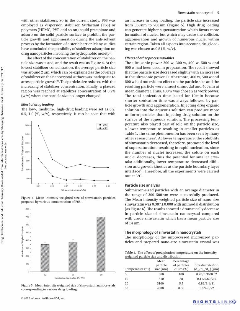
Particle size analysisSubmicron-sized particles with an average diameter in the range of 300–500 nm were successfully produced. The Mean intensity weighted particle size of nano-size simvastatin was 0.387 ± 0.008 with unimodal distribution (as Figure 6). The results showed a dramatically decrease in particle size of simvastatin nanocrystal compared with crude simvastatin which has a mean particle size of 14 μm.
The morphology of simvastatin nanocrystalsThe morphology of the unprocessed micronized par-ticles and prepared nano-size simvastatin crystal was
Figure 4. Mean intensity weighted size of simvastatin particles prepared by various concentration of F68.
Figure 5. Mean intensity weighted size of simvastatin nanocrystals corresponding to various drug loading.
Table 1. The effect of precipitation temperature on the intensity weighted particle size and distribution.
Temperature (°C)
Mean particle
size (nm)
Percentage of particles <1µm (%)
Size distribution (d
25/d
50/d
90) (μm)
3 360 100 0.20/0.36/0.6210 510 88 0.11/0.60/2.020 3100 5.7 0.86/3.1/1130 4600 0.36 1.6/4.6/22
Dru
g D
evel
opm
ent a
nd I
ndus
tria
l Pha
rmac
y D
ownl
oade
d fr
om in
form
ahea
lthca
re.c
om b
y Sh
enya
ng P
harm
aceu
tical
Uni
vers
ity o
n 07
/11/
12Fo
r pe
rson
al u
se o
nly.
6 T. Jiang et al.
Drug Development and Industrial Pharmacy
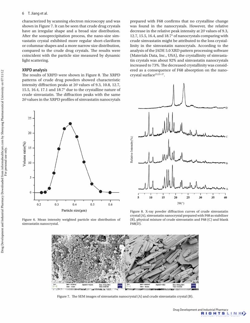
characterized by scanning electron microscopy and was shown in Figure 7. It can be seen that crude drug crystals have an irregular shape and a broad size distribution. After the sonoprecipitation process, the nano-size sim-vastatin crystal exhibited more regular short-claviform or columnar shapes and a more narrow size distribution, compared to the crude drug crystals. The results were coincident with the particle size measured by dynamic light scattering.
XRPD analysisThe results of XRPD were shown in Figure 8. The XRPD patterns of crude drug powders showed characteristic intensity diffraction peaks at 2θ values of 9.3, 10.8, 12.7, 15.5, 16.4, 17.1 and 18.7° due to the crystalline nature of crude simvastatin. The diffraction peaks with the same 2θ values in the XRPD profiles of simvastatin nanocrytals
prepared with F68 confirms that no crystalline change was found in the nanocrystals. However, the relative decrease in the relative peak intensity at 2θ values of 9.3, 12.7, 15.5, 16.4, and 18.7° of nanocrystals comparing with crude simvastatin might be attributed to the loss crystal-linity in the simvastatin nanocrytals. According to the analysis of the JADE 5.0 XRD pattern processing software (Materials Data, Inc., USA), the crystallinity of simvasta-tin crystals was about 92% and simvastatin nanocrystals increased to 73%. The decreased crystallinity was consid-ered as a consequence of F68 absorption on the nano-crystal surface23,25–27.
Figure 6. Mean intensity weighted particle size distribution of simvastatin nanocrystal.
Figure 7. The SEM images of simvastatin nanocrystal (A) and crude simvastatin crystal (B).
Figure 8. X-ray powder diffraction curves of crude simvastatin crystal (A), simvastatin nanocrystal prepared with F68 as stabilizer (B), physical mixture of crude simvastatin and F68 (C) and blank F68(D).
Dru
g D
evel
opm
ent a
nd I
ndus
tria
l Pha
rmac
y D
ownl
oade
d fr
om in
form
ahea
lthca
re.c
om b
y Sh
enya
ng P
harm
aceu
tical
Uni
vers
ity o
n 07
/11/
12Fo
r pe
rson
al u
se o
nly.
Simvastatin nanocrystal 7
© 2012 Informa Healthcare USA, Inc.
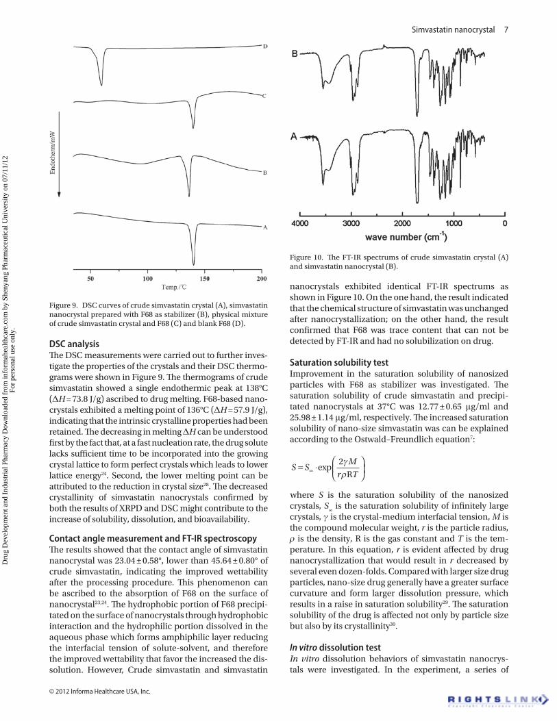
DSC analysisThe DSC measurements were carried out to further inves-tigate the properties of the crystals and their DSC thermo-grams were shown in Figure 9. The thermograms of crude simvastatin showed a single endothermic peak at 138°C (ΔH = 73.8 J/g) ascribed to drug melting. F68-based nano-crystals exhibited a melting point of 136°C (ΔH = 57.9 J/g), indicating that the intrinsic crystalline properties had been retained. The decreasing in melting ΔH can be understood first by the fact that, at a fast nucleation rate, the drug solute lacks sufficient time to be incorporated into the growing crystal lattice to form perfect crystals which leads to lower lattice energy24. Second, the lower melting point can be attributed to the reduction in crystal size28. The decreased crystallinity of simvastatin nanocrystals confirmed by both the results of XRPD and DSC might contribute to the increase of solubility, dissolution, and bioavailability.
Contact angle measurement and FT-IR spectroscopyThe results showed that the contact angle of simvastatin nanocrystal was 23.04 ± 0.58°, lower than 45.64 ± 0.80° of crude simvastatin, indicating the improved wettability after the processing procedure. This phenomenon can be ascribed to the absorption of F68 on the surface of nanocrystal23,24. The hydrophobic portion of F68 precipi-tated on the surface of nanocrystals through hydrophobic interaction and the hydrophilic portion dissolved in the aqueous phase which forms amphiphilic layer reducing the interfacial tension of solute-solvent, and therefore the improved wettability that favor the increased the dis-solution. However, Crude simvastatin and simvastatin
nanocrystals exhibited identical FT-IR spectrums as shown in Figure 10. On the one hand, the result indicated that the chemical structure of simvastatin was unchanged after nanocrystallization; on the other hand, the result confirmed that F68 was trace content that can not be detected by FT-IR and had no solubilization on drug.
Saturation solubility testImprovement in the saturation solubility of nanosized particles with F68 as stabilizer was investigated. The saturation solubility of crude simvastatin and precipi-tated nanocrystals at 37°C was 12.77 ± 0.65 μg/ml and 25.98 ± 1.14 μg/ml, respectively. The increased saturation solubility of nano-size simvastatin was can be explained according to the Ostwald–Freundlich equation7:
S SM
r T= ⋅
∞ exp
2γρR
where S is the saturation solubility of the nanosized crystals, S
∞ is the saturation solubility of infinitely large
crystals, γ is the crystal-medium interfacial tension, M is the compound molecular weight, r is the particle radius, ρ is the density, R is the gas constant and T is the tem-perature. In this equation, r is evident affected by drug nanocrystallization that would result in r decreased by several even dozen-folds. Compared with larger size drug particles, nano-size drug generally have a greater surface curvature and form larger dissolution pressure, which results in a raise in saturation solubility29. The saturation solubility of the drug is affected not only by particle size but also by its crystallinity30.
In vitro dissolution testIn vitro dissolution behaviors of simvastatin nanocrys-tals were investigated. In the experiment, a series of
Figure 9. DSC curves of crude simvastatin crystal (A), simvastatin nanocrystal prepared with F68 as stabilizer (B), physical mixture of crude simvastatin crystal and F68 (C) and blank F68 (D).
Figure 10. The FT-IR spectrums of crude simvastatin crystal (A) and simvastatin nanocrystal (B).
Dru
g D
evel
opm
ent a
nd I
ndus
tria
l Pha
rmac
y D
ownl
oade
d fr
om in
form
ahea
lthca
re.c
om b
y Sh
enya
ng P
harm
aceu
tical
Uni
vers
ity o
n 07
/11/
12Fo
r pe
rson
al u
se o
nly.
8 T. Jiang et al.
Drug Development and Industrial Pharmacy
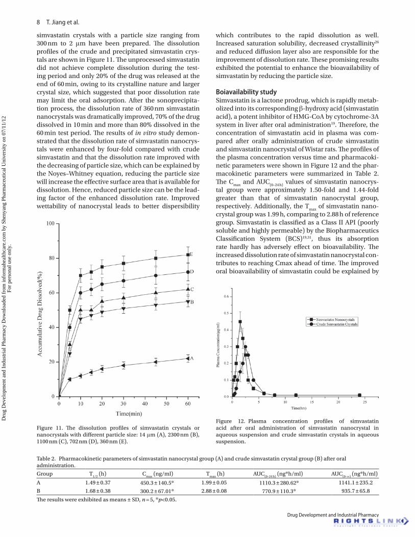
simvastatin crystals with a particle size ranging from 300 nm to 2 μm have been prepared. The dissolution profiles of the crude and precipitated simvastatin crys-tals are shown in Figure 11. The unprocessed simvastatin did not achieve complete dissolution during the test-ing period and only 20% of the drug was released at the end of 60 min, owing to its crystalline nature and larger crystal size, which suggested that poor dissolution rate may limit the oral adsorption. After the sonoprecipita-tion process, the dissolution rate of 360 nm simvastatin nanocrystals was dramatically improved, 70% of the drug dissolved in 10 min and more than 80% dissolved in the 60 min test period. The results of in vitro study demon-strated that the dissolution rate of simvastatin nanocrys-tals were enhanced by four-fold compared with crude simvastatin and that the dissolution rate improved with the decreasing of particle size, which can be explained by the Noyes–Whitney equation, reducing the particle size will increase the effective surface area that is available for dissolution. Hence, reduced particle size can be the lead-ing factor of the enhanced dissolution rate. Improved wettability of nanocrystal leads to better dispersibility
which contributes to the rapid dissolution as well. Increased saturation solubility, decreased crystallinity26 and reduced diffusion layer also are responsible for the improvement of dissolution rate. These promising results exhibited the potential to enhance the bioavailability of simvastatin by reducing the particle size.
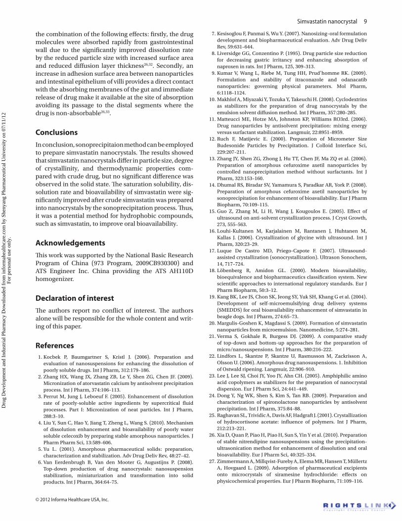
Boiavailability studySimvastatin is a lactone prodrug, which is rapidly metab-olized into its corresponding β-hydroxy acid (simvastatin acid), a potent inhibitor of HMG-CoA by cytochrome-3A system in liver after oral administration19. Therefore, the concentration of simvastatin acid in plasma was com-pared after orally administration of crude simvastatin and simvastatin nanocrystal of Wistar rats. The profiles of the plasma concentration versus time and pharmacoki-netic parameters were shown in Figure 12 and the phar-macokinetic parameters were summarized in Table 2. The C
max and AUC
(0–24 h) values of simvastatin nanocrys-
tal group were approximately 1.50-fold and 1.44-fold greater than that of simvastatin nanocrystal group, respectively. Additionally, the T
max of simvastatin nano-
crystal group was 1.99 h, comparing to 2.88 h of reference group. Simvastatin is classified as a Class II API (poorly soluble and highly permeable) by the Biopharmaceutics Classification System (BCS)19,31, thus its absorption rate hardly has adversely effect on bioavailability. The increased dissolution rate of simvastatin nanocrystal con-tributes to reaching Cmax ahead of time. The improved oral bioavailability of simvastatin could be explained by
Figure 12. Plasma concentration profiles of simvastatin acid after oral administration of simvastatin nanocrystal in aqueous suspension and crude simvastatin crystals in aqueous suspension.
Table 2. Pharmacokinetic parameters of simvastatin nanocrystal group (A) and crude simvastatin crystal group (B) after oral administration.
Group T1/2
(h) Cmax
(ng/ml) Tmax
(h) AUC(0–24 h)
(ng*h/ml) AUC(0–∞)
(ng*h/ml)
A 1.49 ± 0.37 450.3 ± 140.5* 1.99 ± 0.05 1110.3 ± 280.62* 1141.1 ± 235.2
B 1.68 ± 0.38 300.2 ± 67.01* 2.88 ± 0.08 770.9 ± 110.3* 935.7 ± 65.8
The results were exhibited as means ± SD, n = 5, *p<0.05.
Figure 11. The dissolution profiles of simvastatin crystals or nanocrystals with different particle size: 14 μm (A), 2300 nm (B), 1100 nm (C), 702 nm (D), 360 nm (E).
Dru
g D
evel
opm
ent a
nd I
ndus
tria
l Pha
rmac
y D
ownl
oade
d fr
om in
form
ahea
lthca
re.c
om b
y Sh
enya
ng P
harm
aceu
tical
Uni
vers
ity o
n 07
/11/
12Fo
r pe
rson
al u
se o
nly.
Simvastatin nanocrystal 9
© 2012 Informa Healthcare USA, Inc.
the combination of the following effects: firstly, the drug molecules were absorbed rapidly from gastrointestinal wall due to the significantly improved dissolution rate by the reduced particle size with increased surface area and reduced diffusion layer thickness26,32. Secondly, an increase in adhesion surface area between nanoparticles and intestinal epithelium of villi provides a direct contact with the absorbing membranes of the gut and immediate release of drug make it available at the site of absorption avoiding its passage to the distal segments where the drug is non-absorbable26,33.
Conclusions
In conclusion, sonoprecipitaion method can be employed to prepare simvastatin nanocrystals. The results showed that simvastatin nanocrystals differ in particle size, degree of crystallinity, and thermodynamic properties com-pared with crude drug, but no significant difference was observed in the solid state. The saturation solubility, dis-solution rate and bioavailability of simvastatin were sig-nificantly improved after crude simvastatin was prepared into nanocrystals by the sonoprecipitation process. Thus, it was a potential method for hydrophobic compounds, such as simvastatin, to improve oral bioavailability.
Acknowledgements
This work was supported by the National Basic Research Program of China (973 Program, 2009CB930300) and ATS Engineer Inc. China providing the ATS AH110D homogenizer.
Declaration of interest
The authors report no conflict of interest. The authors alone will be responsible for the whole content and writ-ing of this paper.
References 1. Kocbek P, Baumgartner S, Kristl J. (2006). Preparation and
evaluation of nanosuspensions for enhancing the dissolution of poorly soluble drugs. Int J Pharm, 312:179–186.
2. Zhang HX, Wang JX, Zhang ZB, Le Y, Shen ZG, Chen JF. (2009). Micronization of atorvastatin calcium by antisolvent precipitation process. Int J Pharm, 374:106–113.
3. Perrut M, Jung J, Leboeuf F. (2005). Enhancement of dissolution rate of poorly-soluble active ingredients by supercritical fluid processes. Part I: Micronization of neat particles. Int J Pharm, 288:3–10.
4. Liu Y, Sun C, Hao Y, Jiang T, Zheng L, Wang S. (2010). Mechanism of dissolution enhancement and bioavailability of poorly water soluble celecoxib by preparing stable amorphous nanoparticles. J Pharm Pharm Sci, 13:589–606.
5. Yu L. (2001). Amorphous pharmaceutical solids: preparation, characterization and stabilization. Adv Drug Deliv Rev, 48:27–42.
6. Van Eerdenbrugh B, Van den Mooter G, Augustijns P. (2008). Top-down production of drug nanocrystals: nanosuspension stabilization, miniaturization and transformation into solid products. Int J Pharm, 364:64–75.
7. Kesisoglou F, Panmai S, Wu Y. (2007). Nanosizing–oral formulation development and biopharmaceutical evaluation. Adv Drug Deliv Rev, 59:631–644.
8. Liversidge GG, Conzentino P. (1995). Drug particle size reduction for decreasing gastric irritancy and enhancing absorption of naproxen in rats. Int J Pharm, 125, 309–313.
9. Kumar V, Wang L, Riebe M, Tung HH, Prud’homme RK. (2009). Formulation and stability of itraconazole and odanacatib nanoparticles: governing physical parameters. Mol Pharm, 6:1118–1124.
10. Makhlof A, Miyazaki Y, Tozuka Y, Takeuchi H. (2008). Cyclodextrins as stabilizers for the preparation of drug nanocrystals by the emulsion solvent diffusion method. Int J Pharm, 357:280–285.
11. Matteucci ME, Hotze MA, Johnston KP, Williams RO3rd. (2006). Drug nanoparticles by antisolvent precipitation: mixing energy versus surfactant stabilization. Langmuir, 22:8951–8959.
12. Ruch F, Matijevic E. (2000). Preparation of Micrometer Size Budesonide Particles by Precipitation. J Colloid Interface Sci, 229:207–211.
13. Zhang JY, Shen ZG, Zhong J, Hu TT, Chen JF, Ma ZQ et al. (2006). Preparation of amorphous cefuroxime axetil nanoparticles by controlled nanoprecipitation method without surfactants. Int J Pharm, 323:153–160.
14. Dhumal RS, Biradar SV, Yamamura S, Paradkar AR, York P. (2008). Preparation of amorphous cefuroxime axetil nanoparticles by sonoprecipitation for enhancement of bioavailability. Eur J Pharm Biopharm, 70:109–115.
15. Guo Z, Zhang M, Li H, Wang J, Kougoulos E. (2005). Effect of ultrasound on anti-solvent crystallization process. J Cryst Growth, 273, 555–563.
16. Louhi-Kultanen M, Karjalainen M, Rantanen J, Huhtanen M, Kallas J. (2006). Crystallization of glycine with ultrasound. Int J Pharm, 320:23–29.
17. Luque De Castro MD, Priego-Capote F. (2007). Ultrasound-assisted crystallization (sonocrystallization). Ultrason Sonochem, 14, 717–724.
18. Löbenberg R, Amidon GL. (2000). Modern bioavailability, bioequivalence and biopharmaceutics classification system. New scientific approaches to international regulatory standards. Eur J Pharm Biopharm, 50:3–12.
19. Kang BK, Lee JS, Chon SK, Jeong SY, Yuk SH, Khang G et al. (2004). Development of self-microemulsifying drug delivery systems (SMEDDS) for oral bioavailability enhancement of simvastatin in beagle dogs. Int J Pharm, 274:65–73.
20. Margulis-Goshen K, Magdassi S. (2009). Formation of simvastatin nanoparticles from microemulsion. Nanomedicine, 5:274–281.
21. Verma S, Gokhale R, Burgess DJ. (2009). A comparative study of top-down and bottom-up approaches for the preparation of micro/nanosuspensions. Int J Pharm, 380:216–222.
22. Lindfors L, Skantze P, Skantze U, Rasmusson M, Zackrisson A, Olsson U. (2006). Amorphous drug nanosuspensions. 1. Inhibition of Ostwald ripening. Langmuir, 22:906–910.
23. Lee J, Lee SJ, Choi JY, Yoo JY, Ahn CH. (2005). Amphiphilic amino acid copolymers as stabilizers for the preparation of nanocrystal dispersion. Eur J Pharm Sci, 24:441–449.
24. Dong Y, Ng WK, Shen S, Kim S, Tan RB. (2009). Preparation and characterization of spironolactone nanoparticles by antisolvent precipitation. Int J Pharm, 375:84–88.
25. Raghavan SL, Trividic A, Davis AF, Hadgraft J. (2001). Crystallization of hydrocortisone acetate: influence of polymers. Int J Pharm, 212:213–221.
26. Xia D, Quan P, Piao H, Piao H, Sun S, Yin Y et al. (2010). Preparation of stable nitrendipine nanosuspensions using the precipitation-ultrasonication method for enhancement of dissolution and oral bioavailability. Eur J Pharm Sci, 40:325–334.
27. Zimmermann A, Millqvist-Fureby A, Elema MR, Hansen T, Müllertz A, Hovgaard L. (2009). Adsorption of pharmaceutical excipients onto microcrystals of siramesine hydrochloride: effects on physicochemical properties. Eur J Pharm Biopharm, 71:109–116.
Dru
g D
evel
opm
ent a
nd I
ndus
tria
l Pha
rmac
y D
ownl
oade
d fr
om in
form
ahea
lthca
re.c
om b
y Sh
enya
ng P
harm
aceu
tical
Uni
vers
ity o
n 07
/11/
12Fo
r pe
rson
al u
se o
nly.
10 T. Jiang et al.
Drug Development and Industrial Pharmacy
28. Sharma P, Denny WA, Garg S. (2009). Effect of wet milling process on the solid state of indomethacin and simvastatin. Int J Pharm, 380:40–48.
29. Müller RH, Benita S, Böhm BHL. Emulsions and nanosuspensions for the formulation of poorly soluble drugs. Stuttgart: Medpharm Scientific Publishers; 1998.
30. Qiu Y, Chen Y, Zhang GGZ. Developing solid oral dosage forms: pharmaceutical theory and practice. Burlington: Academic; 2009.
31. Ambike AA, Mahadik KR, Paradkar A. (2005). Spray-dried amorphous solid dispersions of simvastatin, a low tg drug: in vitro and in vivo evaluations. Pharm AQ4 m Res, 22:990–998.
32. Hintz RJ, Johnson KC. (1989). The effect of particle size distribution on dissolution rate and oral absorption. Int J Pharm, 51,9–17.
33. Jia L, Wong H, Cerna C, Weitman SD. (2002). Effect of nanonization on absorption of 301029: ex vivo and in vivo pharmacokinetic correlations determined by liquid chromatography/mass spectrometry. Pharm Res, 19:1091–1096.
Dru
g D
evel
opm
ent a
nd I
ndus
tria
l Pha
rmac
y D
ownl
oade
d fr
om in
form
ahea
lthca
re.c
om b
y Sh
enya
ng P
harm
aceu
tical
Uni
vers
ity o
n 07
/11/
12Fo
r pe
rson
al u
se o
nly.