Embed Size (px)
Citation preview
This work is licensed under a Creative Commons Attribution 4.0 International License
Newcastle University ePrints - eprint.ncl.ac.uk
Haagensen EJ, Thomas HD, Schmalix WA, Payne AC, Kevorkian L, Allen RA,
Bevan P, Maxwell RJ, Newell DR.
Enhanced anti-tumour activity of the combination of the novel MEK inhibitor
WX-554 and the novel PI3K inhibitor WX-037.
Cancer Chemotherapy and Pharmacology
2016, 78(6), 1269-1281
Copyright:
© The Author(s) 2016. This article is published with open access at Springerlink.com. This article is
distributed under the terms of the Creative Commons Attribution 4.0 International License
(http://creativecommons.org/licenses/by/4.0/), which permits unrestricted use, distribution, and
reproduction in any medium, provided you give appropriate credit to the original author(s) and the
source, provide a link to the Creative Commons license, and indicate if changes were made.
DOI link to article:
http://dx.doi.org/10.1007/s00280-016-3186-4
Date deposited:
24/01/2017

1 3
Cancer Chemother Pharmacol (2016) 78:1269–1281DOI 10.1007/s00280-016-3186-4
ORIGINAL ARTICLE
Enhanced anti‑tumour activity of the combination of the novel MEK inhibitor WX‑554 and the novel PI3K inhibitor WX‑037
Emma J. Haagensen1 · Huw D. Thomas1 · Wolfgang A. Schmalix2 · Andrew C. Payne3 · Lara Kevorkian3 · Rodger A. Allen3 · Paul Bevan2 · Ross J. Maxwell1 · David R. Newell1
Received: 2 June 2016 / Accepted: 31 October 2016 / Published online: 11 November 2016 © The Author(s) 2016. This article is published with open access at Springerlink.com
the tumour uptake of WX-554. In vivo efficacy studies revealed that the combination of WX-037 and WX-554 was non-toxic and exhibited marked tumour growth inhibition greater than observed with either agent alone.Conclusion These studies show for the first time that com-bination treatment with the novel MEK inhibitor WX-554 and the novel PI3K inhibitor WX-037 can induce syn-ergistic growth inhibition in vitro, which translates into enhanced anti-tumour efficacy in vivo.
Keywords PI3K · MEK · Combination · Synergy; Colorectal cancer
Introduction
The development of numerous targeted small molecule inhibitors represents an important and evolving new approach to cancer therapy. However, as tumours often have defects in multiple oncogenic signalling pathways, single agent anti-tumour activity is modest, and thus com-binations of targeted agents are being investigated. Specifi-cally, the MAPK pathway, a major proliferative pathway, and the PI3K pathway, a major survival pathway, are fre-quently activated in cancer and are being concomitantly targeted.
Many MEK inhibitors, such as PD 0325901 and selu-metinib (AZD6244), have been developed to target the MAPK pathway and have shown potent growth inhibi-tory activity in experimental systems [1–4]. A novel orally available small molecule allosteric MEK inhibitor WX-554 (UCB1366554), which potently inhibits MEK1 and MEK2 with a half maximal inhibitory concentration (IC50) of 4.7 and 11 nM, respectively, has been developed by Wilex and UCB Celltech. WX-554 has demonstrated
Abstract Purpose Tumours frequently have defects in multiple oncogenic pathways, e.g. MAPK and PI3K signalling pathways, and combinations of targeted therapies may be required for optimal activity. This study evaluated the novel MEK inhibitor WX-554 and the novel PI3K inhibitor WX-037, as single agents and in combination, in colorectal carcinoma cell lines and tumour xenograft-bearing mice.Methods In vitro growth inhibition, survival and signal transduction were measured using the Sulforhodamine B, clonogenic and Western blotting assays, respectively, in HCT116 and HT29 cell lines. In vivo anti-tumour efficacy and pharmacokinetic properties were assessed in HCT116 and HT29 human colorectal cancer xenograft tumour-bear-ing mice.Results The combination of WX-554 and WX-037 exhib-ited marked synergistic growth inhibition in vitro, which was associated with increased cytotoxicity and enhanced inhibition of ERK and S6 phosphorylation, compared to either agent alone. Pharmacokinetic analyses indicated that there was no PK interaction between the two drugs at low doses, but that at higher doses, WX-037 may delay
Electronic supplementary material The online version of this article (doi:10.1007/s00280-016-3186-4) contains supplementary material, which is available to authorized users.
* David R. Newell [email protected]
1 Newcastle Cancer Centre, Northern Institute for Cancer Research, Paul O’Gorman Building, Medical School, Newcastle University, Framlington Place, Newcastle-upon-Tyne NE2 4HH, UK
2 Wilex AG, Grillparzerstrasse 18, 81675 Munich, Germany3 UCB Pharma Ltd, 208 Bath Road, Slough SL1 3WE, UK
1270 Cancer Chemother Pharmacol (2016) 78:1269–1281
1 3
marked inhibition of ERK1/2 phosphorylation in HT29 cells, and growth inhibition in a range of cell lines in vitro, and tumour growth delay or stasis in vivo, with increased sensitivity in BRAF or RAS mutant cells and tumours [5]. WX-554 has been shown to be safe and tolerable in a dose escalation study in healthy volunteers [6], and a recent Phase I study indicated that WX-554 has very good bio-availability and that it is able to inhibit MEK signal trans-duction in a dose-dependent manner [3, 7, 8]. WX-554 was investigated in a Phase Ib/II dose escalation study to determine safety and pharmacokinetics/pharmacodynamics in patients with solid tumours [7, 9], which was terminated due to business, rather than clinical, reasons.
Targeting the PI3K/AKT pathway has been investigated with a range of PI3K inhibitors which can be isoform-specific inhibitors, pan class I inhibitors (e.g. pictilisib, GDC-0941) or dual PI3K/mTOR inhibitors (e.g. dactolisib, NVP-BEZ235), and promising anti-cancer efficacy has been reported in pre-clinical models [10–16]. Wilex and UCB Celltech have also developed a novel small molecule pan class I PI3K inhibitor, WX-037 (UCB1370037), from an indole series. WX-037 is a potent inhibitor of the α and δ isoforms of PI3K (IC50 = 4.1 and 2.4 nM, respectively) and a weaker inhibitor of the β and γ isoforms of PI3K and DNA-PK (IC50 = 69, 36 and 28 nM, respectively) with no detectable inhibition of mTOR (IC50 = >20,000 nM). In pre-clinical studies, WX-037 demonstrated strong inhibi-tion of AKT phosphorylation, and promising growth inhi-bition in a range of cell lines in vitro, and tumour growth delay or stasis in vivo, with greater sensitivity observed in PIK3CA mutant or PTEN null cells and tumours [5]. WX-037 was investigated in a Phase I dose escalation study to investigate its safety, pharmacokinetics, pharmacody-namics and clinical activity in patients with solid tumours; however, this trial was also terminated due to business rea-sons [9, 17].
Previous studies have shown that concomitant inhibi-tion of the PI3K and MAPK pathways by PI3K and MEK inhibition yields promising anti-cancer effects in vitro and in vivo [2, 18–26], and the combination of WX-554 and WX-037 demonstrated synergy in vitro and increased effi-cacy in vivo [5]. Consequently, the Phase I trial of WX-037 was designed not just to investigate the efficacy of single agent WX-037, but also the combination of WX-554 and WX-037 [9].
The aim of this study was to determine the in vitro and in vivo activity of the novel MEK inhibitor WX-554 and the novel PI3K inhibitor WX-037, alone and in combination, in colorectal carcinoma cell lines and tumour xenograft-bear-ing mice. These colorectal carcinoma cell lines (HT29 and HCT116) were used as they both contain KRAS/RAF and PI3K pathway mutations. The first objective was to deter-mine the in vitro potency and efficacy of the compounds,
alone and in combination, by measuring growth inhibition using an SRB assay, cytotoxicity via a clonogenic assay and cell signalling by Western blotting, and to investigate if the effects of the combinations on cell growth were syn-ergistic, additive or antagonistic using median effect analy-ses. These results were then used to design in vivo experi-ments to investigate the pharmacokinetic profile of the compounds and their efficacy, alone and in combination.
Methods
Ethics statement
All in vivo experiments were reviewed and approved by the Newcastle University (UK) animal welfare committee and were performed according to the guidelines for the welfare and use of animals in cancer research [27] and national law, under project license (PPL60/4442) issued by the UK Gov-ernment Home Office under the animals (scientific proce-dure) act 1986.
Inhibitors
The MEK inhibitor WX-554 and the PI3K inhibitor WX-037 were kindly supplied by Wilex, Munich, Ger-many. For in vitro studies, the inhibitors were dissolved in anhydrous dimethyl sulphoxide (DMSO) and were stored frozen under light-protected conditions at −20 °C. For in vivo studies, the MEK inhibitor WX-554 was dissolved in 0.9% NaCl (w/v), 10 mM Na-citrate pH 3.0 (w/v) and 0.2% Tween 20 (v/v) in sterile distilled water and the PI3K inhibitor WX-037 was suspended in SMEDDS (self-micro-emulsifying drug delivery system; 25% Capmul MCM EP (glycerol monocaprylocaprate) (v/v), 37.5% Tween 80 (polyoxyethylene(20) sorbitan monooleate) (v/v) and 37.5% PEG 400 (polyethylene glycol 400) (v/v)).
Cell lines and reagents
HCT116 and HT29 human colorectal cancer cells were obtained from the ATCC (American Type Culture Collec-tion). All cell lines were grown in RPMI-1640 medium (supplemented with 10% (v/v) foetal bovine serum, 1% (v/v) penicillin (50 U/ml)—streptomycin (50 mg/ml) and 2 mM L-glutamine) and were confirmed free of myco-plasma contamination by regular testing with Mycoalert (Cambrex, Iowa, USA).
Animals
Animal studies were all carried out using female athymic CD1 nude mice (Charles River, Kent, UK), implanted

1271Cancer Chemother Pharmacol (2016) 78:1269–1281
1 3
with HCT116 or HT29 xenografts (1 × 10⁷ cells in 50 µl media injected subcutaneously into the right flank), main-tained and handled in isolators under specific pathogen-free conditions.
Growth inhibition assay
Exponentially growing cells were grown in media in 96-well format and were exposed to increasing concentra-tions of the single agent inhibitors WX-554 or WX-037, or WX-554 combined with WX-037 at 0.25, 0.5, 1, 2 or 4 times their half maximal growth inhibitory concentra-tion (GI50) in DMSO, or 0.5% DMSO alone, for 72 h. Growth was then measured using the Sulforhodamine B (SRB) method and analysed as described previously [23]. The GI50 concentration was calculated based on a standard point to point curve with 1000 segments using GraphPad Prism software (California, USA). The data were ana-lysed by median effect analysis using CalcuSyn software (Biosoft, Cambridgeshire, UK), which calculates the com-bination index of multiple drugs by an algebraic estimation algorithm.
Cytotoxicity assay
Exponentially growing cells were exposed to increas-ing concentrations of the single agent inhibitors WX-554 or WX-037, or 10 µM of WX-554 combined with 10 µM WX-037 in DMSO or 0.5% (v/v) DMSO alone for 72 h before harvesting and reseeding for colony formation. After growth for 10–14 days, colonies were fixed in metha-nol–acetic acid 3:1 (v/v) and stained with crystal violet (0.4% w/v). Colonies consisting of more than 50 cells were counted on an automated colony counter (Oxford Optronix, Oxford, UK). Two-tailed paired t tests were used to com-pare the different groups. Differences with a p < 0.05 were considered statistically significant.
Western blotting
Cells were treated with WX-554 and WX-037 at 1 or 10 times the half maximal growth inhibitory concentration (GI50) in DMSO, or 0.5% (v/v) DMSO alone, for 24 h. Western blots were prepared, probed with phospho-4EBP1 (Thr37/46) (#2855), phospho-p44/42 MAPK (Thr202/Tyr204) (#4370), phospho-AKT (Ser473) (#4060) or phos-pho-S6 ribosomal protein (Ser235/236) (#4858) monoclo-nal antibodies obtained from Cell Signalling Technology (New England BioLabs (UK) Ltd, Hertfordshire, UK) and developed as described previously [23]. Blots were then stripped (100 mM 2-mercaptoethanol, 2% (w/v) SDS and
62.5 mM Tris pH6.8 at 55 °C for 30 min) and re-probed with the respective total monoclonal antibody (4EBP1 (53H11) (#9644), p44/42 MAPK (ERK1/2) (#4695), AKT (pan) (C67E7) (#4691) or S6 ribosomal protein (5g10) (#2217)) obtained from Cell Signalling Technology (New England BioLabs (UK) Ltd, Hertfordshire, UK) and devel-oped as described above.
Pharmacokinetic (PK) studies
Mice bearing HCT116 or HT29 human tumour xenografts were treated with 1 or 5 mg/kg WX-554, or 20 or 100 mg/kg WX-037, alone or in combination, and were bled by cardiac puncture under terminal anaesthesia at 6 or 24 h post-treatment (3 mice/time point). Blood was collected into heparinized tubes, and plasma was separated and stored at −20 °C until analysed. Tumours were removed, snap frozen in liquid nitrogen and stored at −80 °C prior to PK analyses. Samples were extracted with solid phase extraction (SPE) and analysed with high-performance liq-uid chromatography (HPLC) coupled with tandem mass spectrometry (MS/MS) by Wilex (Munich, Germany). The quantification limit for WX-554 was 1 ng/mL and within and between day variation was <15%. WinNonlin Software Version 4.0.1 (Pharsight Corporation, Peypin, France) was used for PK/PD modelling and non-compartmental analy-sis. Paired t tests were used to compare the different treat-ment groups, and differences with a p value ≤0.05 were considered statistically significant.
Determination of anti‑tumour activity
Mice bearing HCT116 human tumour xenografts were randomized into treatment groups and then treated by oral gavage with either the vehicle (10 ml/kg), 2 mg/kg WX-554, 50 mg/kg WX-037 or the combination of 2 mg/kg WX-554 and 50 mg/kg WX-037 once daily for 14 days. Tumour volume was monitored by calliper measurement using the equation a2 × b/2, where a is the smallest meas-urement and b the largest. Data are presented as median relative tumour volumes (RTV), where the tumour volume in each mouse on the initial day of treatment (day 0) is assigned an RTV value of 1. The time to RTV4 for each individual tumour was calculated based on a standard point to point curve with 1000 segments using GraphPad Prism software (CA, USA). Mann–Whitney U tests were used to compare the different groups, i.e., the control ver-sus each treatment group, the single agents versus each other and each single agent versus their combination. Dif-ferences with a p value ≤0.05 were considered statistically significant.

1272 Cancer Chemother Pharmacol (2016) 78:1269–1281
1 3
Results
The PI3K inhibitor WX‑037 and the MEK inhibitor WX‑554 are synergistic and exhibit increased cytotoxicity in combination in vitro
The growth inhibitory activity of the PI3K inhibitor WX-037 and the MEK inhibitor WX-554, as single agents, in HCT116 and HT29 cells was measured using the SRB assay (Supplementary Figure 1). Both drugs induced over 65% growth inhibition in both the colorectal cell lines. The results were used to determine the half maximal growth inhibitory (GI50) concentration of the drugs after 72-h exposure. The MEK inhibitor WX-554 was found to have GI50 values of 38 and 4.3 nM, whereas the PI3K inhibi-tor WX-037 was less potent with GI50 values of 2934 and 112 nM in the HCT116 and HT29 cell lines, respectively (Supplementary Figure 1).
Studies were then performed to determine the effect of combining the PI3K and MEK inhibitors on colorectal carcinoma cell growth over 72 h. WX-037 and WX-554 were used alone at 0.25x, 0.5x, 1x, 2x and 4x their respec-tive GI50 concentration, as calculated from Supplementary Figure 1, and at equipotent concentrations at the same GI50 ratios in combination. Figure 1 shows that the combination of WX-037 and WX-554 was markedly more growth inhib-itory than either compound alone, completely inhibiting growth at the highest concentrations. Data were then evalu-ated by median effect analysis (CalcuSyn, Biosoft, Great Shelford, UK) to determine whether the greater growth inhibitory activity of the combination of WX-554 and WX-037 reflected an additive or a synergistic effect. The combination of the PI3K inhibitor WX-037 and the MEK inhibitor WX-554 was strongly synergistic when combined
at the GI50 concentration compared to the compounds alone (Supplementary Table 1).
Cell survival after 72-h exposure to the PI3K inhibitor WX-037 and the MEK inhibitor WX-554 was also meas-ured using a clonogenic cytotoxicity assay. Single agent WX-554 showed significant cytotoxicity at 10 µM with 67% cell kill in the HCT116 cell line and 75% in the HT29 cell line; however, the mean lethal concentration (LC50) of 0.6 µM and 1.6 µM WX-554 was approximately 16-fold and 372-fold higher than the corresponding GI50- values in the HCT116 and HT29 cell lines, respectively. WX-037 showed no marked cytotoxicity with less than 50% cell death after 72 h treatment at 10 µM (Supplementary Figure 2).
The cytotoxicity of the PI3K and MEK inhibitors in combination after 72 h treatment was then determined. However, as WX-037 did not produce > 50% cytotoxicity at 10 µM, it was not possible to determine an LC50 value, and hence the highest concentration previously used of 10 µM WX-037 was combined with 10 µM WX-554. There was a statistically significant increase in cytotoxicity when the PI3K and MEK inhibitors were combined, compared to the cytotoxicity induced by the drugs as single agents, in the HCT116 (p = 0.02) and HT29 (p < 0.01) cell lines (Fig. 2). Overall, the interaction of WX-037 and WX-554 resulted in significantly enhanced cell growth inhibition and an increase in cytotoxicity in both cell lines studied.
The effect of 24-h exposure to the PI3K inhibitor WX-037 and the MEK inhibitor WX-554, both as sin-gle agents and in combination, was also investigated by Western blotting to determine the effect on the PI3K/AKT signalling pathway, using total and phospho-specific anti-bodies for AKT, S6 and 4EBP1 and the effect on MAPK signalling, using total and phospho-specific antibodies for
Fig. 1 Growth inhibition induced by the PI3K inhibitor WX-037 and the MEK inhibitor WX-554, alone and in combination, in the HCT116 and HT29 cell lines. HCT116 (a) and HT29 (b) cells were treated with the indicated fractions of the GI50 concentrations of the inhibitors, alone or in combination, derived from Supplementary
Figure 1, for 72 h, and an SRB assay was subsequently performed. Growth is presented as a percentage of the control, in which cells were treated with 0.5% (v/v) DMSO. Points represent the mean of 3 independent experiments ± standard error. Lines were fitted using nonlinear regression analysis

1273Cancer Chemother Pharmacol (2016) 78:1269–1281
1 3
ERK1/2. The compounds were used as single agents or in combination at their respective GI50 concentrations and at 10x the GI50 concentration.
Supplementary Figure 3 shows that treatment with the MEK inhibitor WX-554 reduced ERK1/2 phosphorylation at 1 and 10 times the GI50 concentration in the HCT116 cell line, and at 10 times the GI50 concentration in the HT29 cell line. The reduction in ERK1/2 phosphoryla-tion was enhanced with the combination of WX-554 and WX-037 leading to complete inhibition with 10 times the GI50 concentration in both colorectal carcinoma cell lines (Supplementary Figure 3). Additionally, there was a con-centration-dependent reduction in AKT phosphorylation in the HCT116 cell line after treatment with the single agent PI3K inhibitor WX-037 which was enhanced, to yield com-plete inhibition at the GI50 concentration, after combined WX-037 and WX-554 treatment. In the HT29 cell line, there was a reduction in AKT phosphorylation at 10 times the GI50 concentration; however, this reduction was similar with single agent WX-037 and the combination (Supple-mentary Figure 3).
Single agent WX-037 also caused a concentration-dependent reduction in S6 phosphorylation at 1 and 10 times the GI50 concentration, and treatment with the com-bination of WX-037 and WX-554 enhanced this inhibition,
causing complete inhibition at 10 times the GI50 concentra-tion in both colorectal carcinoma cell lines (Supplementary Figure 3). However, WX-037 had no marked effect on the phosphorylation of 4EBP1 alone or in combination with WX-554 in either colorectal carcinoma cell line (Supple-mentary Figure 3). Hence, overall, the combination of the MEK inhibitor WX-554 and the PI3K inhibitor WX-037 resulted in enhanced inhibition of ERK1/2 and S6 phos-phorylation, and inhibition of AKT phosphorylation, in both colorectal carcinoma cell lines. However, both the single agents and the combination had no major impact on 4EBP1 phosphorylation.
The PI3K inhibitor WX‑037 and the MEK inhibitor WX‑554 exhibit increased tumour growth delay in combination in vivo
A PK study was carried out with samples taken 6 and 24 h after treatment with a single dose of the PI3K inhibi-tor WX-037 and the MEK inhibitor WX-554, alone and in combination, in HCT116 and HT29 human tumour xeno-graft-bearing mice. The concentrations of the drugs in the plasma and the tumour tissue were measured using LC–MS/MS (Figs. 3, 4, Supplementary Figures 4 and 5 and Supplementary Table 2).
Fig. 2 Cell survival after 72-h exposure to 10 µM of the PI3K inhibi-tor WX-037 and 10 µM of the MEK inhibitor WX-554, alone and in combination, in the HCT116 cell line and HT29 cell lines. HCT116 (a) and HT29 (b) cells were treated with a fixed concentration of each inhibitor alone or in combination for 72 h, and cell survival was sub-
sequently determined by clonogenic assay after 10–14 days of colony growth. Survival is presented as a percentage of the control, in which cells were treated with 0.5% (v/v) DMSO. Bars represent the mean of 3 independent replicates ± standard error. *Significantly different from either agent alone, p ≤ 0.05

1274 Cancer Chemother Pharmacol (2016) 78:1269–1281
1 3
After a single dose of 1 or 5 mg/kg WX-554, alone or in combination with 20 or 100 mg/kg WX-037, WX-554 concentrations in the plasma and tumour tissue gener-ally greatly exceeded the in vitro GI50 value of 18 ng/ml (38 nM) in the HCT116 cell line and 2 ng/ml (4 nM) in the HT29 cell line (determined in Supplementary Fig-ure 1) (Fig. 3, Supplementary Figure 4 and Supplemen-tary Table 2A). The exception to this was that plasma WX-554 concentrations were only approximately equal to the HCT116 GI50 value at 6 h and were below the HCT116 GI50 value by 24 h, in the HCT116 tumour xen-ograft-bearing mice after treatment with 1 mg/kg WX-554, alone or in combination with 20 or 100 mg/kg WX-037
(Supplementary Figure 4A and Supplementary Table 2A). Additionally, the plasma WX-554 concentration was also below the HCT116 GI50 value by 24 h after 5 mg/kg WX-554 alone; however, WX-554 concentrations still exceeded the GI50 value when 5 mg/kg WX-554 was com-bined with WX-037 (Supplementary Figure 4C and Supple-mentary Table 2A). However, overall, the absolute plasma and tumour levels of WX-554 were generally similar in the HT29 and HCT116 tumour xenograft-bearing mice (Fig. 3, Supplementary Figure 4 and Supplementary Table 2A).
There was no consistent effect of concomitant dosing with 20 or 100 mg/kg WX-037 on the levels of WX-554 in the tumour or the plasma after administration of 1 mg/
Fig. 3 Concentrations of the MEK inhibitor WX-554 alone and in combination with the PI3K inhibitor WX-037 in tumours from mice bearing HCT116 or HT29 human tumour xenografts. Tumour con-centrations of WX-554 measured by LC–MS/MS from HCT116 (a, c) and HT29 (b, d) tumour xenograft-bearing mice at the indicated time points after a single p.o. dose of 1 mg/kg (a, b) or 5 mg/kg (c,
d) WX-554 alone or combined with 20 or 100 mg/kg WX-037. Data are presented as the mean concentration from 3 mice in each group ± standard error. Horizontal dashed lines indicate the in vitro GI50 con-centration for the respective cell line, calculated from Supplementary Figure 1

1275Cancer Chemother Pharmacol (2016) 78:1269–1281
1 3
kg WX-554 in either HCT116 or HT29 tumour xeno-graft-bearing mice. In contrast, after dosing with 5 mg/kg WX-554, there was generally a WX-037 dose-dependent decrease in the levels of WX-554 at 6 h in the tumour and the plasma upon concomitant treatment with WX-037, which was significant for the tumour data at 6 h (p < 0.05) (Fig. 3, Supplementary Figure 4 and Supplementary Table 2A). These results suggest that WX-037 may delay the tumour uptake of WX-554 at the higher dose (5 mg/kg) compared to when WX-554 is administered alone.
In contrast to the data for WX-554, after treatment with 20 mg/kg WX-037, as a single agent or in combina-tion with 1 or 5 mg/kg WX-554, WX-037 concentrations in plasma and tumour tissue at 6 and 24 h were markedly lower than the in vitro GI50 value of 1414 ng/ml (2934 nM) in the HCT116 cell line (determined in Supplementary Fig-ure 1) (Fig. 4a, Supplementary Figure 5A and Supplemen-tary Table 2B). Plasma and tumour WX-037 concentrations were also markedly below the HCT116 GI50 value 24 h after treatment with 100 mg/kg WX-037, given as a single
Fig. 4 Concentrations of the PI3K inhibitor WX-037 alone and in combination with the MEK inhibitor WX-554 in tumours from mice bearing HCT116 or HT29 human tumour xenografts. Tumour con-centrations of WX-037 measured by LC–MS/MS from HCT116 (a, c) and HT29 (b, d) tumour xenograft-bearing mice at the indicated time points after a single p.o. dose of 20 mg/kg (a, b) or 100 mg/kg
(c, d) WX-037 alone or combined with 1 or 5 mg/kg WX-554. Data are presented as the mean concentration from 3 mice in each group ± standard error. Horizontal dashed lines indicate the in vitro GI50 con-centration for the respective cell line, calculated from Supplementary Figure 1

1276 Cancer Chemother Pharmacol (2016) 78:1269–1281
1 3
agent or in combination with 1 or 5 mg/kg WX-554; how-ever, concentrations in the plasma and tumour tissue were similar to or exceeded the HCT116 GI50 value 6 h after a 100 mg/kg dose of WX-037 (Fig. 4c, Supplementary Fig-ure 5C and Supplementary Table 2B).
After treatment with 20 or 100 mg/kg WX-037, as a sin-gle agent or in combination with 1 or 5 mg/kg WX-554, concentrations in plasma and tumour tissue greatly exceeded the in vitro GI50 value of 54 ng/ml (112 nM) in the HT29 cell line (determined in Supplementary Figure 1) at 6 h. At 24 h, although concentrations were similar to or exceeded the HT29 GI50 value in all tumour samples and in plasma after a 100 mg/kg dose of WX-037, plasma con-centrations had generally declined to below the GI50 value after a 20 mg/kg dose of WX-037 (Fig. 4b–d, Supplemen-tary Figure 5B and D and Supplementary Table 2B). As with the WX-554 data, the absolute plasma and tumour levels of WX-037 were similar in the HCT116 and HT29 tumour xenograft-bearing mice (Fig. 4, Supplementary Fig-ure 5 and Supplementary Table 2B).
There did not appear to be a consistent effect of con-comitant dosing with 1 or 5 mg/kg WX-554 on the levels of WX-037 in the tumour or the plasma at 6 h after dos-ing with 20 mg/kg in either HCT116 or HT29 tumour xen-ograft-bearing mice (Fig. 4a, b, Supplementary Figure 5A and B and Supplementary Table 2B). However, after dos-ing with 100 mg/kg, there appeared to be a dose-depend-ent decrease in the levels of WX-037 in the tumour and the plasma upon concomitant dosing with WX-554 at 6 h, but this effect was only significant in HCT116 tumours (Fig. 4c, d, Supplementary Figure 5C and D and Supple-mentary Table 2B). Hence, concentrations of WX-037 were generally similar regardless of whether it was administered as a single agent or in combination with WX-554, and lev-els were consistently higher at 6 h compared with 24 h.
As the combination of WX-554 and WX-037 was syn-ergistic in the in vitro studies, it may not be necessary for the drug concentrations in the plasma and the tumour to exceed those of the in vitro GI50 values for the single agents to achieve efficacy with the combination in vivo. Based on the in vitro results, in order to achieve half maximal growth inhibition, less than 1/6th and approximately 1/3rd of the single agent GI50 was required for 50% growth inhibi-tion with the drug combination in the HCT116 and HT29 cell lines, respectively, which equates to GI50 values of 3 and <1 ng/ml WX-554 and 219 and 20 ng/ml WX-037 in the HCT116 and HT29 cell lines (calculated from Fig. 1). Therefore, with all the combinations of WX-554 and WX-037, WX-554 levels in the plasma and tumour tissues are at or exceed the GI50 concentration for the combination at both 6 and 24 h (Fig. 3, Supplementary Figures 4 and Supplementary Table 2A). Furthermore, with all combina-tions of WX-554 and 100 mg/kg WX-037, WX-037 levels
in the plasma and tumour tissues are at or exceed the GI50 for the combination at both 6 and 24 h, and with all combi-nations of WX-554 with 20 mg/kg WX-037, WX-037 levels in the plasma and tumour tissues are at or exceed the GI50 for the combination at 6 h, but remain below at 24 h (Fig. 4, Supplementary Figure 5 and Supplementary Table 2B).
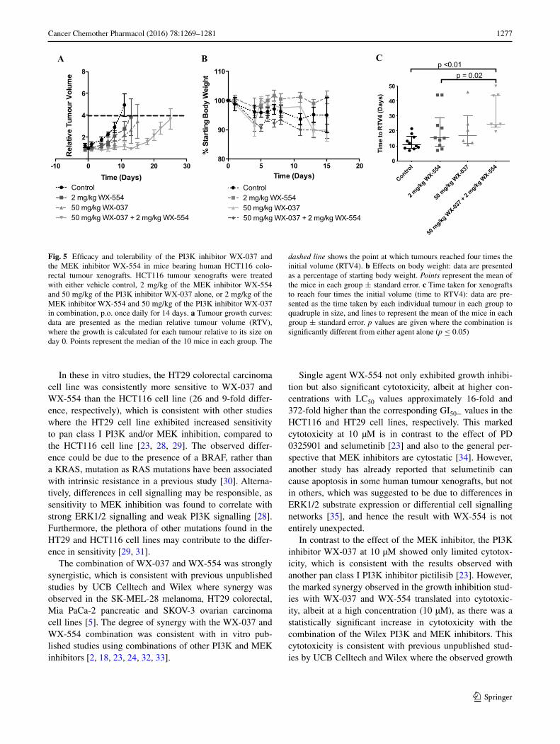
Based on the results of the PK study, the efficacy of 50 mg/kg of the PI3K inhibitor WX-037 and 2 mg/kg of the MEK inhibitor WX-554 given orally, as single agents and in combination, was assessed in HCT116 human tumour xenograft-bearing mice (Fig. 5). The individual doses of the PI3K and MEK inhibitors were chosen to be approxi-mately equiactive, in order to mirror the in vitro conditions under which synergy had been demonstrated (Supplemen-tary Figure 1). In this study, mice were treated daily for 14 days and tumour volumes were measured three times a week. Figure 5a demonstrates that treatment with 50 mg/kg WX-037 and 2 mg/kg WX-554, alone and in combination, caused tumour growth delay compared to vehicle-treated control tumours, and that growth delay was greater with the combination. Additionally, body weight was monitored daily to assess the tolerability of the therapy, and both sin-gle agent and combination treatments were found to be well tolerated as average body weights did not drop below 89% of the starting weight (Fig. 5b). The time for the tumours to quadruple in size (time to RTV4) was calculated (Fig. 5c), and statistical analyses using a Mann–Whitney test dem-onstrated a significant difference between vehicle-treated control tumours and the combination group (p < 0.01), and between the single agent MEK inhibitor and the combina-tion group (p = 0.02).
Discussion
The novel PI3K inhibitor WX-037 and the novel MEK inhibitor WX-554 have demonstrated in vitro activity in a range of cancer cell lines including breast, fibrosarcoma, thyroid, melanoma, colorectal, ovarian and pancreas lines with a broad range of GI50 values, where generally the cell lines most sensitive to WX-037 had PIK3CA mutations or PTEN loss, and to WX-554 had BRAF or RAS muta-tions [5]. Furthermore, the potency of WX-554 determined in the HCT116 and HT29 cell lines in this study (38 and 4.3 nM, respectively) was similar to that determined in the unpublished Wilex studies (29 and 7.2 nM) [5], and to the MEK inhibitor, PD 0325901 (21 and 6.5 nM) using the same assay [23]. Similarly the potency of WX-037 deter-mined here in the HCT116 and HT29 cell lines (2934 and 112 nM, respectively) was similar to that reported in the unpublished Wilex studies (136 nM in the HT29 cell line) [5], and to the pan class I PI3K inhibitor, pictilisib (1081 and 157 nM) using the same assay [23].
1277Cancer Chemother Pharmacol (2016) 78:1269–1281
1 3
In these in vitro studies, the HT29 colorectal carcinoma cell line was consistently more sensitive to WX-037 and WX-554 than the HCT116 cell line (26 and 9-fold differ-ence, respectively), which is consistent with other studies where the HT29 cell line exhibited increased sensitivity to pan class I PI3K and/or MEK inhibition, compared to the HCT116 cell line [23, 28, 29]. The observed differ-ence could be due to the presence of a BRAF, rather than a KRAS, mutation as RAS mutations have been associated with intrinsic resistance in a previous study [30]. Alterna-tively, differences in cell signalling may be responsible, as sensitivity to MEK inhibition was found to correlate with strong ERK1/2 signalling and weak PI3K signalling [28]. Furthermore, the plethora of other mutations found in the HT29 and HCT116 cell lines may contribute to the differ-ence in sensitivity [29, 31].
The combination of WX-037 and WX-554 was strongly synergistic, which is consistent with previous unpublished studies by UCB Celltech and Wilex where synergy was observed in the SK-MEL-28 melanoma, HT29 colorectal, Mia PaCa-2 pancreatic and SKOV-3 ovarian carcinoma cell lines [5]. The degree of synergy with the WX-037 and WX-554 combination was consistent with in vitro pub-lished studies using combinations of other PI3K and MEK inhibitors [2, 18, 23, 24, 32, 33].
Single agent WX-554 not only exhibited growth inhibi-tion but also significant cytotoxicity, albeit at higher con-centrations with LC50 values approximately 16-fold and 372-fold higher than the corresponding GI50− values in the HCT116 and HT29 cell lines, respectively. This marked cytotoxicity at 10 µM is in contrast to the effect of PD 0325901 and selumetinib [23] and also to the general per-spective that MEK inhibitors are cytostatic [34]. However, another study has already reported that selumetinib can cause apoptosis in some human tumour xenografts, but not in others, which was suggested to be due to differences in ERK1/2 substrate expression or differential cell signalling networks [35], and hence the result with WX-554 is not entirely unexpected.
In contrast to the effect of the MEK inhibitor, the PI3K inhibitor WX-037 at 10 µM showed only limited cytotox-icity, which is consistent with the results observed with another pan class I PI3K inhibitor pictilisib [23]. However, the marked synergy observed in the growth inhibition stud-ies with WX-037 and WX-554 translated into cytotoxic-ity, albeit at a high concentration (10 µM), as there was a statistically significant increase in cytotoxicity with the combination of the Wilex PI3K and MEK inhibitors. This cytotoxicity is consistent with previous unpublished stud-ies by UCB Celltech and Wilex where the observed growth
Time (Days)
% S
tart
ing
Bod
y W
eigh
t
0 5 10 15 2080
90
100
110
Control2 mg/kg WX-55450 mg/kg WX-03750 mg/kg WX-037 + 2 mg/kg WX-554
Time (Days)
Rel
ativ
e Tu
mou
r Vol
ume
-10 0 10 20 30
2
4
6
8
Control2 mg/kg WX-55450 mg/kg WX-03750 mg/kg WX-037 + 2 mg/kg WX-554
Tim
e to
RTV
4 (D
ays)
Control
2 mg/kgWX-55
4
50mg/kg
WX-03
7
50mg/kg
WX-037 + 2 mg/kg
WX-55
40
10
20
30
40
50
p = 0.02p <0.01
A B C
Fig. 5 Efficacy and tolerability of the PI3K inhibitor WX-037 and the MEK inhibitor WX-554 in mice bearing human HCT116 colo-rectal tumour xenografts. HCT116 tumour xenografts were treated with either vehicle control, 2 mg/kg of the MEK inhibitor WX-554 and 50 mg/kg of the PI3K inhibitor WX-037 alone, or 2 mg/kg of the MEK inhibitor WX-554 and 50 mg/kg of the PI3K inhibitor WX-037 in combination, p.o. once daily for 14 days. a Tumour growth curves: data are presented as the median relative tumour volume (RTV), where the growth is calculated for each tumour relative to its size on day 0. Points represent the median of the 10 mice in each group. The
dashed line shows the point at which tumours reached four times the initial volume (RTV4). b Effects on body weight: data are presented as a percentage of starting body weight. Points represent the mean of the mice in each group ± standard error. c Time taken for xenografts to reach four times the initial volume (time to RTV4): data are pre-sented as the time taken by each individual tumour in each group to quadruple in size, and lines to represent the mean of the mice in each group ± standard error. p values are given where the combination is significantly different from either agent alone (p ≤ 0.05)

1278 Cancer Chemother Pharmacol (2016) 78:1269–1281
1 3
inhibitory synergy with WX-037 and WX-554 in the SK-MEL-28 melanoma, HT29 colorectal, Mia PaCa-2 pancre-atic and SKOV-3 ovarian carcinoma cell lines was found to correlate with the induction of apoptosis [5]. Similarly, published in vitro studies have shown that combined inhibi-tion of PI3K and MEK resulted in cell death [33, 36, 37].
Cell signalling studies showed that the single agent PI3K inhibitor WX-037 caused a concentration-dependent reduction in AKT and S6 phosphorylation, which was gen-erally enhanced by the MEK inhibitor WX-554, but that 4EBP1 phosphorylation was not inhibited, consistent with the results of published studies using pan class I PI3K, as opposed to mixed PI3K/mTOR, inhibitors [23, 32, 37, 38]. Furthermore, single agent MEK inhibitor WX-554 caused a concentration-dependent reduction in ERK phosphoryla-tion, and the combination of WX-554 and WX-037 resulted in enhanced inhibition of ERK1/2 phosphorylation, as observed with other MEK inhibitors [23, 39, 40] and com-binations [23, 33].
In the pre-clinical studies presented here, and in both pre-clinical and clinical studies reported elsewhere [5, 6], WX-554 has been shown to be non-toxic. Furthermore, sin-gle agent WX-554 induced tumour growth delay at 2 mg/kg in HCT116 colorectal tumour xenograft models, consist-ent with previous unpublished studies using WX-554 and published studies using other MEK inhibitors that reported tumour growth delay or stasis in vivo, with increased sen-sitivity in BRAF or RAS mutant cells and tumours [3–5, 25]. In vivo pharmacokinetic analyses revealed concentra-tions of WX-554 in plasma and tumour tissue increased in a dose-dependent manner and that tumour WX-554 levels generally exceeded the in vitro GI50 concentration, whereas plasma levels were more variable.
Single agent WX-037 was also found to be non-toxic in these studies and those reported elsewhere [5, 17], which is consistent with data for other pan class I PI3K inhibi-tors [41–45]. Furthermore, 50 mg/kg WX-037 generated a tumour growth delay in HCT116 colorectal tumour xeno-graft models, which is consistent with previous unpub-lished studies using WX-037 and published studies using other pan class I PI3K inhibitors that reported tumour growth delay, or in some cases tumour stasis, with greater sensitivity in PIK3CA mutant or PTEN null cells and tumours [5, 14, 15, 46–51]. Pharmacokinetic studies indi-cated that concentrations of WX-037 in plasma and tumour tissue increased dose dependently and that WX-037 tumour and plasma levels generally exceeded the in vitro GI50 con-centration at an early time point (6 h), depending on the cell line and dose, whereas levels generally had decreased below the GI50 value by the later time point (24 h). These pharmacokinetic data are similar to those observed in HCT116 tumour xenograft-bearing mice treated with pic-tilisib [25].
The PK analyses indicated that there was no pharma-cokinetic interaction between WX-037 and WX-554 at lower doses. However, concomitant dosing of WX-037 with the high dose (5 mg/kg) of WX-554 caused a dose-depend-ent decrease in early time point (6 h) WX-554 tumour and plasma concentrations, and a dose-dependent increase in late time point (24 h) WX-554 levels, suggesting that the presence of WX-037 may delay the uptake of WX-554, leading to higher WX-554 levels at 24 h. Furthermore, con-comitant WX-554 dosing with high doses (100 mg/kg) of WX-037 caused a dose-dependent decrease in early time point (6 h) WX-037 tumour and plasma concentrations; however, this effect was only significant in the HCT116 tumour tissue due to variation in the data. The pharma-cokinetic interaction between higher doses of WX-037 and WX-554 is in contrast to the results obtained with the com-bination of PD 0325901 and pictilisib in HCT116 tumour xenograft-bearing mice [25], where there was no signifi-cant pharmacokinetic interaction, and to pre-clinical and clinical reports that there was no pharmacokinetic interac-tion between the PI3K and MEK inhibitors, pictilisib and cobimetinib [52, 53]. However, a recent Phase Ib study using a combination of the PI3K inhibitor buparlisib and the MEK inhibitor trametinib reported a potential pharma-cokinetic interaction, which was suggested to be due to the variability of the accumulation of the MEK inhibitor as no clear mechanism could be identified [54].
In HCT116 colorectal tumour xenografts, the combi-nation of 50 mg/kg WX-037 and 2 mg/kg WX-554 was non-toxic and caused tumour stasis and enhanced tumour growth delay compared to either single agent at the same dose. This improved activity is consistent with previous studies using combinations of PI3K and MEK inhibitors in a range of pre-clinical human tumour xenograft and mouse models [19, 21, 22, 25, 55–58]. In a previous study using the MEK inhibitor, PD 0325901, and the PI3K inhibitor, GDC-0941, the cell line-dependent differences in sensitiv-ity to the inhibitors determined in the in vitro studies did not correlate with the results observed in the in vivo studies [23, 25], suggesting that HT29 xenografts would not neces-sarily of been more sensitive to the inhibitors in vivo. Nev-ertheless, previous studies by UCB and Wilex have dem-onstrated that WX-554 treatment is able to induce tumour growth delay in HT29 xenografts [5].
The improved efficacy using combinations of PI3K and MEK inhibitors in pre-clinical studies is being translated into clinical trials [45, 54, 59], and there are currently at least 7 ongoing or completed Phase 1b clinical studies [9]. However, there have been issues with the tolerability of the combination treatment in two phase 1b trials combining the MEK inhibitor trametinib, with either the pan-PI3K inhibi-tor buparlisib, or the PI3K/mTOR inhibitor GSK2126458, where the former study showed promising anti-tumour

1279Cancer Chemother Pharmacol (2016) 78:1269–1281
1 3
activity but frequent dose interruptions and reductions were necessary due to toxicity [54]. The latter study was termi-nated due to poor tolerability and efficacy [9, 60]. These results suggest that the choice of the PI3K and MEK inhib-itor to be combined will be crucial to the success of the treatment, due to subtle differences in the pharmacology of the inhibitors leading to differing toxicity profiles. Over-all, these studies characterize, for the first time, the in vitro and in vivo efficacy of the PI3K inhibitor WX-037 and the MEK inhibitor WX-554, as single agents and in combina-tion. Furthermore, these studies illustrate that dual targeting of PI3K and MEK can induce synergy in vitro which trans-lates to marked tumour growth delay in vivo.
Acknowledgements The pharmacokinetic studies were performed by employees of Wilex.
Funding HDT, RJM and DRN are funded by a CRUK Drug Discov-ery Programme C240/A7409, and EJH was an MRC CASE Award PhD Student supported by UCB Celltech, Slough, United Kingdom.
Compliance with ethical standards
Conflict of interest Paul Bevan and Wolfgang Schmalix were employees of Wilex at the time that the research was conducted. Andrew Payne, Lara Kevorkian and Rodger Allen are employees of UCB Celltech, and Rodger Allen also consults for UCB Celltech.
Ethical approval All in vivo experiments were reviewed and approved by the Newcastle University (UK) animal welfare committee and were performed according to the guidelines for the welfare and use of ani-mals in cancer research [27] and national law, under project license (PPL60/4442) issued by the UK Government Home Office under the animals (scientific procedure) act 1986.
Open Access This article is distributed under the terms of the Crea-tive Commons Attribution 4.0 International License (http://crea-tivecommons.org/licenses/by/4.0/), which permits unrestricted use, distribution, and reproduction in any medium, provided you give appropriate credit to the original author(s) and the source, provide a link to the Creative Commons license, and indicate if changes were made.
References
1. Hennig M, Yip-Schneider MT, Wentz S, Wu H, Hekmatyar SK, Klein P, Bansal N, Schmidt CM (2010) Targeting mitogen-activated protein kinase kinase with the inhibitor PD0325901 decreases hepatocellular carcinoma growth in vitro and in mouse model systems. Hepatology 51(4):1218–1225
2. Liu D, Xing M (2008) Potent Inhibition of thyroid cancer cells by the MEK Inhibitor PD0325901 and Its potentiation by sup-pression of the PI3K and NF-κB pathways. Thyroid 18(8):853–864. doi:10.1089/thy.2007.0357
3. Akinleye A, Furqan M, Mukhi N, Ravella P, Liu D (2013) MEK and the inhibitors: from bench to bedside. J Hematol Oncol 6:27
4. Templeton IE, Musib L (2015) MEK inhibitors beyond mono-therapy: current and future development. Curr Opin Pharmacol 23:61–67. doi:10.1016/j.coph.2015.05.012
5. Wilex AG (unpublished data) 6. Mala C, Neville NG, Haindl E, Buergle M, Schmalix W, Bevan
P (2010) A phase I, first-in-human single ascending dose study of the MEK inhibitor WX-554 given to healthy male subjects. J Clin Oncol 28((suppl; abstr)):e13666
7. Wilex AG (2012). http://www.wilex.de. Accessed 23/07/12 8. Miller CR, Oliver KE, Farley JH (2014) MEK1/2 inhibitors in
the treatment of gynecologic malignancies. Gynecol Oncol 133(1):128–137. doi:10.1016/j.ygyno.2014.01.008
9. Clinical Trials gov (2016). http://clinicaltrials.gov/. Accessed 22/03/2016
10. Maira S-M, Stauffer F, Brueggen J, Furet P, Schnell C, Fritsch C, Brachmann S, Chene P, De Pover A, Schoemaker K, Fabbro D, Gabriel D, Simonen M, Murphy L, Finan P, Sellers W, Gar-cia-Echeverria C (2008) Identification and characterization of NVP-BEZ235, a new orally available dual phosphatidylinositol 3-kinase/mammalian target of rapamycin inhibitor with potent in vivo antitumor activity. Mol Cancer Ther 7(7):1851–1863. doi:10.1158/1535-7163.mct-08-0017
11. Serra V, Markman B, Scaltriti M, Eichhorn PJA, Valero V, Guz-man M, Botero ML, Llonch E, Atzori F, Di Cosimo S, Maira M, Garcia-Echeverria C, Parra JL, Arribas J, Baselga J (2008) NVP-BEZ235, a Dual PI3K/mTOR inhibitor, prevents PI3K signal-ing and inhibits the growth of cancer cells with activating PI3K mutations. Cancer Res 68(19):8022–8030. doi:10.1158/0008-5472.can-08-1385
12. Baumann P, Mandl-Weber S, Oduncu F, Schmidmaier R (2009) The novel orally bioavailable inhibitor of phosphoinositol-3-ki-nase and mammalian target of rapamycin, NVP-BEZ235, inhib-its growth and proliferation in multiple myeloma. Exp Cell Res 315(3):485–497
13. Maira S-M, Stauffer F, Schnell C, Garcia-echeverria C (2009) PI3K inhibitors for cancer treatment: where do we stand? Bio-chem Soc Trans 37(1):265–272. doi:10.1042/bst0370265
14. Folkes AJ, Ahmadi K, Alderton WK, Alix S, Baker SJ, Box G, Chuckowree IS, Clarke PA, Depledge P, Eccles SA, Friedman LS, Hayes A, Hancox TC, Kugendradas A, Lensun L, Moore P, Olivero AG, Pang J, Patel S, Pergl-Wilson GH, Raynaud FI, Robson A, Saghir N, Salphati L, Sohal S, Ultsch MH, Val-enti M, Wallweber HJA, Wan NC, Wiesmann C, Workman P, Zhyvoloup A, Zvelebil MJ, Shuttleworth SJ (2008) The Iden-tification of 2-(1H-Indazol-4-yl)-6-(4-methanesulfonyl-piper-azin-1-ylmethyl)-4-morpholin-4-yl-thieno[3,2-d]pyrimidine (GDC-0941) as a potent, selective, orally bioavailable inhibitor of class I PI3Kinase for the treatment of cancer. J Med Chem 51(18):5522–5532
15. Raynaud FI, Eccles SA, Patel S, Alix S, Box G, Chuckowree I, Folkes A, Gowan S, De Haven Brandon A, Di Stefano F, Hayes A, Henley AT, Lensun L, Pergl-Wilson G, Robson A, Saghir N, Zhyvoloup A, McDonald E, Sheldrake P, Shuttleworth S, Valenti M, Wan NC, Clarke PA, Workman P (2009) Biological properties of potent inhibitors of class I phosphatidylinositide 3-kinases: from PI-103 through PI-540, PI-620 to the oral agent GDC-0941. Mol Cancer Ther 8(7):1725–1738. doi:10.1158/1535-7163.mct-08-1200
16. Mayer IA, Arteaga CL (2016) The PI3K/AKT pathway as a tar-get for cancer treatment. Annu Rev Med 67:11–28. doi:10.1146/annurev-med-062913-051343
17. Wilex AG (2013). http://www.wilex.de. Accessed 30/03/2016 18. Yu K, Toral-Barza L, Shi C, Zhang WG, Zask A (2008) Response
and determinants of cancer cell susceptibility to PI3K inhibitors: combined targeting of PI3K and Mek1 as an effective anticancer strategy. Cancer Biol Ther 7(2):310–318
19. Engelman JA, Chen L, Tan X, Crosby K, Guimaraes AR, Upad-hyay R, Maira M, McNamara K, Perera SA, Song Y, Chirieac LR, Kaur R, Lightbown A, Simendinger J, Li T, Padera RF,

1280 Cancer Chemother Pharmacol (2016) 78:1269–1281
1 3
Garcia-Echeverria C, Weissleder R, Mahmood U, Cantley LC, Wong K-K (2008) Effective use of PI3K and MEK inhibitors to treat mutant Kras G12D and PIK3CA H1047R murine lung can-cers. Nat Med 14(12):1351–1356
20. Jokinen E, Koivunen JP (2015) MEK and PI3K inhibition in solid tumors: rationale and evidence to date. Ther Adv Med Oncol 7(3):170–180. doi:10.1177/1758834015571111
21. Sos ML, Fischer S, Ullrich R, Peifer M, Heuckmann JM, Koker M, Heynck S, Stückrath I, Weiss J, Fischer F, Michel K, Goel A, Regales L, Politi KA, Perera S, Getlik M, Heukamp LC, Ansén S, Zander T, Beroukhim R, Kashkar H, Shokat KM, Sellers WR, Rauh D, Orr C, Hoeflich KP, Friedman L, Wong KK, Pao W, Thomas RK (2009) Identifying genotype-dependent efficacy of single and combined PI3K- and MAPK-pathway inhibition in cancer. Proc Nat Acad Sci USA 106(43):18351–18356
22. Hoeflich KP, Merchant M, Orr C, Chan J, Den Otter D, Berry L, Kasman I, Koeppen H, Rice K, Yang N-Y, Engst S, Johnston S, Friedman LS, Belvin M (2012) Intermittent administration of MEK inhibitor GDC-0973 plus PI3K inhibitor GDC-0941 trig-gers robust apoptosis and tumor growth inhibition. Cancer Res 72(1):210–219. doi:10.1158/0008-5472.can-11-1515
23. Haagensen EJ, Kyle S, Beale GS, Maxwell RJ, Newell DR (2012) The synergistic interaction of MEK and PI3K inhibitors is modulated by mTOR inhibition. Br J Cancer 106(8):1386–1394
24. Haagensen EJ, Thomas HD, Mudd C, Tsonou E, Wiggins CM, Maxwell RJ, Moore JD, Newell DR (2016) Pre-clinical use of isogenic cell lines and tumours in vitro and in vivo for predic-tive biomarker discovery; impact of KRAS and PI3KCA muta-tion status on MEK inhibitor activity is model dependent. Eur J Cancer (Oxford, England: 1990) 56:69–76. doi:10.1016/j.ejca.2015.12.012
25. Haagensen EJ, Thomas HD, Wilson I, Harnor SJ, Payne SL, Rennison T, Smith KM, Maxwell RJ, Newell DR (2013) The enhanced in vivo activity of the combination of a MEK and a PI3K inhibitor correlates with [18F]-FLT PET in human colo-rectal cancer xenograft tumour-bearing mice. PLoS ONE 8(12):e81763. doi:10.1371/journal.pone.0081763
26. Britten CD (2013) PI3K and MEK inhibitor combinations: exam-ining the evidence in selected tumor types. Cancer Chemother Pharmacol 71(6):1395–1409. doi:10.1007/s00280-013-2121-1
27. Workman P, Aboagye EO, Balkwill F, Balmain A, Bruder G, Chaplin DJ, Double JA, Everitt J, Farningham DAH, Glennie MJ, Kelland LR, Robinson V, Stratford IJ, Tozer GM, Watson S, Wedge SR, Eccles SA (2010) Guidelines for the welfare and use of animals in cancer research. Br J Cancer 102(11):1555–1577
28. Balmanno K, Chell SD, Gillings AS, Hayat S, Cook SJ (2009) Intrinsic resistance to the MEK1/2 inhibitor AZD6244 (ARRY-142886) is associated with weak ERK1/2 signalling and/or strong PI3K signalling in colorectal cancer cell lines. Int J Can-cer 125(10):2332–2341. doi:10.1002/ijc.24604
29. Genomics of Drug Sensitivity in Cancer (2012). http://www.can-cerrxgene.org/. Accessed 05/10/2012
30. Ebi H, Corcoran RB, Singh A, Chen Z, Song Y, Lifshits E, Ryan DP, Meyerhardt JA, Benes C, Settleman J, Wong K-K, Cantley LC, Engelman JA (2011) Receptor tyrosine kinases exert dominant control over PI3K signaling in human KRAS mutant colorectal cancers. J Clin Investig 121(11):4311–4321. doi:10.1172/jci57909
31. Garnett MJ, Edelman EJ, Heidorn SJ, Greenman CD, Dastur A, Lau KW, Greninger P, Thompson IR, Luo X, Soares J, Liu Q, Iorio F, Surdez D, Chen L, Milano RJ, Bignell GR, Tam AT, Davies H, Stevenson JA, Barthorpe S, Lutz SR, Kogera F, Law-rence K, McLaren-Douglas A, Mitropoulos X, Mironenko T, Thi H, Richardson L, Zhou W, Jewitt F, Zhang T, O’Brien P, Bois-vert JL, Price S, Hur W, Yang W, Deng X, Butler A, Choi HG, Chang JW, Baselga J, Stamenkovic I, Engelman JA, Sharma SV, Delattre O, Saez-Rodriguez J, Gray NS, Settleman J, Futreal PA,
Haber DA, Stratton MR, Ramaswamy S, McDermott U, Benes CH (2012) Systematic identification of genomic markers of drug sensitivity in cancer cells. Nature 483(7391):570–575
32. Pitts TM, Newton TP, Bradshaw-Pierce EL, Addison R, Arca-roli JJ, Klauck PJ, Bagby SM, Hyatt SL, Purkey A, Tentler JJ, Tan AC, Messersmith WA, Eckhardt SG, Leong S (2014) Dual pharmacological targeting of the MAP kinase and PI3K/mTOR pathway in preclinical models of colorectal cancer. PLoS ONE 9(11):e113037. doi:10.1371/journal.pone.0113037
33. Ku BM, Jho EH, Bae YH, Sun JM, Ahn JS, Park K, Ahn MJ (2015) BYL719, a selective inhibitor of phosphoinositide 3-Kinase alpha, enhances the effect of selumetinib (AZD6244, ARRY-142886) in KRAS-mutant non-small cell lung cancer. Invest New Drugs 33(1):12–21. doi:10.1007/s10637-014-0163-9
34. Kohno M, Pouyssegur J (2006) Targeting the ERK signaling pathway in cancer therapy. Ann Med 38(3):200–211
35. Davies BR, Logie A, McKay JS, Martin P, Steele S, Jenkins R, Cockerill M, Cartlidge S, Smith PD (2007) AZD6244 (ARRY-142886), a potent inhibitor of mitogen-activated protein kinase/extracellular signal-regulated kinase kinase 1/2 kinases: mecha-nism of action in vivo, pharmacokinetic/pharmacodynamic relationship, and potential for combination in preclinical mod-els. Mol Cancer Ther 6(8):2209–2219. doi:10.1158/1535-7163.mct-07-0231
36. Wee S, Jagani Z, Xiang KX, Loo A, Dorsch M, Yao Y-M, Sell-ers WR, Lengauer C, Stegmeier F (2009) PI3K pathway acti-vation mediates resistance to MEK inhibitors in KRAS mutant cancers. Cancer Res 69(10):4286–4293. doi:10.1158/0008-5472.can-08-4765
37. Edgar KA, Wallin JJ, Berry M, Lee LB, Prior WW, Sampath D, Friedman LS, Belvin M (2010) Isoform-specific phos-phoinositide 3-kinase inhibitors exert distinct effects in solid tumors. Cancer Res 70(3):1164–1172. doi:10.1158/0008-5472.can-09-2525
38. Santiskulvong C, Konecny GE, Fekete M, Chen K-YM, Karam A, Mulholland D, Eng C, Wu H, Song M, Dorigo O (2011) Dual targeting of phosphoinositide 3-kinase and mammalian target of rapamycin Using NVP-BEZ235 as a novel therapeutic approach in human ovarian carcinoma. Clin Cancer Res 17(8):2373–2384. doi:10.1158/1078-0432.ccr-10-2289
39. Haura EB, Ricart AD, Larson TG, Stella PJ, Bazhenova L, Miller VA, Cohen RB, Eisenberg PD, Selaru P, Wilner KD, Gadgeel SM (2010) A phase II study of PD-0325901, an Oral MEK inhibitor, in previously treated patients with advanced non-small cell lung cancer. Clin Cancer Res 16(8):2450–2457. doi:10.1158/1078-0432.ccr-09-1920
40. Yeh TC, Marsh V, Bernat BA, Ballard J, Colwell H, Evans RJ, Parry J, Smith D, Brandhuber BJ, Gross S, Marlow A, Hurley B, Lyssikatos J, Lee PA, Winkler JD, Koch K, Wal-lace E (2007) Biological characterization of ARRY-142886 (AZD6244), a potent, highly selective mitogen-activated protein kinase kinase 1/2 inhibitor. Clin Cancer Res 13(5):1576–1583. doi:10.1158/1078-0432.ccr-06-1150
41. Yap TA, Bjerke L, Clarke PA, Workman P (2015) Drugging PI3K in cancer: refining targets and therapeutic strategies. Curr Opin Pharmacol 23:98–107. doi:10.1016/j.coph.2015.05.016
42. Sarker D, Ang JE, Baird R, Kristeleit R, Shah K, Moreno V, Clarke PA, Raynaud FI, Levy G, Ware JA, Mazina K, Lin R, Wu J, Fredrickson J, Spoerke JM, Lackner MR, Yan Y, Friedman LS, Kaye SB, Derynck MK, Workman P, de Bono JS (2015) First-in-human phase I study of pictilisib (GDC-0941), a potent pan-class I phosphatidylinositol-3-kinase (PI3K) inhibitor, in patients with advanced solid tumors. Clin Cancer Res 21(1):77–86. doi:10.1158/1078-0432.ccr-14-0947
43. Bendell JC, Rodon J, Burris HA, de Jonge M, Verweij J, Birle D, Demanse D, De Buck SS, Ru QC, Peters M, Goldbrunner M,

1281Cancer Chemother Pharmacol (2016) 78:1269–1281
1 3
Baselga J (2012) Phase I, dose-escalation study of BKM120, an oral pan-Class I PI3K inhibitor, in patients with advanced solid tumors. J Clin Oncol 30(3):282–290. doi:10.1200/jco.2011.36.1360
44. Hong DS, Bowles DW, Falchook GS, Messersmith WA, George GC, O’Bryant CL, Vo AC, Klucher K, Herbst RS, Eckhardt SG, Peterson S, Hausman DF, Kurzrock R, Jimeno A (2012) A mul-ticenter phase I trial of PX-866, an oral irreversible phosphati-dylinositol 3-kinase inhibitor, in patients with advanced solid tumors. Clin Cancer Res 18(15):4173–4182. doi:10.1158/1078-0432.ccr-12-0714
45. Shapiro GI, Rodon J, Bedell C, Kwak EL, Baselga J, Brana I, Pandya SS, Scheffold C, Laird AD, Nguyen LT, Xu Y, Egile C, Edelman G (2014) Phase I safety, pharmacokinetic, and phar-macodynamic study of SAR245408 (XL147), an oral pan-class I PI3K inhibitor, in patients with advanced solid tumors. Clin Can-cer Res 20(1):233–245. doi:10.1158/1078-0432.ccr-13-1777
46. Foster P, Yamaguchi K, Hsu PP, Qian F, Du X, Wu J, Won KA, Yu P, Jaeger CT, Zhang W, Marlowe CK, Keast P, Abulafia W, Chen J, Young J, Plonowski A, Yakes FM, Chu F, Engell K, Bentzien F, Lam ST, Dale S, Yturralde O, Matthews DJ, Lamb P, Laird AD (2015) The selective PI3K inhibitor XL147 (SAR245408) inhibits tumor growth and survival and potentiates the activity of chemotherapeutic agents in preclinical tumor models. Mol Can-cer Ther 14(4):931–940. doi:10.1158/1535-7163.mct-14-0833
47. Burger MT, Pecchi S, Wagman A, Ni ZJ, Knapp M, Hendrick-son T, Atallah G, Pfister K, Zhang Y, Bartulis S, Frazier K, Ng S, Smith A, Verhagen J, Haznedar J, Huh K, Iwanowicz E, Xin X, Menezes D, Merritt H, Lee I, Wiesmann M, Kaufman S, Craw-ford K, Chin M, Bussiere D, Shoemaker K, Zaror I, Maira SM, Voliva CF (2011) Identification of NVP-BKM120 as a potent, selective, orally bioavailable class I PI3 kinase inhibitor for treat-ing cancer. ACS Med Chem Lett 2(10):774–779. doi:10.1021/ml200156t
48. Ohwada J, Ebiike H, Kawada H, Tsukazaki M, Nakamura M, Miyazaki T, Morikami K, Yoshinari K, Yoshida M, Kondoh O, Kuramoto S, Ogawa K, Aoki Y, Shimma N (2011) Discovery and biological activity of a novel class I PI3K inhibitor, CH5132799. Bioorg Med Chem Lett 21(6):1767–1772. doi:10.1016/j.bmcl.2011.01.065
49. Liu N, Rowley BR, Bull CO, Schneider C, Haegebarth A, Schatz CA, Fracasso PR, Wilkie DP, Hentemann M, Wilhelm SM, Scott WJ, Mumberg D, Ziegelbauer K (2013) BAY 80-6946 is a highly selective intravenous PI3K inhibitor with potent p110alpha and p110delta activities in tumor cell lines and xenograft models. Mol Cancer Ther 12(11):2319–2330. doi:10.1158/1535-7163.mct-12-0993-t
50. Ihle NT, Williams R, Chow S, Chew W, Berggren MI, Paine-Murrieta G, Minion DJ, Halter RJ, Wipf P, Abraham R, Kirkpat-rick L, Powis G (2004) Molecular pharmacology and antitumor activity of PX-866, a novel inhibitor of phosphoinositide-3-ki-nase signaling. Mol Cancer Ther 3(7):763–772
51. Kong D, Yamori T (2009) Advances in development of phosphatidylinositol 3-kinase inhibitors. Curr Med Chem 16(22):2839–2854
52. Shapiro G, LoRusso P, Kwak EL, Cleary JM, Musib L, Jones C, de Crespigny A, Belvin M, McKenzie M, Gates MR, Chan IT, Bendell JC (2011) Clinical combination of the MEK inhibitor
GDC-0973 and the PI3K inhibitor GDC-0941: a first-in-human phase Ib study testing daily and intermittent dosing schedules in patients with advanced solid tumors. J Clin Oncol 29((suppl; abstr)):3005
53. Choo EF, Ng CM, Berry L, Belvin M, Lewin-Koh N, Mer-chant M, Salphati L (2013) PK-PD modeling of combina-tion efficacy effect from administration of the MEK inhibitor GDC-0973 and PI3K inhibitor GDC-0941 in A2058 xenografts. Cancer Chemother Pharmacol 71(1):133–143. doi:10.1007/s00280-012-1988-6
54. Bedard PL, Tabernero J, Janku F, Wainberg ZA, Paz-Ares L, Vansteenkiste J, Van Cutsem E, Perez-Garcia J, Stathis A, Britten CD, Le N, Carter K, Demanse D, Csonka D, Peters M, Zubel A, Nauwelaerts H, Sessa C (2015) A phase Ib dose-escalation study of the oral pan-PI3K inhibitor buparlisib (BKM120) in combina-tion with the oral MEK1/2 inhibitor trametinib (GSK1120212) in patients with selected advanced solid tumors. Clin Cancer Res 21(4):730–738. doi:10.1158/1078-0432.ccr-14-1814
55. Roper J, Sinnamon MJ, Coffee EM, Belmont P, Keung L, Geor-geon-Richard L, Wang WV, Faber AC, Yun J, Yilmaz OH, Bron-son RT, Martin ES, Tsichlis PN, Hung KE (2014) Combination PI3K/MEK inhibition promotes tumor apoptosis and regression in PIK3CA wild-type KRAS mutant colorectal cancer. Cancer Lett 347(2):204–211. doi:10.1016/j.canlet.2014.02.018
56. Martinelli E, Troiani T, D’Aiuto E, Morgillo F, Vitagliano D, Capasso A, Costantino S, Ciuffreda LP, Merolla F, Vecchione L, De Vriendt V, Tejpar S, Nappi A, Sforza V, Martini G, Ber-rino L, De Palma R, Ciardiello F (2013) Antitumor activity of pimasertib, a selective MEK 1/2 inhibitor, in combination with PI3K/mTOR inhibitors or with multi-targeted kinase inhibitors in pimasertib-resistant human lung and colorectal cancer cells. Int J Cancer 133(9):2089–2101. doi:10.1002/ijc.28236
57. Kinross KM, Brown DV, Kleinschmidt M, Jackson S, Chris-tensen J, Cullinane C, Hicks RJ, Johnstone RW, McArthur GA (2011) In vivo activity of combined PI3K/mTOR and MEK Inhi-bition in a KrasG12D; Pten deletion mouse model of ovarian cancer. Mol Cancer Ther 10(8):1440–1449. doi:10.1158/1535-7163.mct-11-0240
58. Faber AC, Li D, Song Y, Liang M-C, Yeap BY, Bronson RT, Lif-shits E, Chen Z, Maira S-M, Garcia-Echeverria C, Wong K-K, Engelman JA (2009) Differential induction of apoptosis in HER2 and EGFR addicted cancers following PI3K inhibition. Proc Nat Acad Sci 106(46):19503–19508. doi:10.1073/pnas.0905056106
59. Shimizu T, Tolcher AW, Papadopoulos KP, Beeram M, Rasco DW, Smith LS, Gunn S, Smetzer L, Mays TA, Kaiser B, Wick MJ, Alvarez C, Cavazos A, Mangold GL, Patnaik A (2012) The clinical effect of the dual-targeting strategy involving PI3K/AKT/mTOR and RAS/MEK/ERK pathways in patients with advanced cancer. Clin Cancer Res 18(8):2316–2325. doi:10.1158/1078-0432.ccr-11-2381
60. Grilley-Olson JE, Bedard PL, Fasolo A, Cornfeld M, Cartee L, Razak AR, Stayner LA, Wu Y, Greenwood R, Singh R, Lee CB, Bendell J, Burris HA, Del Conte G, Sessa C, Infante JR (2016) A phase Ib dose-escalation study of the MEK inhibitor trametinib in combination with the PI3K/mTOR inhibitor GSK2126458 in patients with advanced solid tumors. Invest New Drugs. doi:10.1007/s10637-016-0377-0