Embed Size (px)
Citation preview

Fax +41 61 306 12 34E-Mail [email protected]
ENETS Consensus Guidelines
Neuroendocrinology 2010;91:341–350 DOI: 10.1159/000287255
ENETS Consensus Guidelines for theManagement of Bone and Lung Metastases from Neuroendocrine Tumors Beata Kos-Kudła a Dermot O’Toole b Massimo Falconi c David Gross d Günther Klöppel e Anders Sundin f John Ramage g Kjell Öberg h Bertram Wiedenmann i Paul Komminoth j Eric Van Custem k Mohandes Mallath l Mauro Papotti m Martyn Caplin n and all other Palma de Mallorca Consensus Conference Participants 1
a Division of Endocrinology, Department of Pathophysiology and Endocrinology, Medical University of Silesia, Katowice , Poland; b Department of Clinical Medicine and Gastroenterology, St. James’s Hospital and Trinity College, Dublin , Ireland; c Medicine and Surgery, University of Verona, Verona , Italy; d Department of Endocrinology and Metabolism, Hadassah University Hospital, Jerusalem , Israel; e Institut für Allgemeine Pathologie und Pathologische Anatomie, Universität Kiel, Kiel , Germany; f Department of Radiology, Uppsala University Hospital, Uppsala , Sweden; g Department of Gastroenterology, North Hampshire Hospital, Basingstoke , UK; h Department of Internal Medicine, Endocrine Unit, University Hospital, Uppsala , Sweden; i Division of Hepatology and Gastroenterology, Department of Internal Medicine, Charité-Universitätsmedizin Berlin, Campus Virchow Klinikum, Berlin , Germany; j Institute for Pathology, Kantonsspital Baden, Baden , Switzerland; k Digestive Oncology Unit, Department of Gastroenterology, University Hospital Gasthuisberg, Leuven , Belgium; l Department of Digestive Diseases, Tata Memorial Hospital, Mumbai , India; m Department of Biological and Clinical Sciences, University of Turin, St. Luigi Hospital, Turin , Italy; n Department of Medicine, Royal Free Hospital, London , UK
Epidemiology
Bone Metastases In a recent study of 668 NET patients in France, bone
metastases were found in 6.4% [6] . A review of 26-year re-cords from the M.D. Anderson Cancer Center database identified 1,633 patients with carcinoid tumors where 18% of patients had metastases to bone and soft tissues [5] . Mei-jer et al. [4] recently reported a retrospective study where bone metastases were found in 12% of carcinoid patients. Generally, metastases to bone are relatively uncommon in carcinoid disease (7–15% of all metastases) and are often reported as multiple [3, 4, 7] . The incidence of bone metas-tases remains underestimated as they are frequently unde-tected or simply not focused on. In a small postmortem study, 42% of patients had bone metastases compared to only 4% detected in live patients with advanced disease [8, 9] . Introduction of new diagnostic modalities (e.g. sensi-
Introduction
Gastroenteropancreatic (GEP) neuroendocrine tu-mors (NET) are rare tumors with a low incidence of at least 3–5/100,000 but a considerably higher prevalence of 35/100,000 [1] . The majority of patients at the time of di-agnosis are found to have an advanced stage of the dis-ease. Bone metastasis is not an infrequent complication in most neoplasms and has been found in 70–85% of can-cer patients at autopsy [2] . GEP-NET bone metastases fre-quently remain undetected. They are often accompanied by widespread extraosseous metastases and are found to occur predominantly in patients with liver metastases [3, 4] . It is thought that lung metastases occur with a similar frequency to bone metastases [5] .
Received: August 25, 2009 Accepted: February 3, 2010 Published online: May 19, 2010
Beata Kos-Kudła Department of Pathophysiology and Endocrinology Division of Endocrinology, Medical University of Silesia Ceglana Str. 35, PL–40-952 Katowice (Poland) Tel./Fax +48 32 358 1366, E-Mail bkoskudla @ sum.edu.pl
© 2010 S. Karger AG, Basel0028–3835/10/0914–0341$26.00/0
Accessible online at:www.karger.com/nen
1 See list at the end of the paper.
© For permitted use only. ANY FURTHER DISTRIBUTION OF THIS ARTICLE REQUIRES WRITTEN PERMISSION FROM S. KARGER AG BASEL AND MAY BE SUBJECT TO A PERMISSION FEE

Kos-Kudła et al. Neuroendocrinology 2010;91:341–350342
tive scintigraphic/PET modalities) may reveal asymptom-atic bone metastases, thus increasing the overall rate of detection of bone metastases in living patients. Bone me-tastases may occur at any time, from long before the diag-nosis to even 20 years after the initial presentation. No clear time pattern has been noted to facilitate prediction of their occurrence [10] . NET bone metastasis distribution is comparable to that found in non-NET tumors [3] . Opin-ions regarding the influence of the primary tumor site on the development of bone metastases vary. According to some authors, bone metastases arise more often from fore-gut or hindgut rather than from midgut tumors. Other studies suggest no preferential primary site [3, 4] . NETs originating from the lungs metastasized (15%) mainly to liver, bone, adrenal glands, and brain [11] . Diagnosis of metastases occurred synchronously with the diagnosis of the primary bronchial carcinoid in all cases [12] .
Lung Metastases GEP-NET lung metastases are relatively uncommon,
occurring in 13.6% based on the US National Cancer In-stitute’s Surveillance, Epidemiology, and End Results (SEER) vast database [13] . In a recent French study [6] lung metastases were found in 5.1% while a review of the M.D. Anderson Cancer Center database identified 6% metastases to the lung [5] .
Minimal Consensus Statements on Epidemiology According to the literature, 4–15% of NET metastases de-
velop in bone; similarly lung metastases represent around 5–14%. In the majority of cases, they are found in the ad-vanced stage of the disease (usually stage IV).
The incidence of bone and lung metastases is almost cer-tainly underestimated as these metastases are often asymp-tomatic and omitted in routine diagnostics.
Clinical Presentation
Bone Metastases Bone metastases are clinically detected in a low per-
centage of patients with advanced NET tumors [8] . Characteristic symptoms for bone metastases are: (1)
pain, principal symptom, which can be accompanied by (2) pathological fractures and/or (3) symptoms from hy-percalcemia [3, 4] .
Pain is the main symptom of bone metastases, but only occurs in a minority and can affect work, locomotion, mood, sleep, relations with others, and enjoyment of life [2] . Bone metastases are more often asymptomatic and
frequently detected incidentally during staging of GEP-NET disease [3, 4] .
Lung Metastases Symptoms associated with lung metastases are rare.
Patients may present with cough, hemoptysis and pneu-monia (classical triad) representing the sequel of luminal obstruction and tumor ulceration [14] . The clinical man-ifestations of endocrine tumors are determined by the functional status of the tumor [15] . In patients with non-functional tumors, symptoms depend on tumor load and the location of the metastases. In bone and lung metasta-ses originating from functional NETs, symptoms can be accompanied by characteristic syndromes which depend on the hypersecretion of the specific hormones from pri-mary tumor such as carcinoid syndrome, hypoglycemic syndrome in insulinoma, Zollinger-Ellison syndrome in gastrinoma, necrolytic migratory erythema in gluca-gonoma or watery diarrhea, hypokalemia and achlorhy-dria syndrome characteristic for VIPoma (see ENETS Guidelines) [14, 16–27] . Ectopic hormone production from lung metastases (i.e. ACTH, cortisol, IGF-1) can also be the cause of some symptoms.
Prognosis
The stage of disease significantly influences the over-all prognosis of well-differentiated GEP-NET. The best 5-year survival rate is for localized disease at 93% and 5-year survival rates in distant metastatic disease are be-tween 20 and 30%. Distant metastases are found in 22% of cases, half of which have unknown primaries [2, 4, 7] . Recent data from the US SEER and the Norwegian Reg-istry of Cancer (NRC) database reported 5-year survival of approximately 55% [1] . Among over 4,000 cases of ma-lignant GEP-NET registered in the UK, relative survival for all NET was 46% at 5 years and 38% at 10 years. Five-year survival was 57% for well-differentiated tumors but a worse prognosis was reported for poorly differentiated tumors (only 5.2% survive for 5 years) [28] .
Bone Metastases Bone metastases are usually accompanied by metasta-
ses at other distant sites; however, literature regarding the direct influence of bone metastases on overall survival is unavailable. Less than 50% of patients with metastatic GEP tumors survive 5 years with hepatic and bone me-tastases being the major causes of death [29] .

ENETS Consensus Guidelines Neuroendocrinology 2010;91:341–350 343
Lung Metastases The type of tumor influences survival, as well as its
histology and differentiation, disease-free interval, num-ber of metastases, and the presence of mediastinal nodal disease [12] . The lowest 5-year survival rate is found among patients with poorly differentiated tumors with distant metastatic disease at diagnosis.
The overall median survival may be as little as 6 months in patients with pulmonary metastases. Accord-ing to Khan et al. [12] , the computed overall 5-year sur-vival is 61% in NET patients with surgically resectable pulmonary metastatic disease.
Minimal Consensus Statements on Clinical Presenta-tion and Prognosis Most bone metastases are asymptomatic. When symp-
toms occur, this may be accompanied by pathological frac-tures and symptoms associated with hypercalcemia. Most lung metastases are asymptomatic; however, a minority of patients may develop cough, hemoptysis or pneumonia.
Bone and lung metastases appear in advanced disease of-ten associated with other distant metastases.
Based on available literature, the direct influence of indi-vidual metastases on NET patients’ prognosis is difficult to evaluate. In patients with resectable lung metastases, surgery may increase the overall 5-year survival to over 60%. Among factors influencing survival are the type of primary tumor, its histology and differentiation, disease-free interval, number of metastases, and the presence of other distant metastases.
Diagnostic Procedures
Imaging
For localization of metastatic disease the combined use of cross-sectional (anatomic) and functional imaging methods is always recommended. Single individual meth-ods are not sensitive and specific enough for NET. High-resolution contrast-enhanced computed tomography (CT) and magnetic resonance imaging (MRI) are excellent for the detection of metastatic disease. Somatostatin receptor scintigraphy (SRS) most commonly with 111 In-pentetreo-tide ( 111 In-Octreoscan) is widely used because most NETs express somatostatin receptors predominantly subtype 2 [30] . The protocol for 111 In- Octreoscan needs to include single photon emission tomography which increases sen-sitivity. Scintigraphy with technetium ( 99m Tc-HYNIC-TOC, 99m Tc-HYNIC-TATE or 99m Tc-depreotide) is also used as a sensitive and cost-effective method [30, 31] . SRS is a sensitive method for localizing radiologically occult
NET, with a reported sensitivity of 80–100% [32] . This whole-body imaging technique may provide important information about unsuspected metastatic disease. No functional imaging test is perfect; therefore, a combina-tion of different imaging modalities may be required to facilitate the diagnosis. Computer-aided fusion of ana-tomic and functional image data has been shown to in-crease the precision of metastatic disease location. Hybrid PET/CT has recently proved to be highly accurate for de-tection of NET metastases [33] . PET as a single modality does not currently play a major role in the imaging of well-differentiated tumor metastases, because of their slow growth and low metabolic rate. FDG PET may be helpful in staging high-grade, often poorly differentiated NETs. Other PET radiotracers, such as fluorodopa 18 F, and 68 Ga-labeled radiopharmaceuticals may significantly improve future diagnostics of NET metastases. However, these ra-diotracers are not commonly available [34, 35] .
Bone Metastases The following imaging methods are useful in the de-
tection of bone metastases: (1) Localized techniques: (a) plain skeletal radiography
and (b) MRI. (2) Whole body imaging: (a) bone scintigraphy (with
99m Tc-labeled radiopharmaceuticals, e.g. diphospho-nates) and (b) SRS [or rarely 131 I-metaiodobenzylguani-dine (MIBG) scintigraphy].
Radiographic signs of bone metastases may be easily missed [3] . MRI is considered the most sensitive tech-nique for demonstrating bone metastases in patients with NETs and it is recommended for precise monitoring of response to therapy [36] . MRI is the most sensitive meth-od of detecting metastases in bone marrow, with a sensi-tivity of nearly 100% [4] ( fig. 1 a, b). If marrow deposits are sclerotic, they generally show low signal intensity on T 2 -weighted images. Bone scintigraphy is the next sensitive and reliable method to detect NET bone metastases. It identifies those metastases with osteoblastic reaction. Scintigraphy performed with 111 In-pentetreotide or less frequently 123 I-MIBG may also be a useful method to de-tect bone metastases; however, its sensitivity is relatively low, being positive in only 50 and 20% of bone metastases, respectively [3] . Intense uptake in spleen, liver and kid-neys may lead to an underestimation of uptake in the area of the vertebral column, especially the last thoracic and the first lumbar vertebra. Intense uptake in liver metas-tases may also lead to nonvisualization of rib metastases [37] . Bone scintigraphy has a high sensitivity of 90–100% for detection of these metastases and therefore it can be

Kos-Kudła et al. Neuroendocrinology 2010;91:341–350344
used in patients with suspicion of bone metastases. It is superior to 111 In-pentetreotide and 131 I-MIBG scintigra-phy [3, 4] . The introduction of computer workstations for image reading has facilitated viewing of CT examina-tions. Different window settings may easily be applied to diagnose lesions in soft tissues, lungs and bone, respec-tively. Also, current routine use of multiple planar refor-matted CT images in the sagittal and coronal view makes it easier to evaluate bone for metastases. The sensitivity for CT when applying current interactive image interpre-tation routines is, therefore, in this respect, probably higher than has been reported in the literature for the previously used hard-copy reading.
MRI has a slightly higher sensitivity and specificity for bone metastases than bone scintigraphy. Both bone scin-tigraphy and SRS have the advantage of imaging the whole body. SRS is the first-line investigation in NET pa-tients suspected to have metastatic disease [4] .
Lung Metastases Typical imaging procedures in lung metastases are:
chest X-ray, CT/MRI with or without contrast medium, SRS, PET, combined SRS or PET with CT/MRI, endobron-chial endoscopy, and transthoracic aspiration bi opsy.
Lung metastases can be detected incidentally on chest radiographs. Plain X-rays are nonspecific; therefore, sus-picious lesions should be confirmed by a CT of the chest to determine the extent of metastases and involvement of mediastinal lymph nodes ( fig. 1 c). MRI is not a routine diagnostic modality for lung imaging but may be useful if there is concern about concomitant neural foramen or brachial plexus involvement [14] . SRS can be used to de-tect all distant NET metastases, including those in lung (once it has been proven positive at the primary site). Combined techniques of SRS or PET with CT or MRI are especially effective (sensitivity 96–100%) for NET detec-tion [30] . The use of 68 Ga-labeled octreotide or octreotate ( 68 Ga-DOTA-TOC or TATE) PET to identify NET has a sensitivity of 97%, a specificity of 92% for whole body staging, and an accuracy of 96% [14, 38] . Invasive mo-dalities can be applied when a suspected lesion in the lung has been identified by noninvasive imaging. The visual appearance evaluation (a firm tumor mass growing into, and possibly obstructing, the lumen of a bronchus) with the use of flexible endobronchial endoscopy remains an important tool in the diagnosis of pulmonary lesions. Other alternatives are: CT-guided, percutaneous trans-thoracic needle biopsy (preferred for peripheral lesions), EUS and biopsy, mediastinoscopy, video-assisted thorac-ic surgery, and thoracotomy [14, 39] .
Minimal Consensus Statement on Imaging MRI is the most sensitive technique for demonstrating
bone metastases in patients with NETs. It has slightly higher sensitivity and specificity than bone scintigraphy; the latter is the most sensitive nuclear imaging technique to detect bone metastases, superior to 111 In-pentetreotide and 131 I-MIBG scintigraphy.
Suspicious lesions found on chest X-ray should be con-firmed by CT. SRS can be generally recommended to stage all NET patients with metastases with a positive primary up-take.
Combined techniques including SRS or PET with CT or MRI are being applied more frequently, which improves sen-sitivity in detecting all NET metastases including lung and bone. Invasive modalities such as endobronchial endoscopy or CT-guided percutaneous transthoracic needle biopsy may be useful in determining the nature of a pulmonary lesion before considering surgery.
Laboratory Tests
In patients with suspected bone and lung metastases of NET origin, the following peptide/endocrine markers can be determined: (1) chromogranin A (CgA) as a nonspe-cific general NET marker, (2) hormones and substances specific for a given functional tumor, depending on char-acteristic clinical symptoms, e.g. 5-hydroxyindoleacetic acid (5-HIAA), serotonin, gastrin, insulin, pancreatic peptide, etc., (3) parathormone, calcium, pituitary hor-mones (in the case of suspected multiple endocrine neo-plasia) [40] , and (4) parathyroid hormone-releasing pep-tide should be considered in patients with hypercalcemia of malignancy and in patients with low parathormone [32] .
Bone Metastases The following markers are often used for evaluation of
bone metabolism although they are not specific for an NET origin: (1) serum bone-specific alkaline phospha-tase (BSAP), (2) serum amino-terminal propeptide of type I procollagen determined as markers of osteoblastic activity or bone formation, and (3) serum concentration of the cross-linked amino-terminal telopeptide of type I collagen (NTx) determined as a marker of osteoclastic activity or bone resorption.
NETs usually grow slowly (i.e. well-differentiated tu-mors) and their influence on metabolism of surrounding normal bone is insignificant. Furthermore, when pa-tients are treated with somatostatin analogues, growth hormone and growth factors are inhibited, which there-fore may affect bone metabolism and its markers. Excess

ENETS Consensus Guidelines Neuroendocrinology 2010;91:341–350 345
of serotonin secretion stimulates formation of collagen and may cause ambiguous small changes in bone colla-gen metabolism. Possibly due to these interfering mecha-nisms, BSAP appeared to be the only marker useful in clinical applications [4] .
Lung Metastases Among the NET markers specified above, the follow-
ing may be most often detected in hormonally active tu-mors: serotonin, urinary 5-HIAA and rarely ACTH, cor-tisol, and IGF-1. Plasma CgA may also be useful as a
a
Fig. 1. Male patient aged 52 years with a well-differentiated neuroendocrine carcinoma of unknown primary origin and metastases to liver, lung and bones. a MRI: demonstrating metastases to L 1 and L 5 vertebrae. b MRI: metastases to the right sacral bone, the iliac and pubic bone on the left iliac ala. c Thoracic CT scan: metastatic foci in the left lung (the largest measures 13 mm in diameter) and numerous enlarged mediastinal lymph nodes.
c
a b

Kos-Kudła et al. Neuroendocrinology 2010;91:341–350346
marker of the response to treatment and in monitoring patients for recurrent disease [14] . These markers may also prove useful for differential diagnosis of broncho-pulmonary lesions.
Minimal Consensus Statements on Laboratory Tests for Diagnosis and Follow-Up Biochemistry utilizing CgA and specific markers depend-
ing on the functional status of primary tumor should be as-sessed if it had not been done earlier. Furthermore, in lung metastases, assessment of ectopic hormone levels is recom-mended. For bone metastases, serum BSAP is useful in clin-ical practice. The follow-up should involve CgA and specific NET markers (if they were previously elevated).
Pathology and Genetics
Histopathology
Bone Metastases They are often not histologically proven. Obtaining
histological proof for the presence of bone metastases is generally impractical, although CT-guided fine-needle aspiration or tru-cut biopsy is occasionally performed where the diagnosis is unclear.
Lung Metastases Histological confirmation of the origin of lung metas-
tases is usually not required and depends on the clinical context. Histology should be done only if it affects man-agement. Usually detection of somatostatin receptor ex-pression in SRS is sufficient in daily practice.
Histopathologic assessment may be required for inde-terminate lesions especially if surgery is being consid-ered. Fine-needle aspiration specimens or thoracocente-sis for patients with pleural effusions can be used to con-firm diagnosis, but cytologic specimens often provide limited information due to the limited ability to perform immunocytochemistry [14, 39] . Immunocytochemistry is necessary for establishing the diagnosis in core biop-sies. It should include CgA and synaptophysin. In all cas-es, mitotic count and proliferative index need to be as-sessed. The following features of the tumors should also be taken into consideration before the therapeutic deci-sion is made: (1) morphologic organ-specific criteria, (2) clinicopathologic classification (pTNM), (3) histological grading system (G), and (4) clinical staging (see relevant chapters of ENETS Guidelines) [16–27, 40] .
Genetics
Hereditary predisposition to the development of bone and lung metastases has not been proven. Some inherited disorders are associated with GEP and pulmonary NET, e.g. multiple endocrine neoplasia type I, von Hippel-Lin-dau disease and neurofibromatosis 1.
Minimal Consensus Statement on Histopathology and Genetics Bone metastases are often not histologically proven and
histopathology of pulmonary metastases is only considered if it may affect further treatment. When histology is indicat-ed, immunohistochemistry should include CgA, synapto-physin and Ki-67 proliferation index. The TNM classifica-tion should be employed to stage tumors, including tumor grading where possible. There are no specific recommenda-tions for applying genetic tests in routine diagnostics of bone and lung NET metastases.
Treatment
The presence of bone and lung metastases has prog-nostic and therapeutic implications. Differentiation be-tween patients with and without distant metastases is im-portant for therapy.
Effective treatment of disseminated NETs requires a multimodal approach because maintaining of the qual-ity of life is a priority and may prolong survival [41] . Op-tions for the treatment of patients with advanced disease (biotherapy, chemotherapy and novel-targeted thera-peutic approaches) have been dealt with elsewhere [16–27] .
Patients with bone metastases should be considered for treatment with bisphosphonates which can be admin-istered either orally or intravenously [42] ; in cases of hy-percalcemia, adequate hydration is also useful. Painful bony metastases should be treated with adequate analge-sics while considering radiotherapy. Patients with lung metastases may need symptomatic inhalers.
Surgery
The influence of surgery on survival in patients with bone and lung metastases has not been formally studied. In this setting, surgery is applied as a palliative method.

ENETS Consensus Guidelines Neuroendocrinology 2010;91:341–350 347
Bone Metastases Surgical therapy for NET bone metastases is rarely
recommended and only for individual lesions; surgery may be indicated for mechanical reasons.
Lung Metastases Surgical resection of pulmonary metastases might be
considered in patients with solitary lung metastasis or a small number of lung metastases; such a consideration would depend on whether or not there were other meta-static sites and the overall management plan.
Among surgical methods thoracoscopic resection for unilateral and bilateral lesions may be considered or pos-terolateral thoracotomy for unilateral lesions and median sternotomy for bilateral lesions [12] .
Wedge resection is performed both in cases of single as well as multiple lesions in the same lobe, provided rea-sonable residual lung function can be preserved. Other-wise, lobectomy is required [12, 43] . Rarely pneumonec-tomy is considered; however, its effect on quality of life and overall outcome needs to be carefully reviewed be-fore proceeding.
Whether patients with lymph node involvement ben-efit from resection requires further study, but in this sce-nario one would be cautious about proceeding to surgery. Occasionally surgery for lung metastases may be effective in treating the metabolic problems associated with NET malignancy such as hypercalcemia and hypertension. It was recently determined that for a limited number and small lung metastases, ablation techniques such as radio-frequency ablation may be considered.
The role of adjuvant chemotherapy is unknown but should be considered, depending on the histology and biology of the individual’s NET disease [12] .
Minimal Consensus Statement on Surgery Surgical therapy for NET bone metastases is rarely recom-
mended. Surgical resection of lung metastases is usually pal-liative but may be considered for patients with no evidence of extrathoracic disease, good control of the primary tumor, no medical contraindication, and with all lesions resectable, pro-vided satisfactory residual pulmonary function is maintained.
Medical Therapy
Biotherapy in Bone and Lung Metastases
Although somatostatin analogues and interferon have a role in the treatment of patients with functionally
active GEP-NETs in the presence or absence of metasta-ses, their use in a similar situation in nonfunctional tu-mors has not been adequately studied. Somatostatin an-alogues may be considered in some cases where SRS is positive [41, 44, 45] .
Chemotherapy
The use and type of chemotherapy depends on tumor grade and origin and the presence of bone or lung metas-tases does not influence the choice of therapy per se. The presence of such metastases usually indicates advanced disease and a systemic form of therapy is usually war-ranted. The active and appropriate regimens in relation to the specific metastatic sites in question are dependent on the origin of the primary tumor [16–27] .
Minimal Consensus Statement on Biotherapy and Chemotherapy Somatostatin analogues and interferon have a role in the
treatment of patients with functionally active GEP-NETs in the presence of metastases, although their use in nonfunc-tional tumors has not been adequately studied. Chemothera-py regimens in relation to specific metastatic sites are depen-dent on the origin of the primary tumor.
Peptide Receptor Radionuclide Therapy and Radiotherapy Bone and Lung Metastases Peptide receptor radionuclide therapy (PRRT) with ei-
ther 177 Lu and 90 Y has been shown to be effective in a lim-ited number of patients with both bone and lung metasta-ses [30, 45] , demonstrating a positive SRS prior to treat-ment. Treatment with 131 I-MIBG may be considered in cases with negative SRS and avid accumulation of MIBG in metastases. It may be effective in alleviating pain and other NET symptoms in these situations. Patients eligible for treatment with radiolabeled SST analogues should have an appropriate expression of SST receptors in metas-tases with SRS and uptake at least equivalent or greater than that of the liver on planar imaging. Patients with an intensive radiotracer accumulation in all the neoplastic foci, including metastases whose dimensions are small and which are characterized by a uniform radiotracer up-take, are good candidates for intensive treatment aimed at objectively reducing the tumor mass. The likelihood of complete remission is, however, estimated at 5%, although partial remission, according to the RECIST (Response

Kos-Kudła et al. Neuroendocrinology 2010;91:341–350348
Evaluation Criteria In Solid Tumors) criteria, may be ex-pected in almost 50% of cases. Overall treatment is pallia-tive and aims to prolong progression-free survival and to reduce NET symptoms and pain [30, 45] .
Treatment with somatostatin analogues labeled with the combination of 90 Y and 177 Lu is planned to be subject to broader clinical trials in the near future. Available data suggest that 177 Lu may be more effective for smaller tu-mors whereas 90 Y may be more effective for larger tumors because of the different mean range of the respective beta decay [44, 46, 47] ( fig. 2 ).
With regard to PRRT for bone and lung metastases, details such as the number of cycles, doses, maximum to-tal activity, and the best time to begin PRRT administra-tion have not yet been established through evidence-based medicine. In patients with large-volume bone metastases, there is a significant risk of bone marrow tox icity.
153 Sm has recently been considered in SRS-negative bone metastases for symptom control [48] . External ra-diotherapy for bone metastases is applied mainly to re-lieve pain and often provides good palliation [49] .
Minimal Consensus Statements on PRRT PRRT is a promising therapeutic option for patients with
a strong uptake of SRS in cases of inoperable or metastatic NETs; a limited number of patients with bone or lung metas-tases can benefit, although this is still being studied. Cur-rently, the radiotracers which can be employed are 177 Lu, 90 Y and in some cases 131 I-MIBG. Radiotherapy should be con-sidered for patients with bone metastases, especially if they are painful, and it may also be used in a prophylactic setting to avoid fractures.
Follow-Up
Bone and Lung Metastases The follow-up is usually every 3–6 months and in-
cludes laboratory tests (CgA, 5-HIAA or other relevant peptides/hormones). Imaging studies include CT, MRI, bone scintigraphy and/or SRS, depending on the clinical situation.
Before PRRT
ANT3.5 h
POST3.5 h
After 6 months After 12 months
POST4 h
ANT4 h
DX SIN SIN DX
ANT POST
Fig. 2. SRS before, during and after 380 mCi of 90 Y/ 177 Lu DOTA-TATE combination in a 63-year-old female patient with a well-differentiated NET and numerous distant metastases (including bone). SRS taken before, 6 months into the treatment and 12 months following PRRT; the majority of lesions had disappeared [reproduced with permission of L. Królicki, Department of Nuclear Medicine, Medical University, Warsaw].

ENETS Consensus Guidelines Neuroendocrinology 2010;91:341–350 349
List of Participants
Rudolf Arnold, Department of Internal Medicine, Philipps University, Munich, Germany; John Buscombe, Royal Free Hospi-tal, London, UK; Yuan-Jia Chen, Peking Union Medical College Hospital, Chinese Academy of Medical Sciences, Beijing, China; Federica Cioppi, University of Florence, Florence, Italy; Wouter de Herder, Erasmus University Medical Center, Rotterdam, The Netherlands; Barbro Eriksson, University Hospital, Uppsala, Swe-den; Nicola Fazio, European Institute of Oncology, Milan, Italy; Ashley Grossman, St. Bartholomew, London, UK; Gregory Kal-tsas, G. Genimatas Hospital, Athens, Greece; Reza Kianmanesh, Bichat-Beaujon-Louis Mourier University Hospital Group, APHP, University of Paris Diderot, Paris, France; Matthew Kulke, Dana-Farber Cancer Institute, Boston, Mass., USA; Dik Kwekkeboom, Erasmus University Medical Center, Rotterdam, The Nether-lands; Rachida Lebtahi, Bichat Hospital, Paris, France; Mickael Lesurtel, Swiss HPB Centre, University Hospital of Zürich, Zürich,
Switzerland; Peter Lind, LKH Klagenfurt, Villach, Austria; Jose Manuel Lopes, Department of Pathology, University of Porto, Porto, Portugal; Ola Nilsson, Sahlgrenska University Hospital, Göteborg, Sweden; Juan O’Connor, Instituto Alexander Fleming, Buenos Aires, Argentina; Ulrich-Frank Pape, Campus Virchow Klinikum, Charité-Universitätsmedizin Berlin, Berlin, Germany; Marianne Pavel, Campus Virchow Klinikum, Charité-Univer-sitätsmedizin Berlin, Berlin, Germany; Aurel Perren, Universität München, Munich, Germany; Ursula Plöckinger, Campus Vir-chow Klinikum, Charité-Universitätsmedizin Berlin, Berlin, Germany; Guido Rindi, Department of Pathology and Laboratory Medicine, University of Parma, Parma, Italy; Philippe Ruszniew-ski, Beaujon University Hospital, Clichy, France, Hironobu Sasa-no, Tohoku University School of Medicine, Sendai, Japan; Jean-Yves Scoazec , Hôpital Edouard Herriot, Lyon, France; IsabelSevilla Garcia, Hospital Clinico Universitario, Malaga, Spain; Thomas Steinmüller, DRK Kliniken Westend, Berlin, Germany.
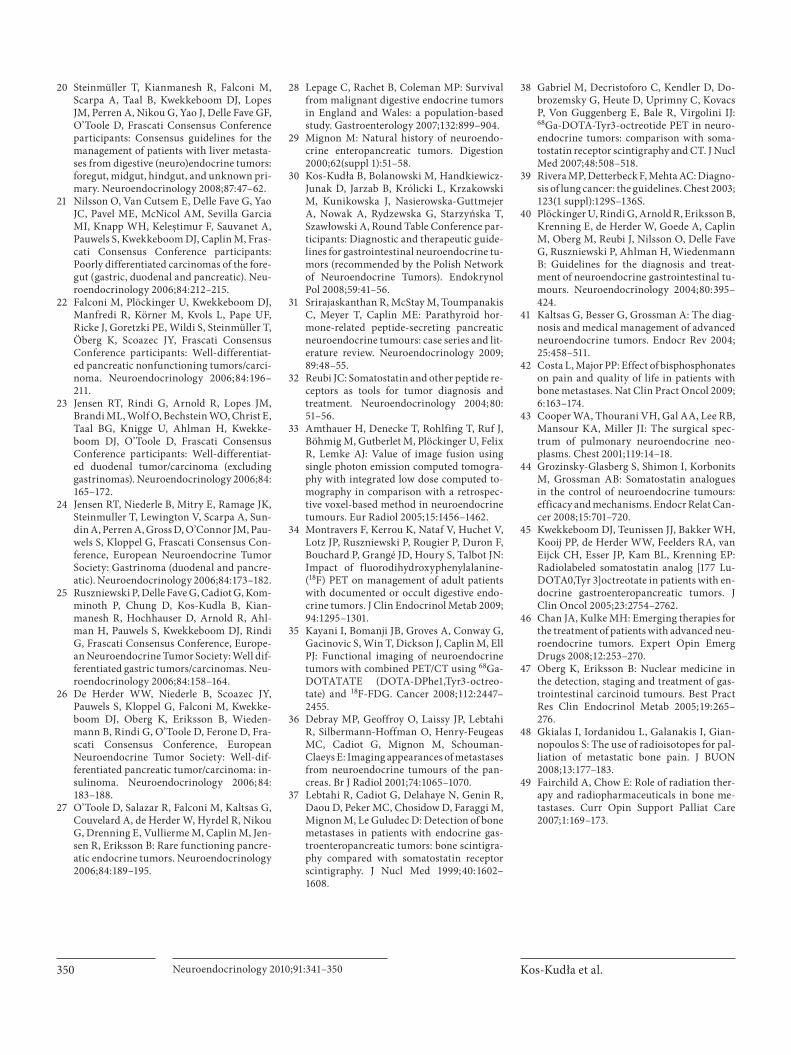
References
1 Hauso O, Gustafsson BI, Kidd M, Waldum HL, Drozdov I, Chan AK, Modlin IM: Neu-roendocrine tumor epidemiology: contrast-ing Norway and North America. Cancer 2008; 113: 2655–2664.
2 Hadi S, Zhang L, Hird A, de Sa E, Chow E: Validation of symptom clusters in patients with metastatic bone pain. Curr Oncol 2008; 15: 211–218.
3 Zuetenhorst JM, Hoefnageli CA, Boot H, Valdés Olmos RA, Taal BG: Evaluation of (111)In-pentetreotide, (131)I-MIBG and bone scintigraphy in the detection and clini-cal management of bone metastases in carci-noid disease. Nucl Med Commun 2002; 23: 735–741.
4 Meijer WG, van der Veer E, Jager PL, van der Jagt EJ, Piers BA, Kema IP, de Vries EG, Wil-lemse PH: Bone metastases in carcinoid tu-mors: clinical features, imaging characteris-tics, and markers of bone metabolism. J Nucl Med 2003; 44: 184–191.
5 Hlatky R, Suki D, Sawaya R: Carcinoid me-tastasis to the brain. Cancer 2004; 101: 2605–2613.
6 Lombard-Bohas C, Mitry E, O’Toole D, Lou-vet C, Pillon D, Cadiot G, Borson-Chazot F, Aparicio T, Ducreux M, Lecomte T, Etienne PL, Cacheux W, Legoux JL, Seitz JF, Rusz-niewski P, Chayvialle JA, Rougier P, FFCD-ANGH-GERCOR: Thirteen-month regis-tration of patients with gastroentero-pancreatic endocrine tumours in France. Neuroendocrinology 2009; 89: 217–222.
7 Zuetenhorst JM, Taal BG: Metastatic carci-noid tumors: a clinical review. Oncologist 2005; 10: 123–131.
8 Janson ET, Holmberg L, Stridsberg M, Eriks-son B, Theodorsson E, Wilander E, Oberg K: Carcinoid tumors: analysis of prognostic factors and survival in 301 patients from a referral center. Ann Oncol 1997; 8: 685–690.
9 Ross EM, Roberts WC: The carcinoid syn-drome: comparison of 21 necropsy subjects with carcinoid heart disease to 15 necropsy subjects without carcinoid heart disease. Am J Med 1985; 79: 339–354.
10 Taal BG, Visser O: Epidemiology of neuro-endocrine tumours. Neuroendocrinology 2004; 80: 3–7.
11 Chong S, Lee KS, Chung MJ, Han J, Kwon OJ, Kim TS: Neuroendocrine tumors of the lung: clinical, pathologic, and imaging findings. Radiographics 2006; 26: 41–58.
12 Khan JH, McElhinney DB, Rahman SB, George TI, Clark OH, Merrick SH: Pulmo-nary metastases of endocrine origin: the role of surgery. Chest 1998; 114: 526–534.
13 Modlin IM, Lye KD, Kidd M: A 5-decade analysis of 13,715 carcinoid tumors. Cancer 2003; 97: 934–959.
14 Gustafsson BI, Kidd M, Chan A, Malfert-heiner MV, Modlin IM: Bronchopulmonary neuroendocrine tumors. Cancer 2008; 113: 5–21.
15 Scarsbrook AF, Ganeshan A, Statham J, Thakker RV, Weaver A, Talbot D, Boardman P, Bradley KM, Gleeson FV, Phillips RR: An-atomic and functional imaging of metastatic carcinoid tumors. Radiographics 2007; 27: 455–477.
16 Eriksson B, Klöppel G, Krenning E, Ahlman H, Plöckinger U, Wiedenmann B, Arnold R, Auernhammer C, Körner M, Rindi G, Wildi S, Frascati Consensus Conference partic-ipants: Consensus guidelines for the man-agement of patients with digestive neuro-endocrine tumors – well-differentiated jejunal-ileal tumor/carcinoma. Neuroendo-crinology 2008; 87: 8–19.
17 Ramage JK, Goretzki PE, Manfredi R, Kom-minoth P, Ferone D, Hyrdel R, Kaltsas G, Ke-lestimur F, Kvols L, Scoazec JY, Garcia MI, Caplin ME, Frascati Consensus Conference participants: Consensus guidelines for the management of patients with digestive neu-roendocrine tumours: well-differentiated colon and rectum tumour/carcinoma. Neu-roendocrinology 2008; 87: 31–39.
18 Plöckinger U, Couvelard A, Falconi M, Sun-din A, Salazar R, Christ E, de Herder WW, Gross D, Knapp WH, Knigge UP, Kulke MH, Pape UF, Frascati Consensus Conference participants: Consensus guidelines for the management of patients with digestive neu-roendocrine tumours: well-differentiated tumour/carcinoma of the appendix and gob-let cell carcinoma. Neuroendocrinology 2008; 87: 20–30.
19 Ahlman H, Nilsson O, McNicol AM, Ruszniewski P, Niederle B, Ricke J, Jensen R, Kos-Kudła B, Öberg K, O’Connor JM, Pavel ME, Vullierme MP, Frascati Consensus Conference participants: Poorly differenti-ated endocrine carcinomas of midgut and hindgut origin. Neuroendocrinology 2008; 87: 40–46.
Kos-Kudła et al. Neuroendocrinology 2010;91:341–350350
20 Steinmüller T, Kianmanesh R, Falconi M, Scarpa A, Taal B, Kwekkeboom DJ, Lopes JM, Perren A, Nikou G, Yao J, Delle Fave GF, O’Toole D, Frascati Consensus Conference participants: Consensus guidelines for the management of patients with liver metasta-ses from digestive (neuro)endocrine tumors: foregut, midgut, hindgut, and unknown pri-mary. Neuroendocrinology 2008; 87: 47–62.
21 Nilsson O, Van Cutsem E, Delle Fave G, Yao JC, Pavel ME, McNicol AM, Sevilla Garcia MI, Knapp WH, Keleştimur F, Sauvanet A, Pauwels S, Kwekkeboom DJ, Caplin M, Fras-cati Consensus Conference participants: Poorly differentiated carcinomas of the fore-gut (gastric, duodenal and pancreatic). Neu-roendocrinology 2006; 84: 212–215.
22 Falconi M, Plöckinger U, Kwekkeboom DJ, Manfredi R, Körner M, Kvols L, Pape UF, Ricke J, Goretzki PE, Wildi S, Steinmüller T, Öberg K, Scoazec JY, Frascati Consensus Conference participants: Well-differentiat-ed pancreatic nonfunctioning tumors/carci-noma. Neuroendocrinology 2006; 84: 196–211.
23 Jensen RT, Rindi G, Arnold R, Lopes JM, Brandi ML, Wolf O, Bechstein WO, Christ E, Taal BG, Knigge U, Ahlman H, Kwekke-boom DJ, O’Toole D, Frascati Consensus Conference participants: Well-differentiat-ed duodenal tumor/carcinoma (excluding gastrinomas). Neuroendocrinology 2006; 84: 165–172.
24 Jensen RT, Niederle B, Mitry E, Ramage JK, Steinmuller T, Lewington V, Scarpa A, Sun-din A, Perren A, Gross D, O’Connor JM, Pau-wels S, Kloppel G, Frascati Consensus Con-ference, European Neuroendocrine Tumor Society: Gastrinoma (duodenal and pancre-atic). Neuroendocrinology 2006; 84: 173–182.
25 Ruszniewski P, Delle Fave G, Cadiot G, Kom-minoth P, Chung D, Kos-Kudla B, Kian-manesh R, Hochhauser D, Arnold R, Ahl-man H, Pauwels S, Kwekkeboom DJ, Rindi G, Frascati Consensus Conference, Europe-an Neuroendocrine Tumor Society: Well dif-ferentiated gastric tumors/carcinomas. Neu-roendocrinology 2006; 84: 158–164.
26 De Herder WW, Niederle B, Scoazec JY,Pauwels S, Kloppel G, Falconi M, Kwekke-boom DJ, Oberg K, Eriksson B, Wieden-mann B, Rindi G, O’Toole D, Ferone D, Fra-scati Consensus Conference, European Neuroendocrine Tumor Society: Well-dif-ferentiated pancreatic tumor/carcinoma: in-sulinoma. Neuroendocrinology 2006; 84: 183–188.
27 O’Toole D, Salazar R, Falconi M, Kaltsas G, Couvelard A, de Herder W, Hyrdel R, Nikou G, Drenning E, Vullierme M, Caplin M, Jen-sen R, Eriksson B: Rare functioning pancre-atic endocrine tumors. Neuroendocrinology 2006; 84: 189–195.
28 Lepage C, Rachet B, Coleman MP: Survival from malignant digestive endocrine tumors in England and Wales: a population-based study. Gastroenterology 2007; 132: 899–904.
29 Mignon M: Natural history of neuroendo-crine enteropancreatic tumors. Digestion 2000; 62(suppl 1):51–58.
30 Kos-Kudła B, Bolanowski M, Handkiewicz-Junak D, Jarzab B, Królicki L, Krzakowski M, Kunikowska J, Nasierowska-Guttmejer A, Nowak A, Rydzewska G, Starzyńska T, Szawłowski A, Round Table Conference par-ticipants: Diagnostic and therapeutic guide-lines for gastrointestinal neuroendocrine tu-mors (recommended by the Polish Network of Neuroendocrine Tumors). Endokrynol Pol 2008; 59: 41–56.
31 Srirajaskanthan R, McStay M, Toumpanakis C, Meyer T, Caplin ME: Parathyroid hor-mone-related peptide-secreting pancreatic neuroendocrine tumours: case series and lit-erature review. Neuroendocrinology 2009; 89: 48–55.
32 Reubi JC: Somatostatin and other peptide re-ceptors as tools for tumor diagnosis and treatment. Neuroendocrinology 2004; 80: 51–56.
33 Amthauer H, Denecke T, Rohlfing T, Ruf J, Böhmig M, Gutberlet M, Plöckinger U, Felix R, Lemke AJ: Value of image fusion using single photon emission computed tomogra-phy with integrated low dose computed to-mography in comparison with a retrospec-tive voxel-based method in neuroendocrine tumours. Eur Radiol 2005; 15: 1456–1462.
34 Montravers F, Kerrou K, Nataf V, Huchet V, Lotz JP, Ruszniewski P, Rougier P, Duron F, Bouchard P, Grangé JD, Houry S, Talbot JN: Impact of f luorodihydroxyphenylalanine-( 18 F) PET on management of adult patients with documented or occult digestive endo-crine tumors. J Clin Endocrinol Metab 2009; 94: 1295–1301.
35 Kayani I, Bomanji JB, Groves A, Conway G, Gacinovic S, Win T, Dickson J, Caplin M, Ell PJ: Functional imaging of neuroendocrine tumors with combined PET/CT using 68 Ga-DOTATATE (DOTA-DPhe1,Tyr3-octreo-tate) and 18 F-FDG. Cancer 2008; 112: 2447–2455.
36 Debray MP, Geoffroy O, Laissy JP, LebtahiR, Silbermann-Hoffman O, Henry-Feugeas MC, Cadiot G, Mignon M, Schouman-Claeys E: Imaging appearances of metastases from neuroendocrine tumours of the pan-creas. Br J Radiol 2001; 74: 1065–1070.
37 Lebtahi R, Cadiot G, Delahaye N, Genin R, Daou D, Peker MC, Chosidow D, Faraggi M, Mignon M, Le Guludec D: Detection of bone metastases in patients with endocrine gas-troenteropancreatic tumors: bone scintigra-phy compared with somatostatin receptor scintigraphy. J Nucl Med 1999; 40: 1602–1608.
38 Gabriel M, Decristoforo C, Kendler D, Do-brozemsky G, Heute D, Uprimny C, Kovacs P, Von Guggenberg E, Bale R, Virgolini IJ: 68 Ga-DOTA-Tyr3-octreotide PET in neuro-endocrine tumors: comparison with soma-tostatin receptor scintigraphy and CT. J Nucl Med 2007; 48: 508–518.
39 Rivera MP, Detterbeck F, Mehta AC: Diagno-sis of lung cancer: the guidelines. Chest 2003; 123(1 suppl):129S–136S.
40 Plöckinger U, Rindi G, Arnold R, Eriksson B, Krenning E, de Herder W, Goede A, Caplin M, Oberg M, Reubi J, Nilsson O, Delle Fave G, Ruszniewski P, Ahlman H, Wiedenmann B: Guidelines for the diagnosis and treat-ment of neuroendocrine gastrointestinal tu-mours. Neuroendocrinology 2004; 80: 395–424.
41 Kaltsas G, Besser G, Grossman A: The diag-nosis and medical management of advanced neuroendocrine tumors. Endocr Rev 2004; 25: 458–511.
42 Costa L, Major PP: Effect of bisphosphonates on pain and quality of life in patients with bone metastases. Nat Clin Pract Oncol 2009; 6: 163–174.
43 Cooper WA, Thourani VH, Gal AA, Lee RB, Mansour KA, Miller JI: The surgical spec-trum of pulmonary neuroendocrine neo-plasms. Chest 2001; 119: 14–18.
44 Grozinsky-Glasberg S, Shimon I, Korbonits M, Grossman AB: Somatostatin analogues in the control of neuroendocrine tumours: efficacy and mechanisms. Endocr Relat Can-cer 2008; 15: 701–720.
45 Kwekkeboom DJ, Teunissen JJ, Bakker WH, Kooij PP, de Herder WW, Feelders RA, van Eijck CH, Esser JP, Kam BL, Krenning EP: Radiolabeled somatostatin analog [177 Lu-DOTA0,Tyr 3]octreotate in patients with en-docrine gastroenteropancreatic tumors. J Clin Oncol 2005; 23: 2754–2762.
46 Chan JA, Kulke MH: Emerging therapies for the treatment of patients with advanced neu-roendocrine tumors. Expert Opin Emerg Drugs 2008; 12: 253–270.
47 Oberg K, Eriksson B: Nuclear medicine in the detection, staging and treatment of gas-trointestinal carcinoid tumours. Best Pract Res Clin Endocrinol Metab 2005; 19: 265–276.
48 Gkialas I, Iordanidou L, Galanakis I, Gian-nopoulos S: The use of radioisotopes for pal-liation of metastatic bone pain. J BUON 2008; 13: 177–183.
49 Fairchild A, Chow E: Role of radiation ther-apy and radiopharmaceuticals in bone me-tastases. Curr Opin Support Palliat Care 2007; 1: 169–173.