Embed Size (px)
Citation preview

FEMS Microbiology Reviews 12 (1993) 125-148 <, 1993 Federation of European Microbiological Societies 0168-6445/93/$15.00 Published by Elsevier
125
FEMSRE 00321
Energy transduction in lactic acid bacteria
B e r t P o o l m a n
Department Of Microbiolo~', Unil,ersity c~[ Groningen, Haren, the Netherlands
Abstract: In the discovery of some general principles of energy transduction, lactic acid bacteria have played an important role. In this review, the energy transducing processes of lactic acid bacteria are discussed with the emphasis on the major developments of the past 5 years. This work not only includes the biochemistry of the enzymes and the bioenergetics of the processes, but also the genetics of the genes encoding the energy transducing proteins. The progress in the area of carbohydrate transport and metabolism is presented first. Sugar translocation involving ATP-driven transport, ion-linked cotransport, heterologous exchange and group translocation are discussed. The coupling of precursor uptake to product product excretion and the linkage of antiport mechanisms to the deiminase pathways of lactic acid bacteria is dealt with in the second section. The third topic relates to metabolic energy conservation by ehemiosmotic processes. There is increasing evidence that precursor/product exchange in combination with precursor decarboxy[ation allows bacteria to generate additional metabolic energy. In the final section transport ot nutrients and ions as well as mechanisms to excrete undesirable (toxic) compounds from the cells are discussed.
Key word.s: Energy transduction; Transport; Bioenergetics; Lactic acid bacteria
Introduction
Lactic acid bacter ia metabol ize ( ferment) sub- strates without net p roduct ion of reducing equiv- alents. As an example may serve the degrada t ion of glucose to lactic acid, resul t ing in the produc- tion of 2 tool of A T P per mol of glucose metabo- lized. Some lactic acid bacteria, however, have l imited capabil i t ies to utilize exogenous electron acceptors for the degrada t ion (oxidation) of sub- strates. Carbohydra te metabol ism in Lactococcus
lactis and Streptococcus thermophilus can be af- fected by the presence of oxygen. U n d e r aerobic condi t ions and slow fe rmen ta t ion rates, L. lactis
Correspondence to: B. Poolman, Department of Microbiology, University of Groningen, Kerklaan 30, 9751 NN Haren, the Netherlands.
is able to convert lactose almost quant i ta t ively to acetic acid (plus carbon dioxide), thereby produc- ing an excess of N A D H [1]. These facultative
anaerobic lactic acid bacter ia conta in flavin-type N A D H oxidases and N A D H peroxidase which obviates the need for pyruvate reduct ion to lac-
tate or reduct ion of acetyl-CoA to ethanol . By forming acetic acid instead of lactic acid, and if an oxidase is used to reoxidizc the N A D H pro- duced, addi t ional A TP can be formed by sub- strate level phosphorylat ion. Theoret ical ly, carbo- hydrate metabol ism by lactic acid bacteria may also yield acetic acid as end product , and thus extra ATP, unde r anaerobic condi t ions if fu- mara te reductase is used. In this scheme elec- trons are t ransferred from a N A D H dehydroge- nase via (mena )qu inones to fumara te reductase. All these componen t s are present in the cytoplas- mic m e m b r a n e of L. lactis [2], but the use of this

126
electron transfer chain in the oxidation of N A D H under anaerobic conditions has never been demonstrated.
Since large quantities of fermentation products arc formed in the cytoplasm of lactic acid bacte- ria, the outwardly directed concentration gradi- ents of these molecules may become high. When these end-product gradients exceed the electro- chemical proton (or sodium ion) gradient, and product efflux occurs carrier-mediated in symport with protons or sodium ions, additional metabolic energy can be generated by this process [3,4]. Metabolic energy will thus be conserved by trans- forming a product gradient into a proton (or sodium ion) gradient which, in turn, can be used to synthesize ATP via the FoF~-ATPase or to drive other endergonic processes.
In addition to the ATP produced in the gly- colytic pathway, some lactic acid bacteria gener- ate additional ATP by arginine or agmatine metabolism via the corresponding deiminase pathways [5-8].
Recently, another mechanism for the genera- tion of metabolic energy in lactic acid bacteria has been discovered. Rather than forming ATP by substrate level phosphorylation, a proton mo- tive force (Ap) is formed by the combined action of a secondary transport mechanism responsible for the uptake of substrate and excretion of prod- uct and a decarboxylation reaction [9-11].
Energy transduction not only refers to pro- cesses that lead to the production of metabolic energy in the form ATP or ion concentration gradients, but also covers the various transport processes that form solute concentration gradi- ents at the expense of metabolic energy. Solute transport systems in bacteria can be subdivided into three distinct mechanisms that differ in the form of energy that is used and the way this energy is transduced in the transloeation process [10], i.e. primary (light, chemical energy), sec- ondary (chemiosmotic energy) and group translo- cation transport (chemical modification concomi- tant with transport). Primary transport in lactic acid bacteria is examplified by the ion- and so- lute-ATPases which utilize the change in free energy upon hydrolysis of ATP (to ADP plus inorganic phosphate) to transport solutes against
+ H
\
Sugars Sugars Amino acids Di/tripeptides K + Amino acids
Oligopeptides Citrate ~ Phosphate
H + / ~ ~ Glvcine. hmain
+ ca2+ Na,
PEP
ATP Vitamins
ATP
BCECF, Drugs? Bacteriocins
+ H ATP
ADP + Pi ~ .~ ,
Sugar
Fig. 1. Solute transport mechanisms in lactic acid bacteria. Primary (ATP-driven) transport systems are indicated as filled icons; secondary symport and antiport systems are indicated as open icons; the PEP-dependent sugar:phosphotransferasc system is indicated by shaded icons. The examples given arc primarily based on experimental evidence from work on L.
lactis (see Table 2).
their concentration gradients (Fig. 1, filled icons). Secondary transport includes the ion-linked so- lute symport and antiport systems as well as the p r e c u r s o r / p r o d u c t exchange systems. These transport systems couple the transport of a solute to the (downhill) movement of a second molecule (often H + or Na ~, or another compound that is structurally related to the substrate) (Fig. 1, open icons). Solutes (e.g. substrates, nutrients) can be concentrated in the cytoplasm by means of so- lute-cation symport, while solute /cat ion antiport systems allow the bacteria to keep the cytoplas- mic concentration of certaio solutes (e.g. un- wanted products) below the medium concentra- tion (Fig. 1, open icons). Group translocation in (lactic acid) bacteria has only been observed for carbohydrates and involves phosphorylation of the sugar concomitant with transport by the phospho- enolpyruvate : sugar phosphotransferase system (PTS) (Fig. 1, shaded icons). The subdivision of transport systems on basis of their mechanism of energy coupling will be followed throughout this manuscript. The first part of thc review focusses on those transport processes that are linked to the pathways of metabolic energy generation, i.e. glycolytic, deiminase and solute decarboxylation

pathways. The P'lb that forms one mechanism by which sugars can be transported into the cell will only be dealt with briefly (details on the PTS of Gram-positive bacteria are given in the review of Hengstenberg et al., this issue). In the second part of the review the nutrient and ion trans- porters, and some new excretion mechanisms of lactic acid bacteria will be discussed.
Sugar transport and initial metabolism
The initial event in the metabolism of mono- and disaccharides involves the translocation of these molecules across the cytoplasmic mem- brane. The systems by which the carbohydrate molecules are transported can be subdivided into (i) sugar transport ATPases, (ii) ion-linked sugar transport and sugar exchange mechanisms, and (iii) PEP:sugar PTS. These different mechanisms of transport have been observed in lactic acid bacteria for a wide variety of sugars but those that mediate lactose (galactoside) transport have been described in greatest detail.
Depending on the transport mechanism the following step in lactose metabolism involves ei- ther hydrolysis of the free sugar by /3-galactosi- dase (LacZ), yielding glucose and galactose, or hydrolysis of lactose 6-phosphate by phospho-/3- galactosidase (LacG), yielding glucose and galac- tose 6-phosphate. The two major pathways of glucose degradation in lactic acid bacteria are the glycolytic (or Embden-Meyerhof-Parnas) and the phosphoketolase pathway [12]. The glycolytic pathway is operative in Lactococcus spp., ho- mofermentative lactobacilli (e.g. Lactobacillus delbrueckii subsp, bulgaricus and heh,eticus), pe- diococci and Streptococcus therrnophilus, whereas the phosphoketolase pathway(s) resulting in heterofermentation is present in obligate hetero- fermentative species like Leuconostoc spp., Lac- tobacillus buchneri and Lactobacillus bret,is [12,13]. In facultative heterofermentative species, like Lactobacillus casei, Lactobacillus plantarurn and Lactobacillus pentosus, both the glycolytic and phosphoketolase pathways can be present [13].
Galactose is metabolized further via the Leloir pathway (if not excreted into the medium, see
127 Lactose-H+symport & Leloir Pathway I
kactOSeou t H + out
+ LactoSein ain l'acZ
Galactose + Glucose ATP ~ /
ADP ~ galK ~ A T P
GaI-1-P "~ ADP
UDPglc J galE l I gait glk
UDPgal GIc-1 -P
pgm~"~~ Gic_6.p b
Glycolysis Fig. 2. Lactose-H + symport and Leloir pathway, lacZ, ~- galactosidase; galE, galactokinase: galT, UDPglucose :galac- tose 1-phosphate uridylyl transferase; galE. UDPglucose 4-ep-
imerase, pgm, phosphoglucomutase: glk, glucokinase.
below), whereas galactose 6-phosphate enters the tagatose 6-phosphate pathway. Similarly, other carbohydrates may enter the cell either as free sugar or phosphorylated derivative, and these molecules can be degraded further following phosphorylation by a kinase or directly, i.e. when transport is catalysed by the PTS or sugar phos- phate / phosphate antiport [ 14-17].
The enzymes of the Leloir pathway of galac- tose metabolism include galactokinase (GalK) which phosphorylates galactose at the C-I posi- tion, UDPglucose :galactose-l-phosphate uridy- lyltransferase (GAIT) which transfers the UDP group from UDPglucose to galactose-l-phos- phate, and UDPglucose 4-epimerase (GalE) which catalyses the epimerization of UDPgalac- tose to UDPglucose (Fig. 2). UDPglucose syn- thetase (GalU, not shown) and phosphoglucomu- tase (Pgm) are also needed for galactose metabolism, but the corresponding genes do not form part of the gal operon. This allows expres-
128
sion of GalU and Pgm when the other Gal cn- zymcs are repressed. In E. colt GalU and Pgm are synthesized constitutively [18]. The reversible reactions of the Leloir pathway allow cells to make UDPgalactose (in the absence of exogenous galactose) which can be used for the synthesis of complex (extraccllular) polysaccharides.
The majority of 'wild-type" strains of S. ther- mol)hilus and L. bulgaricus cannot metabolize galactose, and in case of S. thermol~hihts the GaI phenotype has been attributed to a defect in the induction mechanism of the galactokinasc [19,20]. Under the selective pressure of the chcmostat G a l ' variants of S. thermol)hihts could be isolated, indicating that galK is present m this organism [19]. The level of galactokinasc was many times greater in the G a l ' variants than in the parent strain. The failure of wild-type cells to express GalK results in the excretion into the medium of 1 molecule of galactosc pcr molecule of lactose metabolized.
('arbohydrate tran.v~ort A TPases
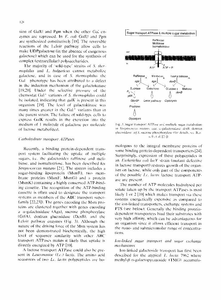
Recently, a binding protein-dependent trans- port system facilitating the uptake of multiple sugars, i.e. the galactosidcs raffinosc and meli- biose, and isomaltotriosc, has been described fi)r Strel)tOCOCClts tlZlltallx [21]. The system includes a sugar-binding lipoprotein (MsmE), two mem- brane proteins (MsmF, MsmG) and a protein (MsmK) containing a highly conserved ATP-bind- ing cassette. The recognition of the ATP-binding cassette is often used to designate the transport systems as membcrs of the ABC transport super- family [22,23]). The genes encoding the Msm pro- teins are clustered together with genes encoding a e~-galactosidase (Aga), sucrose phosphowlasc (GtfA), dextran glucosidase (DexB). and the kcloir pathway enzymes (Fig. 3). Although the nature of the driving fl)rcc of the Msm system has not been demonstrated biochemically, the high level of scquence similarity with other ABC transport ATPases makes it likely that uptakc is directly energized by ATP [10].
A lactose transport ATPase could also be pre- sent in Leuconostoc (Lc.) lactis. The amino acid sequences of two Lc. lactis polypeptides are he-
Sugar transport ATPase & multiple sugar metabolism
Raffinose Melibiose Isomaltotriose
out
Raffinose. Meiibiose.
a ag8
~/ "3 Sucrose
Pi~-.xJ gtfA
GIc-6P +
Fructose
Glycolysis
Fig. 3. Sugar transport t \TPasc and mul l ip lc sugar metabolism ill <~'t#'L'fI[f)L'OCCt(5 #tlllltltlS. ll<l~a, ~-galac/os idasc; dexlY, dc'xliao glucosidasc: t~(fi4, sucrose phosphorylas¢ ( for details, see Rus-
sell ct ;.il, [21i).
I somaltotrioSein
d e x B / ~
Galactose Glucose ~ Maltose
Leloir pathway Glycolysis
mologous to the integral membrane proteins of some binding protein-dependent transporters [24]. Surprisingly, expression of these polypeptides in an Escherichia colt hwY strain (mutant defective in lactose transport) restores growth of the organ- ism on lactose, while only part of the components of the possible Lc. lactis lactose transport ATP- ase are present.
The number of ATP molecules hydrolyscd per solute taken up by the transport ATPases is most likely 1 or 2 [10] which makes transport via these systems energetically expensive as compared to the ion-linked transporters, exchange systems and PTS (see below). Generally the binding protein- dependent transporters bind their substrates with very high affinity, which can be advantageous fl)r an organism since it allows cfficieni transport in the nano- and submicromolar range of concentra- tions.
Ion-linked sugar lran,wort and sugar ~;vchange mechanisms
Ion-linked galactoside transport has first been described for the atypical L. luctis 7962 where methyl-/d-D-galactopyranoside (TMG) aceumula-

tion has been shown to be coupled to the uptake of a proton [25]. The system is highly specific for galactose, T M G and other galactose analogs, but exhibits poor affinity for lactose. A similar system has been described for L. lactis ML3 [26]. Lac- tose transporters driven by the electrochemical proton gradient have been described for S. ther- mophilu,~ and L. hulgaricus [27]. These trans- porters are selective not only for lactose (/3- galactoside) but also for melibiose (~-galactoside). galactosc (monosaccharide) and to a lesser extent raffinosc (trisaccharide) [28]. Deletion of the chromosomal gen t encoding the lactose transport protein (LacS) of S. thermophilus abolishes all modes of facilitated galactoside diffusion, demon- strating that a single system catalyses the uptake, cffiux and exchange of galactosides (J. Knol, B. Mollet and B. Poolman, unpublished results).
Studies on the kinctic mechanism of the lac- tose transport protein ([,acS) of S. thermophilus have indicated that a full translocation cycle in- w)lves stoichiometric t ransmembrane movement of a galactoside and a proton. On the other hand, with saturating concentrations of galactosides (e.g. lactose and galactose) on either side of the mem- brane, release of substrate (and proton) is imme- diately followed by (re)binding of substrate (and proton) rather than by reorientation of the 'empty ' binding site(s) [29]. Under these conditions the carrier protein performs an exchange reaction with no net proton translocation. Since S. ther- mop~if~us and L. hulgaricus strains excrete galac- rose into the medium in amounts stoichiometric with the uptake of lactose, the suggestion has been made that under physiological conditions lactose is taken up in exchange for a galactose molecu,le [27,30]. Evidence in favour of this no- tion has been obtained from kinetic studies of galactoside transport [29] and comparison of in viw) transport and metabolism rates (Poolman and Foucaud, unpublished). The suggestion that the LacS galactoside transporter of S. ther- mophilus is a strict lac tosc /galac tose antiporter [31] is not correct. Although the lac tose /ga lac tose exchange reaction may be fawmred under many conditions, the exchange mode simply reflects partial steps, forward and backward reactions with no net proton translocation, of the complete
12 ~)
translocation cycle which involves sugar and pro- ton uptake on one side and release on the other side of the membrane, and reorientation of loaded and unloaded substrate binding sites [29].
The exchange reaction has some distinct ad- vantages over sugar-H + symport lk~r the uptake of lactose and excretion of galactosc. Firstly, al- though uptake of lactose in symport with H + is stimulated by the t ransmembrane electrochemical proton gradient (..lp), galactose efflux is inhibited and net exit will only occur if the galactose con- centration gradient (A#~;,,~/F) exceeds the dp. In comparison, the linkage of lactose uptake to galactosc excretion via an exchange reaction as- sures tight coupling to the metabolism, which minimizes (excessive) galactose accumulation and possible product inhibition [30]. Secondly, ex- change is less drastically affected by the absolute H + concentrations than sugar-H + symport [29]. The rate of efflux is significantly limited by the release of the proton at the outer surface of the membrane, and, as a consequence, extremely slow below pH 7. Similarly, influx is inhibited by a low intraceIlu[ar pH. Exchange, on the other hand, proceeds maximally between pH 6-7 and with rates at least one order of magnitude faster than the corresponding sugar-H + symport reaction.
The genes encoding the lactose (galactoside) transport proteins (LacS) of S. thermophilus and L. hulgaricus have been cloned, characterized and functionally expressed in E. coli [27,28,32,33]. Both proteins are composed of an amino-termi- nal polytopic hydrophobic membrane domain (carrier domain) and a carboxy-terminal hydro- philic domain. The carrier domain is homologous to the melibiose carrier protein (MelB) of E. coli, the xylose carrier protein (XylP) of Lactohacillus pentosus, and some other carrier proteins that most likely couple the uphill movement of sugars to the downhill movement of a cation (proton, sodium or lithium) (Fig. 4). The hydrophilic (cyto- plasmic) domain of LacS is homologous to vari- {)us IIA protein(s) or protein domains of PTS, previously indicated as enzyme Ill or enzyme III-likc domain (Fig. 4). The chimeric nature of LacS makes this t ransporter unique and suggests that it shares features of both ion-linked trans- porters and PTS.
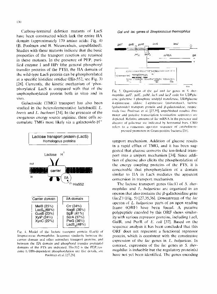
130
Carboxy-terminal deletion mutants of LacS have been constructed which lack the entire IIA domain (approximately 170 amino acids; Fig. 4) (B. Poolman and B. Nieuwenhuis, unpublished). Studies with these mutants indicate that the basic properties of the transport reaction are retained in these mutants. In the presence of PEP, puri- fied enzyme I and HPr (the general phosphoryl transfer proteins of the PTS), the IIA domain of the wild-type LacS protein can be phosphorylated at a specific histidine residue (His-552, see Fig. 3) [28]. Currently, the kinetic mechanism of 'phos- phorylated' LacS is compared with that of the unphosphorylated protein both in vitro and in vivo.
Galactoside (TMG) transport has also been studied in the heterofermentative lactobacilli L. breuis and L. buchneri [34]. In the presence of the exogenous energy source arginine, these cells ac- cumulate TMG most likely via a galactoside-H +
/ Lactose transport protein (LacS)|
Homologous proteins J
Lactose H +
ou,
' in ~ H i s552
Carrier domain
MelB (23%) LacSm(68%) GusB (23%) XylP (24%) XynC (22%)
IIA domain
Crr (34%) NagE (36%) BglF (41%) ScrA (37%) PtsG (36%) LaCSib(48%)
Fig. 4. Model of the lactose transport protein (LacS) of Streptococcus thermophilus. Sequence similarity between the carrier domain and other secondary' transport proteins, and between the IIA domain and phosphoryl transfer protein(s) domains of the PTS are indicated. His-552 is the P E P / e n - zyme l / H P r - d e p e n d e n t phosphorylation site (for details, see
Poolman et al. [27,28].
Gal and lac genes of Streptococcus thermophilus
galE galM lacS lacZ
°niPs1 lacPs mRNA gal
l 7 ~c AGT,'~AAAAACACTG tAAA FI A lq-GA CTGCATAAACC A.a ~ l ITCA
~ 35 Consensus CRO TGWNANCGNTNWCA
NOOOCOOO O~O TAT AATGTAAACGTATTCAAA [ AATA G GAT G MTCCGA,,'~TGG
~ 7~- s~Tart I I
1 kb
Fig. 5. Organization of the gal and lac genes in S. lh(,r- mophih~s, gaiT, ,galE, galM, lacS and lacZ code fl~r UDPglu- cose:galactose I-phosphate uridylyl transferase, UDPglucose 4-epimerase, aldose 1-epimerase (mutarotase) , lactose (galactoside) transport protein and /3-galactosidasc, respec- tively (scc Pooh'nan et al. [27,35]; unpublished results). Pro- moter and putative transcription termination sequences arc depicted. Relative amounts of &c m R N A in the presence and absence of galactose arc indicated by horizontal bars. CRO refers to a consensus operator sequence of catabolite-re-
pressed promoters in Gram-positive bacteria [38].
symport mechanism. Addition of glucose results in a rapid efflux of TMG, and it has been sug- gested that glucose converts the ion-linked trans- port into a uniport mechanism [34]. Since addi- tion of glucose also elicits the phosphorylation of the energy coupling proteins of the PTS, it is conceivable that phosphorylation of a domain similar to I1A in LacS mediates the apparent conversion in transport mechanism.
The lactose transport genes (lacS) of S. ther- mophilus and L. bulgaricus are organized in an operon that also contains the/3-galactosidase gene (lacZ) (Fig. 5) [27,35,36]. Downstream of the lac operon of L. bulgaricus parts of an open reading frame (ORF) have been found. A putative polypeptide encoded by this ORF shows similar- ity with various repressor proteins, including Lacl, GalR, and P u r r of E. coil [37]. Based on the sequence analysis it has been concluded that this ORF does not represent a functional repressor protein, which is consistent with the constitutive expression of the lac genes in L. bulgaricus. In contrast, expression of the lac genes in S. ther- mophilus is inducible but the regulatory protein(s) have not yet been identified. The genes encoding

aldose 1-epimerasc (GalM), UDPglucose 4-epi- merase (GalE) and UDPglucose-hexose-l-phos- phate uridylyltransferase (GAIT) are found up- stream of the lac operon of S. therrnophilus (Fig. 5). GalE and GalT form part of the Leloir path- way of galactose metabolism (Fig. 2), but in S. thermophilus these enzymes most likely function in the synthesis of precursors (UDPsugars) of complex extracellular polysaccharides. Induction of the lac (and gal) genes is effected by galactose and galactose analogs, but not by lactose, whereas glucose represses ([35]; Foucaud and Poolman, unpublished). Sequences strongly resembling those found upstream of eatabolite-repressed genes of Gram-positive bacteria [38] have been detected in the lac promoter region of S. ther- mophilus (consensus CRO, Fig. 5), suggesting that expression of the lac genes is also under glucose (catabolite) repression control.
With the exception of the secondary trans- porters for galactosides described above, there is no further evidence for ion-linked sugar transport or sugar exchange mechanisms in lactic acid bac- teria [16]. Recently, the genes responsible for the ability of L. pentosus to utilize xylose have been cloned and characterized. In addition to D-xylose isomerase (XylA), l)-xylulose kinase (XylB) and a presumed regulatory protein (XylR) [39], an open reading frame (XylP) has been found which dis- plays sequence similarity to the carrier domain of the lactose transport proteins of S. thermophilus and L. bulgaricus, and a number of other sec- ondary transport proteins (Fig. 4). This system most likely transports xylose in symport with H + (J. Martena, M. Posno and B. Poolman, unpub- lished results).
Sugar phosphate/phosphate antiport Sugar 6-phosphate /phosphate antiport has
been demonstrated first in L. lactis [40]. Subse- quent studies have shown that this type of anion- exchange is found in various Gram-positive and Gram-negative bacteria. Since anion-exchange is the topic of a number of recent reviews [17,41], only the basic properties of the antiporter mecha- nism will be discussed. The features of the sugar 6-phosphate /phosphate antiporter of L. lactis are: (i) the system catalyses homologous and het-
131
erologous exchange of phosphate and sugar 6- phosphates; (ii) substrate specificity studies have shown that arsenate can replace phosphate, and that the apparent affinity constants (KAPP t or KApp i) for the organic sugar phosphates are in the order: 2-deoxyglucose 6-phosphate, mannose 6-phosphate, glucose 6-phosphate < fructose 6- phosphate < glucosamine 6-phosphate < ribose 5-phosphate < others; (iii) the V, .... of homolo- gous phosphate exchange is approximately 5-fold faster than the V,~;, x of hetcrologous exchange; (iv) the transport system favours monovalcnt phosphate but takes randomly the available mono- and divalent sugar 6-phosphates; (v) the exchange is elcctroneutral under all conditions; (vi) to maintain electroneutrality during hcterologous exchange, the antiport system catalyses phos- pha te / sugar 6-phosphate exchange with a pH-dc- pendent variable stoichiometry. At pH 7.(t (0.9 pH units above the pK 2 of glucose 6-phosphate) the carrier catalyses exchange of two molecules of monovalent phosphate for one molecule of diva- lent glucose 6-phosphate, whereas at pH 5.2 (0.9 pH units below the pK~ of glucose 6-phosphate) the antiporter takes one molecule of monovalcnt phosphate for one molecule of monovalcnl glu- cose 6-phosphate.
The significance of catalysing sugar 6-phos- pha t e /phospha te exchange is not quite under- stood since sugar phosphates are usually not pre- sent in the bacterial environment. At the same time, cells need to prevent leakage of these en- ergy-rich intermediates. For E. coli and Sahno- nella typhimurium sugar phosphate uptake cou- pled to phosphate excretion could have some relevance in habitats like the intestinal tract of warm-blooded animals, in which phosphorylated sugar intermediates can be found [42]. In lactic acid bacteria, the reverse reaction, i.e. phosphate uptake at the expense of sugar phosphate excre- tion, has been implicated as a defence mecha- nism against unregulated sugar phosphate pro- duction, which can be bacteriocidal [43].
A system catalysing phosphate self-exchange has been identified in Streptococcus pyogenes [44]. At present, no physiological role can be assigned to this transport system since only phosphate and arsenate can serve as substrates.

132
Lactose-PTS & Tagatose 6-phosphate pathway ]
Lactose out
in HPr-P~f" I I A ~
PEP--(" ~ HPr Lac-6P ~"-> EI~P
Pyruvate ~ lacG Gal-6P + Glucose
Tag-6P GIc-6P lacC A T P - ~ q .~AZP
ADP ~ + TDP FDP ~ ADP
lacD I ~ DHAP~ G3P ~
Glycolysis Fig. O. Lactosc-PTS and tagatose 6-pho.,,phatc pathway, htc(;, phospho-/J-gahtctosidasc: lacAB, galactosc B-phosphate iso- mcrase; /act', lagatose 6-phosphate kinasc: lacD, tagatosc 1,6-diphosphatc aldolasc: glk, glucokinase (lot details, scc l)c
Vos ct al. [4tL53]).
PEP." sugar phosphotrans,[~,rease systems
Sugar transport mediated by the PTS involves phosphoryl transfer from phosphoenolpyruvatc via the general cytoplasmic components enzyme 1 and HPr to the sugar specific component(s) I IABC (Fig. 6). The phosphoenzymc intermedi- ates of enzyme I, HPr and IIA of all PTS studied to date carry the phosphoryl group on a histidinc residue (see [45]). In addition to the histidinc, HPr in Gram-posit ive bacteria carries a specific serinc residue that can be phosphorylatcd by an ATP-dependcnt protein kinase [46]. This phos- phorylation site does not play a role in the phos- pho~yl transfer per se but phosphorylation of the scrinc modulates the PTS activity. Biochemically it has bccn demonstrated that phosphory[ation of IIB-mannitol of E. coil is at a specific cystcine residue [45]. This phosphorylation site actually transfers the phosphoryl group to the sugar that is translocated by I1C. By replacing each of the histidinc and cysteine residues of liB(" of the
lactose PTS of Lb. casei, evidence has becn ob- tained that a cysteine (Cys-384), rather than a histidine residue, mediates the final phosphoryl transfer to lactose [47].
In addition to catalysing galactosidc uptake concomitant with phosphorylation of the sugar, IIBC of the lactose PTS is also believed to cata- [yse efflux (expulsion) of intraccllularly formed free galactosidcs [14,46]. This expulsion of galac tosides is phcnomenologically similar to that ob- served in the heterofermentative lactobacilli where a secondary transport mechanism has been implicated in the efflux of frec galactosides (scc above). For details on galactoside cfflux in lactic acid (and other Gram-positive) bacteria the reader is referred to Rcizer [48].
The genes encoding IIBC (or lactose specific cnzyme 11) and l lA (previously, factor Ill ~ .... or enzyme Ill-[at) components of the lactose P'['S and those coding fur the phospho-/3-galactosidasc of L. lactis and L. casei have been cloned and sequenced [47,49-52]. The genes are organized m an operon structure which has best bcen studied for L. lactis. The lactose opcron of L. lactL~' also comprises the tagatose 6-phosphate pathway genes [53] (Fig. 6). The hie genes of L. lactis and Lb. casei arc induced during growth on lactose and galactosc. A rcprcssor protein (LacR)ac t ing at the level of transcription mediates the expres- sion of the lac genes in L. lactZs' [53-55]. "l'hc interaction of LacR with the lac operator region is inhibited by tagatosc 6-phosphate, and this phosphorylated intermediate rather than galac- tosc 6-phosphate most likely functions as inducer of the L. lactis lac operon [56]. LacR belongs to the family of DNA-binding proteins that also includes LacR of StaphyhJcoccus ¢lll?t_'ll5 and the E. coli reprcssors DeoR, FucR and GutR [54].
When lacR of L. lactis is deleted, the lactose PTS and phospho-/J-galaetosidase activities of cells grown on glucose are approximately 511¢,; of those grown in the presence of lactose [57]. This suggests that expression of the lac operon is not only cffected by the LacR reprcssor but is also subject to glucose (catabolite) repression. A re- gion homologous to a sequence involved in catabolite repression in bacilli is lound down- stream of one of the operator elements of the l,.
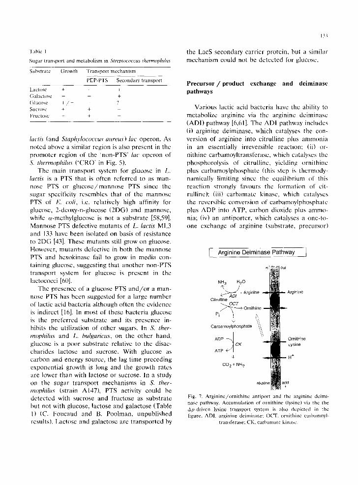
Table I
Sugar transport and metabolism in Streptococcus thermophilus
Substrate Growth Transport mechanism
PEP-PTS Secondary transport
Lactose + - + Gatactose + Glucose + / - - ? Sucrose + + -- Fructose + + -
lactis (and Staphylococcus attreus) lac operon. As noted above a similar region is also present in the promoter region of the 'non-PTS' lac operon of S. thermophilus ( 'CRO' in Fig. 5).
The main transport system for glucose in L. lactis is a PTS that is often referred to as man- nose PTS or g lucose /mannose PTS since the sugar specificity resembles that of the mannose PTS of E. coli, i.e. relatively high affinity for glucose, 2-deoxy-D-glucose (2DG) and mannose, while c~-methylglucose is not a substrate [58,59]. Mannose PTS defective mutants of L. lactis ML3 and 133 have been isolated on basis of resistance to 2DG [43]. These mutants still grow on glucose. However, mutants defective in both the mannose PTS and hexokinase fail to grow in media con- taining glucose, suggesting that another non-PTS transport system for glucose is present in the lactococci [60].
The presence of a glucose PTS a n d / o r a man- nose PTS has been suggested for a large number of lactic acid bacteria although often the evidence is indirect [16]. In most of these bacteria glucose is the preferred substrate and its presence in- hibits the utilization of other sugars. In S. ther- rnophilus and L. bulgaricus, on the other hand, glucose is a poor substrate relative to the disac- charides lactose and sucrose. With glucose as carbon and energy source, the lag time preceding exponential growth is long and the growth rates are lower than with lactose or sucrose. In a study on the sugar transport mechanisms in S. ther- mophilus (strain A147), PTS activity could be detected with sucrose and fructose as substrate but not with glucose, lactose and galactose (Table 1) (C. Foucaud and B. Poolman, unpublished results). Lactose and galactose are transported by
133
the LacS secondary carrier protein, but a similar mechanism could not be detected for glucose.
Precursor/product exchange and deiminase pathways
Various lactic acid bacteria have the ability to metabolize arginine via the arginine dciminase (ADI) pathway [6,61]. The ADI pathway includes (i) arginine deiminase, which catalyses the con- version of arginine into citrulline plus ammonia in an essentially irreversible reaction: (ii) or- nithine carbamoyltransferase, which catalyses the phosphorolysis of citrullinc, yielding ornithine plus carbamoylphosphate (this step is thermody- namically limiting since the equilibrium of this reaction strongly favours the formation of cit- rulline); (iii) carbamate kinase, which catalyses the reversible conversion of carbamoylphosphate plus ADP into ATP, carbon dioxide plus ammo- nia; (iv) an antiporter, which catalyses a one-to- one exchange of arginine (substrate, precursor)
I Arginine Deiminase Pathway ]
NH 3 H20
A ~ D / Arginine Arginine Citrulline
~ O C T Pi ~ Ornithine
Carbamoylphosphate
ADP ~ Ornithine CK Lysine
ATP
H +
CO 2 + NH 3
alk
Fig. 7. Arginine/ornithine antiport and the arginine deimi- nase pathway. Accumulation of ornithine (lysine) via the the Ap-driven lysine transport system is also depicted in the figure. ADI, arginine deiminase; OCT, ornithine carbamoyl-
transferase; CK, carbamate kinasc.

134
for ornithine (product) [5,6] (Fig. 7). Overall, the ADI pathway yields 1 mol of ornithine and car- bon dioxide, 2 tool of ammonia, and I tool of ATP by substrate level phosphorylation per mol of arginine metabolized [6,61]. A similar deimi- nasc pathway is used for the conversion of agma- tine into putrescine in Enterococcus faecalis, in which case an analogous transport system for cationic metabolites is associated with the agma- tinc deiminase (AgmD1) pathway [8]. Since the driving force for the uptake of these cationic substrates (precursors) and excretion of the prod- ucts is supplied by the precursor and product concentration gradients, no additional metabolic energy is required for the translocation of these solutes. Consequently, the net metabolic energy yield of arginine and agmatine metabolism by the deiminase pathways is I tool of ATP per mol of substrate metabolized.
Detailed kinetic analysis of arginine/orni thinc antiport indicates that the carrier possesses a single substrate-binding site which is prcsent al- ternately at the inner and outer surface of the cytoplasmic membrane [62]. The exchange reac- tion catalysed by the antiporter resembles a 'ping-pong' mechanism regarding enzyme kinet- ics. Thus, arginine (substrate) associates at the outer surface of the membranc, the carrier-sub- stratc complex reorients its binding site and argi- nine dissociates at the inner surface. Rapid metabolism of argininc assures that the intracel- lular arginine concentrations are kept low com- pared to those of ornithine [6]. Next, ornithine (product) binds, the carr ier-product complex re- orients its binding site and ornithine is released into the medium. Under conditions that a large fraction of arginine is converted into ornithine, arginine metabolism will slow down because or- nithine competes with binding of arginine at the outer surface of the membrane.
Since uptake of arginine and excretion of or- nithine via the antiporter protein are tightly linked, questions arise with respect to the initia- tion of arginine metabolism and replenishment of the ornithine pool when a fraction of the arginine is used for biosynthetic purposes. L. lactis has solved this dilemma by taking advantage of a Ap-driven lysine transport system that can accept
ornithine with low affinity [63]. Furthermore, the antiporter catalyses heterologous exchange of arginine for lysine in addition to exchange of arginine for ornithine (Fig. 7). Accumulation of lysine via the Ap-driven transport system in com- bination with exchange of lysine for arginine via the antiportcr results in cyclic transport of lysine and net accumulation of arginine at the expense of the Ap. In this scheme a high level of or- nithine can be sustained when part of the argi- nine is used for biosynthesis.
Precursor/product exchange and decarboxyi- ation reactions
Malolaetic fermentation is carried out by some species of the genera Lactobacillus, Lactococcus, Leuconostoc and Pediococcus [64]. In this path- way L-malate enters the cells and is decarboxy[- ated by malolactic enzyme to yield k-lactic acid and carbon dioxide, aftcr which the products leave the cell. Although the decarboxylation reac- tion does not yield metabolic energy directly, the free energy of the decarboxylation reaction can be conserved via an indirect H + pump mecha- nism. It has been shown for L. lactis that l,-malate utilization results in the formation of a Ap that is sufficiently high to drive ATP synthesis via the F0 F1-ATPase [9].
The transport of L-malate has been character- ized in artificial membranes prepared from L. lactis IL1403 and in isogenic mutants, that are defective in either L-malate transport or L-malate decarboxylation [9]. These studies have shown that monoanionic L-malate ( M H ) is taken up either in exchange for L-lactic acid (LH) or as MH uniport (Fig. 8). In both cases a negative charge is translocated from the medium to the cytoplasm which results in the formation of a J 7 t (inside negative). Since charge compensation in the decarboxylation reaction requires the con- sumption of a proton, the cytoplasm is alkalinized relative to the outside medium and a ApH is formed. Thus, the free energy of the decarboxyl- ation reaction is converted into a Ap ( = A ~ - ZApH) simply by compartimentalization of the malolactic fermentation pathway, i.e. stoichio-
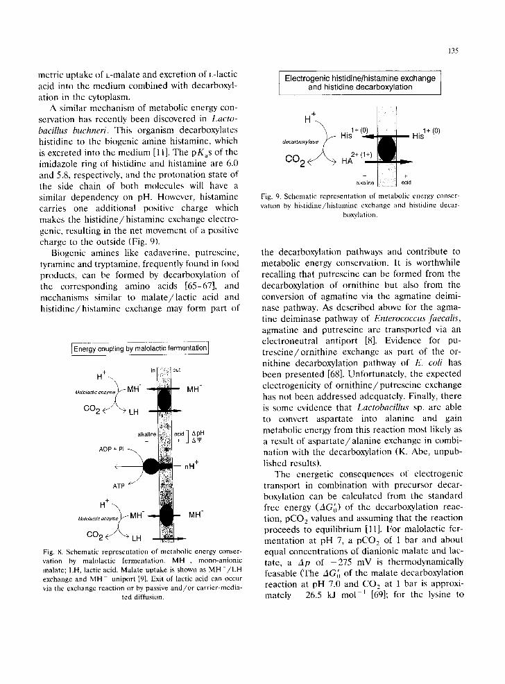
135
metric uptake of L-malate and excretion of L-lactic acid into the medium combined with decarboxyl- ation in the cytoplasm.
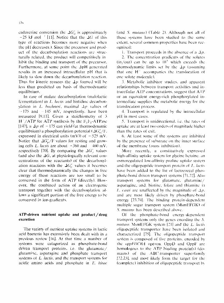
A similar mechanism of metabolic energy con- servation has recently been discovered in Lacto- bacillus buchneri. This organism decarboxylates histidine to the biogenic amine histamine, which is excreted into the medium [11]. The PKaS of the imidazole ring of histidine and histamine are 6.0 and 5.8, respectively, and the protonation state of the side chain of both molecules will have a similar dependency on pH. However, histamine carries one additional positive charge which makes the hist idine/histamine exchange electro- genic, resulting in the net movement of a positive charge to the outside (Fig. 9).
Biogenic amines like cadaverine, putrescine, tyramine and tryptamine, frequently found in food products, can be formed by decarboxylation of the corresponding amino acids [65-67], and mechanisms similar to mala te / lac t ic acid and hist idine/histamine exchange may form part of
I Energy coupling by malolactic fermentation]
H + ~ . x
enzyme~- M H- Malolactic
) , CO2 <-~ ~ LH
out
MH-
ADP + Pi
alkaline acid] APHA,t,
- - nH +
H +~ATP ~ I ~
enzyme? M H- - - ~ ~ M H" Malolactic
CO2 4 ~ ~ LH --
Fig. 8. Schematic representation of metabolic energy conser- vation by malolactie fermentation. MH , mono-anionic malate; LH, lactic acid. Ma]ate uptake is shown as M H - / L H exchange and MH uniport [9]. Exit of lactic acid can occur via the exchange reaction or by passive and/or carrier-media-
ted diffusion.
Electrogenic histidine/histamine exchange and histidine decarboxylation
H+ : ::
deca Oo y,L HisI+ I-- Hg+(O) 0 0 2 4 - ~ ~ HA 2+0+) ., .~ . .
-- +
alkaline : acid
Fig. 9. Schematic representation of metabolic energy conser- vation by histidine/histamine exchange and histidine decar-
box'y]ation.
the decarboxylation pathways and contribute to metabolic energy conservation. It is worthwhile recalling that putrescine can be formed from the decarboxylation of ornithine but also from the conversion of agmatine via the agmatine deimi- nase pathway. As described above for the agma- tine deiminase pathway of Enterococcus faecalis, agmatine and putrescine are transported via an electroneutral antiport [8]. Evidence for pu- t resc ine/orni th ine exchange as part of the or- nithine decarboxylation pathway of E. coli has been presented [68]. Unfortunately, the expected electrogenicity of orn i th ine /put resc ine exchange has not been addressed adequately. Finally, there is some evidence that Lactobacillus sp. are able to convert aspartate into alanine and gain metabolic energy from this reaction most likely as a result of aspar ta te /a lanine exchange in combi- nation with the decarboxylation (K. Abe, unpub- lished results).
The energetic consequences of electrogenic transport in combination with precursor decar- boxylation can be calculated from the standard free energy (AGo) of the decarboxylation reac- tion, pCO 2 values and assuming that the reaction proceeds to equilibrium [11]. For malolactic fer- mentation at pH 7, a pCO 2 of 1 bar and about equal concentrations of dianionic malate and lac- tate, a Ap of - 2 7 5 mV is thermodynamically leasable (The AG~ of the malate decarboxylation reaction at pH 7.0 and CO 2 at 1 bar is approxi- mately -26 .5 kJ mol i [69]; for the lysine to
13¢~
cadaverine convcrsion thc ..IG~'~ is approximatcly - 2 5 kJ too l t [11]. Notice that the _IG of this type of reactions becomes more negative when the pH decreases.). Since the precursor and prod- uct of the decarboxylation reactions arc struc- turally related, the product will competitively in- hibit the binding and transport of the precursor. Furthermore, at some point the ZIpH generated results in an increased intracellular pH that is likely to slow down the decarboxylation reaction. Thus for kinetic reasons the -11~ formed will be less than predicted on basis of thermodynamic equilibrium.
In case of malate decarboxylation (maloh~ctic fermentation) in k. lactis and histidinc decarbox- y/ation in L. bttc/tneH, maximal dp valucs of - 1 7 5 and - 1 4 5 mV, respectively, have been measured [9,11]. Given a stoichiomctry of 3 H + /ATP for ATP synthesis by the F~I:I-ATPase [147], a zip of - 175 can yield (at thermodynamic equilibrium) a phosphorylation potential (ziG~',/F, expressed in electrical units (mV)) of -525 mV. Notice that ziG~/F values tor resting and grow- ing cells L. lactis are about - 3 6 0 and 460 mV, respectively [70]. By comparing the zi(;~'~ values (and also the ziG, at physiologically relevant con- centrations of the reactants) of the decarboxyl- ation reactions with the AG/, values it becomes d e a r that thermodynamically the changes in frcc energy of these reactions are too small to be conserved in the form of ATP (directly). How- ever, the combined action of an elcctrogenic transport together with the decarboxylation al- lows a significant portion of the free encrgy to be conserved in ion-gradients.
ATP-driven nutrient uptake and product/drug excretion
The variety of nutrient uptake systems in lactic acid bacteria has extensively been dealt with in a previous review [16]. At that time a number of systems were catagorized as phosphate-bond driven transport proteins, i.e. the g lu t ama te / glutamine, asparagine and phosphate transport systems of L. lactis, and the transport systems for acidic amino acids and phosphate in E. hirae
(and S. mutans) (Table 2). Although not all of these systems have been studied to the same extent, several common propcrtics have been rec- ognized:
1. Transport procceds in the absencc of a 317. 2. The concentration gradients of the solutes
( in /ou t ) can be up to 10 ~ which cxceeds the thermodynamic limits set by tile J p (assuming that onc H ' accompanies thc translocation of one solute molecule).
3. Metabolic inhibitor studies, and apparent relationships between transport activities and in- tracellular ATP concentrations, suggest that ATP or an equivalent energy-rich phosphorylatcd in- termediate supplies thc metabolic energy for the translocation process.
4. Transport is regulated by the intracellu[ar pH in most cases.
5. Transport is unidirectional, i.e. the rates of uptake are at least two orders of rnagnitude higher than the rates of exit.
6. At least some of the systems are inhibited by the presence of substrate on the im~cr surface of the membrane (tram" inhibition).
More recently, it constitutively expressed high-affinity uptake system for glycine betainc, an osmoregulated low-affinity proline uptake system and the oligopcptide transport protein of L. h;ctis have been added to the list of lactococcal phos- phate-bond driven transport systems [71,72]. Also transport systems for glutamate, g lu tamine / asparaginc, and, biotinc, folate and thiaminc in L. casei arc unaffected by the naagnitude of dp , and arc most likely driven by phosphate-bond energy [73,74]. The binding protein-depcndcnt multiple sugar transport systcm (MsmEFGK) of S. mulans has been described abovc.
Of the phosphate-bond encrgy-depcndcnt transport systems only the genes encoding the S. mutans MsmEFGK system [21] and tile k, lactis oligopeptide transporter have been isolatcd and characterized [75]. Thc oligopcptide transporl system is composed of five proteins, encoded by the oppFDCBA operon. OppD and OppF are homologous to the ATP binding protein(s) (do- mains) of the ABC-transpor ter superfamily [22,23], and most likely l\)rm the target for tile (completc) inhibition of oligopeptide transport by

ortho-vanadate [72]. OppB and OppC are highly hydrophobic proteins, that on basis of hydropathy analyses, are able to span the cytoplasmic mem- brane in ~f-helical configuration six times. OppB and Opp(" are homologous to the corresponding polypcplides of the oligopeptide transporters of S. typhimtu'ium and B. subtilis, and DciAB and DciAC of the dipeptide transport system of Bacillus subtilis. The t ransmembrane domains of OppB and OppC would constitute the pathway that facilitates the translocation of the oligopep- tides across the cytoplasmic membrane. OppA is homologous to the peptidc binding proteins of S. o'phimurium and B. suhtilis.
Since Gram-posit ive bacteria do not have a periplasmie space, the question whether these bacteria possess binding protein-dependent trans- port systems analogous to those found in Gram- negative bacteria could only be addressed after the primaD' sequences of the proteins became available. It has turned out that Gram-positive species have the equivalent systems with binding proteins that are anchored to the membrane by a lipid group, which restricts diffusion to two di- mensions (in the plane of the membrane) [76,77]. The sugar-binding protein MsmE of S. mutans has been shown to be attached to the membrane by means of an amino-terminal lipo-amino acid anchor (Figs. 1 and 3; [78]). Known prokaryotic lipoproteins have a glyceridc linked to a cysteine residue, and a consensus sequence (Leu-Ala- Ala /Gly- ,LCys) around the the cysteine in the prolipoprotein has been proposed {79]. OppA of L. lactis possesses the consensus prolipoprotein cleavage site Leu-Ser-Ala-,1, Cys between residues 22-23 [751.
The functional characteristics of the lactococ- cal Opp system strongly resemble those of the other phosphate-bond-dependent transport sys- tems (Table 2), of which the g lu tamate /g lu tamine transporter of L. lactis has been studied in great- est detail [80-82]. It is conceivable that these systems have a structural complexity similar to Opp, and that a substrate-binding lipoprotein serves as the initial receptor for transport by delivering the substrates to the integral mem- brane components. Only in a few cases ATP hydrolysis has been demonstrated rigorously to
137
energize binding protein-dependent transport [83,84]. The mechanism by which hydrolysis of ATP is coupled to the solute translocation pro- cess is still not clear (fl~r recent reviews, sec [23,85]). The energy requirements of phosphate- bond-dependent transport in lactic acid bacteria (and other Gram-positive species) indicate that ATP plays a role in the energization of transport, but unequivocal evidence is still lacking. Finally, it is worth emphasizing that although these ATP- driven transporters facilitate transport in the ab- sence of a Ap, the systems are highly sensitive to changes in the intracellular pH [80,86,87]. Conse- quently, changes in the _lpH component of Ap are directly reflected in changes in transport ac- tivity and can easily lead to misinterpretations regarding the mechanism of energy coupling to these systems [87].
The ATP-dependent transport systems de- scribed solar only catalyse nutrient uptake and exhibit little or no cfflux activity under physiolog- ical conditions. In a study on the regulation of the intracellular pH in L. lactis, employing the fluo- rescent probe 2',7'-bis-(2-carboxyethyl)-5(and 6)- carboxyfluorescein (BCECF), energy-dependent efflux of BCECF has been observed [88]. A fur- ther characterization of this efflux process indi- cated that most likely ATP serves as energy donor [89]. In search of the physiological function of this BCECF efflux pump, and owing to its functional resemblance to eukaryotic P-g[yeoprotein (Mdrl) , a variety of compounds /d rugs (antibiotics, Mdrl substrates and inhibitors, uncouplers, ionophores etc.) have been tested as possible substrate of the BCECF efflux pump. None of these toxic com- pounds could serve as substrate for the BCECF effiux pump [89]. To some of the compounds tested (e.g. ethidium-bromide), however, L. lactis mutants with increased resistance could be iso- lated [9(I]. These mutants confer cross-resistance towards various toxic compounds such as the Mdrl substrates daunomycin and actinomycin D, the Plasrnodium falciparum multidrug resistance (mdr) substrate chloroquine, the bacterial mdr (Bmr) substrate rhodamine 6-G, and ionophores like nigericin (for a review on bacterial excretion systems see Poolman [10]). Resistance of the cells to these molecules is conferred by a transport

138
Table 2
Transport mechanisms in lactic acid bacteria
Substrate specificity
H + -Symport Leucine, isoleucine, valine Alanine, glycine Serine, threonine Alanine, glycine, serine, threonine Lysine, ornithine Phenylalanine, tyrosine, tyrptophane Di- and tripeptides Citrate Lactose, galactose, melibiose, TMG, raffinose S. Galactose L.
Organisms
I,. lactis, L. plantarum, Lc. mesenteroides, L. casei L. lactis, (S. thermophihts) L. lactis E. hirae, S. pyogenes L. lactis, (L. plantarum) (L. lactis, L. easel) L. lactis Lc. mesenteroides, L. lactis, Lc. lactis
thermophilus i, L. bulgaricus t, lactis
TMG (Xylose)
Na +-Symport Alanine, (glycine) Serine, threonine
H +-Antiport Calcium Sodium
Substrate/product antiport (exchange) Arginine/ornithine Agmatine/putrescine Sugar-phosphate/phosphate Malate/lactic acid Histidine/histamine (Tyrosine/tyramine) (Aspartate/alanine) Phosphate/phosphate
ATP-driven uptake Glutamate, glutamine Glutamate, aspartate Asparagine Proline, glycine betaine Phosphate Oligopeptides Biotine, folate, thiamine Potassium Raffinose, melibiose, isomaltotriose
ATP-driven efflux Proton BCECF Calcium (Lactococcin A) (Nisin) (Pediocin PA-1)
ATP-driven exchange Sodium/potassium
L. lactis, (L. brevis, 1+. buchneri, L. plantarum) I.. pentosus
S. bocis S. bo+'is
L. lactis E. hirae
L. lactis, E. faecalis, S. sanguis, S. milleri E. fiwcalis L. lactis L. lactis ' L. buchneri L. buchneri Lactobacillus sp. S. pyogenes
L. lactis E. hirae, S. mutans, L. casei L. lactis, (L. ~asei) L. lactis L. lactis, E. hirae L. lactis L. cash E. hirae S. sanguis
E. hirae, (various other lactic bacteria) L. lactis E. hirae, S. sanguis, L. lactis L, lactis L. lactis P. acidilactici
E. hirae
Proteins
AIaT "
DtpT CitP LacS
XylP
OppFDCBA
Ktrl MsmEFGK
F0F t
LcnCD NisT PedD
Ktrll

139
system that removes various hydrophobic peptide compounds, drugs, and other (toxic) molecules from the cells. Apparently, lactic acid bacteria possess a number of transport systems that are involved in the excretion of compounds that are normally not encountered by the cells, but pro- vide the cells with mechanisms to clean up the cytoplasmic environment when needed.
The driving force for secretion by the multiple drug resistance pump of L. lactis has not yet been established, but some of its properties are similar to that of eukaryotic Mdrl . Mdrl belongs to the family of ABC transporters by the fact that it has the highly conserved ATP-binding cassette in two separate domains as part of a single polypeptide that also contains the transmem- brahe domains, the two-times-six a-helical seg- ments [23]. Partially purified Mdrl catalyses transport in response to ATP hydrolysis [91], and, similar to the BCECF effiux pump and the oligopeptide transporter of L. lactis [72,89], the eukaryotic multidrug effiux pump is inhibited by ortho-vanadate [92].
Recently, an E. coli strain that is highly sensi- tive to ethidium has been used to select for resistance towards high concentrations of this compound upon complementation with a chro- mosomal library of L. lactis DNA (H. Bolhuis, D. Molenaar, B. Poolman, and W.N. Konings, un- published results). The putative L. lactis mdr gene(s) (lmr) are present on a DNA fragment of less than 3 kb. The lmr gene(s) and their func- tional expression in E. coli are characterized, and insertion inactivation of the chromosomal gene(s) in L. lactis should establish whether the cloned gene(s) correspond with those that upon muta- tion confer a high cross-resistance towards vari- ous toxic compounds to L. lactis.
Molecular analysis of the lactococcin A gene
cluster from L. lactis subsp, lact& biovar diacety- lact& has revealed the presence of two open reading frames, designated LcnC and LcnD, which are homologous to E. coil HIyB and HlyD, respectively [93]. The HlyB protein exports hemolysin across the cytoplasmic membrane, whereas HIyD, which is co-expressed with HIyB, is likely to participate in hemolysin transport across the outer membrane of E. coli [94]. Even though HIyB possesses the ATP-binding cassette typical of the ABC superfamily, hemolysin export by HIyB seems to require a Ap (in addition to ATP) [95]. Despite some uncertainties in the mechanism of energy coupling to these excretion processes, and the precise role of the individual components (e.g. HIyD in E. coli; role of LcnD in L. lactis which does not have an outer mem- brane), it is fair to say that LcnC and LcnD are likely to constitute a secretion pathway for lacto- coccin(s) in L. lactis that is dependent on ATP [96]. A similar secretion machinery composed of proteins homologous to HlyB (LcnB) and HlyD (LcnD) has been proposed for the translocation across the membrane of the antimicrobial peptide subtilin (lantibiotic) of B. subtilis [97]. Also for transport across the cytoplasmic membrane of the lantibiotic nisin from L. lactis [98] and the bacte- riocin PA-1 from Pediococcus acidilactici [99], proteins homologous to the HlyB family arO likely to play a role.
Secretion of hemolysin A by HIyB/HIyD is signal sequence independent as opposed to pro- tein translocation by the Sec machinery. By anal- ogy with hemolysin A secretion, translocation of polypeptides across the cytoplasmic membrane by proteins homologous to HIyB/HIyD is often des- ignated 'signal sequence independent ' . It needs to be stressed that bacteriocins and lantibiotics from lactic acid bacteria are synthesized as pre-
Note to Table 2: " Alanine transport mutants have been isolated on basis of resistance to /3-chloro-alanine [72]. ~' LacS protein that catalyses galactoside-H + symport and (heterologous) sugar exchange; lactose/galactose exchange most likely
dominates during lactose metabolism (see text). c System also catalyses monoanionic L-malate uniport (see Fig. 8).
Brackets indicate preliminary evidence for the transport mechanism in the particular organism. The genes encoding DtpT, CitP, LacS, XylP, OppDFBCA, MsmEFGK, FoFi-ATPase subunits, LcnCD, NisT and PedD have been isolated and sequenced (see text). Transport of sugar by the PTS is not indicated.

141)
cursors, and that the corresponding presequences could play a role in the translocation of thc precursors across the cytoplamic membrane. Since only the mature bacteriocins and lantibiotics have antimicrobial activity, the presequence could also serve as a means to protect the producing strain [,)(,].
ATP-driven cation transport
The major ATPase of lactic acid bacteria is the N,N'-dicyclohexylcarbodiimide (DCCD)-sensitive F¢~FrATPase which pumps protons at the ex- pense of ATP but can also function as ATP synthetase [100]. ATP synthesis by the F0F ~- ATPase occurs when n x Ap > AGe's, in which n represents the number of protons translocated (tl is most likely 3), Ap is the proton motive force and AG~'~ is the phosphorylation potential. Thus when a high Ap is generated by osmotic reac- tions, e.g. product effiux, solute deearboxy[ation reactions (see above), the metabolic energy in the form of an electrochemical H + gradient can bc used to drive ATP synthesis. Under conditions that ATP is formed by substrate level phospho~- lation, e.g. glycolysis, deiminase pathways, the FoF1-ATPase will function as a hydrolase in order to generate a Ap.
Besides serving as a converter of metabolic energy (ATP ~ Ap), the F,F~-ATPase of lactic acid bacteria plays a major role in regulating the intracellular pH as has been shown convincingly for E. hirae [101-105]. In fact, it has been shown that E. hirae can grow under conditions that Ap is .zero, provided the medium is slightly alkaline and contains high concentrations of K ' and nu- trients, while Na ~ concentrations are kept low [106]. This further demonstrates that regulation of the intracelluar pH (by the F~FI-ATPasc and perhaps other ion-translocating enzymes) is cru- cial for the survival of (lactic acid) bacteria [87]. The role of the .Jp in (lactic acid) bacteria is indicated by the severe inhibition of growth in the absence of a Ap when the concentrations of nutrients arc in the sub-millimolar rather than in the millimolar range or when high concentrations of Na ~ are present [106]. Thus rapid growth of
(lactic acid) bacteria requires the accumulation of various essential nutrients and excretion of un- wanted products for which the Ap supplies the driving force. Various kinds of information are available on the function, structure and regula- tion of FoFt-ATPases in (lactic acid) bacteria [102,104,105,107-10q], but these aspects arc not included in this review.
Transport of various cations in lactic acid bac- teria is dependent on ATP or a related phos- phate bond intermediate. These systems will only be summarized here (see also Table 2, reviewed in Konings el al. [16], and include vanadate-sensi- live Ca z ~ efflux systems in L. lactis, E. hirae and Str~7~tococcus sangtds [ 110,111 ], a vanadate-sensi- live K~-ATPase in E. hirae (see also below: [112- 114], a constitutive K + transport system (Ktrl) in E, hirae [115] and a Na+-ATPase in E. hirae (KtrlI) that expels Na ~ in exchange for K + [116- 118]. The Ktrl K ~ uptake system requires ATP in addition to a Ap for transport, and has been claimed lo co-transport K + and H ~ [115]. Ktrl has properties that coincide with the K+-ATPase of E. hirae, which originally has been proposed to function as a Ap-regulated, electrogenic ATP- driven K + uptake system [113,114]. More re- cently, this ATPase has been suggested to be a H + pump [119] and the observed K + transport has been attributed to secondary movements of K ~ via leak pathways [118]. KtrII is induced by Na ~ and although the enzyme catalyses a similar exchange as the ouabain-sensitive Na ~,K*- ATPase of mammalian cells [116,120], the mecha- nism of ion coupling is quite different [118]. Un- like the Na * /K +-ATPase of mammalian cells the E. hirae enzyme is not a P-type ATPase and is not inhibited by ortho-vanadate.
Ion-linked nutrient uptake
Next to E. coli, solute transport in bacteria as a whole has perhaps best been studied in L. lactis, and sofar all cation-linked transport sys- tems in this lactic acid bacterium that have been studied use H + as coupling ion. Evidence for Na+-coupled transport comes from studies in Streptococcus hot'is [ 121 ].

141
Under section 'ATP-driven nutrient uptake and p roduc t / d rug excretion' it has been shown that amino acids with acidic (Glu, Asp) and amide side chains (Gin, Ash) as well as proline arc transported by ATP-driven mechanisms in all lac- tic acid bacteria tested, i . e .L , lactis, E. hirae. S. mutans and L. casei (Table 2). Although these amino acids together with glycine betaine, potas- sium ions, and some other compounds do play a role in the mechanisnr that confers resistance to osmotic stress [71,122], it is not immediately clear why these solutcs in particular arc taken up by ATP-driven mcchanisms whereas (most) other amino acids arc transported by solute-H + sym- port (Table 2). The branched chain aliphatic amino acids (Leu, lie, Val), thc ncutral amino acids (Ala, G[y), the aliphatic amino acids with a hydroxyl side chain (Set, Thr) and the aromatic amino acids (Phe, Tyr, Trp) arc transported by scparate H ~-linked mechanisms in L. lactis [1(),87,123-126]. Transport of branched chain amino acids driven by 2//,, has also been demon- strated for Lc. mesenteroides subsp, devtranicum [127]. In E. hirae and Streptococcus pyogenes, unlike L. lactis, the neutral amino acids glycine, alanine (or ~-aminoisobutyric acid), serinc and thrconine arc transported by a common ,.lp- drivcn systcm [128,129]. Evidcnce, often based on metabolic inhibitor studies, points to H+-linked uptake of these amino acids in organisms like S. thermoldtilus, S. agaluctiae, S. pneumoniae and others (for review, see Konings et al. [16].
Thc H~-Iinked amino acid transportcr that has been analyscd in depth is the branched-chain amino acid carrier of L. hwti.s' [125,126,130-134]. The substraie specificity, the kinctic mechanism of transport, the role of the lipid cnvironmcnt, and some other molccular properties of the par- tially purificd protcin reconstituted into proteoli- posomes have bccn analysed. Several properties of the system have been reviewed in the past [16,87].
In recent years important progess has been made in the study of the peptide transporters of lactic acid bacteria. Whercas peptides of three upto at least six residues arc taken up by an ATP-driven binding l ipoprotein-dependent trans- port system (OppFDCBA) [72,75], di- and tripep-
tides are taken up by an Ap-driven transport protein (DtpT) in L. lactis [135]. Sofar, the DtpT and OppFDCBA peptide transporters together with the proteinase (PrtP) are the only compo- nents of the proteolytic system of L. lactis that are essential for casein utilization and for growth in milk [75,136,137]. The d i / t r ipcpt ide transport system also represents the first demonstration of H ~ coupled peptide transport in bacteria. The genc encoding the d i / t r ipept ide transporter (DtpT) has been isolated, sequenced and func- tionally expressed in E. colt (A. Hagting, B. Pool- man and W.N. Konings, unpublished results). DIpT is a highly hydrophobic protein with a unique primary structure which has no significant similarity to proteins in the MIPS protein data base (release Dec. 1992). The secondary structure of the DtpT protein most likely consists of 12 t ransmcmbranc {~-helical segments which span the membrane in a zig-zag manner. Using the "positive inside' rule [138], the amino- and car- boxy-termini are prcsent on thc outside of the cytoplasmic membrane unlike most secondary transport proteins [139].
The plasmid-encoded citrate transport gene (citP) of L. lactis subsp, diacetvlactis has been isolatcd and functionally expressed in E. coil [140]. The CitP protein is homologous to the ci t ra tc-H*-Na + symporter (CitS) of Kh'hsiella imeumoniae [141]. For the CitS protein of K. lmeumoniae it has been shown that dianionic citrate (H-Cit 2 ) is transported in symport with 1 Na" and at least 2 H + [142]. Despite the homol- ogy between CitP and CitS (ovcrall similarity of 76(:i), there arc no indications that CitP of L. la¢'tis uses Na" as additional coupling ion.
Ion-linked cation transport
Bacteria keep their intracellular Ca 2' and Na ~ concentrations well below that of the surrounding medium as long as metabolic energy is available. Ca 2~ extrusion in lactic acid bacteria is driven either by ATP [110] or ..lp [143]. In fact, for L. lactis it has been shown that subsp, lactis uses a Ca24_ATPase [ 111 ] while subsp, cremoris excretes Ca 2. by CaX~/H + antiport [143]. Sodium extru-
142
sion in bacteria is generally attributed to Na ~/H ~ antiport mechanisms [144]. The sodium gradient formed can serve as a driving force for, for in- stance, transport systems catalysing Na+-solute symport ([139]; e.g. alanine, glycine and serine, threonine transport in S. boris, Table 2). In addi- tion, N a + / H + antiporters are thought to be in- volved in the regulation of the cytoplasmic pH [144,145]. In case of the lactic acid bacteria, N a + / H + antiport activity has only been demon- strated in E. hirae [118,146]. The secondary Na ~/H + antiporter of E. hirae is expressed con- stitutively, in contrast to the ATP-driven Na +/K + exchange system (Ktrll) of E. hirae which is induced in media with high concentrations of Na + [116] (see above).
Concluding remarks
The present manuscript updates our 1989 re- view [16]. Research related to energy-transducing processes in lactic acid bacteria still focusses on two organisms, i .e .L, lactis (formerly Streptococ- cus lactis and S. cremoris) and E. hirae (formerly, Streptococcus faecalis), although work on other species is beginning to emerge. In the last 5 years a number of genes encoding transport proteins from lactic acid bacteria have been isolated, char- acterized and functionally expressed in homolo- gous and heterologous host systems. This has allowed detailed analysis of the molecular prop- erties of these systems both in vivo and in vitro. In this respect research on the lactose transport protein (LacS) of S. thermophilus and the lactose PTS of L. lactis is most advanced. The recent cloning and characterization of the genes encod- ing the two peptide transporters of L. lactis (DtpT and OppFDCBA), and establishment of their es- sential roles in the utilization of casein(-derived peptides), are of eminent importance for the un- raveling of the 'pathway(s)' of casein breakdown in lactic acid bacteria.
The discovery of precursor /product exchange reactions in bacteria, such as sugar phospha te / phosphate, arginine/orni thine, lactose/galac- tose, malate / lact ic acid, hist idine/histamine ex- change and others, has its origin for the greater
part in the research on lactic acid bacteria. Simi- lar systems as well as the chemiosmotic circuits associated with the decarboxylation reactions have also been demonstrated in other bacteria,
For all these studies a variety of techniques has been developed, ranging from protocols for the isolation of membrane vesicles, membrane fusion and reconstitution techniques, to the isola- tion of mutants either by random or targeted mutagenesis (integration) and efficient (electro) transformation protocols. Most of these tools have been developed for L. lactis (and E. hirae), but appear applicable in one way or another to many lactic acid bacteria.
Acknowledgements
The research of the author has been made possible by a fellowship from the Royal Nether- lands Academy of Arts and Sciences. I would like to thank J. Knol, W.N. Konings and B. Ten Brink for critical reading of the manuscript, and A. Haandrikman, R. van Rooyen and W.M. De Vos for supplying information prior to publication.
References
I Smart, J.B. and Thomas, T.D. (1987) Effect of oxygen on lactose metabolism in lactic streptococci. Appl. Environ. Microbiol. 53, 533 541.
20no, R., Lageveen, R.G., Veldkamp, H. and Konings, W.N. (1982) Lactate efflux induced electrical potential in membrane vesicles of Streptococcus cremoris. J. Bacteriol. 146, 733-738.
3 Michels, P.A.M., Michels. J.P.J.2 Boonstra, J. and Kon- ings, W.N. (1979)Generation of electrochemical proton gradient in bacteria by the extrusion of metabolic end products. FEMS Microbiol. Lett. 5, 357-364.
4 Ten Brink, B., Otto, R., Hansen, U.P. and Konings, W.N. (1985) Energy recycling by lactate efflux in growing and nongrowing cells of Streptococcus cremoris. J. Bacteriol. 162, 383 390.
5 Driessen, A.J.M., Poolman, B., Kiewiet, R. and Konings, W.N. (1987) Arginine transport in Streptococcus lactia is catalyzed by a cationic exchanger. Proc. Natl. Acad. Sci. USA 84, 6(193-6097.
6 Poolman, B,, Driessen. A.J.M. and Konings, W.N. (1987) Regulation of arginine-ornithine exchange and the argi- nine deiminase pathway in Streptococcus lactis. J. Bacte- riol, 169, 5597-56(t4.
7 Thompson, J. (1987) Ornithine transport and exchange in Streptococcus lactis. J. Bacteriol. 169, 4147-4153.
8 Driessen, A.J.M., Smid, E.J. and Konings, W.N. 11988) Transport of diamines by Enterococcus faecalis is medi- ated by an agmatine-putrescine antiporter. J. Bacteriol. 1711, 4522-4527.
9 Poolman, B., Molenaar, D., Smid, E.J., Ubbink, T., Abee, T., Renault, P.P. and Konings, W.N. (1991) Malolactic fermentation: electrogenic malate uptake and malate/ lactate antiport generate metabolic energy. J. Bacteriol. 173, 61)31/-6037.
l0 Poolman, B., Molenaar, D. and Konings, W.N. (19921 Diversity of transport mechanisms in bacteria. Handbook of Biomembranes, Vol. 2, (M. Shinitzky, Ed.), pp. 1-50 Balaban Publishers, Rehovot.
11 Molenaar, D., Bosscher, J.S., Ten Brink, B., Driessen. A.J.M. and Konings, W.N. (19931 Generation of a proton motive force by histidine decarboxylation and electro- genic histidine/histamine antiport in Lactobacillus buch- neri. J. Bacteriol. 175, 2864 2870.
12 Kandler, O. (1983) Carbohydrate metabolism in lactic acid bacteria. Ant. Leeuwenhoek 49, 209-224.
13 Hammes, W.P., Weiss, N. and Holzapfel, W. (1991) The genera Lactobacillus and Carnobacterium. In: The Prokaryotes, 2nd edn. (A. Barlows, H.G. Trfiper, M. Dworkin, W. Harder, K.H. Schleifer, Eds.), Springer Ver- lag, Berlin.
14 Thompson, J. (1987) Sugar transport in the lactic acid bacteria. In: Sugar Transport and Metabolism in Gram- positive Bacteria (J. Reizer and A. Peterkofsky, Eds.), pp. 13-38. Ellis ltorwood, Chichester.
15 Thompson, J. (1987) Regulation of sugar transport and metabolism in lactic acid bacteria. FEMS Microbiol. Rev. 46, 221-231.
16 Konings, W.N., Poolman, B. and Driessen, A.J.M. 11989) Bioenergetics and solute transport in lactococci. CRC Cril. Rev. Microbiol. 16, 419-476.
17 Maloney, P.C., Ambudkar, S.V., Anantharam, V., Sonna, L.A. and Varadhachary, A. (1990) Anion-exchange mech- anisms in bacteria. Microb. Rev. 54, 1-17.
18 Adhya, S. 11987)The galactose operon. In: Escherichia colt and Salmonella typhimurium: Cellular and Molecular Biology (F.C. Neidhardt et al., Eds.), Am. Soc. Microbiol., Washington, D.C.
19 Thomas, T.D. and Crow, V.L. (19841 Selection of galac- rose-fermenting Streptococcus thermophilus in lactose- limited chemostat cultures. Appl. Environ. Microbiol. 48, 186-191.
20 Hutkins, R., Morris, H.A. and McKay, L.L. 11985) Galac- tokinase activity in Streptococcus thermophilus. App[. En- viron. Microbiol. 50, 777-780.
21 Russell, R.R.B., Aduse-Opoku, J., Sutcliffe, I.C., Tao, L. and Ferretti, J.J. 11992) A binding protein-dependent transport system in Streptococcus mutans responsible for multiple sugar metabolism. J. Biol. Chem. 267, 4631-4637.
22 Hyde, S.C., Emsley, P., Hartshorn, M.J., Mimmack, M.M., Gileadi, U., Pearce, S.R., Gallagber, M.P., Gill, D.R.,
143
Hubbard, R.E., Hubbard, R.K. and Higgins, C.F. (1990) Structural model of ATP-binding protein associated with cystic fibrosis, multidrug resistance and bacterial trans- port. Nature 346, 362-365.
23 Higgins, C.F. (1992) ABC transporters: from microorgan- isms to man. Annu. Rev. Cell. Biol. 8, 67 113.
24 David, S. (1992) Genetics of mesophilic citrate ferment- ing lactic acid bacteria. Ph.D. thesis, Agricultural Univer- sity of Wageningen, the Netherlands.
25 Kashket, E.R. and Wilson, T.tt. (1973) Proton-coupled accumulation of galactoside in Streptococcus lactis 7962. Proc. Natl. Acad. Sci. USA 70, 2866-2869.
26 Thompson, J. (1980) Galactose transport systems in Streptococcus lactis. J. Bacteriol. 144, 683-691.
27 Poolman, B., Royer, T.J., Mainzer, S.E. and Schmidt, B.F. (1989) Lactose transport system of Streptococcus thermophih~s: a hybrid protein with homology to the melibiose carrier and enzyme 111 of phosphoenol-pyru- vate-dependent phosphotransferase systems. J. Bacteriol. 171,244 253.
28 Poolman, B., Modderman, R. and Reizer, J. 11902) Lac- tose transport sytem of Streptococcus thermol~hihts: The role of histidine residues. J. Biol. Chem. 267, 91511-9157.
29 Foucaud, C. and Poolman, B. 11992) Laciose transport system of Streptococcus thermophilus: Functional reeon- stitution of the protein and characterization of the kinetic mechanism of transport. J. Biol. Chem. 267, 221187-221194.
30 Poolman, B. 11990) Precursor/product antiport in bacte- ria. Molec. Microbiol. 4, 1629-1636.
31 Hutkins, R.W. and Ponne, C. (1991) Lactose uptake driven by galactose effiux in Streptococcus thermophilus: evidence for a galactose-lactose antiporter. Appl. Envi- ron. Microbiol. 57, 941-944.
32 Schmidt, B.F., Royer, T.J., Yoast, S. and Poolman, B. (1990) The lactose transport gene from Lactobacillus delbruckii subsp, bulgaricus: part of the lactose operon as in Streptococcus thermophihts. FEMS Microbiol. Rev. 87, plS.
33 Leong-Morgenthaler, P., Zwahlen, M.C. and Hottinger, H. ( 1991 ) Lactose metabolism in Lactobacillus bul,~,,aricus : Analysis of the primary structure and expression of the genes involved. J. Bacterio[. 173, 1951 1957.
34 Romano, A.H., Brino, G., Peterkofsky, A. and Reizer, J. (1987) Regulation of /3-galactoside transport and accu- mulation in heterofermentative lactic acid bacteria. J. Bacteriol. 169, 5589-5596.
35 Poolman, B., Royer, T.J., Mainzer, S.E. and Schmidt, B.F. (19901 Carbohydrate utilization in Str~7~tococcus thermophilus: Characterization of the genes for aldose I-epimerase (mutarotase) and UDPglucose 4-epimerase. J. Bacteriol. 172, 4037-4047.
36 Schmidt, B.F., Adams, L.M., Requadt, C., Power, S. and Mainzer, S.E. (1989) Expression and nucleotide sequence of the Lactobacillus bulgaricus /3-galactosidase gene cloned in Escherichia co//. J. Bacteriol. 171,625-635.
37 Yoast, S., Schmidt, B.F., Royer, T.J. and Poolman, B. (1990) Characterization of the 3' end of the lactose

144
opuron lrom Lacloha<dlu~ d c l t . m ' u k i i subsp, ht~&at'lcl~,s.
F'[£MS Microbiol. Rev. 87. 1~. 3<'4 Wcickcrt, M.J. and ( 'hambliss, G.t t . (19t)O) Silo directed
IllUlagcnusis 01 a ca[abolJ[c i'Uplussion Op¢l'alOl SUqLlt_'lWt_' in Bauil luv vuhtili~. Proc. Nall. i\caci. ,";ci. t !SA X7. ~23x ¢,242.
~,t) Poslltl. M.. I luuvcinlallS. P . I ' . t lM. . Giczcn, M.J.F.. [.ok- man. B.('., l.uu~. R. and Pouv+cls, P.tt. ~19~)1) ( 'omplc- muti la t ion t~l the inahilit3 t/l l.act+d~acillt+~ strains tt~ utilize i> xylosu with D-xylosc cahibolisni-encc+ding guncs uf l .m tohacil lu~ ida.tar###1. , \ppl . Environ. Microhiol. 57. 27t~4 276h.
40 Miilonuy, P.('.. Alllbtidk;.il..%,V., Thomas. J alld Schiller. I.. (1984) Phosphl i tc :hcxose O-phosphate anliporl in .~'[r('l~If)cotcl#, la(ll~. J. Bautcriol. 15X. 23S 245.
41 Malon%, P.('. (Ig911) Microhus ~.llld l'llUllll'~FallU t",iolog,,. t:EMS Miclobiol. Rex. ,"47. 91 1(12
4.2 Starr, P.M.. l'ri.ipcr, l t .(L, l~,arhva~,. A and .";chlugul, I t . ( i . ( 1 9 S l ) The PlokaIsotcs. Sprmgur Vcilag. Bcllin.
-13 [hlmal~SOn..I, and ( 'hassb. B.M. (It.IN2) Ntlvcl phospho- Cllolpyltl~atu d¢'l)uildunl iuti lu cyulc in Sttcpt(u'o((ut lau
I lS: 2-dCOXV I)gltlU'.lSU LIIICouplu:~ CllCl'g)pl'OdLlClJol/ fl'ltlll grov, th..I. Bautclioi. 151, 1454 1465.
44 Ruizcr. J. and Saicl. MrI I. (1987) MuchalliSln ;illtt re:guilt l iol l o[ phosphatu ll~lllSporl in ,~[#'(7)tOCO((lt% HVfs.kl'lh",. I Bactcriol. 1~19, 297 302.
45 P:ls. t t . t | , and Rol~illurd, ( i . l . (19S8).S Ph~lsphocxstcmu and phosphohislJdinu arc inlurmediaics in the phospho unolpyru\atc-dupcndcnt mannitol l ranspor l catit l)zcd b}, A~uheriuhia i¢Jli El l M,i. Biochumisir,, 27. 5~35 5S3c).
4h RcJzur. J., |)uulschur. J.. ,%tltl[na. ,%., [hOll lpsl/ l l . J. ~ilht Saiur, M.I 1. ( 19~5! ~tl~Cll uucunltllalh~ll in ( hain-posil i~u ltat_'lt21"i~l: CXt.']LlSi{/I1 alltl cxpu]sioI1 111uuh/llliSllls Trt.'l/d~ BiochcnL Sci. II), 32 3:,.
47 Alpcrt . ( ".-A. and ('hassy. B.M. (199(I) MolucLllai chu]ing LIIlct I ) N A scqtlulIcc OI ]~!t'/;, thu gt_'llC ul/ctldillg the lautosu-spccific unzymc I1 ol lhu phtlsl')holl+allslclasu s,.s- Icm ol l a c t o b a ( d l u ~ ~a~u i . . 1 Biol ( 'hum. 2 ~ . 22561 225h~.
4S Ruizcr..I. (19~9) Rugulation oI Stlgal" uplaku Hntl ulf lux ill (h-anl-positivu bauturia. FFMS Microhiu l Ru~. 63+ 14u tSn.
49 Du Vos, \¥.M.. Bocrr igtcl . 1.. "van Ro¢)ijull. 14.1.. P, ciuhu. B. and HuilgMunburg, W. (1991i) ( 'harau lcr i la ih ,n oI lhu lucios¢-sp¢cil'iu uilAynlt.S o[ the pht)spht)llallslt_'r~i,~u s~,.~ luni in Lact<,t<,<~u~ iuutt.~ .I. Biol. ( 'hunl. ]hS. 22554 2251~0.
5(I Boizct. 13.. Vi]lu~.al. l).. Slos. P. Nmul . M.. Novul. M. and Mcrcunicr, :\. { ItJF,,~i Isolation and slructtiral ;inalysis ~ll the phosptlo-,/J-galactosida~,c gcnu trois ,h'lru[)lo(¢)l'l'lls Jill Z~ Z2h,X. Gcnc (~2. 24~ 2t'~l.
51 Alpurt. ('. A. and ('hass~,, B.M. (19H~) Molucuhu cloning and nuclcotidu suqticilcu o l thu faulof 111 I''< gcnu o l I , tut ,dlaui/ l l~ ('~+L~'{)l. (;~'[lC t+2. 277-2,";8.
52 Portur. t .V . and ( 'hass~, B.M. (It)SS) Nuclcotidc ",c- qtiuncu i)I Ihu ~g t>phosphogahicloMdc galacloh~l.ho]asc gunu ol l . a t t ohacdh#s u . w i : uompiirist'lll hi ~lllaIOgtlC$ ld'L'
genes ol o lhcr (h-am-posithu olgani~,ms. (icm_. 62, 263 27n.
53 Van Rooijcn, R.J., SchalG~ijk, S. and du Vos. W.M. (1991) Molcculai cloning, charaulcrizalhm and nuclcolidu suqucnuu of the lagalosc 6-phosphate pathwa), gcnu ulus- lur of lhc lautosc opulon of [_a<t~,<¢~ccu.s ]a('ltL J. Biot. ( 'hcnl. 266. 7176 7181.
54 Van Rooyun. R.J. and l)c Vos. Vv.M. I Iqgl)) Molcuular cloning. IranscrJptiOllal al/alysis, and nuc]cotidc suqucncu o f la<R, a gone cncodhlg the ruprussor ol the hictosc tlhosphollUil>,l¢lasc s~,StUlll oI 1.a( h.,( i)( qHv [d('lis. J. Biol ( 'hem. 205. IH49q I,~5(13.
55 Van Rooijcn. R..I.. ( iasson. M..I. and dc ¥'o~,. ~ . M . (19q2) ( 'haiactcr izal ion of the l .attoco<cu.~ lauli.~ lactose tlpUF()ll plOllli)[Ul; UO[lllibLlliOll oJ' [Jltllkillg suqUCllCt_'s ~llld
l_;lcR lupIusst/l to [;ltllllOlUl acti;it?,. ,I. t3acteriol. 17-1. 227.1 22S0.
5h Van Rooijun. R.J. and I)c Vos. W'.M. {1993) Purilicalion tit tile Laclg,xot{ll,s lacti,s l . l tc i lUplchsoF alld chal-ilutt_'li 4alton ol its I)N,,\ Iqnding situs l a u O l and I . ( [ )2 . J. Biol ( 'hem., in press.
57 Van Rooi.icn, P,.J. and ])u Vos, 'vV.M. (1993) l )dc t i on I,l the /.a<[o('~>cl ItS /acti.s lac[~ I'C[)I'CSNOF ~CIIC and its clfuui (ill [hu regtlialit)ll tfl ]autosu opcroI1 t_'Xl)lUSSi(tn> .1. I'lltclt_'- riol., in plus~,.
>,~ l ' hompson . J. (197S) In ',ivo regulation ol glycolysis and CI1LII+dCIuI'iZLIth)ll O1 sugLtl:l)ho>,pllt)tlLIllSfUFLl% c syMUlllS H/ .S'trcptf)~o( ('zfl la<tis. J. 13at:icrhd. 136. 4h5 47h.
5U Postnla. P.W. and l.ung¢lul. J.~' . (It)SS) Phospho UllOlp}¢l'll~.~llC :carbohydr;llc rlhospholl~lnsiurzlsc s VMUlll ill baului ia MicnH~iol. Rex. 49. 232 269.
0fl l'h~mlpsorl. J.. ( 'hassy. B.M. and I{gall. \V. (Ig85) I aclonc nlCliibolism ill £II'CpIo('faUCltS [a¢'HY: SIuctics wJlh 4 IllLIIilIII lacking glttutlkin;isc LIII(I n/itlllltISC phosphoilLlllS|cF~lSt_" ilk'- t i \ i l ics . . I Baululiol. 1o2. 217 223.
61 ('tJnill. t~,.. (illlnsdtl/ll, N. Picrald, A. and %talon. \ t I¢J,~¢I) l~it)s},nihcsin :rod nlchlholisnl ol argininu in hautc lia. Microhi()l. Ru~. 5(). 314 352.
f~2 I)riusscn. ,\..I.M.. IVlolunaal. 1). ~uld Konings. W.N. t19,'-;9) Kindles and i cgulalion ol urgininc :ornithhlc uxchangu in nlcmhranc \csiclcs ol l . a < t ~ o { u u ~ la~ti,L .I. Biol. ( 'hem. 2¢~4, 10361 I{i37II.
63 l)riusscn. /\..I.M.. van I.ucuv.cn. R and Konings. ~ . N . (lU)S{)l l lilllSpl)rl ol bank' LIIllillO acids by IllCIIlblLIIIC ',C',iCIcs o f l .a(mto~<ut [at t*.~. .1. FJactcriol. 171. 1453 145,',.
64 Kunkcc. R. { l t#J l ) Some iolcs ol nlalic auid in the r'nalLi lactic h:ln/untation in '.~inc mak ing Ft-MS Miurohiol. Rt_'~. ~,";. 55 72.
05 (talc. ILl'. (1946} "[tie hacturial anlulo acid ch_'carl+,ox$1 asu>. Adv. l(nz),mol, n. I 32.
oh Tun Brink, FL. Damink. ( ' . . . Ioostun. II.M.I..J. and ItLlis in'l VcM. J.ll.J. (Itjt){I) ()CCtlrrullCt., and l'l)rnlalioi1 ol biologically acti,<c amines in foods. Int. J. Food. Miclo biol. I I. 73 ~4.
f~7 Rice. ,%i1_> ~l[]d K o c h l c l , P.E. (1976)" lymsinc Lllld hisli dJnu dt.'catrl'~ox~lasc acli\ilJcs ol t'<di, aou<u~ <'vrc¢~wa,'
and Laetohacillus sp. and the production of tyramine in fermented sausages. J. Milk Food Technol. 39, 166 169.
68 Kashiwagi, K., Miyamoto, S., Suzuki, F., Kobayashi, tl. and Igarashi, K. (1992) Excretion of putrescine by the putresc ine-orni th ine antiporter encoded by the potE gene of Escherichia coli. Proc. Natl. Acad. Sci. USA 89, 4529-4533.
69 Kolb, S., Otte, H., Nagel, B. and Schink, B. (1992) Energy conservation in malolactic fermentat ion by Lactohaeillus phmtarum and l,actohacillus sake. Arch. Microbiol. 157, 457-4¢~3
7(} Otto, R., Klont, B., Ten Brink. B. and Konings, W.N. (1984) The phosphate potential, adenylate energy charge and proton motive force in growing cells of Streptococcu.s eremoris. Arch. Microbiol, 139, 338 343.
71 Molenaar, D., Hagting, A.. Alkema, ft., Driessen, A.J.M, and Konings, W.N. (1993) Characteristics and osmoregu- lalolT roles of uptake systems for proline and glycinc betaine in Lactococcus &otis. J. Bacleriol.. in press.
72 Kunji, E.R.S., Smid, E.J., Plapp, R., Poolmam B. and Konings, W.N. (1993) Di / t r i - and oligopeptides are taken up via distinct transport mechanisms in Lactococeus lac- tis. J. Bactcriol. 175, 2t)52 2059.
73 Strobel, tl.J., Russell, J.B., Driessen, A.J.M. and Kon ings, W.N, (1989) Transport of amino acids in Lactobacil- [u,~ casei by proton-motive-force-dependent and non-pro- ton-motive-force-dependent mechanisms. J. Bacteriol. 171. 280-284.
74 Henderson, (}.B., Zevely, E.M. and l tuennekens , F.M. (1979) Mechanism of folate transport in Lactobacillus casei: evidence t\~r a component shared with the thiamine and biotin transport systems. J. Bacteriol. 137,1308 1314.
75 Tynkkynen, S., Buist, G., Kunji, E.R.S., Haandrikman, A., Kok, J., Poolman, B. and Venema, G. (1993) Genetic and biochemical characterization of the oligopeptide transport system of Lactococcus lactis. J. Bacteriol., sub- mitted.
76 Gilson, E., AIIoing, G., Schmidl, T., Claverys, J.-P., Dudler, R. and Hofnung, M. (1988) Evidence fl~r high-af- finity binding-protein-dependent- t ransport systems in Gram-positive bacteria and in Mvcoplasma. EMBO J. 7, 3971 3974.
77 Perego, M., Higgins, C.F., Pearce, S.R., Gallaghcr, M.P., t toch, J.A. (1991) The otigopeptide transport system of Bacillus subtilis plays a role in the initiation of sporula tkm. Mol. Microbiol. 5, 173-185.
78 Sutcliffe, I.C., Tao, L., Ferretli, J.J. and Russell, R.R.B. (1993) MsmE, a lipoprolein involved in sugar transport m .S'trtT)loCoCeux mutatls, J. Bacteriol. 175, 1853-1855.
79 Von tteijne, G. (1989)The structure of signal peptides from bacterial lipoproteins. Prof. Eng. 2, 531 534.
80 Poolman, B., ltellingwerf, K.J. and Konings, W.N. (1987) Regulation of the glulamate--glutamine transport system by intraeellular pH in Streptococcus lactis. J. Baeteriol. 169, 2272-2276.
81 Poolman, B.. Smid, E.J. and Konings, W.N. (1987) Ki-
145
netic properties of a phosphate-bond-driven glutamate glutamine transport system in StrtT~tocoecus' lactis and StrtT~toeoccus cremoris. J. Bacteriol. 169, 2755-2761.
82 Poolman, B. and Konings, W.N. (1988) Relation of growth of Str~7~toeoccus laetis and Str~7~tococeus cremoris' to amino acid transport. J. Bacteriol. 170, 70(1 707.
83 Bishop, L., Agbayani, R., Ambudkar , S.V., Maloney, P.C. and Ames, G.F.-L. (1989) Reconsti tution of a bacterial periplasmic permease in proteoliposomes and dcmonstra- tion of ATP hydrolysis concomitant with transport. Proc. Natl. Acad. Sci. USA 86, 6953-6957.
84 Dean, D.A., Fikes, J.D., Gehring, K., Bassford, P.J. and Nikaido, H. (1989) Active transport in membrane vesicles obtained from Escherichia coil cells producing tethered maltose-binding protein. J. Bacteriol. 171, 503-51(k
85 Nikaido, H, and Saier, M.tt. (1992) Transport protcms in bacteria: Common themes in their design. Science 258, 936-942.
86 Poolman. B., Nijssen, R.M.J. and Konings, W.N. (1987) Dependence of Streptococctes' lactis phosphatc transport on internal phosphate concentration and internal p}|.J. Bacteriol. 169, 5373-5378.
87 Poohnan, B., Driessen, A.J.M. and Konings, W.N. (1987) Regulation of solute transport in Streptococci by external and internal pi t values. Microbiol. Re,.'. 51, 498 5118.
88 Molemmr, D., Abet , T. and Konings, W.N. (1991) Con- t inuous measurement of the cytoplasmic pH in [,aetococ- cus lactis with a fluorescent pH indicator. Biochim. Bio- phys. Acta 1115, 75-83.
89 Molcnaar, D., Bolhuis, 1t., Abee, T., Poolman, B. and Konings, W.N. (1992} The eMux of a fluorescent probe is catalyzed by an ATP-driven cxtrusion systcm in Laelo- coccus lacti,s. ,1. Bacteriol. 174, 3118-3124.
90 Molenaar, D., Bolhuis, ]1., Jongsma, J., Dricsscn, A.J.M., Poolman, B. and Konings. W.N. (1993) The multidrug resistance extrusion system of Lactoeoccu,s laclis confers resistance to typical substratcs of cukaryotic multidrug resistance pumps. J. Bacteriol., submitted.
91 Ambudkar , S.V., Lehmg, I.H.. Zhang, J., Cardarelli, C.O., Gottesman, M.M. and Pastan, 1. (19q2) Partial purifica- tion and rcconstitution of the human mullidrug-resis- tancc pump: characterization of the drug-stimulatable ATP hydrolysis. Proc. Natl. Acad. Sci. LISA 89, 8472 8476.
92 l{orio, M., Gottesman, M.M. and Paslan, I. (1988) ATP- dependent transport of vinblastinc in vesicles from hu- man multidrug resistant cells. Proc. Nail. Acad. Sci. USA S5, 3580 3584.
93 Stoddard, G.W., Pelzel, J.P., Van Belkum, M.. Kok. J. and McKay, L.L. (1992) Molecular analyses of thc lacto- coccin A gene cluster from l,aetocoecu5 lacli5 subsp. lactis biovar diaeeo,laetis WM4. Appl. Environ. Micro- biol. 58, 1952 1961.
94 Hughes, C., Stanley, P. and Koronakis, V. (1992) E. coil hemolysin interactions with prokaryotic and eukaryotic cell membranes. Bioessays 14. 519 525.

146
95 Koronakis, V., Hughes, C. and Koronakis, E. 11991) Energetically distinct early and late stages of HIyB/ HlyD-dependent secretion across both Escherichia coli membranes. EMBO J. 10, 3263-3272.
96 Kok, J., Holo, H., Van Belkum, M., Haandrikman, H. and Nes, I.F. (19931 Non-nisin bacteriocins in lactococci: biochemistry, genetics and mode of action. In: Bacteri- ocins of Lactic Acid Bacteria (L. Steenson and D. ttooker, Eds.), Am. Soc. Microbiol., Washington, D.C.
97 Chung, Y.J., Steen, M.T. and Hansen, J.N. 11992) The subtilin gene of Bacillus subtilis ATCC 6633 is encoded in an operon that contains a homolog of the hemolysin B transport protein. J. Bacteriol. 174, 1417-1422.
98 Engelke, G., Gutowski-Eckel, Z., Hammelman, M. and Entian, K.D. (1992) Biosynthesis of the lantibiotic nisin: genomic organization and membrane localization of the NisB protein. Appl. Environ. Microbiol. 58, 3730-3743.
99 Marugg, J.D., Gonzalez, C.F., Kunka, B.S., Ledeboer, A.M., Pucci, M.J., Toonen, M.Y., Walker, S.A., Zoetmul- der, L.C.M. and Vandenbergh, P.A. (19921 Cloning, ex- pression, and nucleotide sequence of genes involved in production of Pediocin PA-I, a bacteriocin from Pedio- coccus acidilactici PAC1.0. Appl. Environ. Microbiol. 58, 2360-2367.
100 Maloney, P.C. 11982) Energy coupling to ATP synthesis of adenosine 5'-triphosphate in Streptococcus lactis. J. Bacteriol. 67, 1-12.
101 Kobayashi, H., Murakami, N. and Unemoto, T. (1982) Regulation of the cytoplasmic pH in Streptococcus fae- calis. J. Biol. Chem. 257, 13246-13252.
102 Kobayashi, H., Suzuki, T. and Unemoto, T. 11986) Strep- tococcal cytoplasmic pl t is regulated by changes in amount and activity of a proton-transloeating ATPase. J. Biol. Chem. 261,627-630.
1(/3 Kobayashi, H. 119851 A proton translocating ATPase regulates pH of the bacterial cytoplasm. J. Biol. Chem. 260, 72-76.
104 Kobayashi, H. 11987) Regulation of cytoplasmic pH in streptococci. In: Sugar Transport and Metabolism in Gram-positive Bacteria (J. Reizer and A. Peterkofsky, Eds.), pp. 255-269. Ellis Horwood, Chichester.
105 Suzuki, T., Unemoto, T. and Kobayashi, H. (1988) Novel streptococcal mutants defective in the regulation of t t +- ATPase biosynthesis and in F~ complex. J. Biol. Chem. 263, 11840-11843.
1(/6 Harold, F.M. and Van Brunt, J. (1977) Circulation of H " and K + across the plasma membrane is not obligatory for bacterial growt. Science 197, 372-373.
107 Mileykovskaya, E.I., Abuladze, A.N. and Ostrovsky, D.N. 119871 Subunit composition of the H ~-ATPase complex from anaerobic bacterium LactobacUlus casei. Eur. J. Biochem. 168, 703-708.
108 Maloney, P.C. 11987) Coupling to an energized mem- brane: role of ion-motive gradients in the transduction of metabolic energy. In: Escherichia coli and Sabnonella typhimurium: Cellular and Molecular Biology (Ingraham, J., Low, K.B., Magasanik, B., Shaechter, M., Umbarger,
tt .E. and Neidhardt, F., Eds.), pp. 222-243. Am. Soc. Microbiol., Washington, D.C.
109 Shibata, C., Ehara, T., Tomura, K., lgarasbi, K. and Kobayashi, H. (1992) Gene structure of Enterococcus hirae (Streptococcus faecalis) FiF0-ATPase , which func- tions as a regulator of cytoplasmic pH.J. Bacteriol. 174, 6117-6124.
110 Rosen, B.P., Ambudkar, S.V., Borbolla, M.G., Chen, C.-M., Houng, H.-S., Mobley, H.L.T., Tsujibo and Zlot- nick, G.W. (1985) Ion extrusion systems in bacteria. Ann. NY Acad. Sci. 456. 235 244.
111 Ambudkar, S.V., Lynn, A.R., Maloney, P.C. and Rosen, B.P. (1986) Reconstitution of ATP-dependent calcium transport from streptococci. J. Biol. Chem. 261, 15596- 15 600.
112 Hugentobler, G., Held, I. and Solioz, M. 11983) Purifica- tion of a putative K+-ATPase from Streptococcus faecalis. J. Biol. Chem. 258, 761l 7616.
113 Fiirst, P. and Solioz, M. (1986) The vanadate-sensitive ATPase of Streptococcus faecalis pumps potassium in a reconstituted system. J. Biol. Chem. 261, 4302 4308.
114 Solioz, M., Mathews, S. and Fiirst, P. (1987) Cloning of the K+-ATPase of Streptococcus ]becalis. Structural and evolutionary implications of its homology to the KdpB- protein of Escherichia coli. J. Biol. Chem. 262, 7358-7362.
115 Bakker, E.P. and Harold, F.M. (1981/) Energy coupling to potassium transport in Streptococcus faecalis. Interplay of ATP and the proton motive force. J. Biol. Chem. 255, 433 -44//.
116 Harold, F.M. and Kakinuma, Y. (1985) Primary and secondary transport of cations in bacteria. Ann. NY Acad. Sci. 456, 375 383.
117 Kakinuma, Y. and Harold, F.M. 119851 ATP-driven ex- change of Na~ and K + ions by Streptococcus ]'ae~alis. J. Biol. Chem. 261/, 2086-2091.
118 Kakinuma, Y. (19921 K + transport in Enterococcus hi- raed. In: Alkali Cation Transport Systems in Prokaryotes (E.P. Bakker, Ed.), CRC Press, Boca Raton, FL.
119 Apell, H.-J. and Bienz, D. (19901 Electrogenic transport by the Enterococcus hirae ATPase. Biochim. Biophys. Acta 11/17, 221 229.
120 Jorgensen, P.L. (19921 Na,K-ATPase, structure and transport mechanism. In: Molecular Aspects of Transport Proteins. New Comprehensive Biochemistry, Vol. 21 (J.J.H.H.M. de Pont, Ed.), Elsevier, Amsterdam.
121 Russell, J.B., Strobel, H.J., Driessen, A.J.M. and Kon- ings, W.N. (1988) Sodium-dependent transport of neutral amino acids by whole cells and membrane vesicels of Streptococcus boL'is, a ruminal bacterium. J. Bacteriol. 170, 3531-3536.
122 Csonka, L.N. and Hanson, A.D. (1991) Prokaryotic os- moregulation. Genetics and physiology. Annu. Rev. Mi- crobiol. 45, 569-606.
123 Thompson, J. (1976) Characteristics and energy require- ments of an c~-aminoisobutyric acid transport system in Streptococcus lactis. J. Bacteriol. 127, 719-730.
124 Driessen, A.J.M., Kodde, J., de Jong, S. and Konings,

W.N. (1987) Neutral amino acid transport by membrane vesicles of Streptococcus cremoris is subjected to regula- tion by internal pH.J. Bacteriol. 169, 2748-2754.
125 Driessen, A.J.M., Hellingwerf, K.J. and Konings, W.N. (1987) Mechanism of energy coupling to entry and exit of neutral and branched chain amino acids in membrane vesicles of Streptococcus cremoris. J. Biol. Chem. 262, 12438-12443.
126 Driessen, A.J.M., De Jong, S. and Konings, W.N. (1987) Transport of branched chain amino acids in membrane vesicles of Streptococcus cremoris. J. Bacteriol. 169, 5193-5200.
127 Winters, D.A., Poolman, B., Hemme, D. and Konings, W.N. (1991) Branched-chain amino acid transport in cy- toplasmic membranes of Leuconostoc mesenteroides subsp, dextranicum CNRZ 1273. Appl. Environ. Micro- biol. 57, 3350-3354.
128 Asghar, S.S., Levin, E. and Harold, F.M. (19731 Accumu- lation of neutral amino acids by Streptococcus faecalis. Energy coupling by a proton-motive force. J. Biol. Chem. 248, 5225-5233.
129 Reizer, J. and Panos, C. (1982) Transport of o~-aminoi- sobutyric acid by Streptococcus pyogenes and its derived L-form. J. Bacteriol. 149, 211-220.
131/ Driessen, A.J.M., Zheng, T., In't Veld, G., Op den Kamp, J.A.F. and Konings, W.N. (1988) The lipid requirement of the branched-chain amino acid transport system of Laetococcus lactis subsp, cremoris. Biochemistry 27, 865- 872.
131 Zheng, T., Driessen, A.J.M. and Konings, W.N. (1988) Effect of cholesterol on the branched chain amino acid transport system in Streptococcus cremoris. J. Baeteriol. 170, 3194-3198.
132 In't Veld, G., Driessen, A.J.M., Op den Kamp, J.A.F. and Konings, W.N. (19911 Hydrophobic membrane thick- ness and lipid-protein interactions of the leucine trans- port system of Lactococcus laetis. Biochim. Biophys. Acta 11165, 203-212.
133 In't Veld, G., De Vrije, T., Driessen, A.J.M. and Kon- ings, W.N. (1992) Acidic phospholipids are required dur- ing solubilization of amino acid transport systems of Laetococcus lactis. Biochim. Biophys. Acta 1104, 250-256.
134 In't Veld, G., Driessen, A.J.M. and Konings, W.N. (1992)
147
Effect of the unsaturation of phospholipid acyl chains on leucine transport of Lactococcus h:etis and membrane permeability. Biochim. Biophys. Acta 1108, 31 39.
135 Staid, E.J., Driessen, A.J.M. and Konings, W.N. (19891 Mechanism and energetics of dipeptidc transport in membrane vesicles of Lactoeoccus lactis. J. Bacteriol. 171,292-298.
136 Staid, E.J., Plapp, R. and Konings, W.N. (19891 Peptide uptake is essential for growth of Lactococcus laetis on the milk protein casein. J. Bacteriol. 171, 6135 614(I.
137 Kok, J. (1990) Genetics of the proteolytic system of lactic acid bacteria. FEMS Microbiol. Rev. 87, 15-42.
138 Von Heijne, G. (19861 The distribution of positively charged residues in bacterial inner membrane proteins correlates with the trans-membranc topology. EMBO J. 5, 3021-3027.
139 Poolman, B. and Konings, W.N. (1993) Secondar3, solute transport in bacteria. Biochim. Biophys. Acta, in press.
140 David, S., van der Rest, M.E., Driessen, A.J.M., Simons, G. and de Vos, W.M. (1990) Nucleotide sequence and expression in Escheriehia coli of the Lactococetts laetis citrate permease gene. J. Bacteriol. 172, 5789-5794.
141 Van der Rest, M.E., Sicwc, R.M., Abee, T., Schwarz, E., Oesterhelt, D. and Konings, W.N. (1992) Nucleotide se- quence and functional properties of a sodium dependent citrate transport system from Klehsiella pneumoniae. J. Biol. Chem. 267, 8971-8976.
142 Van der Rest, M.E., Molenaar, D. and Konings, W.N. (1992) Mechanism of Na '-dependent citrate transport in Klebsiella pneumoniae. J. Bacteriol. 174, 4893-4898.
143 Driessen, A.J.M. and Konings, W.N. (19861 Calcium transport in membrane vesicles of Streptoeoecus cremoris. Eur. J. Biochem. 159. 149 155.
144 Krulwich, T.A. (1983) Na+/H + antiporters. Biochim. Biophys. Acta 726, 245-264.
145 Booth, I.R. (1985) Regulation of cytoplasmic pH in bac- teria. Microbiol. Rev. 4% 359-378.
146 Kakinuma, Y. (19871 Sodium/proton antiporter in Str~7)- toeoccus faecalis. J. Bacteriol. 169, 3886-38911.
147 Maloney, P.C. (1983) Relationship between phosphoryla- tion potential and electrochemical tl + gradient during glycolysis in Streptococcus lactis. J. Bacterio]. 153, 1461 1470.