Embed Size (px)
Citation preview

PATIENT INFORMATION BOOKLET
Endovascular Stent Grafts:A Treatment forAbdominal Aortic Aneurysms

TABLE OF CONTENTS
Introduction 1Glossary 2Abdominal Aorta 4Abdominal Aortic Aneurysm 5Causes 6Symptoms 6Treatment Options 6
Open Surgery 7Endovascular Stent Grafting 8
Abdominal Stent Graft 9Risks 10Benefits 11Abdominal Stent Graft Procedure 12What Symptoms Should Prompt You to Call YourDoctor After the Procedure? 14Follow-Up 14
Implanted Device Identification Card 14Magnetic Resonance Imaging 15Lifestyle Changes 15Questions You May Want to Discuss with Your Doctor 15Additional Information 17
INTRODUCTION
You have discussed having a stent graft procedure to treat an abdominalaortic aneurysm (AAA) with your doctor. Your doctor has given you thisguide to help you further understand the device and procedure. Only adoctor can determine if you are a good candidate for an abdominal stentgraft procedure.
A Glossary is provided in the next section to help you understand themedical terms used in this book. Words that are bolded in the text aredefined in the Glossary.
S

GLOSSARYAbdominal aortic aneurysm (AAA): A bulging or"ballooning"of aweakened area of the abdominal aorta. This term is often called "AAA.'Anatomy: The study of parts of the body.Aneurysm rupture/Rupture: A tear in the blood vessel wall near or at thelocation of the weakened area of the blood vessel.Aorta: The main artery that carries blood from the heart to the rest of thebody.CT scan: A scan that creates a series of X-rays that form a picture of theaneurysm and nearby blood vessels.Endoleak: Blood flow into the aneurysm (bulge or ballooning of theweakened area of the blood vessel) after placement of a stent graft.Endovascular: Inside or within a blood vessel.Endovascular stent grafting: A procedure in which a tube-shaped deviceis placed inside a diseased vessel without surgically opening the tissuesurrounding the diseased vessel.Exclude: Shutting off or removing from the main part.Femoral arteries: Blood vessels that carry blood to the thigh region of eachleg. Doctors can use these arteries as pathways to reach the aorta.Fluoroscopy: A real-time X-ray image that is viewed on a monitor This is animaging technique generally used by doctors to see the placement of thestent graft during endovascular procedures.Imaging: The use of X-rays, CT scans, MRI scans or other techniques toobtain pictures of the inside of the body.Minimally invasive: Involving a puncture or cut of the skin withoutexposing the internal organs.Magnetic resonance imaging (MRI): A technique that uses magnetic fieldsto form images of structures within the body.Open surgery/Open surgical repair: A type of surgery performed to repairan aneurysm.The doctor repairs the aorta by making a large cut in theabdomen.The weakened area of the aorta is removed and replaced with
S

a fabric graft. The graft is sewn into place and acts as a replacement bloodvessel.Stent graft/Abdominal stent graft: A woven polyester tube supportedby a tubular metal web that is placed inside of a diseased vessel withoutsurgically opening the surrounding tissue. After being placed in the artery,the stent graft expands and relieves the pressure on the aneurysm byproviding a new pathway for blood flow.Ultrasound: An imaging technique that creates an image through the useof high-frequency sound waves.
S

ABDOMINAL AORTAThe aorta is the largest blood vessel in the body. It carries blood away fromthe heart to the rest of the body. The abdominal aorta is the part of theaorta located in the abdomen (see Figure 1).
AbdominalAortoornormal
rSnl ot

ABDOMINAL AORTIC ANEURYSMAn aneurysm is the bulging or ballooning of a weakened area of a bloodvessel.The wall of the aorta can become weak due to age, disease ortrauma.This may cause the aortic wall to bulge, leading to an AAA (seeFigure 2). As the bulge grows, the wall of the aorta becomes weaker. Thismay cause the aorta to rupture and lead to massive internal bleeding.A ruptured aneurysm can cause death and needs immediate medicalattention.
artr
Abdominal AorticAneurysm

CAUSES
The risk of developing an AAA increases with age. AAA usually affectspeople over 50 years of age and is more common in men than in women.Other risks include sioking and high blood pressure. A patient with afamily history of AAA is at higher risk and should consult a doctor.
SYMPTOMSIn most cases, patients have no symptoms of an AAA. However, for thosepatients with symptoms, the most common one is pain in the abdomen,back or chest.The pain may range from mild to severe. Some patients mightfeel the aneurysm as a throbbing mass in their abdomen. An AAA is oftendiscovered during an examination being done for other unrelated healthreasons. Your doctor may feel a bulge or pulsation (throbbing) in yourabdomen. Most often, aneurysms are found during a medical test such as aCT scan or ultrasound.
TREATMENT OPTIONSIf your doctor thinks there is a risk that your AAA may rupture, he/she mayrecommend treatment. There are two primary treatment options availabledepending on your doctor's diagnosis:
OPEN SURGERY or ENDOVASCULAR STENT GRAFTING

OPEN SURGERYIn this treatment option, the doctor repairs the aorta by making a largecut in the abdomen (see Figure 3). The aneurysm section of the aorta isremoved and replaced with a fabric graft.
The fabric graft is sewn into place and acts as a replacement blood vessel.The blood flow through the aorta is stopped while the graft is put in place.
Open surgery is typically performed under general anesthesia. It takesabout three to four hours to complete. Patients typically spend one to twodays in an intensive care unit and typically remain in the hospital for oneweek. Patients may require two to three months to recover completely.Open repair is a proven medical procedure.
Incision rmade in the abdomnento repair the AAA
Open Surgical Repair
S

ENDOVASCULAR STENT GRAFTINGThis is a minimally invasive procedure. A stent graft (such as theabdominal stent graft) is placed inside the aneurysm without surgicallyopening the tissue surrounding it (see Figure 4). The stent graft is a fabrictube supported by a metal framework.
This procedure is typically performed under local, regional or generalanesthesia. It takes about one to three hours to complete. Patients typicallyspend a few hours in the intensive care unit and typically remain in thehospital for one to two days. Patients may require four to six weeks torecover completely.
Minimally invasiv eendovascularaneurysm repair using the
abdominal stent graft
Catheters AAA Kidney
Femoralarteries
EndovascularStent Grafting
Risks and benefits are associated with both treatment options.Patients should talk with their doctors about which option is best forthem.
S

ABDOMINAL STENT GRAFTThe abdominal stent graft is a fabric tube supported by a metalframework (see Figure 5). It is placed in the aorta using a catheter. Thestent graft is designed to exclude the aneurysm. The stent graft reducesthe pressure on the aneurysm and provides a new pathway for blood flow.Thls reduces the risk of rupture.
The abdominal stent grafts manufactured by Medtronic are typically madefrom nitinol (nickel-titanium), polyester and platinum-iridium.
Do not get the abdominal stent graft if:* You have a condition that can infect the stent graft* You are allergic to the stent graft materials
Your doctor can help determine if the abdominal stent graft is suitable foryou.
Woven
Tubular polyester tubemetal web (white portion)
AbdominalStent Graft*
'NOTE.The stent graft shown in the figure above is not representative of the actual size The abdominalstent grafts manufactured by Medtronic typically range in length from 124 mm (4.88 in.) to 185 mm(7.28 in.).

RISKSAs with any endovascular stent graft, the abdominal stent graft comes withrisks. Please discuss all risks with your doctor. Major risks associated withabdominal endovascular stent grafts include, but are not limited to:
* Endoleaks- An endoleak is the leaking of blood around the graft intothe aneurysm. Endoleaks can be detected using CT scans. Most endoleaksdo not require treatment. Your doctor can decide if you need any treatment.
* Stent graft movement- This is the movement of the stent graft from itsoriginal position over time. This can be assessed using imaging techniqueslike CT scans. Your doctor can decide if you need any treatment.
* Device-related issues (for example, breaking sutures or the metal portionofthe stent graft) -These issues may be detected using imaging techniquessuch as X- rays. Your doctor can decide if you need any treatment.
* Aneurysm Rupture
* The use of this device requires fluoroscopy and use of dyes for imaging.Patients with kidney problems may be at risk of kidney failure due to the useof dyes.
* Swelling of the groin area
* Nausea and vomiting
* A hole or a tear of the blood vessels are risks associated with any catheter-based procedure. These risks may increase with the use of large-sizedcatheters.
* Formation of an abnormal passage between your arteries and veins
* Bowel complications including death of a portion of your bowel tissuerequiring surgical removal
* Cramping pain and weakness in the legs and especially the calves
* Formation of blood clots that block the flow of blood to your organs
* Fever and inflammation
-76

* Problems affecting your urinary and reproductive organs includinginfection and tissue death
* Impotence
* Infection of the aneurysm and device access site
* Complications of the nervous system including total or partial paralysis ofthe lower half of the body with involvement of both legs, confusion, stroke,and transient ischemic attack
* Blockage of the device or blood vessel
* Kidney problems
* Liver problems
* Additional endovascular procedures
* Surgical conversion to open repair
* Infection, pain or bleeding in wounds
* Death
BENEFITSThere are a number of benefits' to having an abdominal stent graft procedure.Some of these are listed below:
* The procedure is minimally invasive.
* The procedure can be performed under local anesthesia.
* There is a lower surgical complication rate.
* The patient may lose less blood during the procedure. This reduces therisk of blood transfusion.
* The patient may spend less time in the intensive care unit after theprocedure, and have a short hospital stay.
Based on clinical study data for abdominal stent grafts manufactured by Medtronic.Thelong-term term results of the abdominal stent graft have not yet been established.

ABDOMINAL STENT GRAFT PROCEDUREBefore the procedure:
Prior to the procedure, imaging tests like CT scans are performed. Thesetests allow the doctor to assess the aneurysm.
During the procedure:
This procedure is performed using anesthesia. A small cut is made on bothsides of the groin to prepare for the stent grafting procedure.
Fluoroscopy is used to guide the catheter to the AAA. The catheter isa long, thin tube-like device used to place the stent graft in the aorta.The catheter is advanced through the large vessel in the patient's groin(femoral artery) to reach the abdominal aneurysm (see Figure 6).
Catheters being insertedinto a patient's groin
Ki dney
Femorarteries
Insertion ofthe Catheters
S

The stent graft is slowly released from the catheter into the aorta. Asthe stent graft is released, it expands to its proper size so that it snuglyfits into the aorta both above and below the aneurysm. The catheter isthen removed from the body. The stent graft remains inside the aortapermanently (see Figure 7). Additional stent grafts may be requiredto completely exclude the aneurysm. Imaging procedures are oftenperformed to check whether the stent graft is properly placed.
Abdominal Stent GraftInside the Aneurysm
After the procedure:
Immediately after recovery from the stent grafting procedure, you may berequired to lay flat for four to six hours.This will allow the leg wounds tostart healing. Some mild discomfort may be felt at the wounds in the groin.This usually resolves in two days. Side effects may include swelling of thegroin area, numbness of the legs, nausea, vomiting, leg pain or throbbing,lack of appetite, fever and/or absence of bowel movement for one to threedays.
S

WHAT SYMPTOMS SHOULD PROMPT YOU TO CALLYOUR DOCTOR AFTER THE PROCEDURE?If you experience any of the following symptoms, contact your doctorimmediately:
* Pain, numbness, coldness or weakness in the legs or buttocks* Any back, chest, abdominal or groin pain* Dizziness, fainting, rapid heartbeat or sudden weakness
A doctor should also be called if you need to reschedule a follow-up visit forany reason.
FOLLOW-UPIt is important to schedule regular follow-up visits with your doctor. Long-term results of this stent graft have not yet been established. Mostproblems with endovascular repair do not have symptoms. Thus, follow-up is important to determine the success of your stent graft.
Follow-up visits will help the doctor to check your aneurysm and stentgraft on a regular basis. Some problems that might occur are listed inthe Risks section of this booklet. Your doctor will schedule follow-up visitsdepending on your condition. Most often these will occur at one month,one year and annually thereafter. At each visit, imaging such as CT scanswill be carried out to determine the performance of the stent graft. If youhave poor kidney function, you should ask your doctor about the dyes usedin some of these imaging studies, as they may be harmful.
IMPLANTED DEVICE IDENTIFICATION CARDAfter your abdominal stent graft procedure, your doctor will give youa temporary implanted device identification (ID) card.The temporaryimplanted device ID card will tell you the size and number of yourabdominal aortic stent graft implants.
Medtronic will mail you a permanent implanted device ID)card to carry inyour wallet. Your permanent ID card will list the following information:
* Type of device implanted

* Date of implant* Your doctor's information* Magnetic Resonance Imaging (MRI) information
Be sure to tell all of your healthcare providers that you have the stent graftand show them your implanted device ID card.You should keep yourpatient ID card available at all times.
MAGNETIC RESONANCE IMAGINGAfter being implanted with the abdominal stent graft manufactured byMedtronic, it is still safe to have MRI procedures, under certain conditions.MRI information is provided on your implanted device ID card. Show this IDcard to your healthcare providers.
LIFESTYLE CHANGES
* You will need to go for regular follow up visits to check your stentgraft.
* Please consult your doctor about your ability to perform strenuousphysical activities.
The abdominal stent graft is not expected to trigger any passengerscreening devices such as airport security scanners. Please consult yourdoctor to reschedule any follow up visits if you are traveling.
QUESTIONSYOU MAY WANTTO DISCUSS WITH YOURDOCTOR
* What are the other options for treating AAA?* Which stent grafts are approved for treating AAA?* What are all of the risks associated with an abdominal stent graftprocedure?* What are all of the risks associated with open surgical repair?* Will health insurance pay part or all of the costs associated with thisprocedure?
* After the procedure, how often must a doctor follow up with the
S

patient, and which tests will be performed?* Does a patient have to limit activities after treatment? If so, for howlong?* How long can the stent graft remain implanted in the body?* How many stent graft procedures has this facility performed?
This guide is not a substitute for detailed discussions between youand your doctor. Only your doctor can decide if this procedure issuitable for you. This therapy is not for everyone. Please consult yourdoctor. Prescription is required.

ADDITIONAL INFORMATIONAdditional information regarding AAA can be found at:
www.medlineplus.gov
www.fda.gov
CONTACTING MEDTRONIC:
If you have any questions concerning an abdominal stent graftmanufactured by Medtronic, you should contact your doctor. It isMedtronic's mission to alleviate pain, restore health and extend life. If thereis anything that we as a company can do to assist you, please feel free tocontact us at:
Medtronic3576 Unocal PlaceSanta Rosa, CA 95403USATel: 707.525.0111
Cardiovascular LifeLineCustomer SupportTel: 877.526.7890Tel: 763.526.7890
S

NOTES

NOTES

NOTES

NOTES

www.medtronic.comMedtronic Vascular CardioVascular LifeLine3576 Unocal Place Customer SupportSanta Rosa, CA 95403 Tel: 877.526.7890USA Tel; 763.526.7890Tel: 707.525.0111
Product ServicesTel: 888.283,7868Fax: 800.838.3103
MedronicFor distribution in the USA only.© 2010 Medtronic, inc. All rights reserved. Printed in USA UC201100108EN 11/10

THE ENDURANT STENT GRAFT SYSTEM IFU
Medronic
The Endurant® Stent Graft System
ISTERILE R
Instructions for Use (IFU)
IMPORTANTI
* Do not attempt to use the Endurant Stent Graft System before completely reading and understandingthe information contained in the Instructions for Use.
* Carefully inspect all product packaging for damage or defects prior to use. Do not use product if anysign of damage or breach of the sterile barrier is observed.
* These devices are supplied STERILE for single use only. After use, dispose of the delivery catheters inaccordance with hospital, administrative, or government policies. Do not resterilize.
* Caution: Federal law (USA) restricts this device to sale by or on the order of a physician.

THE ENDURANT STENT GRAFT SYSTEM IFU

THE ENDURANT STENT GRAFT SYSTEM IFU
Explanation of symbols on product labelingRefer to the device labeling to see which symbols apply to this product.
[T ] Consult instructions for use at, www.metroniccom/manuals
Contents: One Device
Do not use if package is damaged
Non-pyrogenic
Peel here
Store at room temperature in a dark, dry place
MR Conditional
CAUTION: Federal (USA) law restricts this device for sale by or onR only order of a physician Manufactured In
STERILE R Sterilized using irradiation
REF Catalogue number
SN Serial number
Use By
Do not reuse
Manufacturer
Manufactured In

THE ENDURANT STENT GRAFF SYSTEM IFU
Endurant, Reliant, Talent, and Xcelerant are registered trademarks of Medtronic, Inc.

THE ENDURANT STENT GRAFF SYSTEM IFU
Table of Contents
1. DEVICE DESCRIPTION 12. INDICATIONS FOR USE 23. CONTRAINDICATIONS 24. WARNINGS AND PRECAUTIONS 25. ADVERSE EVENTS 66. SUMMARY OF CLINICAL STUDY 77. PATIENT SELECTION AND TREATMENT 218. HOW SUPPLIED 219. CLINICAL USE INFORMATION 2210. IMPLANT INSTRUCTIONS 2411. BAIL-OUT TECHNIQUES 3012. FOLLOW-UP IMAGING RECOMMENDATIONS 3113. ADDITIONAL SURVEILLANCE AND TREATMENT 3214. DISCLAIMER OF WARRANTY 3215. DEVICE REGISTRATION 32

THE ENDURANT STENT GRAFT SYSTEM IFU

THE ENDURANT STENT GRAFT SYSTEM IFU
1 DEVICE DESCRIPTIONThe Endurant* Stent Graft System is designed to treat infrarenal abdominal aortic or aorto-iliac aneurysms using anendovascular approach. When placed within the aneurysm, the Endurant Stent Graft provides a permanent, alternativeconduit for blood flow within the patient's vasculature.
The stent graft system is comprised of 2 main components: the implantable Endurant Stent Graft and the disposableEndurant Delivery System. The stent graft is preloaded into the delivery system and advanced to the aneurysm usingfluoroscopic guidance. Upon deployment, it self-expands to conform to the shape and size of the seal zones above andbelow the aneurysm.
1.1 Stent GraftThe Endurant Stent Graft (Figure 1) has 2 main components: an aorto-iliac bifurcated component and a contralateral limb.Additional components include aortic and iliac extensions. After placement of the bifurcated component, the contralaterallimb and additional components are introduced separately into the vessel and mated with the implanted component(s).
All stent graft components are composed of nitinol stents sewn to a fabric graft. Radiopaque markers are sewn onto eachcomponent of the stent graft to aid in visualization and to facilitate accurate placement. The Nitinol stents are also visibleunder fluoroscopy.
Stent graft components should be oversized to be larger than the measured vessel inner diameter (aortic components areoversized approximately 10-20%; limb components are oversized approximately 10-25%). Section 9.2 contains detailedsizing information for all stent graft components, including available ranges of length and diameter. Table 1 contains asummary of the stent graft materials.
10
Figure 1: Endurant Stent Graft Components
Note: This and all other product graphics appearing in this manual are not drawn to scale.
1. Radiopaque Marker2. 'e' Marker3. Radiopaque Gate Marker4. Aortic Extension5. Bifurcated Component6. Iliac Extension7. Contralateral Limb
1 S.

THE ENDURANT STENT GRAFT SYSTEM IFU
Table 1: Stent Graft Materials
Component MaterialStents Nickel-Titanium (Nitinol) AlloyButton Radiopaque Markers Platinum-Iridium Alloy.e" Radiopaque Marker PlatinumContralateral Gate Marker GoldGraft Fabric PolyesterSuture Polyester and Polyethylene
The Endurant Stent Graft System does not contain natural rubber latex; however, during the manufacturing process, itmay have incidental contact with latex.
1.1.1 Bifurcated ComponentThe proximal section of the bifurcated component deploys into the proximal neck and upper section of the aneurysm. Theproximal aortic section of the bifurcated component is composed of Nitinol stents sewn to a fabric graft. The suprarenalportion of the proximal stent is not covered with graft fabric (Figure 1). The suprarenal stent has anchor pins to fix thestent graft in place.
The sortic section distally bifurcates into 2 smaller tubes: an ipsilateral single iliac limb and a short contralateral leg. Theipsilateral limb stents are sewn to the outside of the graft fabric creating a smooth inner lumen. The contralateral legstents are sewn to the inside of the graft fabric (Figure 1).
1.1.2 Contralateral Limb ComponentThe proximal end of the contralateral limb component deploys within the short contralateral leg of the bifurcatedcomponent, while the distal end of the contralateral limb component deploys into the contralateral iliac artery. Theproximal section of the contralateral limb component has an open web configuration (Figure 1), which contains no graftmaterial in its stent valleys.
1.1.3 Iliac Extension ComponentIf additional distal stent graft length is needed, iliac extension components are available. The iliac extension componenthas an open web configuration on its proximal end.
1.1.4 Aortic Extension ComponentIf additional proximal stent graft length is needed, aortic extension components are available. The aortic component has abare proximal suprarenal stent with anchor pins.
1.2 Delivery SystemThe Endurant Delivery System, based on the Xcelerant Delivery System consists of a single-use, disposable catheter,with an integrated handle to provide accurate, controlled deployment. The catheter assembly is flexible and compatiblewith a 0.035 in (0.89 mm) guidewire. There are 2 types of Endurant delivery systems: the Endurant Aortic Delivery System(Figure 2) delivers the bifurcated component and aortic extension. The Endurant Iliac Delivery System (Figure 3) deliversthe contralateral limb and iliac extension. The aortic delivery system features a tip capture mechanism, which is notpresent in the iliac delivery system.

2
39 10
11
4
Figure 2: Aortic Delivery System
1. Rear Handle2. Back-End Wheel3. Screw Gear4. External Slider5. Trigger6. Front Grip7. Graft Cover8. Markerband9. Spindle10. Sleeve11. Tapered Tip
4
5
3
6
7
8
Figure 3: Iliac Delivery System
1. Rear Handle2. Screw Gear3. External Slider4. Trigger5. Front Grip6. Graft Cover7. Markerband8. Tapered Tip
3

THE ENDURANT STENT GRAFT SYSTEM IFU
2 INDICATIONS FOR USEThe Endurant Stent Graft System is indicated for the endovascular treatment of infrarenal abdominal aortic or aorto-iliacaneurysms in patients with the following characteristics:
* Adequate iliac or femoral access that is compatible with vascular access techniques, devices, or accessories
* Proximal neck length of 210 mm
* Infrarenal neck angulation of 560*
* Distal fixation length of 215 mm
* Aortic neck diameters with a range of 19 to 32 mm
* Iliac diameters with a range of 8 to 25 mm
* Morphology suitable for aneurysm repair
3 CONTRAINDICATIONSThe Endurant Stent Graft System is contraindicated in:
* Patients who have a condition that threatens to infect the graft.* Patients who are senstive to or have allergies to the device materials listed in Table 1.
Also consider the information in Section 4.2, Patient Selection.
4 WARNINGS AND PRECAUTIONSCaution: Read all instructions carefully. Failure to properly follow the instructions, warnings, and precautions may lead toserious consequences or injury to the patient.
4.1 General* The Endurant Stent Graft System should only be used by physicians and teams trained in vascular interventional
techniques, including training in the use of this device. Specific training expectations are described in Section 9.1,Physician Training Requirements.
* Always have a vascular surgery team available during implantation or reintervention procedures in the event thatconversion to open surgical repair is necessary.-
4.2 Patient Selection* The long-term safety and effectiveness of the Endurant Stent Graft System has not been established.* Do not use the Endurant Stent Graft System in patients unable to undergo, or who will not be compliant with, the
necessary preoperative and postoperative imaging and implantation procedure described in Sections 9 - 12.* The Endurant Stent Graft System is not recommended in patients who cannot tolerate contrast agents necessary for
intra-operative and post-operative follow-up imaging.* The Endurant Stent Graft System is not recommended in patients exceeding weight and/or size limits necessary to
meet imaging requirements.* Key anatomic elements that may affect successful exclusion of the aneurysm include severe proximal neck
angulation (>60); short proximal aortic neck (<10 mm); and thrombus and/or calcium formation at the arterialimplantation sites, specifically the proximal aortic neck and distal iliac artery interface. Irregular calcification and/orplaque may compromise the fixation and sealing of the implantation sites. Necks exhibiting these key anatomicelements may be more conducive to graft migration.
* Iliac conduits may be used to ensure the safe insertion of the delivery system if the patient's access vessels, asdetermined by treating physician, preclude safe insertion of the delivery system.
* Inappropriate patient selection may result in poor device performance or device performance not otherwise inaccordance with the specifications.
* The safety and effectiveness of the Endurant Stent Graft System has not been evaluated in patients who:* Are less than 18 years of age* Are pregnant or lactating* Have an aneurysm that is:
o Suprarenal0 Juxta-renal/ Para-renal0 Isolated ilio-femoralo Mycotico Inflammatory0 Pseudoaneurysm
* Have a dominant patent inferior mesenteric artery and an occluded or stenotic celiac and/or superior mesentericartery
* Have an untreated thoracic aneurysm >4.5 cm in diameter* Requires emergent aneurysm treatment, e.g., trauma or rupture* Have a history of bleeding diathesis or coagulopathy
4

THE ENDURANT STENT GRAFT SYSTEM IFU
* Have had a myocardial infarction (MI) or cerebral vascular accident (CVA) within 3 months prior to implantation* Have a reversed conical neck defined as a >4 mm distal increase over a 10 mm length* Have a known hypersensitivity or contraindication to anticoagulants, antiplatelets, or contrast media, which is
not amenable to pre-treatment* Have significant (typically >25% of vessel circumference of aortic neck and iliac artery, and/or >50% of the
length of the iliac artery) aortic mural thrombus at either the proximal or distal attachment centers that wouldcompromise fixation and seal of the device bilaterally
* Have ectatic iliac arteries requiring bilateral exclusion of hypogastric blood flow* Have arterial access site that is not expected to accommodate the diameter of the device (14F-20F) due to size
or tortuosity* Have active infection at the time of the index procedure documented by pain, fever, drainage, positive culture
and/or leukocytosis (WBC >11,000 mm') that is treated with antimicrobial agents (nonprophylactic)* Have congenital degenerative collagen disease, e.g., Marfan's Syndrome* Have a creatinine >2.0 mg/dl (or >182 pmoVlL)* Are on dialysis* Have connective tissue disorder
* All patients should be advised that endovascular treatment requires lifelong, regular follow-up to assess the healthand performance of the implanted endovascular stent graft. Patients with specific clinical findings (e.g. endoleaks,enlarging aneurysms or changes in the structure or position of the endovascular graft) should receive enhancedfollow-up. Specific follow-up guidelines are described in Section 12, Follow-up Imaging Recommendations.
* Patients experiencing reduced blood flow through the graft limb and/or leaks may be required to undergo secondaryinterventions or surgical procedures.
* Intervention or conversion to standard open surgical repair following initial endovascular repair should be consideredfor patients experiencing enlarging aneurysms and/or endoleak. An increase in aneurysm size and/or persistentendoleak may lead to aneurysm rupture.
4.3 Before Implant* Pre-operative planning for access and placement should be performed before opening the device packaging.* Carefully inspect the Endurant Stent Graft System packaging and system for damage or defects prior to use. Do not
use product if any sign of damage or breach of the sterile barrier is observed. Do not attempt to resterilize theEndurant Delivery System or the Endurant Stent Graft Components.
* Do not bend, kink, or otherwise alter the Endurant Delivery System prior to implantation because it may causedeployment difficulties.
* To reduce the risk of thrombotic problems, an additional bolus of IV heparin should be administered before insertingthe device.
4.4 During Implant* Exercise care in handling and delivery technique to help prevent vessel rupture.* Studies indicate that the danger of micro-embolization increases with increased procedure duration.* Renal complications may occur:
o from an excess use of contrast agentso as a result of embolic or misplaced stent graft
* Do not deploy the stent graft components in a location that could cause an endoleak or occlude arteries necessary tosupply bloodflow to organs or extremities. This could necessitate surgical removal of the device.
* Use fluoroscopic guidance to advance the delivery system and to detect kinking or alignment problems with the stentgraft components. Do not use excessive force to advance or withdraw the delivery system when resistance isencountered. If the delivery system kinks during insertion, do not attempt to deploy the stent graft component.Remove the device and insert a new delivery system.
* Do not continue to torque the delivery system without tip response.* Exercise particular care in difficult areas, such as areas of stenosis, intravascular thrombosis, or in calcified or
tortuous vessels. Consider performing balloon angioplasty at the site of a narrowed or stenotic vessel, and thenattempt to gently reintroduce the catheter delivery system.
* Inadequate seal zone may result in increased risk of leakage into the aneurysm or migration of the stent graft.* Systemic anticoagulation should be used during the implantation procedure based on hospital or physician protocol.
If heparin is contraindicated, an alternative anticoagulant should be considered.* Stent graft components cannot be replaced or drawn back into the delivery system, even if the stent graft component
is only partially deployed.* If the graft cover is accidentally withdrawn, the device will prematurely deploy and may be incorrectly positioned.* When deploying the stent graft, be sure to hold the front grip of the delivery system stationary.* If a balloon catheter is used, do not over-inflate or inflate outside the graft material. Follow all manufacturer
instructions regarding catheter operation.* High pressure injections of contrast media made at the edges of the stent graft immediately after implantation may
cause endoleak.
5 13

THE ENDURANT STENT GRAFT SYSTEM IFU
4.5 Treatment and Follow-up* Any endoleak left untreated during the implantation procedure must be carefully monitored after implantation.* Additional treatment including endovascular treatment or surgical conversion should be strongly considered in the
following cases:o Aneurysm growth >5 mm (with or without endoleak) since last follow-up.o Change in aneurysm pulsatility (with or without growth or endoleak).o Persistent endoleak (with or without aneurysm growth).o Stent graft migration resulting in an inadequate seal zone.o Decrease in renal function due to renal artery occlusion (migration or poor placement).
* All patients with endovascular aneurysm repair should undergo periodic imaging to evaluate the stent graft,aneurysm size, and occlusion of vessels in the treatment area. Significant aneurysm enlargement (>5 mm), theappearance of a new endoleak, evidence of perigraft flow, change in aneurysm pulsatility, or migration resulting in aninadequate seal zone should prompt further investigation and may indicate the need for additional intervention orsurgical conversion.
* Non-clinical testing has demonstrated that the Endurant Stent Graft System is MR Conditional. It can be scannedsafely in both 1.5T and 3.0T MR systems only using the specific testing parameters listed in Section 9.6,MRIInformation.
5 ADVERSE EVENTS5.1 Observed Adverse Events
Major adverse events observed in the clinical study supporting approval of the device are provided in Section 6.5.1,Tables 10 through 13.
5.2 Potential Adverse EventsAdverse events that may occur or require intervention include, but are not limited to:
* Amputation* Anesthetic complications and subsequent attendant problems (e.g., aspiration)* Aneurysm enlargement* Aneurysm rupture and death* Aortic damage, including perforation, dissection, bleeding, rupture and death* Arterial or venous thrombosis or pseudoaneurysm* Arteriovenous fistula* Bleeding, hematoma or coagulopathy* Bowel complications (e.g., ileus, transient ischemia, infarction, necrosis)* Cardiac complications and subsequent attendant problems (e.g., arrhythmia, myocardial infarction, congestive heart
failure, hypotension, hypertension)* Claudication (e.g., buttock, lower limb)* Death* Edema* Embolization (micro and macro) with transient or permanent ischemia or infarction* Endoleak* Fever and localized inflammation* Genitourinary complications and subsequent attendant problems (e.g., ischemia, erosion, fistula, incontinence,
hematuria, infection)* Hepatic failure* Impotence* Infection of the aneurysm, device access site, including abscess formation, transient fever and pain* Lymphatic complications and subsequent attendant problems (e.g., lymph fistula)* Neurologic local or systemic complications and subsequent attendant problems (e.g., confusion, stroke, transient
ischemic attack, paraplegia, paraparesis, paralysis)* Occlusion of device or native vessel* Pulmonary complications and subsequent attendant problems* Renal complications and subsequent attendant problems (e.g., artery occlusion, contrast toxicity, insufficiency,
failure)* Stent graft: improper component placement; incomplete component deployment; component migration; suture break;
occlusion; infection; stent fracture; graft twisting or kinking; insertion and removal difficulties; graft material wear;dilatation; erosion; puncture and perigraft flow
* Surgical conversion to open repair* Vascular access site complications, including infection, pain, hematoma, pseudoaneurysm, arteriovenous fistula,
dissection.* Vascular spasm or vascular trauma (e.g., iliofemoral vessel dissection, bleeding, rupture, death)* Vessel damage* Wound complications and subsequent attendant problems (eg, dehiscence, infection, hematoma, seroma, cellulitis)
6
THE ENDURANT STENT GRAFT SYSTEM IFU
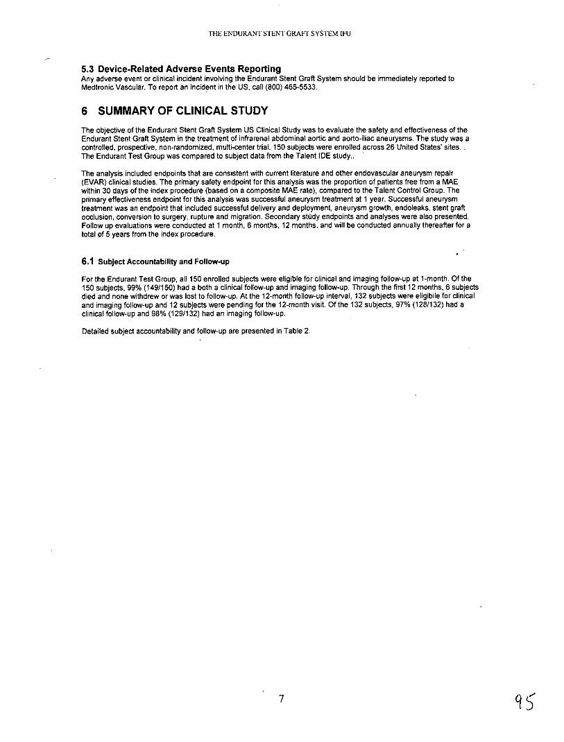
5.3 Device-Related Adverse Events ReportingAny adverse event or clinical incident involving the Endurant Stent Graft System should be immediately reported toMedtronic Vascular. To report an incident in the US, call (800) 465-5533.
6 SUMMARY OF CLINICAL STUDYThe objective of the Endurant Stent Graft System US Clinical Study was to evaluate the safety and effectiveness of theEndurant Stent Graft System in the treatment of infrarenal abdominal aortic and aorto-iliac aneurysms. The study was acontrolled, prospective, non-randomized, multi-center trial. 150 subjects were enrolled across 26 United States' sites. .The Endurant Test Group was compared to subject data from the Talent IDE study..
The analysis included endpoints that are consistent with current literature and other endovascular aneurysm repair(EVAR) clinical studies. The primary safety endpoint for this analysis was the proportion of patients free from a MAEwithin 30 days of the index procedure (based on a composite MAE rate), compared to the Talent Control Group. Theprimary effectiveness endpoint for this analysis was successful aneurysm treatment at 1 year. Successful aneurysmtreatment was an endpoint that included successful delivery and deployment, aneurysm growth, endoleaks, stent graftocclusion, conversion to surgery, rupture and migration. Secondary stody endpoints and analyses were also presented.Follow up evaluations were conducted at 1 month, 6 months, 12 months, and will be conducted annually thereafter for atotal of 5 years from the index procedure.
6.1 Subject Accountability and Follow-up
For the Endurant Test Group, all 150 enrolled subjects were eligible for clinical and imaging follow-up at 1-month. Of the150 subjects, 99% (149/150) had a both a clinical follow-up and imaging follow-up. Through the first 12 months, 6 subjectsdied and none withdrew or was lost to follow-up. At the 12-month follow-up interval, 132 subjects were eligibile for clinicaland imaging follow-up and 12 subjects were pending for the 12-month visit. Of the 132 subjects, 97% (1281132) had aclinical follow-up and 98% (129/132) had an imaging follow-up.
Detailed subject accountability and follow-up are presented in Table 2.
79

THE ENDURANT STENT GRAFT SYSTEM IFU
Table 2: Subject and Imaging Accountability - Endurant Test Group'
Subjectswit Subjects with adequate
Subject follow-up imaging imaging to assess the Subject events occurring beforeperfumeg parameter next visitperformed (oeLb(Core(Core Lab)
0. 0a
b Mo0th 0 0E E 0
05dc 0 0
Z0 >
Interval 00 a 0~5
(Day ~ ~ Z-0 9% (9)(8 ) a a9% (99%
(Analysis Window) W UU
Originally Enrolled 1501 01
Events after implantbut before a I Month 0 0 0 0 0visit
I Month 149 149 147 124 143 149(Day 1-90) (99/6) (990/6) (98%)) (83%/) (95%) (99%)
Events after I Month
visit but before a 6 0 2 0 0 0Month visit
6 Month 143 138 135 134 132 129 132 138(Day 9 1-304) (97%) (93%) (9 1%) (9 1%) (89%) (87%) (89%) (93%)
Events after 6 Monthvisit but before a 12 0 4 0 0 12Month visit
12 Month 32 128 129 128 125 127 123 125 129( Day 305') (97%) (98%) (97%) (95%) (96%) (93%) (95%) (98%)
TData analysis sample size varies for each of the timepoints above and in the following tables. This variability is due to subject availabilityfor follow-up, as well as, quantity and quality of images available from specific timepoints for evaluation. For example, the number andquality of images available for evaluation of endoleak at 6 months is different than the number and quality of images available at 12 monthsdue to variation in the number of image exams performed, the number of images provided from the clinical site to the Core Lab, or thenumber of images with acceptable evaluation quality.2 Technical observations assessed by imaging include stent-graft kinking, stent-graft twisting, stent-graft wireform fracture, suprarenal barestent fracture, anchor pin fracture, and stent-graft stenosis.3In cases where 12 month imaging follow-up data were not available, subsequent imaging follow-up data were used.
6.2 Study Demographics and Baseline Medical History
The demographics between the Endurant Test Group and Talent Control Group were comparable. The mean age andsex/gender distribution were similar between the 2 study groups. In addition, the baseline medical history were alsosimilar with high prevalence of hypertension, chronic obstructive pulmonary disease and tobacco use in the past 10 yearsin both study groups. The baseline SVS/AAVS risk classifications were also similar with over 80% subjects with SVS 2 orabove in both study groups.
Table 3 through Table 5 provides the demographics, baseline medical history and SVS risk classification of the EndurantTest Group and the Talent Control Group.

THE ENDURANT STENT GRAFT SYSTEM IFU
Table 3: Subject Demographics
Endurant Talent ControlParameter Statistics/Category Test Group p-value
(N=150) Group (N=166)
Age (years)
Mean+ SD 73.1 ± 8.0 74.1 ± 7.5 0.255
Median 73.0 76.0
Min, max 52, 88 51, 89
Sex/Gender % (m/n).
Male 91.3% (137/150) 91.6% (152/166) >0.999
Race % (m/n)
White 98.7% (148/150) 92.8% (154/166) 0.013
Non-white 1.3% (2/150) 7.2% (12/166)
'p-values were based on t-tests for continuous variables and Fisher's Exact test for categorical variables.
Table 4: Baseline Medical History
Endurant Talent ControlBody System / Condition Test Group Group p-value'
% (m/n) % (m/n)
Cardiovascular
Angina 18.0% (27/150) 16.9% (28/166) 0.882
Arrhythmia 39.3% (59/150) 44.0% (73/166) 0.426
Congestive heart failure 16.0% (24/150) 28.3% (47/166) 0.010
Hypertension 86.7% (130/150) 83.7% (139/166) 0.528
Myocardial infarction 30.0% (45/150) 38.6% (64/166) 0.124
Peripheral vascular disease 22.7% (34/150) 46.4% (77/166) <0.001
Renal
Renal insufficiency 28.7% (43/150) 33.1% (55/166) 0.397
Other abnormal body systems
Chronic obstructive pulmonary disease 35.3% (53/150) 39.2% (65/166) 0.488
Diabetes 26.7% (40/150) 15.7% (26/166) 0.019
Tobacco use in the last 10 years . 44.0% (66/150) 44.6% (74/166) >0.999
p-values were based on Fisher's Exact test.

THE ENDURANT STENT GRAFT SYSTEM IFU
Table 5: Baseline Modified SVS Classification
SVSMAAVS Endurant TalentTest Group Control Group p-value'
Classification%(r/)%(/) __________
% (m/n) % (m/n)
SVS O 0.0% (0/150) 0.6% (1/166) 0.802
SVS 1 16.0% (24/150) 15.7% (26/166)
SVS 2 54.7% (82/150) 55.4% (92/166)
SVS 3 29.3% (44/150) 28.3% (47/166)
p-value was based on the Cochran-Mantel-Haenzel test for mean score differences in SVS classification
6.3 Baseline Aneurysm Characteristics
Table 6 and Table 7 provide the baseline aneurysm and anatomical measurements of the Endurant Test Group andTalent Control Group.
Table 6: Baseline Aneurysm Characteristics (Corelab Reported)
D Endurant TalentDimension Statistics Test Group Control Group p-value
Maximum aneurysm 150 156diameter (mm)
Mean + SD 55.9 + 8.7 55.0 ± 9.3 0.359
Median 54 53
Min, Max 39. 103 38,88
Proximal neck diameter 150 156(mm)
Mean ± SD 23.5 ± 3.0 25.3 ± 3.6 <0.001
Median 23 26
Min, Max 17,31 16,32
Right iliac diameter (mm) n 148 148
Mean ± SD 14.2 ± 4.2 14.5 ± 3.6 0.447
Median 14 14
Min. Max 9, 48 7, 39
Left iliac diameter (mm) n 150 153
Mean SD 13.9 ± 3.1 14.3 L 3.8 0.347
Median 14 14
Min, Max 8, 24 8, 38
Proximal neck length n 150 154(mm)
Mean ± SD 31.0 ± 14.3 22.9+ 12.5 <0.001
Median 29 21
10

THE ENDURANT STENT GRAFT SYSTEM IFU
Dimension Statistics Endurant Talent 2Test Group Control Group p-valu
Min, Max 53 , 74 3, 75
Infrarenal neck angle (0) n 150 127
Mean ± SD 35.2 ± 13.7 30.5 ± 15.8 0.009
Median 34 30
Min, Max 5, 73 0, 72
Suprarenal neck angle (0) n 150 NA
Mean ± SD 16.0 ± 10.3 NA NA
Median 14 NA
Min, Max 2, 58 NA
'Number of subjects with readable scans.2 p-values were based on a two-sample t-test
' Based on Core Lab measurements, two (2) subjects had proximal neck length measurements <10 mm and were outside the margin of error;however, the site reported measurements were > 10 mm.
Table 7: Distribution of Aneurysm Diameters (Corelab reported)
Statistics/ Endurant Test Talent ControlCategory Group Group
Maximum Aneurysm <30 0.0% (0/150) 0.0% (0/156)Diameter 0/(mrd/n)
30 mm - <40 mm 0.7% (1/150) 1.3% (2/156)
40 mm - < 50 mm 16.0% (24/150) 26.3% (41/156)
50 mm - < 60 mm 63.3% (95/150) 44.2% (69/156)
60 mm - < 70 mm 13.3% (20/150) 20.5% (32/156)
70 mm - <80mm 4.0% (6/150) 5.8% (9/156)
80 mm - <90 mm 1.3% (2/150) 1.9% (3/156)
90 tn - < 100 mm 0.7% (1/150) 0.0% (0/156)
100 mm - < 110 mm 0.7% (1/150) 0.0% (0/156)
110 mm 0.0% (0/150) 0.0% (0/156)
Aneurysm Diameter /o(m/n) < 50 mm 16.7% (25/150) 27.6% (43/156)
Aneurysm Diameter %(m/n) 50 mm 83.3% (125/150) 72.4% (113/156)
n= number of subjects with readable scans.
11

THE ENDURANT STENT GRAFT SYSTEM LFU
6.4 Devices Implanted
Table 8 provides a breakdown of the number of Endurant Stent Grafts devices implanted at the index procedure persubject.
Table 8: Total Number of Devices Implanted at Initial Procedure
Number of Devices Endurant Test GroupImplanted on a Subject (%m/n)'
1 0.7% (1/150)
2 40.0% (60/150)
3 30.0% (45/150)
4 25.3% (38/150)
5 3.3% (5/150)
6 0.7% (1/150)
> 7 0.0% (01150)
Denominator includes all subjects who received the test device.
Sizes of Devices Implanted
Table 9 below shows the distribution of sizes of the bifurcated stent graft used in the Endurant US Clinical Study.
Table 9: Devices Implanted by Size at Index Procedure
Stent Graft Proximal Diameter Endurant(Main Bifurcated, mm) % (mn/)'
23 10.7% (16/150)
25 26.0% (39/150)
28 36.7% (55/150)
32 22.0% (33/150)
36 4.7% (7/150)
'Denominator includes all subjects who received the main bifurcated test device.
6.5 Study Results: Safety Endpoints
6.5.1 Major Adverse Events (MAEs) Free Rate within 30 Days
Table 10 through Table 11 provide an analysis of the MAEs within 30 days. 96.0% subjects in the Endurant TestGroup were MAE-free as compared to 89.2% subjects in the Talent Control Group.
Table 10: MAE free-rate within 30 Days
Endurant TalentMAE Free Rate Test Group Control Groupwithin 30 Days (%m/n) (%m/n)
MAEs free-rate within 30 96.0% (144/150) 89.2% (148/166)Days
12

THE ENDURANT STENT GRAFT SYSTEM IFU
Table 11: MAE Components within 30 Days
Endurant TalentMajor Adverse Event (MAE) Test Group Control Groupwithin 30 Days' (%m/n) (%m/n)
MAE within 30 days 4.0% (6/150) 10.8% (18/166)
All-cause Death 0.0% (0/150) 1.8% (3/166)
Myocardial Infarction 0.7% (1/150) 1.8% (3/166)
Renal Failure 0.7% (1/150) 1.8% (3/166)
Respiratory Failure 1.3% (2/150) 3.0% (5/166)
Paraplegia 0.0% (0/150) 0.0% (0/166)
Stroke 0.7% (1/150) 1.2% (2/166)
Bowel Ischemia 1.3% (2/150) 0.6% (1/166)
Procedural Blood Loss 1000cc 0.7% (1/150) 5.4% (9/166)
'A subject may report multiple MAEs; hence, number of subjects with any MAE may not be the sum of those in eachMAE category.
13 101

THE ENDURANT STENT GRAFT SYSTEM IFU
6.5.2 Major Adverse Events (MAEs) Free Rate within 12 Months
Table 12 through Table 13 provide an analysis of the MAEs within 12 months. 89.2% subjects in the Endurant TestGroup were MAE-free as compared to 80.4% subjects in the Talent Control Group.
Table 12: MAE free-rate within 12 Months
Endurant TalentTest Group Control Group
MAEs free rate within 12 months (%m/n)' (%m/n)'
MAEs free-rate within 12 months 89.2% (124/139) 80.4% (123/153)
'Denominator includes all subjects who had MAE(s) within 365 days or those were followed for at least 305 days.
Table 13: Major Adverse Events through 12 Months
Endurant TalentTest Group Control Group
MAEs within 12 months' (%m/n)2 (%m/n)z
MAE within 12 months 10.8% (15/139) 19.6% (30/153)
All-cause Death 4.3% (6/139) 6.5% (10/153)
Myocardial Infarction 1.4% (2/139) 3.9% (6/153)
Renal Failure 2.2% (3/139) 3.3% (5/153)
Respiratory Failure 2.2% (3/139) 3.9% (6/153)
Paraplegia 0.0% (0/139) 0.0% (0/153)
Stroke 2.9% (4/139) 2.6% (4/153)
Bowel Ischemia 1.4% (2/139) 0.7% (1/153)
Procedural Blood Loss > 1000cc 0.7% (1/139) 5.9% (9/153)
A subject may report multiple MAEs; hence, number of subjects with any MAE may not be the sum of those ineach MAE category.2 Denominator includes all subjects who had MAE(s) within 365 days or were followed for at least 305 days.
14 10)2

THE ENDURANT STENT GRAFT SYSTEM IFU
In addition a Kaplan-Meier analysis of freedom from MAEs was performed and is plotted below in Figure 4. Kaplan-Meier analysis predicts a freedom from MAE rate within 12 months of 89.3% in the Endurant Test Group ascompared to 81.3% in the Talent Control Group. The data used in the Kaplan-Meier analysis is presented below.
1.1
) 0.9
* 0.8
E
>0.7 Endlurant: 89.3% ± 1.96*2.6%Talent Control: 81.3% ± 1.96*3.1%
i 0.6
E 0.30w 0.2 Top=Endurant, Bottom=Talent Controlu. Number of subjects at risk:
0.1 160 144 140 70166 142 136 120
0,0 30 60 90 120 150 180 210 240 270 300 330 360 390
Days from Initial ProcedureFigure 4: Kaplan-Meier Analysis: Freedom from MAEs through 12 Months
Table 14: Kaplan-Meier Estimates of Freedom from MAEs hrough 12 Months
Endurant Test Group Talent Control Group
Treatment 31 to 182 183 to 365 Treatment 31 to 182 183 to 365to 30 days days days to 30 days days days
No. at Risk' 150 144 140 166 142 136
No. of Events 6 4 5 18 4 8
No. Censored2 0 0 65 6 2 8
Kaplan-MeierEstimate' 0.960 0.933 0.893 0.891 0.866 0.813
Standard Error 0.016 0.020 0.026 0.024 0.027 0.031
Number of subjects at risk at the beginning of interval.2 Subjects are censored because their last follow-up has not reached the end of the time interval or because they arelost to follow-up.3 Estimate made at end of time interval.
15 103

THE ENDURANT STENT GRAFT SYSTEM IFU
6.5.3 Aneurysm-related Mortality (ARM) Free-Rate within 12 Months
The ARM free rate within 12 months was 100% in the Endurant Test Group compared to 97.9% in the Talent ControlGroup as shown in Table 15.
Table 15: Aneurysm-related Mortality Free-Rate within 12 Months
Endurant TalentAneurysm-Related Mortality Free Test Group Control GroupRate % (m/n)' % (m/n)'
Aneurysm-Related Mortality Free 100.0% (133/133) 97.9% (143/146)Rate within 12 Months
'Denominators included all subjects who had the event within 365 days or those were followed for at least 305 days.
In addition a Kaplan-Meier analysis of freedom from ARM was performed and is plotted below in Figure 5. Kaplan-Meier analysis predicts a freedom from ARM rate within 12 months of 100% in the Endurant Test Group ascompared to 98.2% in the Talent Control Group. The data used in the Kaplan-Meier analysis is presented below.
1.1
1. ---------------------------------------------
o 0.9
0.8
0 0.7 Endurant: 100% ± 1.96*0.0%Talent Control: 98.2% ± 1.96*1.0%
E 0.6
S0.5
0.4E0
0.3Eo 0.2 Top=Endurant, Bottom=Talent Control0 Number of subjects at risk:2 0.1 150 150 147 75u. 166 157 151 139
0,0 30 60 90 120 150 180 210 240 270 300 330 360 390
Days from Initial Procedure
Figure 5: Kaplan-Meler Analysis: Freedom from Aneurysm-Related Mortality within 12 Months
Table 16: Kaplan-Meier Estimates of Freedom from Aneurysm-Related Mortality through 12 months
Endurant Test Group Talent Control Group
Treatment 31 to 182 183 to 365 Treatment 31 to 182 183 to 365to 30 days days days to 30 days days days
No. at Risk' 150 150 147 166 157 151
No. of Events 0 0 0 3 0 0
No. Censored' 0 3 72 6 6 12
Kaplan-Meier 1.000 1.000 1.000 0.982 0.982 0.982Estimate
Standard Error 0.000 0.000 0.000 0.010 0.010 0.010
Number of subjects at risk at the beginning of interval.2 Subjects are censored because their last follow-up has not reached the end of the time interval or because they arelost to follow-up.Estimate made at end of time interval.
16

THE ENDURANT STENT GRAFT SYSTEM IFU
6.5.4 All-cause Mortality Free Rate within 30 Days
Table 17 provides the all-cause mortality free rate within 30 days for the Endurant Test Group and Talent ControlGroup.
Table 17: All-cause Mortality Free Rate within 30 Days
Endurant TalentTest Group Control Group
All-Cause Mortality Free Rate (%m/n) (%m/n)
All-Cause Mortality Free Rate within 30 Days 100.0% (150/150) 98.2% (163/166)
6.5.5 All-cause Mortality Free Rate within 12 Months
Table 18 provides the all-cause mortality free rate within 12 months for the Endurant Test Group and Talent ControlGroup.
Table 18: All-Cause Mortality Free Rate within 12 Months
Endurant TalentTest Group Control Group
All-cause Mortality Free Rate (%m/n) (%m/n)
All-cause Mortality Free Rate within 12 Months 95.7% (132/138) 93.4% (141/151)
'Denominators included all subjects who had the event within 365 days or those were followed for at least 305 days.
In addition, a Kaplan-Meier analysis of freedom from All-cause Mortality was performed and plotted below inFigure 6. Kaplan-Meler analysis predicts a freedom from all-cause mortality within 12 months of 95.8% in theEndurant Test Group as compared to 93.7% in the Talent Control Group. The data used in the Kaplan-Meieranalysis is presented below.
1.1
1.0 rit---------
0.9
o 0.8a,
0.7 Endurant: 95.8% ± 1.96*1.7%Talent Control: 93.7% ± 1.96*1.9%
90.6
0.5E~0S0.4Eo 0.3
0.2 Top=Endurant, Bottom=Talent ControlNumber of subjects at risk:
0.1 150 150 147 75166 157 151 139
0 - I I I
0 30 60 90 120 150 180 210 240 270 300 330 360 390Days from Initial Procedure
Figure 6: Kaplan-Meier Analysis: Freedom from All-cause Mortality within 12 Months
17

THE ENDURANT STENT GRAFT SYSTEM IFU
Table 19:Kaplan-Meier Estimates of Freedom from All-Cause Mortality through 12 Months
Endurant Test Group Talent Control Group
Treatment 31 to 182 183 to 365 Treatment 31 to 182 183 to 365to 30 days days days to 30 days days days
No. at Risk' 150 150 147 166 157 151
No. of Events 0 2 4 3 3 4
No. Censored2 0 1 68 6 3 8
Kaplan-MeierEstimate' 1.000 0.987 0.958 0.982 0.963 0.937
Standard Error 0.000 0.009 0.017 0.010 0.015 0.019
Number of subjects at risk at the beginning of interval.2 Subjects are censored because their last follow-up has not reached the end of the time interval or because they arelost to follow-up.'Estimate made at end of time interval.
6.6 Study Results: Effectiveness Endpoints
6.6.1 Technical Success
During the index procedure, 99.3% subjects in the Endurant Test Group were recorded as having successful deliveryand deployment of the Endurant Bifurcated Stent Graft compared to 97.6% of the Talent Control Group. One subjectin the Endurant Test Group had the main bifurcated body implanted but the physician was not able to cannulate thecontralateral gate due to a pre-existing challenging anatomy. The subject was ultimately converted to aorto-uni-iliacin-situ and a femoral to femoral bypass was performed.
Table 20: Technical success
Endurant TalentTest Group Control Group
99.3% 97.6%Technical Success' (149/150) (162/166)
Defined as the successful delivery and deployment of the stent graft.
6.6.2 Successful Aneurysm Treatment
The overall succcessful aneurysm treatment rate through 12 months in the Endurant Test was 97.5% as comparedto 87.1% in the Talent Control Group as shown in Table 21.
Successful aneurysm treatment was an endpoint that included delivery and deployment of the graft and surrogatemarkers that represented treatment success. This included aneurysm growth, endoleak, occlusion, conversion tosurgery, rupture and migration. The information on these endpoints is presented in the sections below.
There were three subjects in the Endurant Test Group that were considered treatment failures. In addition to thetechnical failure noted above, one subject experienced an aneurysm rupture at the index procedure and the otherhad a stent graft occlusion necessitating a femoral to femoral bypass.
Table 21: Successful Aneurysm Treatment
Endurant TalentTest Group Control Group
% (m/n)' % (m/n)'
Successful Aneurysm 97.5% (118/121) 87.1% (108/124)Treatment
'Denominator is number of subjects evaluable for this endpoint
18

THE ENDURANT STENT GRAFT SYSTEM IFU
6.6.3 Change in Aneurysm Diameter
Table 22 provides the change in aneurysm diameter as identified by Core Lab from 1 month to 12 months. In theEndurant Test Group, there were no aneurysm diameter increase >5 mm whereas the Talent Control Group reportedthat 2.3% of subjects had an aneurysm growth > 5 mm. About 50% subjects had decrease of aneurysm size greaterthan 5 mm.
Table 22: Aneurysm Diameter Change from I Month to 12 Months (Core Lab)
Endurant TalentChange in Maximum Aneurysm Diameter Test Group Control Groupfrom I Month to 12 Months' % (m/n)2 % (m/n)'
Increase more than 5 mn 0.0% (0/127) 2.3% (3/128)
Stable3 50.4% (64/127) 64.8% (83/128)
Decrease more than 5 mm. 49.6% (63/127) 32.8% (42/128)
Change in aneurysm diameter is based on I-month imaging. When I-month imaging was not available, the pre-discharge imaging was used as the baseline.2 Denominator is number of subjects evaluable for this endpoint.'Stable refers to no change (increase or decrease) of more than 5 mm.
6.6.4 Endoleak by Visit
Table 23 shows all types of endoleak as identified by Core Lab at 1 month, 6 months and 12 months for Endurantand Talent. There were no Type I and/or II endoleaks at 1 month, 6 months and 12 months in the Endurant group.
Table 23: All Endoleaks at 1-Month, 6-Months and 12-Months (Core Lab)
I Month 6 Months . 12 Months
Endurant Talent Endurant Talent Endurant TalentTest Control Test Control Test Control
Group Group Group Group Group GroupEndoleaks % (m/n') % (m/n1 ) % (m/n') % (m/n') % (m/n') % (m/n')
0.0% 9.3% 0.0% 4.2% 0.0% 2.5%Type 1
(0/143) (14/151) (0/129) (5/118) (0/123) (3/122)
Type 11 16.1% 8.6% 11.6% 8.5% 8.9% 6.6%
(23/143) (13/151) (15/129) (10/118) (11/123) (8/122)
0.0% 0.0% 0.0% 0.0% 0.0% 0.0%Type III
(0/143) (0/151) (0/129) (0/118) (0/123) (0/122)
0.0% 0.0% 0.0% 0.0% 0.0% 0.0%Type IV
(0/143) (0/151) (0/129) (0/118) (0/123) (0/122)
0.0% 1.3% 0.0% 1.7% 0.8% 0.8%Indeterminate
(0/143) . (2/151) (0/129) (2/118) (1/123) (1/122)
Subjects had 16.1% 19.2% 11.6% 14.4% 9.8% 9.8%endoleaks of any type2 (23/143) (29/151) (15/129) (17/118) (12/123) (12/122)
' Denominator is the number of subjects who had readable images at the time of assessment.'A subject may have more than I type of endoleaks; hence, number of subjects with any type may not be the sum ofthose in each type.
THE ENDURANT STENT GRAFT SYSTEM IFU
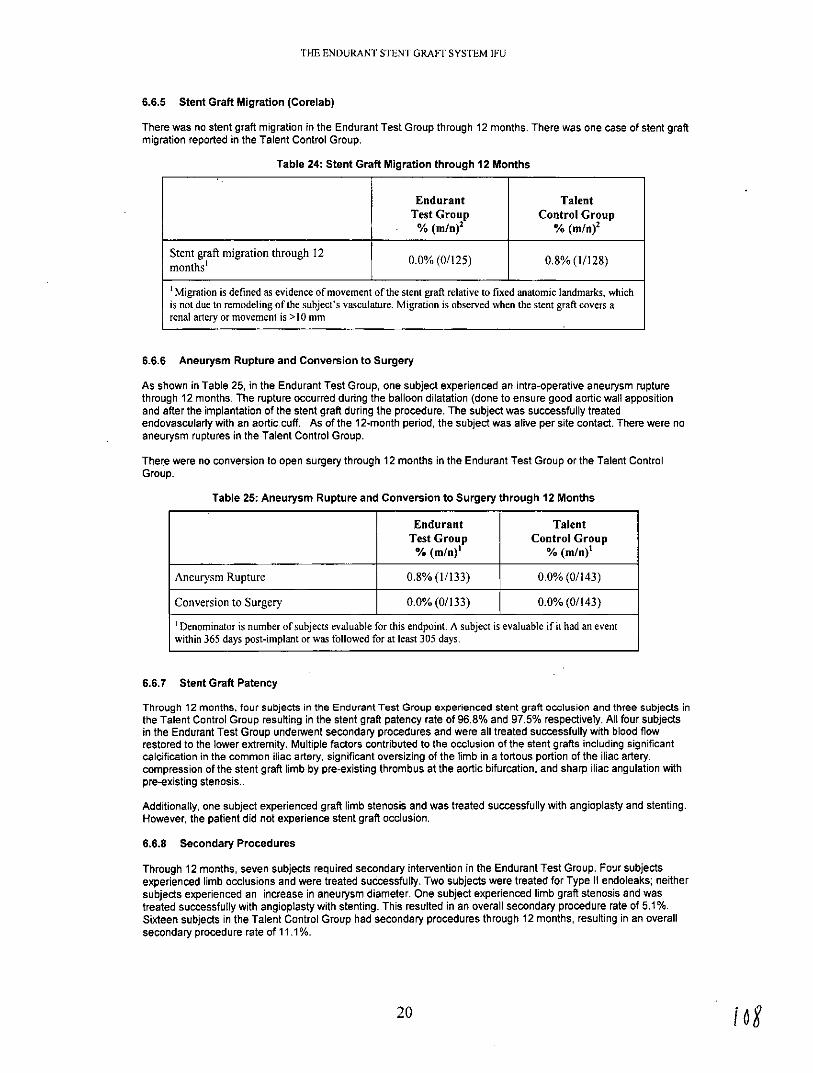
6.6.5 Stent Graft Migration (Corelab)
There was no stent graft migration in the Endurant Test Group through 12 months. There was one case of stent graftmigration reported in the Talent Control Group.
Table 24: Stent Graft Migration through 12 Months
Endurant TalentTest Group Control Group% (m/n) 2 % (m/n)2
Stent graft migration through 12monhs'0.0% (0/125) U.% (1/128)months'
'Migration is defined as evidence of movement of the stent graft relative to fixed anatomic landmarks, whichis not due to remodeling of the subject's vasculature. Migration is observed when the stent graft covers arenal artery or movement is >10 mm
6.6.6 Aneurysm Rupture and Conversion to Surgery
As shown in Table 25, in the Endurant Test Group, one subject experienced an intra-operative aneurysm rupturethrough 12 months. The rupture occurred during the balloon dilatation (done to ensure good aortic wall appositionand after the implantation of the stent graft during the procedure. The subject was successfully treatedendovascularly with an aortic.cuff. As of the 12-month period, the subject was alive per site contact. There were noaneurysm ruptures in the Talent Control Group.
There were no conversion to open surgery through 12 months in the Endurant Test Group or the Talent ControlGroup.
Table 25: Aneurysm Rupture and Conversion to Surgery through 12 Months
Endurant TalentTest Group Control Group% (m/n)' % (m/n)'
Aneurysm Rupture 0.8% (1/133) 0.0% (0/143)
Conversion to Surgery 0.0% (0/133) 0.0% (0/143)
'Denominator is number of subjects evaluable for this endpoint. A subject is evaluable if it had an eventwithin 365 days post-implant or was followed for at least 305 days.
6.6.7 Stent Graft Patency
Through 12 months, four subjects in the Endurant Test Group experienced stent graft occlusion and three subjects inthe Talent Control Group resulting in the stent graft patency rate of 96.8% and 97.5% respectively. All four subjectsin the Endurant Test Group underwent secondary procedures and were all treated successfully with blood flowrestored to the lower extremity. Multiple factors contributed to the occlusion of the stent grafts including significantcalcification in the common iliac artery, significant oversizing of the limb in a tortous portion of the iliac artery,compression of the stent graft limb by pre-existing thrombus at the aortic bifurcation, and sharp iliac angulation withpre-existing stenosis..
Additionally, one subject experienced graft limb stenosis and was treated successfully with angioplasty and stenting.However, the patient did not experience stent graft occlusion.
6.6.8 Secondary Procedures
Through 12 months, seven subjects required secondary intervention in the Endurant Test Group. Four subjectsexperienced limb occlusions and were treated successfully. Two subjects were treated for Type 11 endoleaks; neithersubjects experienced an increase in aneurysm diameter. One subject experienced limb graft stenosis and wastreated successfully with angioplasty with stenting. This resulted in an overall secondary procedure rate of 5.1%.Sixteen subjects in the Talent Control Group had secondary procedures through 12 months, resulting in an overallsecondary procedure rate of 11.1%.
20
THE ENDURANT STENT GRAFT SYSTEM IFU
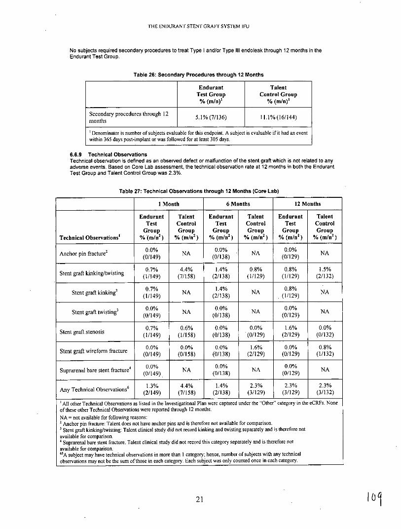
No subjects required secondary procedures to treat Type I and/or Type III endoleak through 12 months in theEndurant Test Group.
Table 26: Secondary Procedures through 12 Months
Endurant TalentTest Group Control Group% (m/n)' % (m/n)'
Secondary procedures through 12 5.1% (7/136) 11.1% (16/144)months
'Denominator is number of subjects evaluable for this endpoint. A subject is evaluable if it had an eventwithin 365 days post-implant or was followed for at least 305 days.
6.6.9 Technical ObservationsTechnical observation is defined as an observed defect or malfunction of the stent graft which is not related to anyadverse events. Based on Core Lab assessment, the technical observation rate at 12 months in both the EndurantTest Group and Talent Control Group was 2.3%.
Table 27: Technical Observations through 12 Months (Core Lab)
1 Month 6 Months 12 Months
Endurant Talent Endurant Talent Endurant TalentTest Control Test Control Test Control
Group Group Group Group Group GroupTechnical Observations' I (m/n2 ) % (n/n 2 ) % (m/n2) % (m/n 2) % (m/n2) % (m/n 2 )
Anchor pin fracture 0.0% NA 0.0% NA 0.0% NA(0/149) (0/138) (0/129)
0.7% 4.4% 1.4% 0.8% 0.8% 1.5%Stent graft kinking/twisting (1/149) (7/158) (2/138) (1/129) (1/129) (2/132)
0.7% NA1.4% NA0.8% NStent graft kinking, .7 NA 1.%NA 0.%NA
(1/149) (2/138) . (1/129)
Stent graft twisting 0.0% NA 0.0% NA 0.0% NA(0/149) (0/138) (0/129)
0.7% 0.6% 0.0% 0.0% 1.6% 0.0%Stent graft stenosis (1/149) (1/158) (0/138) (0/129) (2/129) (0/132)
0.0% 0.0% 0.0% 1.6% 0.0% 0.8%Stent graft wireform fracture (0/149) (0/158) (0/138) (2/129) (0/129) (1/132)
Supraenal bare stent fracture 4 0.0% NA 0.0% 0.0% NA(0/149) (0/138) NA (0/129)
Any Technical Observations6 1.3% 4.4% 1.4% 2.3% 2.3% 2.3%(2/149) (7/158) (2/138) (3/129) (3/129) (3/132)
'All other Technical Observations as listed in the Investigational Plan were captured under the "Other" category in the eCRFs. Noneof these other Technical Observations were reported through 12 months.NA = not available for following reasons:2 Anchor pin fracture: Talent does not have anchor pins and is therefore not available for comparison.' Stent graft kinking/twisting: Talent clinical study did not record kinking and twisting separately and is therefore notavailable for comparison.
Suprarenal bare stent fracture: Talent clinical study did not record this category separately and is therefore notavailable for comparison."A subject may have technical observations in more than I category; hence, number of subjects with any technicalobservations may not be the sum of those in each category. Each subject was only counted once in each category.
21

THE ENDURANT STENT GRAFT SYSTEM IFU
6.7 Acute Procedural Data
Table 28 compares the clinical utility measures of the Endurant Test Group to the Talent Control Group. Acuteprocedural outcomes for the Endurant Test Group and the Talent Control Group with respect to procedure duration,blood loss, blood transfusion, time in the Intensive Care Unit (ICU) and length of stay in the hospital are presentedbelow.
Table 28: Acute Procedural Data
Endurant Talent ControlAcute Procedural Data Statistics Test Group Group
Duration of procedure (min) N 150 166
Mean ± SD 101.5 ± 46.2 167.3 ± 53.2
Median 91.0 155.0
Min, Max 34. 318 85, 417
Subjects receiving general % (m/n) 83.3% (125/150) 40.4% (67/166)anesthesia
Estimated blood loss (cc) N 149 165
Mean ± SD 184.9 ± 167.9 335 ± 282.4
Median 150.0 250.0
Min, Max 0,1450 25, 1750
Subjects requiring blood % (m/n) 0.7% (1/150) 18.2% (30/165)transfusion
Time in ICU (hours) N 150 166
Mean ± SD 6.2 ± 19.4 19.3 ± 73.9
Median 0.0 0.0
Min, Max 0, 135 0,864
Overall hospital stay (days) N 150 166
Mean ± SD 2.1 + 2.3 3.6 ± 6.4
Median 1.0 2.0
Min, Max 1,17 1,79
22

THE ENDURANT STENT GRAFT SYSTEM IFU
7 PATIENT SELECTION AND TREATMENT
7.1 Individualization of TreatmentEach Endurant Stent Graft Component must be ordered in a size appropriate to fit the patient's anatomy. Proper sizing ofthe device is the responsibility of the physician. The stent graft component should be oversized to be larger than thevessel inner diameter (aortic components are oversized approximately 10-20%; limb components are oversizedapproximately 10-25%). Refer to section 9.2, Recommended Device Sizing, for further details. The Endurant Stent Graftcomponents cover aortic diameters ranging from 19 mm to 32 mm and iliac diameters from 8 mm to 25 mm. Therecommended overall length of the Endurant Stent Graft, including multiple deployed components, should extend from thelowest renal artery to just above the internal iliac (hypogastric) artery. All lengths and diameters of the stent graftcomponents necessary to complete the procedure should be available to the physician, especially when pre-operativecase planning measurements (treatment diameters and lengths) are not certain. Use of this approach allows for greaterintraoperative flexibility to achieve optimal procedural outcomes.
Medtronic may consult with physicians to determine proper stent graft component dimensions based on the physician'sassessment of the patient's anatomical measurements. The benefits and risks previously described must be consideredfor each patient before use of the Endurant Stent Graft System.
Caution: Vessel over-distension and damage, or partial stent graft infolding, may be caused by excessive oversizing ofthe stent graft in relation to the diameter of the blood vessel. Also, due to the nature of the design and the flexibility of theEndurant Stent Graft System, the overall length of each stent graft component may be shorter when deployed.
7.2 Patient Counseling InformationThe physician should review the following risks and benefits when counseling the patient about this endovascular deviceand procedure:* age and life expectancy* risks and benefits related to open surgical repair* risks and benefits related to endovascular repair* risks related to noninterventional treatment (medical management)* risks of aneurysm rupture compared to endovascular repair* possibility that subsequent endovascular or open surgical repair of the aneurysm may be required* the long-term safety and effectiveness of the Endurant Stent Graft System has not been established* long-term, regular follow-up is needed to assess patient's health status and stent graft performance* patients with specific clinical findings (eg, endoleaks, enlarging aneurysms) should be monitored closely* symptoms of aneurysm rupture
Medtronic recommends that the physician disclose to the patient, in written form, all risks associated with treatment usingthe Endurant Stent Graft System. Details regarding risks occurring during and after implantation of the device areprovided in Section 5, Adverse Events. Additional counseling information can be found in the Patient Information Booklet.
8 HOW SUPPLIED
8.1 SterilityEach Endurant Stent Graft Component (bifurcated, contralateral limb, and aortic and iliac extensions) is individuallycontained within an Endurant Delivery System, which is sterilized using Electron Beam sterilization. The Endurant StentGraft System is supplied sterile for single use only.* Do not reuse or attempt to resterilize.* If the device is damaged or the integrity of the sterilization barrier has been compromised, do not use the product
and contact your Medtronic Vascular representative for return information.
8.2 Contents* One Endurant Stent Graft System* One Set of Patient Tracking Materials
8.3 StorageStore the system at room temperature in a dark, dry place.
23

THE ENDURANT STENT GRAFT SYSTEM IFU
9 CLINICAL USE INFORMATION
9.1 Physician Training RequirementsAll Physicians should complete in-service training prior to using the Endurant Stent Graft System.
Caution: The Endurant Stent Graft System should only be used by physicians and teams trained in vascularinterventional techniques and in the use of this device.
Below are the skill/knowledge requirements for physicians using the Endurant Stent Graft System:
* Natural history of AAA and aorto-iliac aneurysms, and co-morbidities associated with AAA repair* Radiographic, fluoroscopic, and angiographic image interpretation* Appropriate use of radiographic contrast material* Arterial cutdown, arteriotomy, and repair* Percutaneous access and closure techniques* Nonselective and selective guidewire and catheter techniques* Fluoroscopic and angiographic image interpretation* Embolization* Angioplasty* Endovascular stent placement* Snare techniques* Techniques to minimize radiation exposure* Device selection and sizing
9.2 Recommended Device SizingThe Endurant Stent Graft System components are available in the sizes described in Tables 29-32. If you have questionsabout the product or sizing, refer to contact information in the back of the manual.
Table 29: Sizing Chart - Bifurcated Stent Graft
Proximal x Distal Diameter (mm x Covered Length Vessel inner00 (Fr) mm) (mm) diameter (mm)
36x2036x16 145, 166 29-32
32x2020 32x16 26-28
28x2028x16 23-2528x13 124, 145, 16625x16
2 1-2218 25x13
23x1619-2 0
23x13
Table 30: Sizing chart - Aortic Extension
Proximal x Distal Diameter (mm x Covered Length Vessel innerOD (Fr) mm) (mm) diameter (mm)
36x36 29-3220 32x32 26-28
28x28 49/70 23-2525x25 21-22
1 23x23 19-20
Table 31: Sizing Chart - Contralateral Limb
Proximal x Distal Diameter (mm x Covered Length Vessel innerOD (Fr) mm) (mm) diameter (mm)
16x28 23-2516 16x24 19-22
16x20 15-1816x16 82,93, 124 12-14
14 16x13 10-1116x10 8-9
24
THE ENDURANT STENT GRAFT SYSTEM IFU
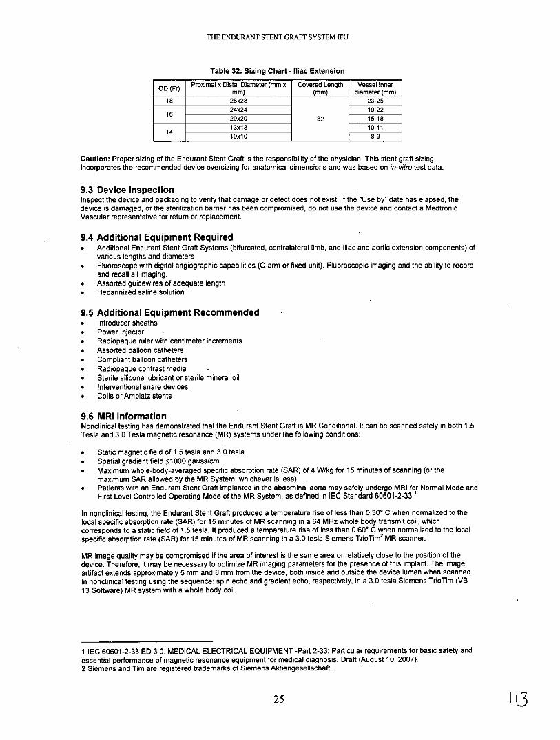
Table 32: Sizing Chart - Iliac Extension
OD (Fr) Proximal x Distal Diameter (mm x Covered Length Vessel innermm) (mm) diameter (mm)
18 28x28 23-2524x24 19-2216 20x20 82 15-1813x13 10-1114 lOx1O 8-9
Caution: Proper sizing of the Endurant Stent Graft is the responsibility of the physician. This stent graft sizingincorporates the recommended device oversizing for anatomical dimensions and was based on in-vitro test data.
9.3 Device InspectionInspect the device and packaging to verify that damage or defect does not exist. If the "Use by" date has elapsed, thedevice is damaged, or the sterilization barrier has been compromised, do not use the device and contact a MedtronicVascular representative for return or replacement.
9.4 Additional Equipment Required* Additional Endurant Stent Graft Systems (bifurcated, contralateral limb, and iliac and aortic extension components) of
various lengths and diameters* Fluoroscope with digital angiographic capabilities (C-arm or fixed unit). Fluoroscopic imaging and the ability to record
and recall all imaging.* Assorted guidewires of adequate length* Heparinized saline solution
9.5 Additional Equipment Recommended* Introducer sheaths* Power Injector* Radiopaque ruler with centimeter increments* Assorted balloon catheters* Compliant balloon catheters* Radiopaque contrast media* Sterile silicone lubricant or sterile mineral oil* Interventional snare devices* Coils or Amplatz stents
9.6 MRI InformationNonclinical testing has demonstrated that the Endurant Stent Graft is MR Conditional. It can be scanned safely in both 1.5Tesla and 3.0 Tesla magnetic resonance (MR) systems under the following conditions:
* Static magnetic field of 1.5 testa and 3.0 tesla* Spatial gradient field 51000 gauss/cm* Maximum whole-body-averaged specific absorption rate (SAR) of 4 W/kg for 15 minutes of scanning (or the
maximum SAR allowed by the MR System, whichever is less).* Patients with an Endurant Stent Graft implanted in the abdominal aorta may safely undergo MRI for Normal Mode and
First Level Controlled Operating Mode of the MR System, as defined in IEC Standard 60601-2-33.1
In nonclinical testing, the Endurant Stent Graft produced a temperature rise of less than 0.30* C when normalized to thelocal specific absorption rate (SAR) for 15 minutes of MR scanning in a 64 MHz whole body transmit coil, whichcorresponds to a static field of 1.5 tesla. It produced a temperature rise of less than 0.60* C when normalized to the localspecific absorption rate (SAR) for 15 minutes of MR scanning in a 3.0 tesla Siemens TrioTim 2 MR scanner.
MR image quality may be compromised if the area of interest is the same area or relatively close to the position of thedevice. Therefore, it may be necessary to optimize MR imaging parameters for the presence of this implant. The imageartifact extends approximately 5 mm and 8 mm from the device, both inside and outside the device lumen when scannedin nonclinical testing using the sequence: spin echo and gradient echo, respectively, in a 3.0 tesla Siemens TrioTim (VB13 Software) MR system with a whole body coil.
1 IEC 60601-2-33 ED 3.0. MEDICAL ELECTRICAL EQUIPMENT -Part 2-33: Particular requirements for basic safety andessential performance of magnetic resonance equipment for medical diagnosis. Draft (August 10, 2007).2 Siemens and Tim are registered trademarks of Siemens Aktiengesellschaft.
25

THE ENDURANT STENT GRAFT SYSTEM IFU
10 IMPLANT INSTRUCTIONS
10.1 Vasular Access and Device PreparationCorrect sizing of the aorta and iliac vessels must be determined before implantation of the bifurcated and iliac stent graftcomponents using contrast-enhanced computer-aided tomography (CT), as well as angiograms of both the iliac arteriesand aorta. 3D imaging may also be beneficial. Refer to section 9.2, Recommended Device Sizing. These images shouldbe available for review during the procedure. Vascular instruments and other surgical supplies needed to gain access tothe artery should also be available.
To reduce the risk of thromboembolism, it is recommended that the patient be heparinized for the duration of theprocedure.
Caution: Do not retract the graft cover of the delivery system until it is accurately placed within the vasculature and readyfor deployment.
Caution: Never advance or retract equipment from the vasculature without the use of fluoroscopy.
10.1.1 Vascular Accessa. Following aseptic procedure, perform a vascular access at the femoral arteries.b. Place a guidewire in the ipsilateral femoral artery and advance it above the renal arteries.c. From the contralateral side femoral artery, place a second guidewire directed to the abdominal aorta.d. Over this guidewire, place an angiography catheter above the renal arteries.e. Take an angiogram.Note: There is a possibility that an additional incision might be necessary to access the common iliac artery.
10.1.2 Device Preparationa. Prior to insertion, view the delivery system under fluoroscopy to visualize the radiopaque markers on the stent graft.
The radiopaque markers indicate the position of the proximal and distal edges of the graft material.b. Turn the graft cover to align the radiopaque marker on the short stub leg (of the bifurcated component) with the
patient's contralateral iliac artery.c. Flush the guidewire lumen with heparinized saline.
d. Wet the graft cover of the delivery system to activate the hydrophilic coating
10.2 Delivery ProcedureMedtronic Vascular recommends using an appropriate caliber introducer sheath to perform diagnostic tests.
Warning: To prevent thrombotic problems, a second bolus of IV heparin is recommended before inserting the device.
10.2.1 Introduction of Bifurcated ComponentWarning: Do not advance the delivery system without first having placed a guidewire.a. Slowly insert the aortic delivery system.b. Advance over the guidewire so that the proximal most stents and the radiopaque markers are visualized in the target
proximal aortic neck (Figure 7).c. Inject contrast media into the abdominal aorta and mark the position of the target location, either on the imaging
screen or on the patient's body.d. Adjust the position of the bifurcated stent graft component so that the top edge of the graft fabric is just below the
lowest renal artery. (The edge of the graft fabric is 0.5 mm -1.0 mm above the top edge proximal radiopaquemarkers.)
Note: If the top edge of the graft fabric is to be placed very close to the renal arteries, contrast media may be injected toidentify the location of the lower renal artery and verify the position before full deployment.Caution: Once proximal position has been identified, do not move the patient or imaging equipment, as it maycompromise accuracy of stent graft placement.Caution: The angiographic catheter can be removed prior to deployment. However, if the angiographic catheter is notremoved until after deployment, ensure that the tip is straightened (pigtail catheter) with a guidewire before removal sothat the stent graft is not pulled down.Caution: When aligning the position of stent graft, be sure the fluoroscope is angled perpendicularly to the center line ofthe infrarenal aorta to avoid parallax or other source of visualization error. Some cranial caudal angulation of the I-I tubemay be necessary to achieve this, especially if there is anterior angulation of the aneurysm neck.
26

THE ENDURANT STENT GRAFT SYSTEM IFU
Figure 7: Introduction of Delivery System
10.2.2 Confirm Positiona. Ensure that the distal portion of the contralateral stub leg is above the aortic bifurcation and within the aneurysmal
sac, and not within the iliac vessel.b. Rotate the handle until the radiopaque marker on the distal-most stent of the contralateral stub leg is aligned with the
contralateral iliac artery.Note: When attempting to rotate the system, if a tip response is not observed, pull back the system and re-position untilthe intended position is achieved.
10.2.3 Deploy Promimal End of Bifurcated Componenta. With 1 hand on the front grip, hold the delivery system stationary.b. With the other hand, slowly withdraw the graft cover by rotating the slider counterclockwise (in the direction of the
slider arrow), until the constrained suprarenal stent is released and 2-3 of the covered stents have been fullydeployed (Figure 8).
c. Use angiography to verify position of the bifurcated component in relation to the renal arteries.If needed, gently push the entire delivery system proximally or pull distally until the proximal end of the graft materialis even with the distal edge of the lowest renal artery.
Note: In the unlikely event of delivery system failure that results in partial stent graft deployment due to graft coverseverance, the "handle disassembly" technique may permit successful deployment of the stent graft. Refer to Section 11,Bail-Out Techniques.Caution: Do not rotate the graft cover during deployment as this may torque the device and cause it to spin ondeployment.Caution: If the graft cover is accidentally withdrawn, the stent graft component will prematurely deploy and may beincorrectly positioned.Warning: Failure to properly align the radiopaque markers may result in improper deployment of the stent graft.
Figure 8: Deploying Proximal End of Bifurcated Component
10.2.4 Deploy Contralateral Leg of Bifurcated Componenta. While continuing to hold the delivery system stationary with 1 hand on the front grip, slowly rotate the slider
counterclockwise until the contralateral leg is released from the delivery sheath (Figure 9).
27

THE ENDURANT STENT GRAFT SYSTEM IFU
Figure 9: Deploy the Contralateral Leg
10.2.5 Release Proximal End of Suprarenal Stenta. Use angiography to verify the position of the bifurcated component in relation to the renal arteries.b. Continue to hold the Endurant Delivery System stationary with 1 hand on the front grip.c. With the other hand, rotate the back-end wheel clockwise (in the direction of the arrow), moving the tapered tip
forward to release the proximal end of the suprarenal stent (Figure 10).d. Observe the release of the suprarenal stent under fluoroscopy and continue turning the back-end wheel until it is
completely clear of the delivery system spindle.Note: In the unlikely event that the proximal end of the suprarenal stent cannot be released, refer to Section 11, Bail-OutTechniques.Caution: In the unlikely event that the back-end wheel separates during wheel rotation, reassemble the wheel. Refer toSection 11, Bail-Out Techniques, if appropriate.
Figure 10: Release Proximal End of Suprarenal Stent
10.2.6 Deploy Distal End of Bifurcated Componenta. Either continue to rotate the slider counterclockwise or while holding the front grip of the delivery system stationary,
use thumb to pull the trigger on the slider and pull the slider back all the way to finish deploying bifurcatedcomponent.
b. Withdraw the graft cover until the distal stent of the ipsilateral limb is completely deployed (Figure 11).Note: Retract the graft cover past the flexible stent stop tip (approximately 10 mm) to ensure that the graft cover edgedoes not disturb the graft position during forward advancement of the catheter for tip recapture.Caution: When using the trigger to rapidly deploy the stent graft, be sure to hold the delivery system stationary. Do notrotate the delivery system during stent graft deployment.
28

THE ENDURANT STENT GRAFT SYSTEM IFU
Figure 11: Deploy Distal End of Bifurcated Component
10.2.7 Recapture Spindle in Tapered Tipa. Continue to hold the delivery system stationary with 1 hand on the front grip.b. Confirm the spindle has fully separated from the suprarenal stent; gently torque the delivery system if it has not fully
separated.c. Gently torque and push the entire delivery system approximately 3 cm proximally so that the tapered tip and spindle
are completely clear of the suprarenal stent.d. With the other hand, rotate the back-end wheel counterclockwise (opposite the direction of the arrow) recapturing the
spindle in the tapered tip (Figure 12).e. Observe the recapture of the spindle within the sleeve of the tapered tip under fluoroscopy.f. Continue turning the back-end wheel counterclockwise until the spindle has been completely recaptured and the
back-end wheel is at the bottom (Figure 12).Note: When pushing the delivery system forward, be careful not to displace the distal end of the ipsilateral limb.Note: Ensure that the suprarenal stent is fully disengaged from the spindle before pushing the delivery system forward.Note: If the spindle catches on the suprarenal stent during advancement, completely advance the back-end wheelclockwise. Using a gentle in-and-out motion with the delivery system, rotate the delivery system until the spindle slips pastthe suprarenal stent. Then continue with the withdrawal process.Caution: Make sure to stop rotating the back-end wheel when you reach the bottom of the back-end screw gear.Warning: Failure to adequately advance the delivery system to recapture the spindle can result in the trapping of asuprarenal crown within the tapered tip sleeve. This will alter the proximal landing zone during delivery system withdrawal.
L
Figure 12: Recapture Spindle in Tapered Tip
10.2.8 Remove Delivery Systema. Continue to hold the delivery system stationary with 1 hand on the front grip and the other hand on the slider.b. Gently torque and withdraw the delivery system until the spindle is retracted into the fabric portion of the stent graft.c. Pull back the slider trigger and hold the slider stationary while bringing the front grip to the slider (Figure 13).d. Use continual fluoroscopy and watch the top of the bifurcated component while slowly pulling back the tapered tip
into the graft cover of the delivery system.e. Gently remove the delivery system. Use fluoroscopy to ensure that the bifurcated component does not move during
withdrawal.Note: Maintain vessel access until all stent graft components are in place.
29

THE ENDURANT STENT GRAFT SYSTEM IFU
Figure 13: Remove Delivery System
10.2.9 Deploy Contralateral Limb Componenta. Prepare the iliac stent graft system as described in Section 10.1.2, Device Preparation,b. On the patient's contralateral side, insert a guidewire through the contralateral leg and aortic neck of the previously
placed bifurcated component.c. Place the delivery system over the guidewire and into the contralateral leg of the bifurcated component.d Insert the contralateral limb component into the contralateral leg of the bifurcated component. The proximal
radiopaque marker of the contralateral limb component should be aligned to the radiopaque marker at the bifurcationof the bifurcated component.
e Ensure there is a 3-stent overlap (Figure 14).
Figure 14: 3-Stent Overlap
f With 1 hand on the front grip, hold the delivery system stationary.g With the other hand, slowly withdraw the graft cover by rotating the slider counterclockwiseh. At any point, pull slider trigger and pull the slider back all the way to finish deploying the contralateral limb
component (Figure 15).i Remove the delivery system as described in Section 10.2.8.Note: In the unlikely event of delivery system failure that results in partial stent graft deployment, the "handledisassembly" technique may permit the successful deployment of the stent graft component. Refer to Section 11, Bail-OutTechniques.Caution: Do not rotate the delivery system during deployment.
30

THE ENDURANT STENT GRAFT SYSTEM IFU
Figure 15: Deploy Contralateral Limb Component
10.2.10 Iliac or Aortic Extension Componentsa. If an aortic extension component is needed, ensure that there is a minimum 3-stent overlap between the aortic
extension component and the bifurcated component.b. Follow the bifurcated component deployment process, except rotate the handle to open the extension component
entirely before releasing the proximal end of the suprarenal stent of the aortic component.c. If an iliac extension component is needed, ensure that there is a minimum 3-stent overlap between the iliac
extension component and the component it is inserted into.d. Follow the contralateral limb component deployment process described in Section 10.2.9.
10.2.11 Smoothing Stent Graft Fabric and Modeling Stent GraftThe Reliant Stent Graft Balloon Catheter (packaged separately) can be used to assist in stent graft implantation bymodeling the covered portion of the stent graft and removing wrinkles and folds from the graft material, as needed. Usethe balloon catheter to model the proximal and distal seal zones as well as any overlapping connection (or junction) areasbetween the stent graft components. Sub-optimal expansion of the self-expanding stent graft components may also beimproved by use of the balloon catheter. Refer to the Reliant Stent Graft Balloon Catheter IFU for specific instructions.Note: The Reliant Stent Graft Balloon Catheter is recommended for use with the Endurant Stent Graft System. Data is notavailable for the use of other balloon catheters in remodeling stent grafts.Note: Care should be taken when inflating the balloon, especially with calcified, tortuous, stenotic, or otherwise diseasedvessels. Inflate slowly. It is recommended that a backup balloon be available.Caution: Over inflation of balloon can cause graft tears and/or vessel dissection or rupture.Warning: When expanding a vascular prosthesis, there is an increased risk of vessel injury and/or rupture, and possiblepatient death, if the balloon's proximal and distal radiopaque markers are not completely within the covered (graft fabric)portion of the prosthesis.Warning: Do not use the Reliant Stent Graft Balloon Catheter in the treatment of dissections.
10.2.12 Seal Entry Sitesa. Remove the introducer and the guidewire.b. Repair the entry site with standard closure technique.
10.2.13 Verify Placement and Seala. At the completion of the procedure, perform angiography to assess the stent graft for proximal and distal endoleaks
and to verify position of the implanted stent graft in relation to the aneurysm and renal arteries.b. Leaks at the attachment or connection sites should be treated using a balloon catheter to remodel the stent graft
against the vessel wall.c. Major leaks that cannot be corrected by re-ballooning may be treated by adding aortic or iliac extension components
to the previously placed stent graft components.Caution: Any leak left untreated during the implantation procedure must be carefully monitored after implantation.
31

THE ENDURANT STENT GRAFT SYSTEM IFU
11 BAIL-OUT TECHNIQUESIn the unlikely event of a delivery system failure, the following bail-out techniques may be used.
11.1 Screw Gear Handle DisassemblyIf partial stent graft component deployment due to graft cover severance occurs, the screw gear handle disassemblytechnique may permit successful deployment of the stent graft.a. Pull back the trigger and fully retract the slider,b. Stabilize the delivery system.c. Insert the tips of a pair of hemostats into each of the screw gear handle disassembly ports on the front grip.d. Disengage the front grip from the screw gear by pressing the tips of the hemostats into the handle disassembly ports
simultaneously advancing the front grip away from the screw gear.e. Advance the front grip until it fully clears the screw gear.f. Separate the screw gear halves in order to identify the location of graft cover severance.g. Manually retract the graft cover with your fingers or with hemostats until the stent graft is fully deployed.h. Follow the instructions for tip capture deployment and delivery system removal.
11.2 BallooningIf the captured proximal tip of the suprarenal stent cannot be deployed and the back-end wheel section still works, theballooning technique may permit successful deployment of the suprarenal stent.a. Use a compliant or semi-compliant balloon (Reliant Balloon recommended).b. Insert the balloon and move it to the bifurcated component aortic section.c. Inflate the balloon inside the stent graft to vessel size to stabilize stent graft.d. Follow the instructions for tip capture deployment and delivery system removal.
11.3 Back-End Handle DisassemblyIf no or partial deployment of the proximal end of the suprarenal stent occurs due to back-end wheel failure, the back-endhandle disassembly technique may permit the successful deployment of the suprarenal stent.a. Use hemostats to depress the exposed tabs to disassemble the back-end wheel.b. Insert the tips of hemostats into each of the rear handle disassembly ports.c. Disengage the rear handle by pressing the tips of the hemostats into the handle disassembly ports simultaneously
retracting the rear handle from the delivery system.d. Stabilize the delivery system.e. Manually push up the back-end T-tube to deploy the tip captured suprarenal stent.f. Manually pull back the back-end T-tube to recapture the tapered tip after deployment.g. Follow the instructions for delivery system removal.h. Hold the back-end T-tube so that it remains retracted and the tapered tip recaptured during delivery system removal.
11.4 Snare the Tapered TipIf the back-end handle disassembly technique is unsuccessful due to an excessively high deployment force, a snare thetapered tip technique may permit successful deployment of the suprarenal stent.a. Use a snare device.b. Advance the snare device to the delivery system tapered tip section through upper torso access (i.e., brachial).c. Utilize fluoroscopy to snare the edge of the delivery system tapered tip.d. Stabilize the delivery system, especially the back-end section.e. Pull the snare device to separate the suprarenal stent from the tip capture.f. Manually pull back the back-end T-tube to recapture the tapered tip after deployment.g. Follow the instructions for delivery system removal.h. Ensure that the back-end T-tube remains retracted and the tapered tip recaptured during delivery system removal.
32
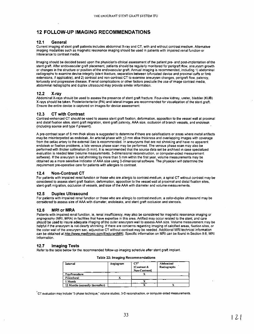
THE ENDURANT STENT GRAFT SYSTEM IFU
12 FOLLOW-UP IMAGING RECOMMENDATIONS
12.1 GeneralCurrent imaging of stent graft patients includes abdominal X-ray and CT, with and without contrast medium. Alternativeimaging modalities such as magnetic resonance imaging should be used in patients with impaired renal function orintolerance to contrast media.
Imaging should be decided based upon the physician's clinical assessment of the patient pre- and post-implantation of thestent graft. After endovascular graft placement, patients should be regularly monitored for perigraft flow, aneurysm growthor changes in the structure or position of the endovascular graft. Annual imaging is recommended, including 1) abdominalradiographs to examine device integrity (stent fracture, separation between bifurcated device and proximal cuffs or limbextensions, if applicable), and 2) contrast and non-contrast CT to examine aneurysm changes, perigraft flow, patency,tortuosity and progressive disease. If renal complications or other factors preclude the use of image contrast media,abdominal radiographs and duplex ultrasound may provide similar information.
12.2 X-rayAbdominal X-rays should be used to assess the presence of stent graft fracture. Four-view kidney, ureter, bladder (KUB)X-rays should be taken. Posterior/anterior (PA) and lateral images are recommended for visualization of the stent graft.Ensure the entire device is captured on images for device assessment.
12.3 CT with ContrastContrast-enhanced CT should be used to assess stent graft fixation, deformation, apposition to the vessel wall at proximaland distal fixation sites, stent graft migration, stent graft patency, AAA size, occlusion of branch vessels, and endoleak(including source and type if present).
A pre-contrast scan of 5 mm thick slices is suggested to determine if there are calcifications or areas where metal artifactsmay be misinterpreted as endoleak. An arterial phase with <3 mm slice thickness and overlapping images with coveragefrom the celiac artery to the external iliac is recommended. In aneurysms that are not shrinking and have no apparentendoleak or fixation problems, a late venous phase scan may be performed. The venous phase scan may also beperformed with thicker collimation (5 mm). It is recommended that the source data set be archived in case specializedevaluation is needed later (volume measurements, 3-dimensional reconstruction, or computer-aided measurementsoftware). If the aneurysm is not shrinking by more than 5 mm within the first year, volume measurements may beobtained as a more sensitive indicator of AAA size using 3-dimensional software. The physician will determine therequirement pre-operative care for patients with allergies to contrast.
12.4 Non-Contrast CTFor patients with impaired renal function or those who are allergic to contrast medium, a spiral CT without contrast may beconsidered to assess stent graft fixation, deformation, apposition to the vessel wall at proximal and distal fixation sites,stent graft migration, occlusion of vessels, and size of the AAA with diameter and volume measurements.
12.5 Duplex UltrasoundFor patients with impaired renal function or those who are allergic to contrast medium, a color-duplex ultrasound may beconsidered to assess size of AAA with diameter, endoleaks, and stent graft occlusion and stenosis.
12.6 MRI or MRAPatients with impaired renal function, ie, renal insufficiency, may also be considered for magnetic resonance imaging orangiography (MRI, MRA) in facilities that have expertise in this area. Artifact may occur related to the stent, and careshould be used to insure adequate iraging of the outer aneurysm wall to assess AAA size. Volume measurement may behelpful if the aneurysm is not clearly shrinking. If there are concerns regarding imaging of calcified areas, fixation sites, orthe outer wall of the aneurysm sac, adjunctive CT without contrast may be needed. Additional MRI technical informationcan be obtained at htto://www.medtronic.com/EndurantMRI. Specific information on MRI can be found in Section 9.6, MRIInformation.
12.7 Imaging TestsRefer to the table below for the recommended follow-up imaging schedule after stent graft implant.
Table 33: Imaging Recommendations
Interval Angiogram CT Abdominal[Contrast & RadiographsNon-Contrastl
Pre-Procedure X
Procedural X
I Month X
12 Months (annually thereafter) X X
CT evaluation may include "3-phase technique,' volume studies, 3-0 reconstruction, or computer-aided measurements.
33

THE ENDURANT STENT GRAFT SYSTEM IFU
12.8 Supplemental ImagingNote: Additional radiological imaging may be necessary to further evaluate the stent graft in situ based on findingsrevealed by one (1) of the surveillance programs. The following recommendations may be considered.
* If there is evidence of poor or irregular position of the stent graft, severe angulation, kinking, or migration of the stentgraft on abdominal X-rays, a spiral CT should be performed to assess aneurysm size and the presence or absenceof an endoleak.
* If a new endoleak or increase in AAA size is observed by spiral CT, adjunctive studies such as 3-D reconstruction orangiographic assessment of the stent graft and native vasculature may be helpful in further evaluating any changesof the stent graft or aneurysm.
* Spiral CT without contrast, MRI or MRA may be considered in select patients who cannot tolerate contrast media orwho have renal function impairment. For centers with appropriate expertise, gadolinium or C02 angiography may beconsidered in patients with renal function impairment requiring angiographic assessment.
13 ADDITIONAL SURVEILLANCE AND TREATMENTAdditional endovascular repair or open surgical aneurysm repair should be considered for patients with evidence ofsuboptimal stent graft fixation, proximal endoleak, distal endoleak, junction endoleak, unknown origin of persistentperigraft flow, or increase in AAA size > 5mm.
14 DISCLAIMER OF WARRANTYALTHOUGH THE MEDTRONIC VASCULAR ENDURANT STENT GRAFT AND DELIVERY SYSTEM, HEREAFTERREFERRED TO AS THE 'PRODUCT', HAS BEEN MANUFACTURED UNDER CAREFULLY CONTROLLEDCONDITIONS, MEDTRONIC, INC., MEDTRONIC VASCULAR, INC. AND THEIR RESPECTIVE AFFILIATES,(COLLECTIVELY "MEDTRONIC") HAVE NO CONTROL OVER THE CONDITIONS UNDER WHICH THIS PRODUCTIS USED. MEDTRONIC, THEREFORE, DISCLAIMS ALL WARRANTIES, BOTH EXPRESSED AND IMPLIED, WITHRESPECT TO THE PRODUCT, INCLUDING, BUT NOT LIMITED TO, ANY IMPLIED WARRANTY OFMERCHANTABILITY OR FITNESS FOR A PARTICULAR PURPOSE. MEDTRONIC SHALL NOT BE LIABLE TO ANYPERSON OR ENTITY FOR ANY MEDICAL EXPENSES OR ANY DIRECT, INCIDENTAL OR CONSEQUENTIALDAMAGES CAUSED BY ANY USE, DEFECT, FAILURE OR MALFUNCTION OF THE PRODUCT, WHETHER ACLAIM FOR SUCH DAMAGES IS BASED UPON WARRANTY, CONTRACT, TORT OR OTHERWISE. NO PERSONHAS ANY AUTHORITY TO BIND MEDTRONIC TO ANY REPRESENTATION OR WARRANTY WITH RESPECT TOTHE PRODUCT.
The exclusions and limitations set out above are not intended to, and should not be construed so as to contravenemandatory provisions of applicable law. If any part or term of this Disclaimer of Warranty is held to be illegal,unenforceable or in conflict with applicable law by a court of competent jurisdiction, the validity of the remaining portions ofthis Disclaimer of Warranty shall not be affected, and all rights and obligations shall be construed and enforced as if thisDisclaimer of Warranty did not contain the particular part or term held to be invalid.
15 DEVICE REGISTRATION
The Endurant Stent Graft System is packaged with additional specific information which includes:
-Temporary Device Identification Card that includes both patient and stent graft information. Physicians shouldcomplete this card and instruct the patient to keep it in their possession at all times. The patients should refer to this cardanytime they visit additional health practitioners, particularly for additional diagnostic procedures (e.g. MRI). Thistemporary identification card should only be discarded when the permanent identification card is received.
* Device Tracking Form to be completed by the hospital staff and forwarded to Medtronic for the purposes of tracking allpatients who received an Endurant Stent Graft (as required by Federal Regulation). The hospital's submission of thedevice tracking form to Medtronic is also required for a patient to receive the permanent identification card.
Upon receipt of the device tracking form, Medtronic will mail the patient a permanent device identification card.This card includes important information regarding the implanted stent graft. Patients should refer to this card anytimethey visit health practitioners, particularly for any diagnostic procedures (e.g., MRI). Patients should carry this card withthem at all times. If a patient does not receive their permanent device identification card, or requires changes to the card,call 1-800-551-5544. In addition a patient information booklet (PIB) will be provided to the physicians during training andadditional copies will be available upon request. The PIB will also be available online on the Medtronic website(www.medtronic.com). This booklet provides patients with basic information on abdominal aortic aneurysms andendovascular repair therapy.
34

THE ENDURANT STENT GRAFT SYSTEM IFU

THE ENDURANT STENT GRAFT SYSTEM IFU

THE ENDURANT STENT GRAFT SYSTEM IFU

THE ENDURANT STENT GRAFT SYSTEM IFU
MANUFACTURER:MEDTRONIC, INC.710 Medtronic ParkwayMinneapolis, MN 55432-5604USATel: (763) 514-4000Fax: (763) 514-4879www.medtronic.com
US CUSTOMER SERVICE/PRODUCT INQUIRIES:Tel: (800) 961-9055Fax: (800) 929-2133
© 2010 Medtronic, Inc.
Protected by one or more of the following United States patents: 6,306,141; 6,911,039; 7,105,016; 7,264,632; and7,655,034.
*M720548B001* RevB