Embed Size (px)
Citation preview

Endourologic management of renal transplant complications1
Stevan B. Streem, M.D. Technical complications requiring reoperation following renal transplantation continue to be significant factors contributing to graft loss and subsequent death. Furthermore, repeated surgical procedures are poorly tolerated in these immunosuppresséd and often ázotemic patients. In the past few years, nonoperative en-dourologic procedures have been employed to temporize or even definitively manage many high-risk urologie patients with obstruc-tive uropathy or urinary fistulas. While the exact indications and long-term results of such management are generally still unknown, these endourologic procedures are now being increasingly applied to selected renal-transplant recipients.
I n d e x term: Kidney, transplantation
Cleve Clin Q 51:349-355, Summer 1984
1 Section of Endourology, Department of Urology, The Cleveland Clinic Foundation. Submitted for pub-lication Dec 1983; accepted Jan 1984. sb
0009-8787/84/02/0349/07/$2.75/0
Copyright® 1984, The Cleveland Clinic Foun-dation.
Despite improving results after renal transplantation over the past decade, technical complications continue to contribute to the morbidity and mortality of transplant recipients. Since these patients are immunosuppressed and often azotemic, such complications are potentially life threatening and require early diagnosis and management to enhance subsequent survival of the patient and graft.
Endourologic procedures have become invaluable for the treatment of a wide variety of disorders, including obstructive uropathy and urinary fistulas, and further re-finements of these techniques now allow their application to renal transplantation. While exact indications have yet to be defined and the long-term results are often still unknown, endourologic techniques are increasingly being used as both temporizing and definitive procedures.
3 4 9
on March 18, 2022. For personal use only. All other uses require permission.www.ccjm.orgDownloaded from

2 * 3 5 0 C l e v e l a n d Cl in ic Q u a r t e r l y Vol . 5 1 , N o . 2
Fig. 1. A. Antegrade nephrostogram reveals complete obstruction at the proximal third of the transplanted ureter. The indwelling percutaneous nephrostomy tube was left in place to allow resolution of the obstructive uropathy prior to open surgical repair.
B. Antegrade nephrostogram, two weeks following ureterolysis and repeat ureteroneocystostomy, reveals resolution of the obstruction and no evidence of extravasation.
Obstructive uropathy Obstructive uropathy occurs in approx imate ly
10% of renal a l lograf t recipients. W h e n hydro-nephros is is suggested by u l t rasound o r com-pu ted t omography , in t ravenous or r e t r o g r a d e pye lography is general ly a t t e m p t e d to f u r t h e r de f ine the level a n d etiology of the obs t ruc t ion . O f t e n , however , adequa te visualization on an excre to ry u r o g r a m is p rec luded by the impai red renal func t ion . Addit ional ly, r e t r o g r a d e pyelog-raphy is o f t en technically impossible fol lowing t ransplan t ure te roneocys tos tomy. In such cases, pe r cu t aneous a n t e g r a d e pyelography has p roved most valuable and allows precise ana tomic delin-eat ion of the site of obstruct ion. 1 T h e p r o c e d u r e is general ly p e r f o r m e d with u l t rasound o r com-pu t ed - tomograph i c guidance using s t andard in-te rvent ional rad iographic methods . Convers ion to t e m p o r a r y pe rcu t aneous nephros tomy drain-age is t hen possible using one of several available t echn iques . 2
T h e m a n a g e m e n t of ure tera l obs t ruc t ion by t e m p o r a r y pe rcu t aneous nephros tomy d ra inage o f fe r s several advantages . T h e ex ten t of recover-able renal func t ion can be assessed a f t e r pro-longed dra inage , and renal func t ion p r io r to de-finitive surgical repa i r can be opt imized. In se-lected cases when the funct ional significance of an a p p a r e n t obs t ruc t ion is unclear , a Whi t ake r test may be p e r f o r m e d for a defini t ive diagnosis. Initial pe r cu t aneous nephros tomy is also invalu-
able for the t r e a t m e n t of u r inary tract infection associated with the obs t ruc t ion for d ra inage of a t ransplant pyohydronephros i s . 3 In those cases when an open surgical p r o c e d u r e will ultimately p rove necessary, the pe rcu t aneous nephros tomy can remain postoperat ively to provide proximal diversion and pro tec t ion of anastomoses. A fur-the r advan tage in this set t ing is that this provides access to p e r f o r m a n t e g r a d e studies postopera-tively to exclude extravasat ion or persistent ob-struct ion at the anas tomot ic site.
Case report A 28-year-old man received an HLA identical kidney
f rom his sister. Urinary continuity was restored by a s tandard ant i ref luxing ureteroneocystostomy. His immedi-ate postoperative course was uneventful , and a nadir creat-inine level of 1.2 m g / d L was reached. He was readmit ted to the hospital four weeks af te r the transplantation with acute pulmonary edema and renal failure, with a serum creatinine level of 8.6 m g / d L . Ultrasound revealed marked hydronephrosis of the t ransplanted kidney. Percutaneous nephrostomy drainage was immediately established. Renal function was regained, and the pulmonary edema resolved. An antegrade pyelogram revealed complete obstruction at the proximal third of the ureter (Fig. 1A). Exploration revealed acute angulation of the ureter at that site. Ureter-olysis and repeat ureteroneocystostomy were per fo rmed , and the percutaneous nephros tomy tube was left in place for proximal diversion. T w o weeks following that proce-dure , a repeat an tegrade pyelogram showed complete res-olution of the obstruction and no extravasation (Fig. IB). T h e nephrostomy tube was removed, and the patient re-mains well with a serum creatinine level of 1.1 m g / d L seven months af te r transplantat ion.
on March 18, 2022. For personal use only. All other uses require permission.www.ccjm.orgDownloaded from

S u m m e r 1 9 8 4 M a n a g e m e n t of r e n a l t r a n s p l a n t c o m p l i c a t i o n s 3 5 1
2A, B
Fig. 2. A. Antegrade pyelogram reveals a long segment of distal ureteral stenosis one year following a repeat ureteroneocystostomy. Percutaneous nephrostomy drainage was established.
B. Adequate urinary drainage provided by an indwelling double-J ureteral stent passed antegrade via the percuta-neous nephrostomy tract. (Reprinted from Streem and Novick' with the permission of W. B. Saunders, Co., Philadelphia, Pa.)
Comment: In selected cases, ureteral obstruc-tion may be managed entirely by endouro-logic techniques. T h e percutaneous management of non-transplant ureteral stenosis using tempo-rary indwelling ureteral stents has previously been described,4 and such therapy is now being evaluated for renal allograft recipients. Fur ther-more, an tegrade placement of indwelling ure-teral stents is of ten accomplished easily following percutaneous nephros tomy in transplant pa-tients, even when re t rograde catheterization is technically impossible.
Case report A 12-year-old girl was the recipient of a cadaver renal
transplant in September 1978. Her postoperative course was complicated by two episodes of acute rejection in the first four months; this was treated with methylprednisolone administered intravenously. Following the second rejection
episode, she remained azotemic with a serum creatinine level of 3.2 m g / d L . An acute rise in the serum creatinine value to 5.4 m g / d L was noted two years a f te r transplanta-tion and fu r the r evaluation revealed hydronephrosis second-ary to distal ureteral stenosis. A repeat ureteroneocystos-tomy was pe r fo rmed , and the results of a renal biopsy specimen obtained at that t ime indicated chronic rejection. T h e serum creatinine level stabilized at 4.5 m g / d L , but then slowly rose. She was readmit ted one year later with a serum creatinine value of 8.4 m g / d L . Hydronephrosis was again demonst ra ted by ul t rasound. An antegrade pyelogram (Fig. 2A) revealed a long segment of distal ureteral stenosis. Percutaneous nephros tomy drainage was established. T h e serum creatinine level improved slightly, but stabilized at 7.2 m g / d L . In view of the associated chronic rejection and poor prognosis for the graft , repeat operative intervention was not deemed appropr ia te . Ret rograde ureteral catheter-ization and stenting was a t tempted, but was technically unsuccessful. Consequently, a double-J silastic ureteral stent was easily placed an tegrade via the nephrostomy tract and left in position without complications (Fig. 2B). Fur ther deteriorat ion of renal function over the next six months
on March 18, 2022. For personal use only. All other uses require permission.www.ccjm.orgDownloaded from
2 * 3 5 2 C l e v e l a n d Cl in ic Q u a r t e r l y Vol . 5 1 , N o . 2
3A, B
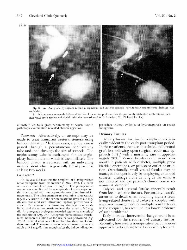
Fig. 3. A. Antegrade pyelogram reveals a segmental mid-ureteral stenosis. Percutaneous nephrostomy drainage was established.
B. Percutaneous antegrade balloon dilatation of the ureter performed via the previously established nephrostomy tract. (Reprinted from Streem and Novick1 with the permission of W. B. Saunders, Co., Philadelphia, Pa.)
ultimately led to a graf t nephrectomy at which time a pathologic examination revealed chronic rejection.
Comment: Alternatively, an a t t emp t may be m a d e to t rea t t ransplant ure tera l stenosis using bal loon dilatat ion.5 In these cases, a gu ide wire is passed t h r o u g h a pe rcu taneous nephros tomy tube a n d then t h r o u g h the site of stenosis. T h e neph ros tomy tube is exchanged fo r an angio-plasty balloon di la tor which is then inflated. T h e bal loon di lator is replaced with an indwell ing ure te ra l stent which is generally left in place for at least two weeks.
Case report An 18-year-old man was the recipient of a living-related
renal t ransplant f rom his mother in May 1982. His nadir serum creatinine level was 1.0 mg/dL . T h e postoperative course was complicated by one episode of acute rejection; this was treated with methylprednisolone administered in-travenously. T h e subsequent serum creatinine value was 3.4 m g / d L . A later rise in the serum creatinine level to 5.3 m g / d l , was evaluated with ultrasound; hydronephrosis was re-vealed. Percutaneous nephrostomy drainage was estab-lished, and the serum creatinine level decreased to 3.6 mg / dl . . An an tegrade pyelogram revealed segmental stenosis of the mid-ureter (Fig. 3A). Antegrade percutaneous translu-minal balloon dilatation of the ureter was pe r fo rmed (Fig. 3B). A ureteral stent was left in place for three weeks and then removed. The serum creatinine level currently remains stable at 3.8 m g / d L nine months af te r the balloon-dilatation
p rocedure without evidence of hydronephrosis on repeat sonograms.
Urinary Fistulas Urinary fistulas a re m a j o r complications gen-
erally evident in the early post- t ransplant per iod. In these patients , the ra te of technical fa i lure and graf t loss following open surgical repai r may ap-proach 30%, 6 with a mortal i ty ra te of approxi-mately 20%. ' Vesical Fistulas occur m o r e com-monly in pat ients with diabetes, mult iple p r io r b ladder operat ions , o r persis tent out let obstruc-tion. Occasionally, small vesical fistulas may be m a n a g e d nonopera t ive ly by employing ex tended ca the te r d ra inage a lone as long as the ur ine is n o t infected and the pa t ien t ' s clinical course re-mains satisfactory.8
Calyceal and ure te ra l fistulas generally result f r o m local ischemic factors . For tunate ly , carefu l a t t en t ion to detail when obta in ing kidneys f r o m living-related d o n o r s a n d cadavers, coupled with improved m a n a g e m e n t of mult iple renal ar ter ies in the recipient , has resul ted in a lower incidence of such problems. 9
Early operat ive in tervent ion has generally been advocated for the t r e a t m e n t of ur inary fistulas. Recently, however , a nonopera t ive pe rcu taneous app roach has been employed successfully for such
on March 18, 2022. For personal use only. All other uses require permission.www.ccjm.orgDownloaded from

S u m m e r 1 9 8 4 M a n a g e m e n t o f r e n a l t r a n s p l a n t c o m p l i c a t i o n s 3 5 3
Fig. 4. A. Antegrade pyelogram reveals extravasation from the distal ureter. Percuta-neous nephrostomy drainage was established.
B. Antegrade nephrostogram six weeks later revealed resolution of the extravasation, although distal ureteral obstruction was evident. (Courtesy of Dr. Stuart Flechner)
fistulas in non-transplant patients and such tech-niques are current ly being applied to renal allo-graf t recipients. A recent retrospective study noted the value of preliminary open nephros-tomy diversion alone in the management of se-lected patients with transplant urinary fistulas,6
and that approach has now been modified so that percutaneous drainage, ra ther than open ne-phrostomy drainage, can be employed with or without ureteral stenting.
Case report A four-year-old boy was the recipient of a living related
renal transplant f rom his mother . T h e kidney was placed intraperitoneally. Excellent function was obtained in the period immediately af ter transplantation; the nadir serum creatinine level was 0.5 mg/dL . T w o weeks af te r the oper-ation, fever, ileus, and anuria developed. T h e serum creat-inine level rose to 2.6 mg /dL , and a renal scan suggested urinary extravasation. An antegrade pyelogram was ob-tained and revealed distal ureteral extravasation (Fig. 4A).
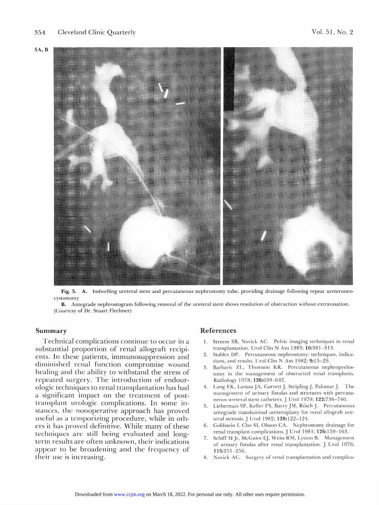
Percutaneous nephrostomy drainage was then instituted; the fever, ileus, and azotemia resolved. An an tegrade ne-phros togram six weeks later revealed total distal ureteral occlusion (Fig. 4B). Repeat ureteroneocystoscopy was then pe r fo rmed; both a ureteral stent and the previously posi-t ioned percutaneous nephros tomy tube were left in place (Fig. 5A). T h e ureteral stent was then removed two months later, at which time a repeat an tegrade nephros togram revealed no obstruction or extravasation (Fig. 5B). T h e nephrostomy tube was then removed and the results of excretory urography pe r fo rmed six months later were nor-mal (Fig. 6). T h e serum creatinine value is currently 0.5 mg/dL..
Comment. In some cases, this approach has proved definitive and has obviated the need for subsequent surgical repair . In one report , five of six fistulas closed spontaneously one day to 10 weeks a f te r percutaneous nephrostomy drainage. While ureteral strictures developed in 3 of those patients, 2 of them responded to dilatation by percutaneous stent placement.1 0
on March 18, 2022. For personal use only. All other uses require permission.www.ccjm.orgDownloaded from
2*354 Cleveland Clinic Quar ter ly Vol. 51, No. 2
5A, B
Fig. 5. A. Indwelling ureteral stent and percutaneous nephrostomy tube, providing drainage following repeat ureteroneo-cystostoiny
B. Antegrade nephrostogram following removal of the ureteral stent shows resolution of obstruction without extravasation. (Courtesy of Dr. Stuart Flechner)
Summary Technica l complicat ions con t inue to occur in a
substantial p ropo r t i on of renal a l lograf t recipi-ents. In these patients , immunosuppress ion and diminished renal func t ion compromise wound heal ing and the ability to wi ths tand the stress of repea ted surgery. T h e in t roduc t ion of endour -ologic techniques to renal t ransplanta t ion has had a significant impact on the t r e a tmen t of post-t ransplant urologic complications. In some in-stances, the nonopera t ive app roach has p roved useful as a t empor iz ing p rocedu re , while in oth-ers it has p roved definit ive. While many of these techniques a re still be ing evaluated and long-t e rm results a r e o f t en u n k n o w n , the i r indications appea r to be b r o a d e n i n g a n d the f r equency of the i r use is increasing.
References 1. Streem SB, Novick AC. Pelvic imaging techniques in renal
transplantation. Urol Clin N Am 1983; 10:301-313. 2. Stables DP. Percutaneous nephrostomy: techniques, indica-
tions, and results. Urol Clin N Am 1982; 9:15-29. 3. Barbaric ZL, Thomson KR. Percutaneous nephropyelos-
tomy in the management of obstructed renal transplants. Radiology 1978; 126:639-642.
4. Lang EK, LanasaJA, Garrett J, StriplingJ, Palomar J. The management of urinary fistulas and strictures with percuta-neous ureteral stent catheters. J Urol 1979; 122:736-740.
5. Lieberman SF, Keller FS, Barry JM, Rósch J. Percutaneous antegrade transluminal ureteroplasty for renal allograft ure-teral stenosis. .] Urol 1982; 128:122-124.
6. Goldstein I, Cho SI, Olsson CA. Nephrostomy drainage for renal transplant complications. J Urol 1981; 126:159-163.
7. Schiff M Jr, McGuire EJ, Weiss RM, Lytton B. Management of urinary fistulas after renal transplantation. J Urol 1976; 115:251-256.
8. Novick AC. Surgery of renal transplantation and complica-
on March 18, 2022. For personal use only. All other uses require permission.www.ccjm.orgDownloaded from

Summer 1984 Management of renal t ransplant complications 355
tions. [In] Novick AC, Straffon RA, eds. Vascular Problems in Urologie Surgery. Philadelphia, WB Saunders, 1982, pp 233-260.
9. Salvatierra C) Jr, Kountz SL, Beizer FO. Prevention of ure-teral fistula after renal transplantation. J Urol 1974; 112:445-448.
10. Lieberman RP, Glass NR, Crummy AB, Sollinger HW, Beizer FO. Nonoperative percutaneous management of urinary fis-tulas and strictures in renal transplantation. Surg Gynecol Obstet 1982; 155:667-672.
Fig. 6. Excretory urogram six months following removal of the percutaneous nephrostomy tube reveals normal transplant anat-omy. (Courtesy of Dr. Stuart Flechner)
on March 18, 2022. For personal use only. All other uses require permission.www.ccjm.orgDownloaded from