Embed Size (px)
Citation preview
.
713ISSN 0372-5480Printed in Croatia
VeterINarSkI arhIV 87 (6), 713-729, 2017
Endoparasites of wildcats in Croatia
Franjo Martinković1, Magda Sindičić2*, Snježana Lučinger1, Iva Štimac1, Miljenko Bujanić3, Tatjana Živičnjak1, Dagny Stojčević Jan1,
Nikica Šprem5, Ratko Popović4, and Dean Konjević3
1Department of Parasitology and Parasitic Diseases with Clinic, Faculty of Veterinary Medicine, University of Zagreb, Zagreb, Croatia
2Department for Game Biology, Pathology and Breeding, Faculty of Veterinary Medicine, University of Zagreb, Zagreb, Croatia
3Department of Veterinary Economics and Epidemiology, Faculty of Veterinary Medicine, University of Zagreb, Zagreb, Croatia
4Croatian Forest, Department Sisak, Sisak, Croatia5Department of Fisheries, Beekeeping, Game Management and Special Zoology,
Faculty of Agriculture, Zagreb, Croatia________________________________________________________________________________________MaRTinKović, F., M. SinDičić, S. LučingeR, i. ŠTiMac, M. BuJanić, T. ŽivičnJaK, D. SToJčević Jan, n. ŠPReM, R. PoPović, D. KonJević: Endoparasites of wildcats in Croatia. vet. arhiv 87, 713-729, 2017.
ABSTRACTreports on the parasitic fauna of wildcats (Felis silvestris silvestris) are rare and often based on a small
sample size, therefore the goal of this research was to investigate the prevalence of endoparasites in wildcats in Croatia. Necropsy was conducted on 34 adult wildcats killed in traffic or provided by hunters following regular hunting operations. all animals tested negative for rabies. the contents of the stomach and intestine were examined under a microscope. Feces from the rectum were analyzed using flotation with a saturated ZnSO4 solution, while the diaphragm was examined using artificial digestion. Direct immunofluorescence was used for the first time to detect Giardia sp. cysts in wildcats. all animals were infected with at least one species of parasites, while the most diverse infestation included six different species of parasites in a single animal. the following parasite species were found (% of prevalence of adult parasites and their developmental stages in all analyzed samples): Taenia taeniaeformis (55.9%), Capillaria sp. (50%), Toxocara cati (50%), Isospora sp. (29.4%), Strongyloides sp. (23.5%), Giardia sp. (17.6%), Ancylostoma tubaeformae (14.7%), Physaloptera sp. (11.8%), hymenolepididae (8.8%), Alaria alata (5.9%), Aelurostrongylus abstrusus (5.9%), Toxascaris leonina (5.9%), Trichinella sp. (5.9%), Mesocestoides lineatus (5.9%), anoplocephalidae (2.9%), Dipylidium caninum (2.9%), Trichuris sp. (2.9%), Isospora felis (2.9%), Eimeria sp. (2.9%) and Sarcocystis sp. (2.9%). among
*Corresponding author:Magda Sindičić, Assistant Professor, DVM, PhD, Department for Game Biology, Pathology and Breeding, Faculty of Veterinary Medicine University of Zagreb, Heinzelova 55, 10000 Zagreb, Croatia, Phone: +385 1 2390 156; Fax: +385 1 2441 390; E-mail: [email protected]
doi: 10.24099/vet.arhiv.170127

714 Vet. arhiv 87 (6), 713-729, 2017
F. Martinković et al.: Endoparasites of wildcats in Croatia
those, Eimeria sp., Trichuris sp. eggs, anoplocephalid and hymenolepidid type eggs are spurious parasites, coming from ingested prey. Four of the identified species have never been previously reported in wildcats - Giardia sp., Strongyloides sp., Sarcocystis sp. and Dipylidium caninum.
Key words: parasites, wildcat, Felis silvestris silvestris, Croatia________________________________________________________________________________________
introduction the wildcat (Felis silvestris silvestris) is the most common and widely-distributed
wild felid, found throughout most of africa, europe, southwest and central asia. In europe the species is native to most countries, except Fennoscandia, but today the species has a sporadic distribution throughout the continent (YAMAGUCHI et al., 2015). In Croatia the wildcat is present throughout the country, except on the islands (JaNICkI et al., 2007). Unfortunately, detailed distribution maps are not available for Croatia, and also there are no scientific data about population size and trends, demographic dynamics, ecology, and diseases.
the effects of pathogens and diseases on the conservation of carnivores are of major concern, but knowledge of their parasitic fauna is scarce (FUNk et al., 2001). Comprehensive parasitological studies depend on the ability to collect a large number of samples. Carcasses are the best source for research of gastrointestinal fauna, while copromicroscopic surveys may be a useful alternative (NAPOLI et al., 2016). In protected species (like the wildcat in most european countries, including Croatia since 2013) it may be difficult to obtain carcasses, as they are rarely found in nature (apart from road kills). Studying wildcat gastrointestinal parasites from feces is also challenging because, due to the low population densities and their solitary way of life, it may be challenging to find fresh samples. as a result, studies on parasites of wildcats are not only rare, but also based on a limited sample size (BURT et al., 1980; SCHUSTER et al., 1993; PAPADOPOULOS et al., 1997; RODRIGUEZ and CARBONELL, 1998; KRONE et al., 2008; KIRKOVA et al., 2011; NAPOLI et al., 2016; VERONESI et al., 2016).
In Croatia, there are no data about parasites in wildcats. therefore, the aim of this survey was to investigate the prevalence of gastrointestinal and Trichinella parasites in wildcats in Croatia.
Materials and methodsSamples. a total of 34 entire carcasses or intestines with diaphragm of adult wildcats
killed in traffic accidents or culled during regular game management activities (prior to the hunting ban in 2013) were collected and stored at -20 °C. DNA analysis confirmed all the samples were from wildcats and that there were no domestic cats in the sampling (SINDIČIĆ et al., 2016).
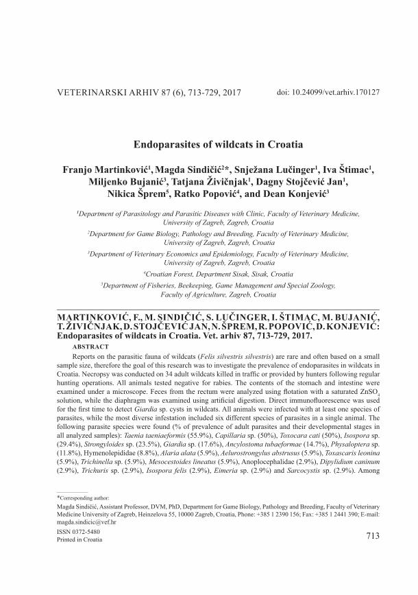
Study area. the origin of samples collected is presented on Fig. 1.
715Vet. arhiv 87 (6), 713-729, 2017
F. Martinković et al.: Endoparasites of wildcats in Croatia
Fig. 1. Location of origin of wildcat samples. The size of the circle corresponds to the number of samples from each location.
Necropsy and parasitological examination. Before necropsy, each animal was tested for rabies and all the tests were negative. Necropsies were conducted at the Faculty of Veterinary Medicine, University of Zagreb, according to a standard veterinary protocol. the gastrointestinal tract (stomach and intestines) was removed from the animal, stored in separated plastic containers, opened along its length and flushed with water, while its mucosa was scraped and separated in another plastic container. From some wildcats only the intestines and diaphragms were available.
the contents of the stomach and intestines were examined in Petri dishes on a dark background under a stereomicroscope, while the scraped mucosa was checked for parasites under the microscope. Parasites were then washed out with saline to remove the gastrointestinal contents in order to facilitate definition to the genus or species level, depending on the condition.
Copromicroscopic examination. Feces was removed from the rectum and separated into two parts of 5 g each. One part was analyzed using the flotation method with a saturated ZnSO4 solution (S.G. = 1.3), and the other was used to detect Giardia sp. cysts. Fecal samples were first prepared using the flotation technique with sucrose, and then analyzed by the direct immunofluorescence method (Meridian Bioscience, Inc., Merifluor® Cryptosporidium/Giardia).
Artificial digestion. the presence of Trichinella sp. parasites was analyzed using diaphragm samples (10g) by the reference method for artificial digestion of Trichinella in meat (the european Community regulation (eC) No. 2075/2005).
Parasite determination. The parasite identifications and staining were based on the available descriptions (MEHLHORN et al., 1993; HRČKOVA et al., 2011; BRIANTI et al., 2012; SChMÄSChke, 2013). Briefly, adult parasites were stained with lactophenol (nematodes,
Croatia
Slovenia
Bosna and herzegovina Serbia
hungary
716 Vet. arhiv 87 (6), 713-729, 2017
F. Martinković et al.: Endoparasites of wildcats in Croatia
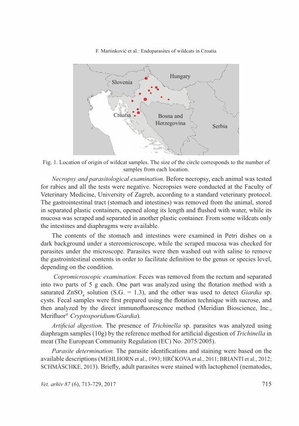
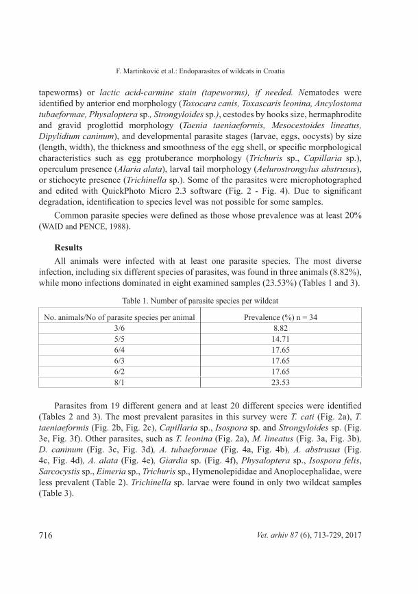
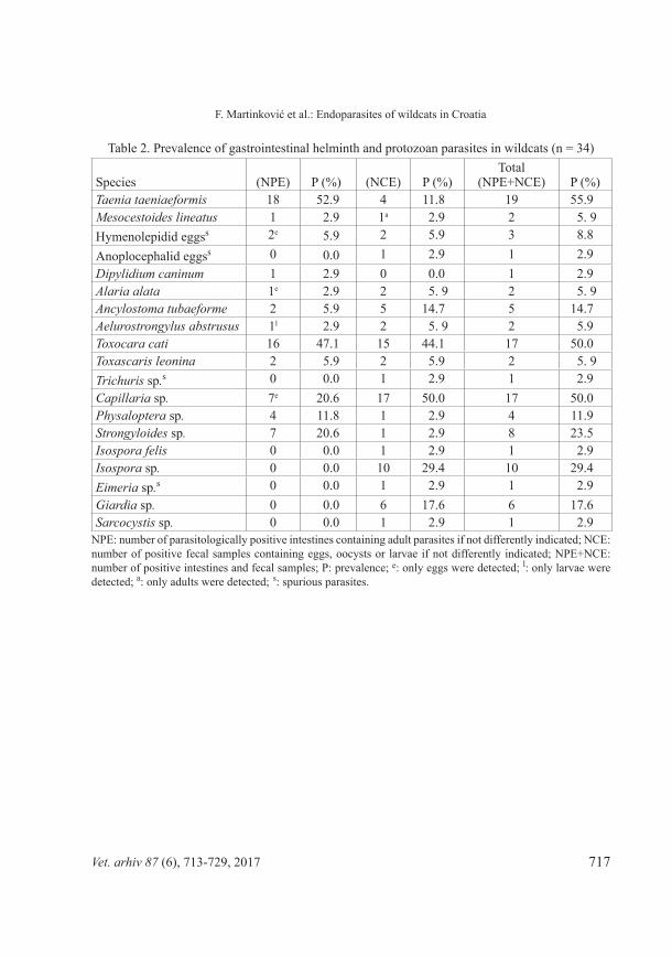
tapeworms) or lactic acid-carmine stain (tapeworms), if needed. Nematodes were identified by anterior end morphology (Toxocara canis, Toxascaris leonina, Ancylostoma tubaeformae, Physaloptera sp., Strongyloides sp.), cestodes by hooks size, hermaphrodite and gravid proglottid morphology (Taenia taeniaeformis, Mesocestoides lineatus, Dipylidium caninum), and developmental parasite stages (larvae, eggs, oocysts) by size (length, width), the thickness and smoothness of the egg shell, or specific morphological characteristics such as egg protuberance morphology (Trichuris sp., Capillaria sp.), operculum presence (Alaria alata), larval tail morphology (Aelurostrongylus abstrusus), or stichocyte presence (Trichinella sp.). Some of the parasites were microphotographed and edited with QuickPhoto Micro 2.3 software (Fig. 2 - Fig. 4). Due to significant degradation, identification to species level was not possible for some samples.
Common parasite species were defined as those whose prevalence was at least 20% (WAID and PENCE, 1988).
Resultsall animals were infected with at least one parasite species. the most diverse
infection, including six different species of parasites, was found in three animals (8.82%), while mono infections dominated in eight examined samples (23.53%) (tables 1 and 3).
table 1. Number of parasite species per wildcat
No. animals/No of parasite species per animal Prevalence (%) n = 34 3/6 8.825/5 14.716/4 17.656/3 17.656/2 17.658/1 23.53
Parasites from 19 different genera and at least 20 different species were identified (tables 2 and 3). the most prevalent parasites in this survey were T. cati (Fig. 2a), T. taeniaeformis (Fig. 2b, Fig. 2c), Capillaria sp., Isospora sp. and Strongyloides sp. (Fig. 3e, Fig. 3f). Other parasites, such as T. leonina (Fig. 2a), M. lineatus (Fig. 3a, Fig. 3b), D. caninum (Fig. 3c, Fig. 3d), A. tubaeformae (Fig. 4a, Fig. 4b), A. abstrusus (Fig. 4c, Fig. 4d), A. alata (Fig. 4e), Giardia sp. (Fig. 4f), Physaloptera sp., Isospora felis, Sarcocystis sp., Eimeria sp., Trichuris sp., hymenolepididae and anoplocephalidae, were less prevalent (table 2). Trichinella sp. larvae were found in only two wildcat samples (table 3).
717Vet. arhiv 87 (6), 713-729, 2017
F. Martinković et al.: Endoparasites of wildcats in Croatia
Table 2. Prevalence of gastrointestinal helminth and protozoan parasites in wildcats (n = 34)
Species (NPe) P (%) (NCe) P (%)total
(NPE+NCE) P (%)Taenia taeniaeformis 18 52.9 4 11.8 19 55.9Mesocestoides lineatus 1 2.9 1a 2.9 2 5. 9hymenolepidid eggss 2e 5.9 2 5.9 3 8.8anoplocephalid eggss 0 0.0 1 2.9 1 2.9Dipylidium caninum 1 2.9 0 0.0 1 2.9Alaria alata 1e 2.9 2 5. 9 2 5. 9Ancylostoma tubaeforme 2 5.9 5 14.7 5 14.7Aelurostrongylus abstrusus 1l 2.9 2 5. 9 2 5.9Toxocara cati 16 47.1 15 44.1 17 50.0Toxascaris leonina 2 5.9 2 5.9 2 5. 9Trichuris sp.s 0 0.0 1 2.9 1 2.9Capillaria sp. 7e 20.6 17 50.0 17 50.0Physaloptera sp. 4 11.8 1 2.9 4 11.9Strongyloides sp. 7 20.6 1 2.9 8 23.5Isospora felis 0 0.0 1 2.9 1 2.9Isospora sp. 0 0.0 10 29.4 10 29.4Eimeria sp.s 0 0.0 1 2.9 1 2.9Giardia sp. 0 0.0 6 17.6 6 17.6Sarcocystis sp. 0 0.0 1 2.9 1 2.9
NPE: number of parasitologically positive intestines containing adult parasites if not differently indicated; NCE: number of positive fecal samples containing eggs, oocysts or larvae if not differently indicated; NPE+NCE: number of positive intestines and fecal samples; P: prevalence; e: only eggs were detected; l: only larvae were detected; a: only adults were detected; s: spurious parasites.
718 Vet. arhiv 87 (6), 713-729, 2017
F. Martinković et al.: Endoparasites of wildcats in Croatia
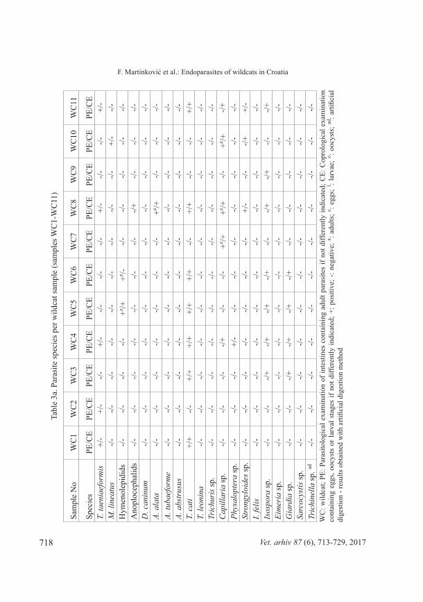
tabl
e 3a
. Par
asite
spec
ies p
er w
ildca
t sam
ple
(sam
ples
WC
1-W
C11
)
Sam
ple
No
WC
1W
C2
WC
3W
C4
WC
5W
C6
WC
7W
C8
WC
9W
C10
WC
11
Spec
ies
Pe/C
ePe
/Ce
Pe/C
ePe
/Ce
Pe/C
ePe
/Ce
Pe/C
ePe
/Ce
Pe/C
ePe
/Ce
Pe/C
eT.
taen
iaef
orm
is+/
-+/
--/-
+/-
-/--/-
-/-+/
--/-
-/-+/
-M
. lin
eatu
s-/-
-/--/-
-/--/-
-/--/-
-/--/-
+/-
-/-h
ymen
olep
idid
s-/-
-/--/-
-/-+e /+
+e /--/-
-/--/-
-/--/-
ano
ploc
epha
lids
-/--/-
-/--/-
-/--/-
-/--/+
-/--/-
-/-D
. can
inum
-/--/-
-/--/-
-/--/-
-/--/-
-/--/-
-/-A.
ala
ta-/-
-/--/-
-/--/-
-/--/-
+e /+-/-
-/--/-
A. tu
baef
orm
e-/-
-/--/-
-/--/-
-/--/-
-/--/-
-/--/-
A. a
bstr
usus
-/--/-
-/--/-
-/--/-
-/--/-
-/--/-
-/-T.
cat
i+/
+-/-
+/+
+/+
+/+
+/+
-/-+/
+-/-
-/-+/
+T.
leon
ina
-/--/-
-/--/-
-/--/-
-/--/-
-/--/-
-/-Tr
ichu
ris s
p.-/-
-/--/-
-/--/-
-/--/-
-/--/-
-/--/-
Cap
illar
ia sp
.-/-
-/--/-
-/+-/-
-/-+e /+
+e /+-/-
+e /+-/+
Phys
alop
tera
sp.
-/--/-
-/-+/
--/-
-/--/-
-/--/-
-/--/-
Stro
ngyl
oide
s sp.
-/--/-
-/--/-
-/--/-
-/-+/
--/-
-/++/
-I.
felis
-/--/-
-/--/-
-/--/-
-/--/-
-/--/-
-/-Is
ospo
ra sp
.-/-
-/--/+
-/+-/+
-/+-/-
-/+-/+
-/--/+
Eim
eria
sp.
-/--/-
-/--/-
-/--/-
-/--/-
-/--/-
-/-G
iard
ia sp
.-/-
-/--/+
-/+-/+
-/+-/-
-/--/-
-/--/-
Sarc
ocys
tis sp
.-/-
-/--/-
-/--/-
-/--/-
-/--/-
-/--/-
Tric
hine
lla sp
. ad-/-
-/--/-
-/--/-
-/--/-
-/--/-
-/--/-
WC
: wild
cat;
PE: P
aras
itolo
gica
l exa
min
atio
n of
inte
stin
es c
onta
inin
g ad
ult p
aras
ites
if no
t diff
eren
tly in
dica
ted;
CE:
Cop
rolo
gica
l exa
min
atio
n co
ntai
ning
egg
s, oo
cyst
s or
larv
al s
tage
s if
not d
iffer
ently
indi
cate
d; +
: pos
itive
; -: n
egat
ive;
a : adu
lts; e : e
ggs;
l : lar
vae;
o : ooc
ysts
; ad: a
rtific
ial
dige
stio
n - r
esul
ts o
btai
ned
with
arti
ficia
l dig
estio
n m
etho
d

719Vet. arhiv 87 (6), 713-729, 2017
F. Martinković et al.: Endoparasites of wildcats in Croatiata
ble
3b. P
aras
ite sp
ecie
s per
wild
cat s
ampl
e (s
ampl
es W
C12
-WC
22)
Sam
ple
No
WC
12W
C13
WC
14W
C15
WC
16W
C17
WC
18W
C19
WC
20W
C21
WC
22Sp
ecie
sPe
/Ce
Pe/C
ePe
/Ce
Pe/C
ePe
/Ce
Pe/C
ePe
/Ce
Pe/C
ePe
/Ce
Pe/C
ePe
/Ce
T. ta
enia
efor
mis
+/-
-/--/-
+/-
-/-+/
-+/
++/
--/-
+/-
+/-
M. l
inea
tus
-/--/-
-/--/-
-/--/-
-/--/-
-/--/-
-/-h
ymen
olep
idid
s-/-
-/--/-
-/--/-
-/--/-
-/--/-
-/--/-
ano
ploc
epha
lids
-/--/-
-/--/-
-/--/-
-/--/-
-/--/-
-/-D
. can
inum
-/--/-
-/--/-
-/--/-
-/--/-
+/-
-/--/-
A. a
lata
-/--/-
-/--/-
-/--/-
-/--/-
-/--/-
-/-A.
tuba
efor
me
-/--/-
-/--/-
-/--/-
-/+-/-
-/--/-
-/+A.
abs
trus
us-/-
-/--/-
-/--/-
-/--/-
-/++l /+
-/--/-
T. c
ati
-/--/-
+/+
-/--/-
+/-
+/-
-/--/-
+/+
+/+
T. le
onin
a-/-
-/--/-
-/--/-
-/--/-
-/--/-
-/--/-
Tric
huri
s sp.
-/--/-
-/--/-
-/--/-
-/--/-
-/--/+
-/-C
apill
aria
sp.
-/+-/-
-/+-/-
-/--/+
+e /++e /+
+e /+-/+
+e /+Ph
ysal
opte
ra sp
.-/-
-/-+/
--/-
-/-+/
--/-
-/--/-
-/--/-
Stro
ngyl
oide
s sp.
-/-+/
-+/
--/-
-/--/-
+/-
+/-
+/-
-/--/-
I. fe
lis-/-
-/--/-
-/--/-
-/--/-
-/--/-
-/--/-
Isos
pora
sp.
-/--/-
-/--/-
-/--/+
-/--/+
-/--/-
-/+Ei
mer
ia sp
.-/+
-/--/-
-/--/-
-/--/-
-/--/-
-/--/-
Gia
rdia
sp.
-/--/-
-/--/-
-/--/-
-/--/-
-/--/-
-/-Sa
rcoc
ystis
sp.
-/--/-
-/--/-
-/-- /-
-/--/-
-/--/-
-/-Tr
ichi
nella
sp. ad
-/-+l
-/--/-
+l-/-
-/--/-
-/--/-
-/-W
C: w
ildca
t; PE
: Par
asito
logi
cal e
xam
inat
ion
of in
test
ines
con
tain
ing
adul
t par
asite
s if
not d
iffer
ently
indi
cate
d; C
E: C
opro
logi
cal e
xam
inat
ion
cont
aini
ng e
ggs,
oocy
sts
or la
rval
sta
ges
if no
t diff
eren
tly in
dica
ted;
+: p
ositi
ve; -
: neg
ativ
e; a : a
dults
; e : egg
s; l : l
arva
e; o : o
ocys
ts; ad
: arti
ficia
l di
gest
ion
- res
ults
obt
aine
d w
ith a
rtific
ial d
iges
tion
met
hod
720 Vet. arhiv 87 (6), 713-729, 2017
F. Martinković et al.: Endoparasites of wildcats in Croatia
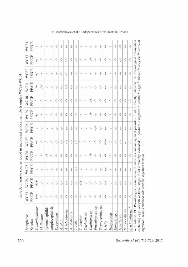
tabl
e 3c
. Par
asite
spec
ies f
ound
in in
divi
dual
wild
cat s
ampl
e (s
ampl
es W
C22
-WC
34)
Sam
ple
No
WC
23W
C24
WC
25W
C26
WC
27W
C28
WC
29W
C30
WC
31W
C32
WC
33W
C34
Spec
ies
Pe/C
ePe
/Ce
Pe/C
ePe
/Ce
Pe/C
ePe
/Ce
Pe/C
ePe
/Ce
Pe/C
ePe
/Ce
Pe/C
ePe
/Ce
T. ta
enia
efor
mis
+/+
+/+
-/--/-
+/-
-/-+/
--/-
-/++/
-+/
--/-
M. l
inea
tus
-/--/-
-/--/-
-/--/-
-/--/-
-/+a
-/--/-
-/-hy
men
olep
idid
s-/-
-/--/-
-/+-/-
-/--/-
-/--/-
-/--/-
-/-an
oplo
ceph
alid
s-/-
-/--/-
-/--/-
-/--/-
-/--/-
-/--/-
-/-D
. can
inum
-/--/-
-/--/-
-/--/-
-/--/-
-/--/-
-/--/-
A. a
lata
-/--/-
-/--/-
-/--/-
-/--/-
-/--/+
-/--/-
A. tu
baef
orm
e-/-
-/--/-
-/--/-
-/--/-
-/-+/
+-/+
+/+
-/-A.
abs
trus
us-/-
-/--/-
-/--/-
-/--/-
-/--/-
-/--/-
-/-T.
cat
i-/-
-/-+/
+-/-
+/+
-/-+/
+-/-
-/-+/
+-/+
-/-T.
leon
ina
+/+
+/+
-/--/-
-/--/-
-/--/-
-/--/-
-/--/-
Tric
huri
s sp.
-/--/-
-/--/-
-/--/-
-/--/-
-/--/-
-/--/-
Cap
illar
ia sp
.-/-
-/--/-
-/+-/-
-/--/+
-/+-/-
-/--/+
-/-Ph
ysal
opte
ra sp
.-/-
-/--/-
-/--/-
+/+
-/--/-
-/--/-
-/--/-
Stro
ngyl
oide
s sp.
-/--/-
-/--/-
-/--/-
-/--/-
-/--/-
-/--/-
I. fe
lis-/-
-/--/-
-/-+o /+
-/--/-
-/--/-
-/--/-
-/-Is
ospo
ra sp
.-/-
-/--/-
-/--/-
-/--/-
-/--/-
-/--/-
-/-Ei
mer
ia sp
.-/-
-/--/-
-/--/-
-/--/-
-/--/-
-/--/-
-/-G
iard
ia sp
.-/-
-/--/-
-/--/-
-/--/-
-/--/-
-/--/+
-/+Sa
rcoc
ystis
sp.
-/--/-
-/--/-
-/+-/-
-/--/-
-/--/-
-/--/-
Tric
hine
lla sp
. ad-/-
-/--/-
-/--/-
-/--/-
-/--/-
-/--/-
-/-W
C: w
ildca
t; PE
: Par
asito
logi
cal e
xam
inat
ion
of in
test
ines
con
tain
ing
adul
t par
asite
s if
not d
iffer
ently
indi
cate
d; C
E: C
opro
logi
cal e
xam
inat
ion
cont
aini
ng e
ggs,
oocy
sts
or la
rval
sta
ges
if no
t diff
eren
tly in
dica
ted;
+: p
ositi
ve; -
: neg
ativ
e; a : a
dults
; e : egg
s; l : l
arva
e; o : o
ocys
ts; ad
: arti
ficia
l di
gest
ion
- res
ults
obt
aine
d w
ith a
rtific
ial d
iges
tion
met
hod
721Vet. arhiv 87 (6), 713-729, 2017
F. Martinković et al.: Endoparasites of wildcats in Croatia
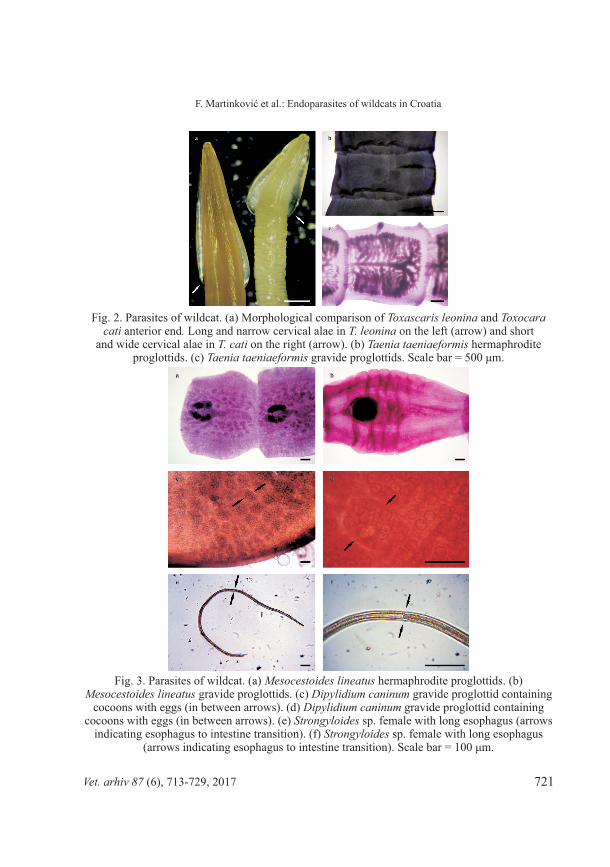
Fig. 2. Parasites of wildcat. (a) Morphological comparison of Toxascaris leonina and Toxocara cati anterior end. Long and narrow cervical alae in T. leonina on the left (arrow) and short
and wide cervical alae in T. cati on the right (arrow). (b) Taenia taeniaeformis hermaphrodite proglottids. (c) Taenia taeniaeformis gravide proglottids. Scale bar = 500 μm.
Fig. 3. Parasites of wildcat. (a) Mesocestoides lineatus hermaphrodite proglottids. (b) Mesocestoides lineatus gravide proglottids. (c) Dipylidium caninum gravide proglottid containing
cocoons with eggs (in between arrows). (d) Dipylidium caninum gravide proglottid containing cocoons with eggs (in between arrows). (e) Strongyloides sp. female with long esophagus (arrows
indicating esophagus to intestine transition). (f) Strongyloides sp. female with long esophagus (arrows indicating esophagus to intestine transition). Scale bar = 100 μm.
722 Vet. arhiv 87 (6), 713-729, 2017
F. Martinković et al.: Endoparasites of wildcats in Croatia
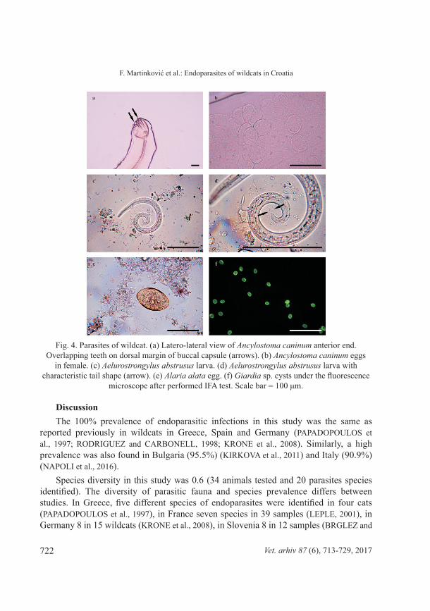
Fig. 4. Parasites of wildcat. (a) Latero-lateral view of Ancylostoma caninum anterior end. Overlapping teeth on dorsal margin of buccal capsule (arrows). (b) Ancylostoma caninum eggs
in female. (c) Aelurostrongylus abstrusus larva. (d) Aelurostrongylus abstrusus larva with characteristic tail shape (arrow). (e) Alaria alata egg. (f) Giardia sp. cysts under the fluorescence
microscope after performed IFA test. Scale bar = 100 μm.
Discussionthe 100% prevalence of endoparasitic infections in this study was the same as
reported previously in wildcats in Greece, Spain and Germany (PAPADOPOULOS et al., 1997; RODRIGUEZ and CARBONELL, 1998; KRONE et al., 2008). Similarly, a high prevalence was also found in Bulgaria (95.5%) (KIRKOVA et al., 2011) and Italy (90.9%) (NAPOLI et al., 2016).
Species diversity in this study was 0.6 (34 animals tested and 20 parasites species identified). The diversity of parasitic fauna and species prevalence differs between studies. In Greece, five different species of endoparasites were identified in four cats (PAPADOPOULOS et al., 1997), in France seven species in 39 samples (LEPLE, 2001), in Germany 8 in 15 wildcats (KRONE et al., 2008), in Slovenia 8 in 12 samples (BRGLEZ and

723Vet. arhiv 87 (6), 713-729, 2017
F. Martinković et al.: Endoparasites of wildcats in Croatia
ŽELEZNIK, 1976), in Italy 10 species in 121 samples (NAPOLI et al., 2016), in Bulgaria 11 in 22 samples (KIRKOVA et al., 2011) in Spain 15 in 19 samples (RODRIGUEZ and CARBONELL, 1998); while the present survey had the highest diversity in terms of the different species identified (20 from 34 samples).
among all parasites detected, the common parasites, with a prevalence of 20% or higher (WAID and PENCE, 1988) were T. taeniaeformis, T. cati, Capillaria sp., Isospora sp., and Strongyloides sp. Taenia taeniaeformis, M. lineatus, T. cati, T. leonina, Capillaria sp., A. tubaeformae, A. abstrusus, Physaloptera sp., Trichinella sp., I. felis, Isospora sp. have been commonly found in wildcat endoparasite surveys across europe with differing prevalence, and the findings of this survey are in accordance with other studies. Four types of spurious parasites were found by copromicroscopic analysis: Eimeria sp., Trichuris sp. eggs, anoplocephalid and hymenolepidid type eggs. It may be assumed that they all originate from prey, rodents or lagomorphs, as previously reported by RODRIGUEZ and CARBONELL (1998) and KIRKOVA et al. (2011).
In this study, Trichinella spp. was found in two out of 34 analyzed samples (5.88%). In other studies conducted in different european countries, Trichinella was found in a higher prevalence, with the highest prevalence in Bulgaria (BRGLEZ and ŽELEZNIK, 1976; KIRKOVA et al., 2011; BLAGA et al., 2009; BADAGLIACCA et al., 2016). Infection of wildlife with Trichinella spp. is widespread throughout europe. there are records of T. britovi and T. spiralis infection in wildcats, where T. britovi infection prevails by 73% vs. 27% (POZIO et al., 2009). In Croatia, Trichinella sp. infection in wildcats has not been detected until now, but is already known in other wild carnivores and omnivores including wolves, badgers, wild boars, bear, and lynx (BECK et al., 2009).
Four parasite species were identified which to our knowledge have previously never been reported in free-living wildcats: D. caninum, Sarcocystis sp., Strongyloides sp., and Giardia sp.
Strongyloides sp. was detected in eight wildcat samples (23.5%) in the current study and this is the first finding of this parasite in Felis silvestris silvestris. Strongyloides species are known, but rare parasites of felines worldwide, and up to now several different species have been described (THAMSBORG et al., 2016). there are a few reports about the low prevalence of Strongyloides sp. infection in domestic cats in europe (takeUChI-STORM at al., 2015a; MIRCEAN et al., 2010). also S. stercoralis was found in one sample in Lynx pardinus in Spain (RODRIGUEZ and CARBONELL, 1998).
according to SPEARE and TINSLEY (1987), when fewer feces are used in routine diagnostics, the chances of finding Strongyloides sp. are slight. this problem can be avoided by using the Baerman test and greater quantities of feces. The females present in the intestine are very thin, and can be found only by dissecting the mucosa, a procedure not done consistently in any former studies. In this study, adults were found in seven
724 Vet. arhiv 87 (6), 713-729, 2017
F. Martinković et al.: Endoparasites of wildcats in Croatia
intestines, with rhabditid larvae in only one separate fecal smear (table 3a-table 3c). the reason for the low Strongyloides sp. prevalence within fecal samples, besides the preservation state of samples, could also be due to the amount of feces used. It could be concluded that Strongyloides sp. parasites are present in Croatian wildcats in a higher prevalence than was detected by fecal examination. In the future, more attention should be paid to avoiding false negative results, especially if only examination of feces is used for monitoring the parasites.
A. alata eggs were found in feces, which is a species previously reported but it is rare in wildcats (takaCS et al., 2011). the species has been reported in domestic cats in several publications - adult flukes were identified in Uruguay (CASTRO et al. 2009), eggs in upstate New York (LUCIO-FORSTER and BOWMAN, 2011) and on Gran Canaria island (RODRÍGUEZ-PONCE et al., 2016); while mesocercariae were identified in domestic cats in Denmark (TAKEUCHI-STORM et al., 2015b). recently, among all the surveys conducted on wildcats (BURT et al., 1980; SCHUSTER et al., 1993; PAPADOPOULOS et al., 1997; RODRIGUEZ and CARBONELL, 1998; KRONE et al., 2008; KIRKOVA et al., 2011; NAPOLI et al., 2016), only takaCS et al. (2011) recorded A. alata. Adult flukes were found in the duodenum and eggs in intestinal scrapings in one male wildcat. Alaria alata was previously described in Croatia in other species: the presence of A. alata mesocercariae was described in wild boar (JAKŠIĆ et al., 2002), while adult flukes were recorded in foxes (RAJKOVIĆ-JANJE et al., 2002) and golden jackals (SINDIČIĆ et al., 2017).
It has never been proven that felids can act as definitive hosts for A. alata in europe, and it is still uncertain if cats can act as paratenic or final hosts of A. alata, as they can host Alaria marcinae. In A. marcinae infection, both cat sexes are final hosts; while lactating females are primarily paratenic hosts, and mesocercarie can be transmitted through milk to their offspring (SHOOP and CORKUM, 1987). Unfortunately, adult flukes were not found in wildcat intestines in this investigation. among animals analyzed in this study, there were also male cats infected with A. alata, indicating the possibility that felids can act as final hosts for A. alata. additionally, reports from takaCS et al. (2011), TAKEUCHI-STORM et al. (2015b) and RODRÍGUEZ-PONCE et al. (2016) and the present findings indicate that A. alata could behave in the same way as A. marcinae in feline hosts. Certainly, further investigation is needed to elucidate if wildcats can be definitive hosts for A. alata or not.
Both D. caninum and Sarcocystis sp. were found in only one sample (2.94%). to the authors’ knowledge these species have not been previously reported in wildcat studies, but both of these parasites are a common finding in domestic cats, reaching up to 83.3% for D. caninum and 0.8-1% for Sarcocystis sp. in some surveys (KNAUS et al., 2011; LUCIO-FORSTER and BOWMAN, 2011; MIRCEAN et al., 2010). In general, the life cycle of Sarcocystis sp. exclusively includes intermediate and final hosts, where the former
725Vet. arhiv 87 (6), 713-729, 2017
F. Martinković et al.: Endoparasites of wildcats in Croatia
behaves as a prey and the latter as a predator. Development to mature stages and the patent period of this parasite in cats are usually transient as most of the oocysts are shed during an approximately two-week period (eCkert et al., 2005). It could be assumed that this short patent period is the reason why the parasite has never been identified in previous studies, because the sample has to be collected exactly during this short period when the parasite is present in the intestines.
Giardia sp. is a common parasite of domestic cats in Europe. During the 2001 - 2014 period, 29 surveys on domestic cats were conducted with prevalence ranging from 0% - 37%, depending on study area and method used (BOUZID et al., 2015). the current survey results showed the presence of Giardia sp. cysts in six wildcat samples (17.6%); while to the authors’ knowledge no other studies conducted on wildcats detected the presence of Giardia sp. Infection with Giardia sp. in wild carnivores in Croatia, other than wildcats, was described by BECK et al. (2011a) and ranged from 4.5% in foxes, 10% in wolves, and up to 12.5% in jackals. also, there is a report of Giardia sp. infection in captive felines from the Croatian Zagreb ZOO (BECK et al., 2011b) and in domestic cats (CaCCIÒ et al., 2010). the low prevalence of Giardia sp. infection in this study could indicate that wildcats ingest Giardia sp. cysts by feeding on rodents. rodents carry Giardia muris, Giardia microti, or Giardia duodenalis assemblage G, which may be unable to establish infection in wildcats, as was described for foxes (BECK et al., 2011a). Still, the true origin of Giardia sp. in wildcats remains to be discovered.
the results of the intestine examination coincide with the copromicroscopic examination in the majority of samples (table 2, table 3). there are some exceptions, e.g. T. taeniaeformis eggs were less prevalent in feces than the adults in intestine. this result was expected because the sensitivity of the flotation method is only 60-80% when more than 1000 eggs/g of feces are present. With lower fecal egg content, the results could be a false negative (eCkert et al., 2005). Other exceptions (Table 2, Table 3) are due to the sample preservation state. Namely, some wildcats were collected as road kill, then frozen until examined, and thawed. as previously reported (RODRIGUEZ and CARBONELL, 1998; KIRKOVA et al., 2011), carcass degradation caused by environmental conditions could considerably influence the results, as some parasites (e. g. tapeworms, nematodes, nematode larvae, oocysts) could deteriorate over time and could be difficult to identify.
To the authors’ knowledge this is the first report of the wildcat helminth and protozoan fauna in Croatia. also, the free-living wildcat is reported as a host for Giardia sp., Sarcocystis sp., Strongyloides sp. and D. caninum for the first time.
ReferencesBADAGLIACCA, P., D. DI SABATINO, S. SALUCCI, G. ROMEO, M. CIPRIANI, N. SULLI,
F. DALL’ACQUA, M. RUGGIERI, P. CALISTRI, D. MORELLI (2016): The role of the wolf
726 Vet. arhiv 87 (6), 713-729, 2017
F. Martinković et al.: Endoparasites of wildcats in Croatia
in endemic sylvatic Trichinella britovi infection in the abruzzi region of Central Italy. Vet. Parasitol. 231, 124-127.
BECK, R., A. BECK, J. KUSAK, Z. MIHALJEVIĆ, S. LUCINGER, T. ZIVICNJAK, D. HUBER, A. GUDAN, A. MARINCULIĆ (2009): Trichinellosis in wolves from Croatia. Vet. Parasitol. 159, 308-311.
BECK, R., H. SPRONG, S. LUČINGER, E. POZIO, S. M. CACCIÒ (2011a): A large survey of Croatian wild mammals for Giardia duodenalis reveals a low prevalence and limited zoonotic potential. Vector Borne Zoonotic Dis. 11, 1049-1055.
BECK, R., H. SPRONG, I. BATA, S. LUČINGER, E. POZIO, S. M. CACCIÒ (2011b): Prevalence and molecular typing of Giardia spp. in captive mammals at the zoo of Zagreb, Croatia. Vet. Parasitol. 175, 40-46.
BLAGA, R., C. GHERMAN, V. COZMA, A. ZOCEVIC, E. POZIO, P. BOIREAU (2009): Trichinella species circulating among wild and domestic animals in romania. Vet. Parasitol. 159, 218-21.
BOUZID, M., K. HALAI, D. JEFFREYS, P. R. HUNTER (2015):. The prevalence of Giardia infection in dogs and cats, a systematic review and meta-analysis of prevalence studies from stool samples. Vet. Parasitol. 207, 181-202.
BRGLEZ, J., Z. ŽELEZNIK (1976): Ein Übersicht über die Parasiten der Wildkatze (Felis silvestris Schreber) in Slowenien. Z. Jagdwiss. 22, 109-112.
BRIANTI, E., G. GAGLIO, S. GIANNETTO, G. ANNOSCIA, M. S. LATROFA, F. DANTAS-TORRES, D. TRAVERSA D. OTRANTO (2012): Troglostrongylus brevior and Troglostrongylus subcrenatus (Strongylida: Crenosomatidae) as agents of broncho-pulmonary infestation indomestic cats. Parasit. Vectors. 5, 178.
BURT, M. D., a. W. PIke, L. K. CORBETT (1980): helminth parasites of wild cats in north-east Scotland. J. helminthol. 54, 303-308.
CACCIÒ, S. M., R. BECK, A. ALMEIDA, A. BAJER, E. POZIO (2010): Identification of Giardia species and Giardia duodenalis assemblages by sequence analysis of the 5.8S rDNA gene and internal transcribed spacers. Parasitology 137, 919-925.
CASTRO, O., J. M. VENZAL, M. L. FÉLIX (2009): Two new records of helminth parasites of domestic cat from Uruguay: Alaria alata (Goeze, 1782) (Digenea, Diplostomidae) and Lagochilascaris major Leiper, 1910 (Nematoda, Ascarididae). Vet. Parasitol. 160, 344-347.
ECKERT, J., K. T. FRIEDHOFF, H. ZAHNER, P. DEPLAZES (2005): Lehrbuch der Parasitologie für die Tiermedizin. Enke Verlag Stuttgart, 2. Aufl., 632p.
FUNK, S. M., C. V. FIORELLO, S. CLEAVELAND, M. E. GOMPPER (2001): The role of disease in carnivore ecology and conservation. In: Carnivore Conservation (Gittleman, J. L., S. M. Funk, D. Macdonald and R. K. Wayne, Eds). Cambridge University Press, Cambridge, pp. 443-466.
HRČKOVA, G., M. MITERPÁKOVÁ, A. O’CONNOR, V. ŠNÁBEL, P. D. OLSON (2011): Molecular and morphological circumscription of Mesocestoides tapeworms from red foxes (Vulpes vulpes) in central europe. Parasitology 138, 638-647.
727Vet. arhiv 87 (6), 713-729, 2017
F. Martinković et al.: Endoparasites of wildcats in Croatia
JAKŠIĆ, S., S. UHITIL, M. VUČEMILO (2002): Nachweis von Mesozerkarien des Saugwurm Alaria alata im Wildschweinefleisch. Z. Jagdwiss. 48, 203-207.
JANICKI, Z., A. SLAVICA, D. KONJEVIĆ, K. SEVERIN (2007): Game zoology. Faculty of Veterinary Medicine, University of Zagreb, Zagreb.
KIRKOVA, Z., E. RAYCHEV, D. GEORGIEVA (2011): Studies on feeding habits and parasitological status of red fox, golden jackal, wild cat and stone marten in Sredna Gora, Bulgaria. J. Life Sci. 5, 264-270.
KNAUS, M., I. KUSI, D. RAPTI, D. XHAXHIU, R. WINTER, M. VISSER, S. REHBEIN (2011). Endoparasites of cats from the Tirana area and the first report on Aelurostrongylus abstrusus (railliet, 1898) in albania. Wien. klin. Wochenschr. 123 (Suppl 1), 31-35.
KRONE, O., O. GUMINSKY, H. MEINIG, M. HERRMANN, M. TRINZEN, G. WIBBELT (2008): endoparasite spectrum of wild cats (Felis silvestris Schreber, 1777) and domestic cats (Felis catus L.) from the Eifel, Pfalz region and Saarland, Germany. Eur. J. Wildl. Res. 54, 95-100.
LEPLE, D. (2001): Parasitologie compareé du chat forestier (Felis silvestris silvestris, Schreber 1777) et du chat domestique (Felis catus, Linne 1758), Ecole Nat. These de Doctorat Vét, Lyon.
LUCIO-FORSTER, A, D. D. BOWMAN (2011): Prevalence of fecal-borne parasites detected by centrifugal flotation in feline samples from two shelters in upstate New York. J. Feline Med. Surg. 13, 300-303.
MEHLHORN, H., D. DÜWEL, W. RAETHER (1993): Diagnose und Therapie der Parasitosen von Haus-, Nutz- und Heimtieren, Gustav Fischer Verlag Stuttgart, Stuttgart, Jena New York, 529 p.
MIRCEAN, V., A. TITILINCU, C. VASILE (2010): Prevalence of endoparasites in household cat (Felis catus) populations from transylvania (romania) and association with risk factors. Vet. Parasitol. 171, 163-166.
NAPOLI, E., S. ANILE, C. ARRABITO, D. SCORNAVACCA, M. V. MAZZAMUTO, G. GAGLIO, D. OTRANTO, S. GIANNETTO, E. BRIANTI (2016): Survey on parasitic infections in wildcat (Felis silvestris silvestris Schreber, 1777) by scat collection. Parasitol. res. 115, 255-261.
PAPADOPOULOS, H., C. HIMONAS, M. PAPAZAHARIADOU, k. ANTONIADOU-SOTIRIADOU (1997): helminths of foxes and other wild carnivores from rural areas in Greece. J. helminthol. 71, 227-231.
POZIO, E., L. RINALDI, G. MARUCCI, V. MUSELLA, F. GALATI, G. CRINGOLI, P. BOIREAU, G. LA ROSA (2009): Hosts and habitats of Trichinella spiralis and Trichinella britovi in europe. Int. J. Parasitol. 39, 71-79.
RAJKOVIĆ-JANJE, R., A. MARINCULIĆ, S. BOSNIĆ, M. BENIĆ, B. VINKOVIĆ, Ž. MIHALJEVIĆ (2002): Prevalence and seasonal distribution of helminth parasites in red foxes (Vulpes vulpes) from the Zagreb County (Croatia). Z. Jagdwiss. 48, 151-160.
RODRIGUEZ, A., E. CARBONELL (1998): Gastrointestinal parasites of the Iberian lynx and other wild carnivores from central Spain. acta Parasitol. 43, 128-136.
728 Vet. arhiv 87 (6), 713-729, 2017
F. Martinković et al.: Endoparasites of wildcats in Croatia
RODRÍGUEZ-PONCE, E., J. F. GONZÁLEZ, M. CONDE DE FELIPE, J. N. HERNÁNDEZ, J. RADUAN JABER (2016): Epidemiological survey of zoonotic helminths in feral cats in Gran Canaria island (Macaronesian archipelago-Spain). acta Parasitol. 61, 443-450.
SCHMÄSCHKE, R. (2013): Die koproskopische Diagnostik von Endoparasiten in der Veterinärmedizin, Schlütersche Verlag, Hannover, p. 152.
SCHUSTER, R., D. HEIDECKE, K. SCHIERHORN (1993): Contributions to the parasite fauna of local hosts. 10. On the endoparasitic fauna of Felis silvestris. appl. Parasitol. 34, 113-120.
SHOOP, W. L., K. C. CORKUM (1987): Maternal transmission by Alaria marcianae (trematoda) and the concept of amphiparatenesis. J. Parasitol. 73, 110-115.
SINDIČIĆ, M, M. JAGIĆ, N. ŠPREM, V. SLIJEPČEVIĆ, R. POPOVIĆ, D. KONJEVIĆ (2016): Diversity of mitochondrial DNA in wildcats in Croatia - preliminary results. Proceedings of Sixth Croatian Veterinary Congress with International Participation, 26 - 29 October, Opatija, Croatia pp. 333-340.
SINDIČIĆ, M., F. MARTINKOVIĆ, M. BUJANIĆ, N. TUŠKAN, M. ŠPEHAR, D. KONJEVIĆ (2017): Morphological and molecular identification of golden jackal parasites. Book of abstracts. 7th International Congress „Veterinary Science and Profession“, 5 - 7 September, Zagreb, Croatia, p. 77.
SPEARE, R., D. J. TINSLEY (1987): Survey of cats for Strongyloides felis. aust. Vet. J. 64, 191.TAKÁCS, A., L. SZEMETHY, M. HELTAI, A. A. TAKÁCS (2011): Data on the parasitological
state of wild cats (Felis silvestris Schreber 1777) and of their hybrids with feral domestic cats (Felis silvestris catus L. 1758) on Hungarian hunting areas. Hungar. Vet. J. 133, 670-674.
TAKEUCHI-STORM, N., H. MEJER, M. N. AL-SABI, C. S. OLSEN, S. M. THAMSBORG, H. L. ENEMARK (2015a): Gastrointestinal parasites of cats in Denmark assessed by necropsy and concentration McMaster technique. Vet. Parasitol. 214, 327-332.
TAKEUCHI-STORM, N., M. N. AL-SABI, S. M. THAMSBORG, H. L. ENEMARK (2015b): Alaria alata Mesocercariae among Feral Cats and Badgers, Denmark. Emerg. Infect. Dis. 21, 1872-1874.
THAMSBORG, S. M., J. KETZIS, Y. HORII, J. B. MATTHEWS (2016): Strongyloides spp. infections of veterinary importance. Parasitology 144, 1-11.
DOI: 10.1017/S0031182016001116 VERONESI, F., D. TRAVERSA, E. LEPRI, G. MORGANTI, F. VERCILLO, D. GRELLI, R.
CASSINI, M. MARANGI, R. IORIO, B. RAGNI, A. DI CESARE (2016): Occurrence of lungworms in european wildcats (Felis silvestris silvestris) of central Italy. J. Wildl. Dis. 52, 270-278.
WAID, D. D., D. B. PENCE (1988): Helminths of mountain lions (Felis concolor) from southwestern texas, with a redescription of Cylicospirura subaequalis (Molin, 1860) Vevers, 1922. Can. J. Zool. 66, 2110-2117.
729Vet. arhiv 87 (6), 713-729, 2017
F. Martinković et al.: Endoparasites of wildcats in Croatia
YAMAGUCHI, N., A. KITCHENER, C. DRISCOLL, B. NUSSBERGER (2015): Felis silvestris. The IUCN Red List of Threatened Species 2015, e.T60354712A50652361. DOI: 10.2305/IUCN.UK.2015-2.RLTS.T60354712A50652361.en
received: 27 January 2017accepted: 15 May 2017
________________________________________________________________________________________MaRTinKović, F., M. SinDičić, S. LučingeR, i. ŠTiMac, M. BuJanić, T. ŽivičnJaK, D. SToJčević Jan, n. ŠPReM, R. PoPović, D. KonJević: endoparaziti u divljih mačaka u Hrvatskoj. vet. arhiv 87, 713-729, 2017.
SaŽeTaK Znanstveni radovi o parazitima divljih mačaka (Felis silvestris silvestris) rijetki su i najčešće rađeni
na malom broju uzoraka te je stoga naš cilj bio istražiti učestalost endoparazita divljih mačaka u Hrvatskoj. Pregledali smo lešine 34 divlje mačke, stradale u prometu ili u sklopu provedbe lovnogospodarskih osnova. Sve su životinje bile negativne na bjesnoću. Sadržaj želudca i crijeva pregledali smo pod mikroskopom. Izmet iz debelog crijeva pregledan je metodom flotacije pomoću zasićene otopine ZnSO4, dok je dijafragma pregledana pomoću umjetne probave. Upotrijebili smo izravnu imunofluorescenciju za utvrđivanje cista Giardia sp., što je prvo takvo istraživanje kod divljih mačaka. Sve pregledane životinje bile su invadirane barem jednom vrstom parazita, dok je najraznolikija invazija uključivala šest različitih vrsta parazita utvrđenih kod jedne životinje. Sljedeće su vrste parazita identificirane (% prevalencije odraslih parazita i razvojnih stadija u svim analiziranim uzorcima): Taenia taeniaeformis (55,9 %), Capillaria sp. (50 %), Toxocara cati (50 %), Isospora sp. (29,4 %), Strongyloides sp. (23,5 %), Giardia sp. (17,6 %), Ancylostoma tubaeformae (14,7 %), Physaloptera sp. (11,8 %), Hymenolepididae (8,8 %), Alaria alata (5,9 %), Aelurostrongylus abstrusus (5,9 %), Toxascaris leonina (5,9 %), Trichinella sp. (5,9 %), Mesocestoides lineatus (5,9 %), Anoplocephalidae (2,9 %), Dipylidium caninum (2,9 %), Trichuris sp. (2,9 %), Isospora felis (2,9 %), Eimeria sp. (2,9 %) i Sarcocystis sp. (2,9 %). Među njima, Eimeria sp., Trichuris sp., jaja anoplocefalida i himenolepidida su pseudoparaziti, koji potječu od plijena. Četiri roda koje smo identificirali do sada nisu nikada opisani kod divljih mačaka - Giardia sp., Strongyloides sp., Sarcocystis sp. i Dipylidium caninum.
Ključne riječi: paraziti, divlja mačka, Felis silvestris silvestris, hrvatska________________________________________________________________________________________

.


