Embed Size (px)
Citation preview
Contents
Introduction . . . . . . . . . . . . . . . . . . . . . . . . 220
Operative Technique, Indications . . . . . . . . . . . . 220Type I: simple Drainage (Fig. 24.1A) . . . . . . . . . 220Type II a/b: Extended Drainage (Fig. 24.1B–D) . . . 220Type III: Endonasal Median Drainage (Fig. 24.1E,F) 224Results of Endonasal Frontal Sinus Surgery . . . . . 226Postoperative Care . . . . . . . . . . . . . . . . . . . 228
Packing . . . . . . . . . . . . . . . . . . . . . . . . 228Local Postoperative Therapy . . . . . . . . . . . . 228General Postoperative Medication . . . . . . . . . 228
Failures . . . . . . . . . . . . . . . . . . . . . . . . . . . 229Postoperative Frontal Sinusitis after Type I and Type II Drainage . . . . . . . . . . . . . . . . . . 229Reclosure after Type III Drainage . . . . . . . . . . . 229
Complications . . . . . . . . . . . . . . . . . . . . . . . 229
General Guidelines for Surgical Therapy of Frontal Sinus Inflammatory Diseases not Responding to Conservative Measures . . . . . . . 230
How to treat frontal sinusitis, which has not responded to conservative measures nor has been operated before? . . . . . . . . . . . . . . . . . 230What to do in cases of chronic postoperative frontal sinusitis after one or even several previous operations,otherwise referred to as “iatrogenic” sinusitis [12, 27, 33]? . . . . . . . . . . . . . . . . . . . . . . . 230How “radical” the extent of primary surgery should be in patients with extensive polyposis of the frontal sinus? . . . . . . . . . . . . . . . . . . . 230Should patients with so-called spontaneous or postoperative mucoceles of the frontal sinus, be operated endonasally or via an external approach? . 230Is a Pott’s puffy tumor always an indication for an external procedure? . . . . . . . . . . . . . . . 231
Conclusion . . . . . . . . . . . . . . . . . . . . . . . . . 231
References . . . . . . . . . . . . . . . . . . . . . . . . . 231
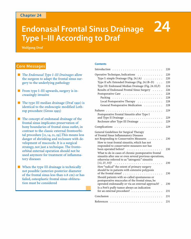
Chapter 24
Endonasal Frontal Sinus DrainageType I–III According to DrafWolfgang Draf
24
Core Messages
� The Endonasal Type I–III Drainages allowthe surgeon to adapt the frontal sinus sur-gery to the underlying pathology
� From type I–III upwards, surgery is in-creasingly invasive
� The type III median drainage (Draf 1991) isidentical to the endoscopic modified Loth-rop procedure (Gross 1995)
� The concept of endonasal drainage of thefrontal sinus implicates preservation ofbony boundaries of frontal sinus outlet, incontrast to the classic external frontoorbi-tal procedure [11, 14, 21, 29].This means lessdanger of shrinking and reclosure with de-velopment of mucocele. It is a surgicalstrategy, not just a technique. The fronto-orbital external operation should not beused anymore for treatment of inflamma-tory diseases
� When the type III drainage is technicallynot possible (anterior-posterior diameterof the frontal sinus less than 0.8 cm) or hasfailed, osteoplastic frontal sinus oblitera-tion must be considered

Introduction
Endonasal surgery of the paranasal sinuses began,apart from a couple of earlier reports, some hundredyears ago [5, 6, 12, 30, 31]. Only a few skilled surgeonshave been able to perform endonasal ethmoidectomyand adequate drainage of the frontal sinus using justa headlight and the naked eye, whereas others creat-ed serious complications such as CSF leak, meningi-tis, brain abscess, and encephalitis ending in the pre-antibiotic era mostly with the death of the patient.This is why for decades, until the 1970s, endonasal si-nus surgery was not accepted in most leading institu-tions.
The renaissance of endonasal surgery was due to sev-eral advances:
� New optical aids such as the microscope andendoscope
� Improved understanding of the physiologyand pathophysiology of nasal and paranasalsinus mucosa
� Patients no longer accepting the sometimesserious sequelae of external operations in ad-dition to an unsatisfactory outcome
� Remarkable progress in anesthesiology pro-viding the endonasal surgeon with an almostbloodless field
Between 1980 and 1984, an endonasal surgical con-cept with different degrees of frontal sinus openingwas worked out and intensively tested before beingpublished [2].
With increasing experience and referrals of diffi-cult frontal sinus cases, it became obvious that not allproblems can be solved via an endonasal route.Therefore the osteoplastic obliterative frontal sinusoperation [34] was included in the concept, in orderto deal with all different kinds of frontal sinus prob-lems. In difficult revision cases, the endonasal opera-tion sometimes has to be combined with the osteo-plastic, mostly obliterative procedure [2].
Operative Technique, Indications
For the endonasal frontal sinus, an operation usingsome type of general anesthesia is required. In addi-tion, topical decongestion helps to provide a dryfield.
Surgery on the frontal recess is usually preceded atleast by an anterior, more often than not by a com-plete ethmoidectomy. Exceptions are those caseswhere a complete ethmoidectomy has already beenperformed. It is important to remove agger nasi cellsand to visualize the attachment of the middle turbi-nate medially, the lamina papyracea laterally, and theanterior skull base with the anterior ethmoidal arterysuperiorly.
Type I: Simple Drainage (Fig. 24.1A)
The type I drainage is established by ethmoidectomyincluding the cell septa in the region of the frontal re-cess. The inferior part of Killian’s infundibulum andits mucosa is not touched. This approach is indicatedwhen there is only minor pathology in the frontal si-nus and the patient does not suffer from ‘prognosticrisk factors” like aspirin intolerance and asthma,which are associated with poor quality of mucosaand possible problems in outcome (Table 24.1). In themajority of cases the frontal sinus heals because ofthe improved drainage via the ethmoid cavity.
Type II a/b: Extended Drainage(Fig. 24.1B–D)
Extended drainage is achieved after ethmoidectomyby resecting the floor of the frontal sinus between thelamina papyracea and the middle turbinate (type IIa) or the nasal septum (type II b) anterior to the ven-tral margin of the olfactory fossa.
In the classification of May and Schaitkin [22]type IIa corresponds with NFA II (nasofrontal ap-proach) and type IIb with NFA III. Hosemann et al.[7, 8, 9] showed in a detailed anatomical study thatthe maximum diameter of a neo-ostium of the fron-tal sinus (type IIa), which could be gained using aspoon or a curette, was 11 mm with an average of
Wolfgang Draf220
24

Chapter 24Endonasal Frontal Sinus Drainage Type I–III 221
Fig. 24.1A–F. Endonasal frontal sinus drainage 2. (A) Type Idrainage (Simple drainage, right side). aea, anterior ethmoidalartery; lp, lamina papyracea; mt, middle turbinate; ns, nasalseptum; oc, olfactory cleft. (B) Type II a drainage (enlargeddrainage, a, right side). Opening of frontal sinus between lam-ina papyracea and middle turbinate. Mostly possible withoutdrill. (C) Type IIb drainage (enlarged drainage, b, right side).Drainage of the frontal sinus between lamina papyracea and
nasal septum. Usually medially drill necessary. (D) Type IIbdrainage Detail with identification of the first olfactory fiber(detail of 1c; of, olfactory fiber). (E) Type III drainage (mediandrainage) with “Frontal T” (red) and first olfactory fiber onboth sides (View from left inferior). (F) Type III drainage (me-dian drainage) sagittal view: removal of the frontal sinus floorin front of the olfactory cleft

Wolfgang Draf222
24
Fig. 24.1E,F

5.6 mm. They also presented an excellent criticalevaluation and results [9].
If one needs to achieve a larger drainage openinglike type II-b, a drill is used because of the increasingthickness of the bone medially towards the nasal sep-tum. During drilling with the diamond burr, bonedust fogs the endoscope, demanding repeated clean-ing. At this point the microscope is useful, allowingone to work with two hands, while an assistant holdsa simple self-retracting speculum according to Cho-lewa (1888) [1]. The endoscopic four–hand technique,introduced by May [21], is also a useful alternative, al-lowing the surgeon to work with two hands while anassistant holds the endoscope.
In revision cases after incomplete ethmoidectomy,it is recommended that a wide approach to the eth-moid sinuses is created using a microscope and drillor punch when possible. Punches and through-cutting instruments [23] help preserve the mucosa,whereas the drill is more destructive in this respect.The wide approach to the ethmoid is obtained by ex-posing the lacrimal bone and reducing it, as well asparts of the agger nasi and part of the frontal processof the maxilla, until the lamina papyracea is clearlyseen through the microscope. This facilitates bettervisualization of the frontal recess to allow furtherwork on the frontal sinus floor, but also makes the
postoperative treatment less painful. As soon thefrontal recess is identified using the middle turbinateand where identifiable, the anterior ethmoidal arteryas landmarks, the frontal infundibulum is exposedand the anterior ethmoidal cells are resected. Duringsurgery, repeated considerations of the pre-operativeCT scans will establish the presence of so-called fron-tal cells [17] (Fig. 24.2, see also Chapter 2) which candevelop far into the frontal sinus, giving the surgeonthe erroneous impression that the frontal sinus hasbeen properly opened. Sagittal CT slices and naviga-tion may be helpful in difficult situations. Whenfrontal cells are present, a procedure called “uncap-ping the egg” by Stammberger [33] uses a 45° tele-scope and results in a type IIa drainage.
An alternative when the middle turbinate hasbeen retracted laterally after previous surgery and isobstructing the frontal sinus drainage is the so-called“frontal sinus rescue procedure” [16] (see Chapter26). The decision should be left to the patient as towhether or not he desires a more conservative proce-dure like this, which has a relatively higher frequencyof recurrence and need for re-operation.
If, after a type IIa drainage has been performed,further widening to produce a type IIb is required,the diamond burr is introduced into the clearly vis-ible gap in the infundibulum and drawn across the
Chapter 24Endonasal Frontal Sinus Drainage Type I–III 223
Table 24.1. Indications for endonasal frontal sinus drainage Type I–III
(A) Indications type I drainageAcute sinusitis – failure of conservative surgery
– orbital and endocranial complicationsChronic sinusitis – first time surgery
– no risk factors (aspirin intolerance, asthma, triad )– revision after incomplete ethmoidectomy
(B) Indications type IIa drainage – serious complications of acute sinusitis– medial muco- pyocele– tumor surgery ( benign tumors)– good quality mucosa
Indications type IIb drainage – all indications of type IIa, if the resulting IIa is smaller than 5×7 mm. For type II bdrill necessary
(C) Indications type III drainage – difficult revision surgery– primarily in patients with prognostic risk factors and severe polyposis particular-
ly patients with triad mucoviscidosis– mucoviscidosis– Kartagener’s syndrome– ciliary immotility syndrome– benign and malignant tumors(see text)

bone in a medial direction. Care is taken to ensurethat the frontal sinus opening is bordered by bone onall sides and that mucosa is preserved at least on onepart of the circumference. To also safely create medi-ally the widest possible opening of the frontal sinusfloor, one should identify the first ipsilateral olfacto-ry fiber (for details see type III drainage, Fig 2B,C).Atthe end a rubber finger stall can be introduced intothe frontal sinus for about 5 days.
The indications for one or the other Type II drain-age in general are listed in Table 24.1B. If the surgeonfeels that the type IIa drainage is too small in regardto the underlying pathology, it is better to performthe type II b drainage procedure.
Some authors [11, 27, 39] advocated the use of soft,flexible silicone stents in cases of a frontal sinus neo-ostium less than 5 mm in diameter, since more rigidsilicone tubes have not given satisfying results [24,29]. So far the techniques using soft silicone drainagedevices showed promising results although longterm observation is still lacking.
Type III: Endonasal Median Drainage(Fig. 24.1E,F)
Endonasal median drainage or type III: The extendedIIb opening is enlarged by resecting portions of thesuperior nasal septum in the neighborhood of thefrontal sinus floor. The diameter of this openingshould be about 1.5 cm. This is followed by resectionof the frontal sinus septum or septa, if more than oneare present. Starting on one side of the patient, onecrosses the midline until the contralateral laminapapyracea is reached.
To achieve the maximum possible opening of thefrontal sinus, it is very helpful to identify the first ol-factory fibers on both sides: the middle turbinate isexposed and is resected in very small pieces from ananterior to posterior direction, along its origin at theskull base. After about 5 mm one will see the first ol-factory fiber coming out of a small bony hole, slight-ly medial to the origin of the middle turbinate. Thesame is done on the contralateral side. Finally the so-called “Frontal T” [3] (Fig. 24.1E) results. Its long crusis represented by the posterior border of the perpen-dicular ethmoid lamina resection, and the shorterwings on both sides are provided by the posteriormargins of the frontal sinus floor resection.
After that, the ethmoidectomy on the left side isperformed exactly as it was on the right.
To perform the type III drainage in the technicallymost efficient way, it is helpful to interchange the useof the endoscope and microscope. Alternatively, thisprocedure can be done with the endoscope alone,though it is more time-consuming. Curved drills ofdifferent angles used with the shaver motor are help-ful [41]. They allow a more superior reach into thefrontal sinus and resection of the interfrontal sinusseptum or septa, if more than one are present. Theyalso allow removal of superiorly located frontal cellswhen present, and thus they help achieve a morecomplete operation. These measures help to createexcellent landmarks for the anterior border of the ol-factory fossa on both sides, which allow for an easierand safer complete resection of the frontal sinus flooras far posteriorly as the location of the first olfactoryfiber.
Finally, a rubber finger stall is placed into eachfrontal sinus, and two more are placed in the ethmoid
Wolfgang Draf224
24
Fig. 24.2. Type III drainage 1 year postoperatively. ssf, septumsinuum frontalium; rfs, right frontal sinus; lfs, left frontal si-nus; sn, septum nasale; re, right ethmoid; le, left ethmoid

cavity and the inferior nasal meatus bilaterally. Thispacking is left for 7 days (!) under prophylactic anti-biotic treatment. The rubber finger stalls do not stickto the surrounding tissue and are therefore easily andpainlessly removed. The packing allows re-epithelial-ization of a major portion of the surgical cavity,which simplifies the postoperative treatment.
In difficult revision cases, one can begin the typeIII drainage primarily from two starting points, ei-ther from the lateral side as already described orfrom medially. The primary lateral approach is rec-ommended if the previous ethmoidal work was in-complete and the middle turbinate is still present as alandmark. One should adopt the primary medial ap-proach, if the ethmoid has been cleared and/or if themiddle turbinate is absent.
The medial approach begins with the partial re-section of the perpendicular plate of the nasal sep-tum, followed by identification of the first olfactoryfiber on each side as already described.
The endonasal median drainage is identical withthe NFA IV [21] and the “modified Lothrop proce-dure” [4]. Lothrop [18, 19] himself warned against us-ing the endonasal route, judging it as too dangerousduring his time; he performed the median drainagevia an external approach. Halle [6] in 1906 created a
large drainage from the frontal sinus directly to thenose using the endonasal approach, and using only aheadlight and the naked eye.
The principle difference between the endonasalmedian frontal sinus drainage and the classic exter-nal Jansen, Lothrop, Ritter, Lynch, and Howarth oper-ations is that the bony borders around the frontal si-nus drainage are preserved. This makes it morestable in the long term and reduces the likelihood ofreclosure by scarring, which may lead to recurrentfrontal sinusitis or a mucocele, not to mention theavoidance of external scar.
The endonasal median drainage (type III) is indi-cated (Table 24.1C) after one or several previous si-nus operations have not resolved the frontal sinusproblem, including an external frontoethmoidecto-my. It is also justified as a primary procedure in pa-tients with severe polyposis and other prognostic“risk factors” affecting outcome, such as aspirin in-tolerance, asthma, Samter’s triad (aspirin hypersen-sitivity, asthma, and allergy), Kartagener’s syndrome,mucoviscidosis, and ciliary dyskinesia syndrome(Table 24.1C). Its use in patients with severe polypo-sis without these risk factors is undetermined andneeds to be evaluated. It seems that patients withgeneralized polyposis but who still show air on coro-
Chapter 24Endonasal Frontal Sinus Drainage Type I–III 225
Fig. 24.3.Frontal sinus view from above aftercoronal osteoplastic revision. Severalfrontal cells of different sizes narrowthe drainage into the nose. (fc, fron-tal cell)

nal CT (“halo sign”) in the periphery of the sinusesalong the skull base have a comparatively betterprognosis than those without, and can be managedby a more conservative technique. The procedure isalso useful for removal of benign tumors in the fron-tal and ethmoid sinuses, as long as the main portionof the tumor in the frontal sinus is medial to a verti-cal line through the lamina papyracea. In addition,the use of the type III drainage makes the removal ofmalignant tumors which are just reaching the frontalsinus safer.
Results of Endonasal Frontal Sinus Surgery
Judging the results of endonasal frontal sinus surgeryrequires a postoperative follow-up of ten or moreyears [14, 25, 26]. The failure rate of Neel et al. [14]with a modified Lynch procedure grew from 7% at amean follow-up of 3.7 years to 30% at 7 years.
Kikawada et al. [16] presented a retrospective re-view of 22 consecutive cases of extended frontal sinussurgery (Draf type III) in patients with obstructivefrontal sinusitis caused by postoperative scarring,with a follow-up time of at least 12 months after sur-gery. Of the 16 patients who underwent the type IIIprocedure, in 14 (88%) the patency of the newly
created frontal sinus drainage and an aerated sinuswere confirmed. Of 12 sides in nine patients whounderwent Draf type II procedure, 5 sides (42%)were also confirmed as cured. In the opinion of theauthors, the median drainage operation (type III) onthe frontal sinus showed excellent long-term resultscompared with the type II procedure.
Weber et al. [36, 40] carried out two studies in 1995and 1996. In the first retrospective study, patientswho underwent endonasal frontal sinus drainage(471 type I drainages, 125 type II drainages, and 52type III drainages) between 1979 and 1992 were eval-uated. From these groups, random patients were ex-amined: 42 patients with type I drainage, 43 with typeII drainage, and 47 with type III drainage were in-cluded in the study. In each patient, the indication forsurgery was chronic polypoid rhinosinusitis, exceptin five patients with type III drainage, in whom an or-bital complication presented associated with acutesinusitis. The follow-up period was between 1 yearand 12 years with a median of 5 years. The subjectiveestimation of operative results by the patients isshown in Figure 24.4a–c.Applying subjective and ob-jective criteria to evaluate the success of endonasalfrontal sinus drainage (Grade 1 = endoscopically nor-mal mucosa, independent of the subjective com-plaints; Grade 2 = subjectively free of symptoms, butwith endoscopically visible inflammatory mucosalchanges; Grade 3 = no subjective improvement andpathologic mucosa = failure), it was possible toachieve a success rate of 85.7% with type I drainage,83.8% with Type II drainage, and 91.5% with type IIIdrainage. This means that, despite the choice of prog-nostically unfavorable cases, type III drainages ap-peared to show the best results, though this was notstatistically significant among the three groups.
In the second study [39], endoscopic and CT ex-aminations were systematically carried out (Figs.24.5,24.6). After 12–98 months follow-up of patientswith type II drainage, 58% of 83 frontal sinuses wereventilated and normal.A ventilated frontal sinus withhyperplastic mucosa was seen in 12%. Scar tissue oc-clusion with total opacification on CT was evident in14%. In 16%, total opacification was due to recurrentpolyposis. Seventy-nine percent of the patients werefree of symptoms or had only minor problems.
Twelve to 89 months following type III drainage,59% of 81 frontal sinuses were ventilated and normal.
Wolfgang Draf226
24
Table 24.2. Indications for the osteoplastic flap procedure
1. Correctly performed Type III drainage failed
2. Patients after many endonasal and various externalfrontal sinus operations, so-called problem frontal si-nus, sometimes in combination with complete endona-sal ethmoidectomy
3. Type III drainage technically not possible (anterior-posterior diameter less than 8 mm)
4. Laterally located muco-pyocele
5. Major destruction of posterior wall
6. Inflammatory complications after trauma (e.g. allo-plastic material, without or with obliteration)
7. Aesthetic correction of pneumatosinus dilatans fron-talis (mostly without obliteration)
8. Major benign tumors (e.g. osteoma) without or withobliteration

A ventilated frontal sinus with hyperplastic mucosawas seen in 17%. Scar tissue occlusion with totalopacification on CT was obvious in 7% and, in 16%,there was total opacification due to recurrent poly-posis. Ninety-five percent of the patients were free ofsymptoms or had only minor problems. Already this
first series of re-evaluation of long-term results dem-onstrates the value of the endonasal frontal sinus sur-gery.
In a retrospective study Mertens et al. [22] com-pared the results of 236 patients operated on between1985 and 1993 using different techniques. After fol-low-up of 3–10 years only 8% of patients needed revi-sion. The lowest revision rate was seen after endona-sal technique according Draf ’s classification (5.9%)compared to the osteoplastic techniques according toJansen-Ritter (Lynch) and Riedel (10.6%).
Chapter 24Endonasal Frontal Sinus Drainage Type I–III 227
Fig. 24.4A–C. Subjective judgment of results of frontal sinussurgery 1 to 12 years after surgery. (A) Type I drainage. (B) TypeII drainage. (C) Type III drainage
Fig. 24.6. Synopsis of CT and endoscopy 12 to 98 months fol-lowing Type III drainage 41.
Fig. 24.5. Synopsis of CT and endoscopy 12 to 98 months fol-lowing Type II drainage 41.

Postoperative Care
There are different ways of providing postoperativecare.Within the years the following standards provedto be efficient:
Packing
Rubber finger stalls (Rhinotamp; Vostra Aachen)filled with sponge have stood the test of time. Theyprovide safe hemostasis, are a stimulator of re-epi-thelialization of bare bone, are cost-effective andpainless to remove. In cases of type I-type IIb drain-age, the packing is left between 2–5 days maximumwithout antibiotic prophylaxis. It is of utmost impor-tance to fix the rubber finger stalls with threads at thenasal dorsum to avoid aspiration. The more stablethe middle turbinate at the end of operation is, theshorter the time of uncomfortable packing can be.The risk of adhesions and synechiae is low becausethis type of packing suppresses the development ofgranulations
After a type III drainage, we leave the packing inplace for 7 days postoperatively as recommended byToffel [35].
Leaving rubber finger stalls for one week carriesthe following advantages [40]:
1. The fibrinoid phase of wound healing is some-how overcome. Reclosure of the large drainageby scars is remarkably reduced, since barebone is re-epithelialized almost completely.
2. Sedation and general anesthesia are not neces-sary for packing removal. Rubber finger packsdo not bind to the wound. Removal of the tam-ponade does not lead to renewed tissue trau-ma. The patients are prepared preoperativelyfor a somewhat uncomfortable postoperativetime. This is by far compensated by the opti-mal wound healing and easy postoperativecare with less crusting.
Postoperative Therapy
The question of whether postoperative intensive me-chanical cleansing is necessary or the wound cavity isself-cleaning without external measures is very con-troversial.
In an obstructed nose or sinus, when the patienthas complaints that can be explained with occlusionof the sinus ostial region by crusts, mechanical clean-ing must be done. However, since each cleaning leadsto injury, freshly granulating tissue, and partial re-moval of new epithelium, a rather controlled andconservative approach to instrument cleaning seemsappropriate.
The patients are given the following instructions toensure proper healing:
1. Irrigate the nasal cavities with saline solutionat least once a day, sometimes more frequently.
2. Use one of the corticosteroid sprays 1–3 times/day.
3. The recommendation is made to use peanut oil1 hour after the use of corticosteroid spray, forgeneral care of the mucosa.
In patients with extreme crusting, the physicianshould inquire about the use of medications becauseas a side effect, they cause mucosal desiccation. Theseinclude psychotropic medications or beta-blockers.Spectacular improvement is possible once thesemedications are changed.
General Postoperative Medication
1. Antibiotics: They are indicated in the postop-erative period for 1–2 weeks in cases of acuteor purulent sinusitis. In type III drainage, werecommend prophylactic antibiotic use, aslong as the tamponade is in place.
Wolfgang Draf228
24

2. Antiallergic medical therapy: This is recom-mended for 6 weeks postoperatively if allergyis diagnosed by history or specific tests. Thepresence of a large number of eosinophils inthe inflamed tissue may provide additionalguidance in the decision-making process. Inless severe cases we prescribe day antihista-mines. In severe allergy patients (e.g. Samter’striad), the combination of antihistamines with low-dose corticosteroid medication for 6 weeks is helpful to prevent early recurrenceof polyps.
Failures
Postoperative Frontal Sinusitis after Type Iand Type II Drainage
Sometimes after ethmoidectomy and type I as well astype II drainage, the patients may develop moreproblems in the frontal sinus than before surgery.Postoperative sinus CT will provide information iffrontal sinusitis has developed.
The pathogenesis of recurrent frontal sinusitis af-ter surgery can involve various mechanisms. Eitherremnant ethmoidal cells developed recurrent sinus-itis or mechanical irritations of the mucosa in thefrontal recess can result in a severe scar aroundKillian’s infundibulum. Both pathologies may resultin blockage of the frontal sinus drainage.
This can be avoided by performing at least a com-plete anterior ethmoidectomy and using extremelyatraumatic handling of the frontal recess mucosa. Fortreatment we recommend the following procedures:a type IIa drainage if a type I procedure was per-formed previously, a type IIb drainage if a type IIaprocedure was performed previously, and a type IIIdrainage after a previous type IIb.
Reclosure after Type III Drainage
Several technical details can lead to this problem:
a) The “chimney” between the anterior ethmoidand the frontal sinus has not been opened well.It is important that after the anterior ethmoid-al artery is identified, the surgeon proceedsalong the skull base medial to the lamina pap-yracea to enter into the frontal sinus.
b) The anterior-posterior opening of the frontalsinus floor, particularly in the midline, is toosmall. The identification of the first olfactoryfiber bilaterally and the creation of the “Fron-tal T” are very helpful to avoid this problem.
c) The resection of the septum/a sinuum frontali-um has been missed or was not performed to asatisfying degree. The new curved drillsbetween 15° and 60° angle are ideal for thispurpose.
d) The resection of the superior nasal septumwas too small. The diameter of resection mustbe 1.5 cm just in front of the “Frontal T” andbelow the frontal sinus floor.
e) The packing between the ethmoid and thefrontal sinus was not left long enough. 7 daysproved to be the best time frame for using rub-ber finger packings.
Complications
In principle, the complication rate of endonasal fron-tal sinus drainage procedures is low and similar tothe frequency of complications of endonasal pansi-nus operations.
An evaluation of complications with special re-spect to endonasal frontal sinus surgery was not per-formed. The operation can be classified as very safe,even with identification of the first olfactory fibers,when optical aids such as microscope and/or endo-scope are used and the techniques described are fol-lowed.
We have analyzed the complications of our endo-nasal micro-endoscopic pansinus operations in twostudies [37, 38].
Chapter 24Endonasal Frontal Sinus Drainage Type I–III 229

The significant complications were:
1. Injury to the periorbit in 14% of cases. Thishad no further consequences except in one pa-tient, who developed periorbital hematoma.No cases of blindness or other orbital lesionslike muscle injury with double vision or lacri-mal drainage obstruction occurred.
2. Dural injury occurred in 2.3% of cases. Thesubsequent course was uneventful and free ofcomplications after immediate plastic closureof the defect with preserved fascia and fibringlue. Persistent CSF leakage or meningitis wasnot observed.
3. A postoperative disturbance of the sense ofsmell was confirmed by a smell test in only onepatient.
General Guidelines for Surgical Therapy of Frontal Sinus Inflammatory Diseases not Responding to Conservative Measures
How to treat frontal sinusitis, which has not responded to conservative measuresnor has been operated before?
Depending on the individual situation, in most of thecases, the endonasal type I or type II drainage will besufficient, whereas in severe polyposis with Samter’striad or other risk factors, a primary type III opera-tion may be indicated.
What to do in cases of chronic postoperative frontal sinusitis after one or even several previous operations,otherwise referred to as “iatrogenic”sinusitis [12, 27, 33]?
The term “iatrogenic sinusitis”suggests that previoussurgeons have made a mistake. Since many other fac-tors may have contributed to an unsatisfactory surgi-
cal outcome, such as a particular anatomic variant, itseems more appropriate to use the term “postopera-tive sinusitis”(see also above) indicative that such apatient has already undergone surgery, an importantfact in further decision-making.
In many patients one can prove with CT that theethmoidectomy was incomplete. Inflamed residualanterior ethmoid cells often cause symptoms ofchronic frontal sinusitis, whereas the more posterior-ly located, well-drained parts of the sinus system areaerated. In this situation completion of the ethmoi-dectomy in combination with a type IIa/b procedureis indicated. In cases of Samter’s triad and otherprognostic risk factors, the type III drainage proce-dure is the best choice. If the patient had numerousprior operations and wishes to have only one more“final” sinus surgery, the surgeon has to choosebetween the type III drainage and the osteoplasticflap procedure with obliteration. If the frontal sinusis large enough and has an anterior-posterior diame-ter of at least 0.8 mm, the type III drainage may be at-tempted. If the frontal sinus has a smaller diameter,the frontal sinus fat obliteration is the safer tech-nique, although more extensive.
How “radical” the extent of primary surgery should be in patients with extensive polyposis of the frontal sinus?
In this situation, particularly in the presence of aspi-rin hypersensitivity or/and asthma, experience hasshown that the primary type III drainage is mostlikely to be successful. In cases of recurrence and se-vere frontal sinus symptoms, the osteoplastic opera-tion is indicated.
Should patients with so-called spontaneous or postoperative mucoceles of the frontal sinus, be operated endonasally or via an external approach?
As long as the endonasal route using a type II or typeIII drainage procedures provides a sufficient openingand not a bottleneck situation, the endonasal marsu-
Wolfgang Draf230
24

pialization is reliable and the least traumatic meas-ure. However, if the medial border of the mucocele islaterally to a vertical line through the lamina papyra-cea, the endonasal approach is rarely possible. This isalso the case if, usually after several previous opera-tions, multiple mucoceles are diagnosed. In this situ-ation the frontal sinus obliteration is usually indicat-ed. The final decision is made on the basis of a multi-planar sinus CT, often in combination with MR, sinceMR gives the best diagnostic information of a mu-copyocele. A previous Jansen-Ritter, Howarth, orLynch operation is not a contraindication to endona-sal drainage.
Is a Pott’s puffy tumor always an indicationfor an external procedure?
If the anterior-posterior diameter of the frontal sinusis 0.8 mm or greater, the likelihood of a successfultype III drainage is high enough to be tried. Long-term postoperative antibiotic therapy is mandatory.
However, these general guidelines cannot replacepersonal experience!
Conclusion
The endonasal frontal sinus type I–III drainage proce-dures provide suitable surgical options for the treat-ment of frontal sinus disease. In cases where the en-donasal approach is not possible or is unsuccessful,the osteoplastic flap procedure with or without oblit-eration may provide a solution. The chance of com-plete reepitheliazation of eventually bare bone isvery likely with the endonasal frontal sinus opera-tions,since they respect the outer osseous borders ofthe newly created frontal sinus drainage and mini-mize the danger of frontal sinus outlet shrinking,thus preventing mucocele formation. This concepthas revolutionized frontal sinus surgery, so that theclassic external frontoorbital frontal sinus operationsaccording to Jansen-Ritter or Lynch or Howarth areconsidered obsolete for the treatment of chronic in-flammatory diseases of the frontal sinus.
References
1. Cholewa ER (1888) Cited after Tange RA, Pirsig W Hetneusspeculum. Door de eeuwen heen. Universiteitsmu-seum van de Universiteit van Utrecht. Glaxo BV
2. Draf W (1991) Endonasal micro-endoscopic frontal sinussurgery. The Fulda concept. Op Tech Otolaryngol HeadNeck Surg 2 : 234–240
3. Draf W, unpublished data4. Gross WE, Gross CW, Becker D, Moore D, Phillips D (1995)
Modified transnasal endoscopic Lothrop procedure as analternative to frontal sinus obliteration. Otolaryngol HeadNeck Surg 113 : 427–434
5. Halle M (1915) Die intranasalen Operationen bei eitrigenErkrankungen der Nebenhoehlen der Nase.Arch LaryngolRhinol 29 : 73–112
6. Halle M (1906) Externe und interne Operation der Neben-hoehleneiterungen. Berl klin Wschr 43 : 1369–1372, 1404–1407
7. Hosemann W, Gross R, Goede U, Kuehnel T (2001) Clinicalanatomy of the nasal process of the frontal bone (spinanasalis interna). Otolaryngol Head Neck Surg 125 : 60–65
8. Hosemann W, Kuehnel T, Held P, Wagner W, Felderhoff A(1997) Endonasal frontal sinusotomy in surgical manage-ment of chronic sinusitis : A critical evaluation. Am J Rhin11 : 1–9
9. Hosemann WG, Weber RK, Keerl RE, Lund VJ (2000) Min-imally invasive endonasal sinus surgery. Thieme, Stuttgart,pp 54–59
10. Howarth WG (1921) Operations on the frontal sinus. J La-ryngol Otol 36 : 417–421
11. Hoyt III WH (1993) Endoscopic stenting of nasofrontalcommunication in frontal sinus disease. ENT Journal 72 :596–597
12. Ingals EF (1905) New operation and instruments for drain-ing the frontal sinus.Ann Otol Rhinol Laryngol 14 : 513–519
13. Jansen A (1894) Zur Eroeffnung der Nebenhoehlen der Nasebei chronischer Eiterung.Arch Laryng Rhinol(Berl) 135–157
14. Kennedy DW, Senior BA (1997) Endoscopic sinus surgery.A review. Otolaryngol Clin North Am 30 : 313–329
15. Kikawada T, Fujigaki M, Kikura M, Matsumoto M Kikawa-da K (1999) Extended endoscopic frontal sinus surgery tointerrupted nasofrontal communication caused by scar-ring of the anterior ethmoid. Arch Otolaryngol Head NeckSurg 125 : 92–96
16. Kuhn FA, Javer AR, Nagpal K, Citardi MJ (2000) The fron-tal sinus rescue procedure : early experience and three-year follow-up. Am J Rhinol 14 : 211–216
17. Lang J (1989) Clinical anatomy of the nose nasal cavity andparanasal sinuses. Thieme, Stuttgart, pp 58–59
18. Lothrop HA (1914) Frontal sinus suppuration. Ann Surg59 : 937–957
19. Lothrop HA (1899) The anatomy and surgery of the frontalsinus and anterior ethmoidal cells. Ann Surg 29 : 175–215
20. Lynch RC (1921) The technique of a radical frontal sinusoperation which has given me the best results. Laryngo-scope 31 : 1–5
Chapter 24Endonasal Frontal Sinus Drainage Type I–III 231
t

21. May M, Schaitkin B (1995) Frontal sinus surgery : endona-sal drainage instead of an external osteoplastic approach.Op Tech Otolaryngol Head Neck Surg 6 : 184–192
22. Mertens J, Eggers S, Maune S (2000) Langzeitergebnissenach Stirnhoehlenoperationen: Vergleich extranasalerund endonasaler Operationstechniken. Laryngorhinootol79 : 396–399
23. Moriyama H, Fukami M,Yanagi K, Ohtori N, Kaneta K(1994) Endoscopic endonasal treatment of ostium of thefrontal sinus and the results of endoscopic surgery. Am JRhinol 8 : 67–70
24. Neel HB III, Whitaker JH, Lake CF (1976) Thin rubbersheeting in frontal sinus surgery : Animal and clinicalstudies. Laryngoscope 86 : 524–536
25. Neel HB, McDonald TJ, Facer GW (1987) Modified Lynchprocedure for chronic frontal sinus diseases : Rationale,technique, and long term results. Laryngoscope 97 : 1274
26. Orlandi RR, Kennedy DW (2001) Revision endoscopicfrontal sinus surgery. Otolaryngol Clin North Am 34 :77–90
27. Rains III BM (2001) Frontal sinus stenting. OtolaryngolClini North Am 34 : 101–110
28. Ritter G (1906) Eine neue Methode zur Erhaltung der vor-deren Stirnhoehlenwand bei Radikaloperationen chronis-cher Stirnhoehleneiterungen. Dtsch Med Wochenschr 32 :1294–1296
29. Schaefer SD (1990) Close LG Endoscopic management offrontal sinus disease. Laryngoscope 100 : 155–160
30. Schaeffer M (1890) Zur Diagnose und Therapie der Er-krankungen der Nebenhoehlen der Nase mit Ausnahmedes Sinus maxillaris. Dtsch Med Wschr 19 : 905–907
31. Spiess G (1899) Die endonasale Chirurgie des Sinus fron-talis. Arch Laryngol 9 : 285–291
32. Stammberger H (1991) Functional endoscopic sinus sur-gery, the Messerklinger technique. BC Decker, Philadelphia
33. Stammberger H (2000) F.E.S.S., “uncapping the egg”. Theendoscopic approach to frontal recess and sinuses. StorzCompany Prints
34. Tato JM, Bergaglio OE (1949) Surgery of frontal sinus. Fatgrafts: A new technique. Otolaryngologica 3 : 1
35. Toffel PH (1995) Secure endoscopic sinus surgery withmiddle meatal stenting. Oper Tech Otolaryngol Head NeckSurg 6 : 157–162
36. Weber R, Draf W, Keerl R, Schick B, Saha A (1997) Micro-endoscopic pansinusoperation in chronic sinusitis. Re-sults and complications. Am J Otolaryngol 18 : 247–253
37. Weber R, Draf W (1992) Die endonasale mikro-endo-skopische Pansinusoperation bei chronischer Sinusitis. II.Ergebnisse und Komplikationen. OtorhinolaryngologiaNova 2 : 63–69
38. Weber R, Draf W (1992a) Komplikationen der endonasa-len mikro-endoskopischen Siebbeinoperation. HNO 40 :170–175
39. Weber R, Hosemann W, Draf W, Keerl R, Schick B, SchinzelS (1997) Endonasal frontal sinus surgery with long-termstenting of the nasofrontal duct. Laryngorhinootologie 76 :728–734
40. Weber R, Keerl R, Huppmann A, Draf W, Saha A (2000)Wound healing after endonasal sinus surgery in time-lapse video : a new way of continuous in vivo observationand documentation in rhinology. In: Stamm A, Draf W(ed) Micro-endoscopic surgery of the paranasal sinusesand skull base. Springer, Berlin, Chapter 26 pp 329–345
41. Wormald PJ, Ananda A, Nair S (2003) Modified endoscop-ic Lothrop as a salvage for the failed osteoplastic flap withobliteration. Laryngoscope 11 : 1988–1992
Wolfgang Draf232
24