Embed Size (px)
Citation preview
Manghera and Douville Retrovirology 2013, 10:16http://www.retrovirology.com/content/10/1/16
REVIEW Open Access
Endogenous retrovirus-K promoter: a landingstrip for inflammatory transcription factors?Mamneet Manghera1 and Renée N Douville1,2*
Abstract
Humans are symbiotic organisms; our genome is populated with a substantial number of endogenous retroviruses(ERVs), some remarkably intact, while others are remnants of their former selves. Current research indicates that notall ERVs remain silent passengers within our genomes; re-activation of ERVs is often associated with inflammatorydiseases. ERVK is the most recently endogenized and transcriptionally active ERV in humans, and as such maypotentially contribute to the pathology of inflammatory disease. Here, we showcase the transcriptional regulation ofERVK. Expression of ERVs is regulated in part by epigenetic mechanisms, but also depends on transcriptionalregulatory elements present within retroviral long terminal repeats (LTRs). These LTRs are responsive to both viraland cellular transcription factors; and we are just beginning to appreciate the full complexity of transcription factorinteraction with the viral promoter. In this review, an exploration into the inflammatory transcription factor siteswithin the ERVK LTR will highlight the possible mechanisms by which ERVK is induced in inflammatory diseases.
Keywords: Endogenous retrovirus (ERV), Long terminal repeat (LTR), Transcription factor, Inflammation, Promoter,Interferon-stimulated response element (ISRE), Nuclear factor κB (NF-κB), Human Immunodeficiency Virus (HIV).
ReviewBackgroundThe human genome contains thousands of genetic para-sites called endogenous retroviruses (ERVs) (reviewed in[1]). These genomic invaders endogenated through infec-tion of germ-line cells; this gave rise to gametes containingintegrated proviruses and viable progeny in a symbiotic re-lationship with the virus. The symbiogenesis between thehuman genome and these DNA parasites has been a majorcontributing factor to genetic and transcriptional changesduring hominid evolution [2,3]. Some ERVs confer bio-logical benefits to humans, and have been retained in ourgenome for a considerable period of time. For instance,the env (envelope) genes of ERVW encode syncytin pro-teins which contribute to the differentiation of syncytio-trophoblast in chorionic villi, aiding in normal placentaldevelopment during pregnancy [4,5]. At the same timeother ERVs, notably ERVK, may be deleterious to the hostconsidering its capacity to express viral RNA, proteins,and under select conditions, intact virions. Isolation ofmature ERVK virions from primary cancer cells and cell
* Correspondence: [email protected] of Biology, The University of Winnipeg, Winnipeg, MB, Canada2Department of Immunology, University of Manitoba, Winnipeg, MB, Canada
© 2013 Manghera and Douville; licensee BioMCreative Commons Attribution License (http:/distribution, and reproduction in any medium
lines reveals expected genomic viral RNA and proteins,although infectivity has yet to be demonstrated empirically[6,7]. Thus, the pathological role of ERVK remains specula-tive [8-10]. It is clear however, that ERVK is transcriptionallyactive in inflammatory diseases including Rheumatoid Arth-ritis (RA) [11], Systemic Lupus Erythematosus (SLE) [12],Schizophrenia [13], Amyotrophic Lateral Sclerosis (ALS)[14], and multiple types of cancers [15]. Several infectiousdiseases are also characterized by enhanced ERVK expres-sion, including Human Immunodeficiency Virus (HIV) in-fection [16,17]. These associations are suggestive of sharedmechanisms by which ERVK expression may be regulatedunder inflammatory conditions. Epigenetic factors can playa large role in the control of these retroelements, and arereviewed extensively elsewhere [18]. In contrast, this reviewaims to highlight the current literature in regards to cellu-lar transcription factors which modulate ERVK expressionby interacting with ERVK long terminal repeats (LTRs).
Importance of the LTR in driving ERVK expressionThe gene expression of ERVK is under the direct con-trol of its long terminal repeats (LTRs). The 50 LTR pro-motes sense transcription of the viral genome (Figure 1).It remains unclear if the 30 LTR influences anti-sense
ed Central Ltd. This is an Open Access article distributed under the terms of the/creativecommons.org/licenses/by/2.0), which permits unrestricted use,, provided the original work is properly cited.

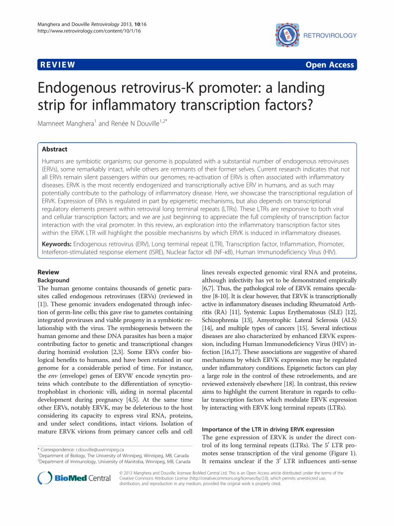
TATA box
MITF-M
LEF-1
NFAT-1
MITF-M
GC box1 GC box2* *
* *GC box4GC box3
Consensus
ERVK10ERVK109ERVK115ERVK108ERVK113
ARE
1
3
2
Figure 1 In silico examination of the conserved transcription factor binding sites and response elements within five endogenousretrovirus-K (ERVK) 50-LTRs using ALGGEN-PROMO software [25]. The ERVK LTR consensus sequence was constructed using individual ERVKLTRs in the following order (GenBank accession numbers in brackets): ERVK-10 (M12854.1), ERVK-9 (former HERV-K109) (AF164615.1), ERVK-8(former HERV-K115) (AY037929.1), ERVK-6 (former HERV-K108) (AF074086.2) and ERVK-113 (JF742069.1). GC boxes 1 to 4, indicated by asterisks,were adapted from [22]. Abbreviations used include: PRE = Progesterone Response Element, CRE = CREB Response Element, ERE = EstrogenResponse Element, ARE = Androgen Response Element, E-box = Enhancer Box, GRE = Glucocorticoid Response Element, ISRE = Interferon-Stimulated Response Element, GC box, GA box, ORE = Oct Response Element. Conventional (793bp) [22] and three alternative (460, 570, and 826 bp)[21,23] transcriptional start sites are depicted by black and grey arrows, respectively. Sequence alignment and annotation were performed usingGeneious software [26].
Manghera and Douville Retrovirology 2013, 10:16 Page 2 of 11http://www.retrovirology.com/content/10/1/16
transcription of the provirus, as seen with other retro-viruses such as HIV and HTLV-1 [19-21]. Each flankingviral LTR consists of a U3, R, and U5 regions in 50 to 30
direction. U3 region is the most important as it containsall the sequences – TATA independent promoter, enhan-cers, and transcription factor binding sites – required forinitiation of transcription of ERVK genes. Additionally,alternative transcriptional start sites have been proposed[21-23], perhaps allowing for differential transcripts undera variety of physiological conditions. ERVK LTR sub-groups exhibiting specific base insertions may also in-fluence the transcriptional regulation of these elements[24]. We are just beginning to appreciate the full complex-ity of ERVK transcriptional regulation, and despite a fullunderstanding, it is clear that both cellular transcription
factors and virally encoded proteins can be transcriptionalactivators of the ERVK LTR. A complete view of the pu-tative transcription factor binding sites and regulatoryelements within the ERVK LTR (Figure 1) allows us toconfirm and speculate upon the involvement of severaltranscription factors in the pathogenesis of inflammatorydiseases through induction of ERVK gene expression.
Overcoming epigenetic silencing of ERVKERVs have been transcriptionally silenced over evolution-ary time from accumulation of point mutations anddeletions. Additionally, epigenetic mechanisms, particu-larly CpG methylation and cytosine deamination of theLTR, control the basal expression of ERVK in various celltypes and tissues; these have been extensively reviewed

Manghera and Douville Retrovirology 2013, 10:16 Page 3 of 11http://www.retrovirology.com/content/10/1/16
elsewhere [18]. The methylation status of U3 region ofthe ERVK LTR has been shown to correlate with itstranscriptional activity; low levels of LTR methylationhave been shown to result in high levels of ERVK expres-sion [27,28]. Methylation of CpG dinucleotides in genomicDNA serves to repress transcription of genes by interfer-ing with the binding of sequence-specific transcription fac-tors [27]. Similarly, CpG methylation of the ERVK LTRprovides a natural defence against intragenomic parasitessuch as ERVK.The activity of ERVK LTRs has been further repressed
by deamination of methylated CpG dinucleotides. Spon-taneous deamination is a major source of abundant G toA and C to T mutations observed in many ERVK LTRs[29], which render them incapable of transcription. GCbox 1 and GC box 4 in the LTR (Figure 1) exhibit G toA and C to T conversions, respectively [29].APOBEC3G-mediated cytidine deamination is another
common source of G to A, and less frequently C to T,mutations in the ERVK LTR [29]. This process occursafter retroviral infection but prior to the integration ofproviral DNA within the human genome [29]. In manyERVK LTRs, APOBEC3G has been responsible for exces-sive G to A mutations [29-31]. APOBEC3G targets GGdinucleotides as well as GGG trinucleotides, efficiently mu-tating Tryptophan codons [29]. G to A mutation of Trypto-phan codons generates new stop codons, which is lethal toERVK when present within coding regions. However, whenthis mutation is present within non-coding regions, itcan prevent the binding of crucial transcription factorsrecognizing that region. In fact, GC box 2 (Figure 1) onthe ERVK LTR displays a plus-strand GG to AG change[22,29,31], which likely inhibits the binding of Sp1 andSp3 transcription factors to this GC box.Despite the layers of epigenetic control, basal and in-
ducible ERVK expression is evident in normal physi-ology and inflammatory disease [32,33], indicating thatadditional regulatory machinery is involved in ERVKtranscription. Together, cell type-dependent epigeneticprogramming and expression of transcriptional regula-tory factors are expected to guide the overall ERVK tran-scriptome, and is apt to become critical in disease stateswhere methylation-mediated silencing is compromised.
Influence of cellular transcription factors on ERVKexpressionOther than epigenetic factors, cellular transcription fac-tors are also crucial for regulating the activity of theERVK LTR. Different types, expression levels and activityof key transcription factors may be required to achievedistinct tissue-specific ERVK expression. However, rela-tively little is known about the transcriptional regulationof ERVK when pertaining to tissue specificity, conditionsof inflammation and disease states. Interestingly, many
ERVK LTRs have intact and conserved binding sites forhuman transcription factors (Table 1), yet few transcrip-tion factors have been experimentally shown to modu-late ERVK LTR activity (Table 2).
Influence of Sp1 and Sp3 on ERVK expressionSp1 and Sp3 are ubiquitously expressed transcriptionfactors, which have been pathologically implicated in can-cers, neuroinflammation, and rheumatic diseases [91-93].Induced by oxidative stress, the expression of Sp1 and Sp3is enhanced in inflammatory diseases such as ALS andSLE [69,70,94]. Over-expression of these transcription fac-tors may explain high levels of ERVK transcripts in thesedisease states, as Sp1 and Sp3 have been shown to driveERVK expression by binding to G-rich elements, such asGC boxes found in the proviral LTR [22] (Figure 1). It hasbeen shown that Sp1 and Sp3 mediate the transcriptionalactivity of the ERVK LTR as knockdown of these tran-scription factors using siRNA resulted in a significant lossof LTR activity [22]. Supershift assays have indicated thatSp1 and Sp3 probably bind as a heteromer to the GCboxes on the ERVK LTR, and mutation of these GC boxesresulted in downregulation of the LTR activity [22].Several mechanisms have been depicted by which Sp1
and Sp3 may promote transcription of ERVK. Sp1 inter-acts with TFIID, a complex consisting of TATA bindingprotein (TBP) and other associated general transcriptionfactors, and thus tethers the transcription pre-initiationcomplex to the TATA-independent promoter of the ERVKLTR [22]. Formation of the pre-initiation complex is acrucial step required for initiation of transcription at apromoter as it allows RNA polymerase to bind to thepromoter and begin transcription. Sp1 also protects CpGislands from methylation, aiding in chromatin remodelingand creating a nucleosome free region [22], to facilitatetranscription. Chromatin immunoprecipitation experimentshave demonstrated that when Sp1 and Sp3 bind to adjacentnucleosomes upstream the transcription start site (Figure 1),that is, to the GC boxes 1 and 3, these regions are accessibleto restriction enzymes, indicated by cleavage at these points[22]. This could only be possible if nucleosome free regionswere present at the sites where Sp1 and Sp3 were bound tothe LTR. Thus, expression of ERVK can be induced by thebinding of Sp1 and Sp3 to the GC boxes on the TATA-independent promoter region of the ERVK LTR.Interestingly, Sp3 may also repress ERVK expression.
Since Sp3 and Sp1 are closely related and have similar affin-ity for the GC boxes [95], Sp3 can prevent Sp1 binding andthus may repress Sp1 mediated activation of the ERVKLTR. Sp3 has a transferable repression domain with theamino acid triplet KEE required for its repressive activity[95]. However, Sp1 also has a similar repression domain[95], yet it does not repress the activity of the LTR [22].This implies that other characteristics of the LTR determine

Table 1 Cellular transcription factors predicted to bind to their respective putative consensus sequences on the ERVK LTR
Transcription Factor Consensus sequence References
Activating Protein 1 (AP-1); c-Jun/c-Fos TGA(G/C)TCA [34]
Activating Protein 2 (AP-2α) GCCNNNGGC [35]
Androgen receptor (AR) GG(A/T)ACANNNTGTTCT (ARE) [36]
cAMP Response Element Binding (CREB) protein T(G/T)ACGTCA (CRE) [37]
cAMP Response Element Modulator (CREM-α) T(G/T)ACGTCA (CRE) [37]
CCAAT-Enhancer Binding Protein (C/EBP) N(A/G)CCAAT [38,39]
Cellular Myeloblastosis virus protein (c-Myb) (T/C)AAC(G/T)G [40]
Cellular Myelocytomatosis virus protein (c-Myc) NNNCACGTGNN (E-box) [41,42]
Early B-cell Factor (EBF) CCCNNGGG [43]
Estrogen Receptor (ER-α; ER-β) GGTCANNNTGACC (ERE) [44]
E-twenty six (ETS-1; ETS-2) GGA(A/T) [45]
ETS-like Transcription Factor 1 (Elk-1) GGA(A/T) [45]
GATA binding protein (GATA) (A/T)GATA(A/G) [46]
Glucocorticoid Receptor (GR-α; GR-β) GGTACANNNTGTTC (GRE) [47]
Ikarose-1 (Ik-1) TGGGA(A/T) [48]
Interferon Regulatory Factor (IRF-1; IRF-3; IRF-7) GAAANN repeats (ISRE) [49,50]
Lymphoid Enhancer-binding Factor 1 (LEF-1) CTTTGAA [51]
Microphthalmia-associated Transcription Factor-M (MITF-M) CA(C/T)GTG (E-box) [23]
Monocyte Enhancer Factor-2 (MEF-2A) CT(A/T)(A/T)AAATAG [52]
Myc Associated Zinc finger protein (MAZ) GGGAGGG [53]
Nuclear Factor of Activated T cells (NFAT-1) GGAGAA [54]
Nuclear Factor I (NF-I) TTGGCNNNNNGCCAA [55]
Nuclear Factor Kappa B (NF-kB) GG(G/A)(G/A)NN(C/T)(C/T)CC [56]
Octamer-1 (OCT-1) ATGCAAAT (ORE) [57]
Polyomavirus Enhancer Activator 3 (PEA-3) GGA(A/T) [45]
Progesterone Receptor (PR-A; PR-B) GNACANNNTGTNC (PRE) [58]
Protein 53 (p53) CATTAG [59]
Recombination signal Binding Protein-Jk (RBP-Jk) (C/T)GTGGGAA [60]
Sex-determining Region Y (SRY) (A/T)(A/T)CAA(A/T) [61]
Signal Transducers and Activators of Transcription (STAT) TTCNNNNGAA [62]
Specificity Protein (Sp-1; Sp-3) GGGCGG (GC-box) [22]
TATA Binding Protein (TBP) TATAAA (TATA box) [63]
T cell Factor 1 (TCF-1) (G/C)ATCAAAGG [64]
Transcription Factor II D (TFII-D) TATAAA (TATA box) [63]
Transcription Factor II I (TFII-I) CANNTG [65]
Upstream Transcription Factor 1 (USF-1) CACGTG (E-box) [41]
Vitamin D Receptor (VDR) G(G/T)TCA [66]
X-box binding protein (XBP-1) CCACG [67]
Yin Yang 1 (YY1) GCCATNTT [68]
Manghera and Douville Retrovirology 2013, 10:16 Page 4 of 11http://www.retrovirology.com/content/10/1/16
whether repressive action of Sp3 will occur or not. Thestructure and the arrangement of GC boxes on theLTR may determine whether Sp3 will repress the ERVKLTR or not. For instance, promoters with multiplebinding sites often do not or weakly respond to Sp3
[95]. Since the ERVK promoter region has four GCboxes, the deactivating effect of Sp3 may be minimized.Nonetheless, the features that determine whether Sp3acts as a repressor or activator of transcription are notwell understood.

Table 2 Transcription factors which have been experimentally shown to influence ERVK LTR activity
Transcription factor Cellular function Implicated diseases Effect on ERVK LTR References
Sp-1, Sp-3 Implicated in the regulation of genes that control multiplecellular processes, including cell cycle, apoptosis, and DNAdamage.
ALS, SLE, RAAlzheimer’s Disease,Huntington’s Disease
Stimulate [22,69-72]
YY1 Positive and negative regulator of genes involved inbiological processes such as differentiation, replication, andcellular proliferation.
Cancers, SLE,neurodegeneration
Stimulate [73-76]
NF-kB Involved in cytoplasmic/nuclear signalling in response tostimuli such as stress, cytokines, free radicals, ultravioletirradiation, oxidized LDL, and bacterial or viral antigens;activates transcription of a variety of genes encodingimmunologically relevant proteins.
HIV infection, ALS, SLE,MS, Rheumatic disease,Cancers
Stimulate [17,69,77-80]
NFAT-1 Plays a key role in the regulation of cytokine genetranscription during the immune response.
HIV infection,Alzheimer’s Disease,Autoimmune diseases
Stimulate [17,81,82]
MITF-M Induces genes essential for melanin synthesis, melanosomeformation, cell cycle progression, and cell survival; essentialfor development of retinal pigmented epithelium andneural crest derived melanocytes.
Melanoma Stimulate [23]
PR Mediates the effects of progesterone on mammary glanddevelopment.
Breast cancer Stimulate [83,84]
ER Mediates the effects of estrogen on reproductive organs,bone, and brain.
SLE, Breast cancer Stimulate [83,85]
AR Mediates embryonic sexual differentiation and required formaintenance of spermatogenesis
Prostate cancer, breastcancer, Kennedy’sdisease
Stimulate [86-90]
Manghera and Douville Retrovirology 2013, 10:16 Page 5 of 11http://www.retrovirology.com/content/10/1/16
Furthermore, other members of the Sp family are alsoclosely related to Sp3 and Sp1. These include Sp4, BTEB1,TIEG1, and TIEG2 [95]. The critical amino acids withinthe three zinc fingers of these members are conserved;they include KHA, RER, and RHK within the first, second,and third zinc fingers, respectively [95]. As a result, thesefour Sp members also recognize classical GC boxes andbind to them with a relatively similar affinity as that ofSp1 and Sp3 [95]. Thus, it can be speculated that variousmembers of the Sp family other than Sp1 and Sp3 mayalso be able to induce ERVK expression by binding to theGC boxes on the LTR.
Influence of YY1 on ERVK expressionYY1 is a ubiquitous transcription factor, which is fre-quently overexpressed in cancers, degenerating neurons,and rheumatic diseases [69,73]; hence, it may be involvedin causing ERVK expression documented in many inflam-matory diseases. In fact, the 5′ terminus of the U3 regionof the ERVK LTR binds to the YY1 enhancer complex(Figure 1). The binding of YY1 to this region, betweennucleotides 62 and 83, has been shown to activate theERVK expression in many cell lines including GH, Tera2,HepG2, and HeLa [74]. This implies that activation ofERVK LTR by YY1 may not be cell-type dependent.
Mutation of this YY1 binding site has been shown tocause a 50% reduction in the activity of the ERVK LTR[74]. Moreover, addition of a functional YY1 bindingsite to an engineered active ERVK LTR sequence contain-ing functional GC boxes has been observed to increasethe ERVK promoter activity to 80% [22]. This indicatesthat in addition to essential GC boxes, transcription fac-tor binding sites, such as those for YY1, are also crucialfor activity of the ERVK LTR.
Influence of MITF-M on ERVK expressionMelanoma, a type of skin cancer, frequently exhibitsenhanced expression of ERVK env and rec proteins [96].Recent studies support the notion that increased ERVKexpression and massive production of ERVK viral-likeparticles contribute to melanocyte malignancy [97]. Mel-anoma specific microphthalmia-associated transcriptionfactor (MITF-M) is an oncogene of melanoma [23], andhas been implicated in activating the ERVK LTR. Re-cently, it was shown that the ERVK-6 LTR has threeMITF-M responsive sequences (E boxes) in the U3 region[23] (Figure 1), which are arranged along with TATA boxand Initiator (Inr) sites in such a way that they togetherconstitute a typical enhancer/promoter structure for RNApolymerase II found in all retroviral LTRs [23]. As a result,

Manghera and Douville Retrovirology 2013, 10:16 Page 6 of 11http://www.retrovirology.com/content/10/1/16
MITF-M is able to induce the ERVK LTR by binding toand inducing the core enhancer/promoter region.
Alternative transcription factors that may modulate ERVKexpressionThe influence of only Sp1, Sp3, YY1 and MITF-M onERVK LTR activation has been documented. Unfortu-nately, the inductive or repressive ability of many othertranscription factors – Oct-1, AP-1, CREB, NF-κB, IRFs,etc. (Table 1) – all of which have potentially intact bind-ing sites on the consensus ERVK LTR (Figure 1), haveyet to be studied. For example, Oct-1 and the membersof the bZip family of transcription factors, AP-1 andCREB, have been shown to induce ERVK indirectly as aconsequence of exogenous viral infections, which will bediscussed in the following sections. Moreover, AP-1 andCREB are often over-expressed in inflammatory diseases,suggesting their potential role in disease pathogenesis byinduction of ERVK. Further research is warranted inorder to precisely determine the influence of these vari-ous transcription factors on the activity of ERVK LTRs.
Putative role of interferon and inflammatory transcriptionfactors in ERVK inductionTranscription factors associated with the innate immuneresponse, especially NF-κB, IRF-1, IRF-3 and IRF-7, mayalso be able to influence the activity of the ERVK LTR.During anti-viral responses and inflammation, many ofthese transcription factors become up-regulated and post-translationally activated. NF-κB is known to be a key regu-lator of exogenous retrovirus transcription [98]. Often,oxidative stress is also implicated in the up-regulation ofNF-κB in neurodegenerative and rheumatic diseases as aresult of the protective cellular response [69,77,78]. Sincethe ERVK LTR has several NF-κB binding sites (Figure 1),this transcription factor is likely to directly influence ERVKexpression.Although inflammatory transcription factors have yet to
be shown to influence ERVK LTR, they are known to con-trol other retroviral LTRs. NF-κB binding to the HIV-1LTR has been shown to stimulate HIV-1 production about50-fold [99]. The members of the interferon regulatoryfactor (IRF) family have been shown to interact with themembers of the NF-κB family; indeed, IRF-1 is requiredfor full NF-κB transcriptional activity at the HIV-1 LTRenhancer [79]. Accordingly, overlapping binding sites forIRFs and NF-kB have been identified in HIV-1 LTR [79].The ERVK LTR also contains overlapping binding sites forthese transcription factors (Figure 1), suggesting functionalcommonality among LTR responsive elements in humanretroviruses.As shown in Figure 1, a conserved feature of ERVK LTRs
is the presence of two ISRE-like motifs (50-GAAANNGAAANN-30), located at nt379 and nt563. These conserved
motifs may accommodate IRF binding, in conjunctionwith NF-κB, fulfilling the transcriptional priming of trad-itional interferon-stimulated genes (reviewed in [101,102]).Of note, at both ISRE sites, the proximal GAAA half-sitemotifs are mutated, likely favouring IRF-7 over IRF-3binding [103]. IFNα signalling can directly lead to IRF-7activation [104], and several reports indicate that in-duction of ERVK-18 superantigen by herpesviruses inPBMC may be mediated through IFNα [105-107]. Thisnot only establishes a link between exogenous virus in-fection and the induction of the ERVs, but may suggestthe involvement of IRF activity in mediating ERVK tran-scription. To date, there is a lack of empirical evidence tosupport that other ERVK members are induced by anti-viral signalling pathways or activation of select IRFs.In contrast, pro-inflammatory cytokines, such as TNFα
and IL-6, have been shown to modulate ERVK transcrip-tional activity [11]. TNFα can engage an autocrine signal-ling loop that culminates in IRF-1 activation, a sustainedlow-level IFNβ response and IRF-7 expression in macro-phages [108]. In addition, TNFα is a strong activator ofNF-κB and AP-1. Together, these factors may contributeto the enhancement of ERVK expression. Similarly, IL-6enhances IRF-1 transcription, and can affect IRF-1 DNAbinding in select cell types [109]. IL-6 signalling alsodrives the activation of STAT3, which can bind ISREcis-elements, another potential mechanism to activatethe ERVK LTR.
An extra layer of control: modulation of ERVK expressionby hormonal regulationThe effect of estrogen and progesterone on ERVK expres-sion has been exclusively studied in breast cancer tissues.Most breast cancer cell lines and many breast tumortissues exhibit significantly higher levels of ERVK envexpression as compared to normal breast tissues [110,111].The expression of env transcripts has been shown to beup-regulated 5 to 10-fold in breast cancer cell linesupon estradiol treatment followed by progesterone[83,112], suggesting the presence of functional hormoneresponse elements in the ERVK LTR. In fact, several es-trogen, androgen and progesterone response elements(ERE, ARE and PRE, respectively) are predicted in theU3 region of the LTR (Figure 1) [83,86,112]. BesidesERVK env expression, enzymatically active ERVK reversetranscriptase protein has also been detected in breasttumor biopsies and the breast cancer cell line T47D [112].Again, estradiol/progesterone treatment of T47D cells leadto an increased level of ERVK reverse transcriptase proteinexpression, as well as its enzymatic activity [112]. Interest-ingly, stimulation of ERVK expression has not beendemonstrated by treatment with estradiol or progesteronealone, but specifically with estradiol followed by progester-one [83]. This suggests that estradiol has a priming effect

Manghera and Douville Retrovirology 2013, 10:16 Page 7 of 11http://www.retrovirology.com/content/10/1/16
on the ERVK LTR, whereby it may alter basal transcrip-tion factor affinity for the LTR, making it more easilyaccessible to progesterone-receptor complexes. Thus,these studies strongly point to the notion that steroidhormones contribute to the regulation of ERVK LTRs.The gene expression of ERVK may not only be influ-
enced directly by cellular and viral transcription factors, butalso indirectly by various pharmaceutical agents [71,100]which act on (or counteract) the transcription factors thatcan bind to the LTR of ERVK. In particular, the extent ofhormone-responsive elements (Figure 1; ERE, PRE, GRE)in the ERVK promoter suggests a susceptibility to theaction of endocrine disruptors [113]. For example, theendocrine disrupting compound bisphenol-A (BPA) isknown to mimic estrogen signalling pathway [114], andmay modulate estrogen receptor targets such as ERVK.Endocrine disruption leading to ERVK re-activation maybridge the often speculative association between envir-onmental exposures and the establishment of chronicinflammatory disease.
Influence of viral proteins on expression of ERVKBesides exploiting cellular transcription factors, ERVKcan also utilize virally-encoded proteins for its induction.This versatility in part explains the up-regulation of ERVKby exogenous viruses such as Human ImmunodeficiencyVirus-1 (HIV-1), Human T-Lymphotrophic Virus-1 (HTLV-1), Herpes Simplex Virus-1 (HSV-1) and Epstein BarrVirus (EBV) [115-117]. These viruses provide viral pro-teins that increase the affinity of transcription factors fortheir binding sites on the ERVK LTR, thereby trans-activating ERVK.
Induction of ERVK by exogenous retrovirusesThe expression of ERVK is often abnormally elevated inHIV-1 infected individuals, reflecting vastly increasedviral RNA titres in their plasma [17,118,119]. However,the mechanism underlying this phenomenon had remainedunknown until recently. It had long been proposed thatHIV-1 proteins Vif and Tat may induce the ERVK LTRdirectly or indirectly [117,118,120]. HIV-1 accessory pro-tein Vif has been shown to impair the translation ofAPOBEC3G mRNA and accelerate its post-translationaldegradation [121]. In the absence of APOBEC3G activity,there is an enhancement of de novo ERVK infectivity, asdemonstrated experimentally in vitro using virions derivedfrom reconstituted elements [29,31]. However, it remainsunclear whether HIV Vif interaction with APOBEC3Gexerts a regulatory effect on ERVK expression [119,122].Moreover, the direct interaction of Tat with nascent
ERVK RNA, and thus the induction of viral transcript ex-pression, had always been suspected. It has only recentlybeen demonstrated that HIV-1 proteins Vif and Tat inde-pendently activate ERVK expression [17,122]. Transfection
of Jurkat T cells and 293FT cell lines with plasmids encod-ing functional Tat and Vif proteins significantly up-regulated ERVK gag RNA by 21- and 15-fold, respectively[17]. The levels of rec and np-9 transcripts and the expres-sion of the ERVK capsid protein also increased in the pres-ence of Tat in several cell lines, as well as in primarylymphocytes that are major targets of HIV-1 infection [17].Similarly, HTLV-1 Tax protein also promotes ERVK tran-scription in Jurkat T cells [123].Several mechanisms have been proposed by which HIV-1
Tat may trans-activate ERVK. Tat activates transcriptionfrom the HIV-1 promoter by interacting with Cyclin T1and recruiting the host positive transcription elongationfactor b (P-TEFb) to its LTR [124]. The Tat induced activa-tion of ERVK expression also occurs at the level of theERVK LTR, but does not involve its interaction with CyclinT1 or P-TEFb [17]. Mutations in the transactivationdomain of Tat, which rendered it either unable to bind toCyclin T1 or increased its binding to P-TEFb, had no meas-urable effect on Tat’s capacity to drive ERVK LTR activity.HIV-1 LTR can be activated in a TAR-independent man-
ner; this effect occurs through the interaction of Tat withSp1 sequences in the U3 region of HIV-1 LTR as well aswith NF-κB [17,98]. Thus, Tat may activate ERVK pro-moter by interacting with GC boxes and NF-κB. In fact,activation of the ERVK LTR by Tat has been demonstratedto be mediated by its interaction with NF-κB and NFAT-1cellular transcription factors [17]. Chemical inhibition ofNF-κB and NFAT-1 repressed Tat mediated activation ofERVK promoter, significantly diminishing Tat-mediatedERVK gag RNA levels. ChIP assays further confirmedthat NF-κB and NFAT-1 were activated and interactedwith multiple binding sites on ERVK LTR (Figure 1) inresponse to Tat. Interestingly, these intact and activeNF-κB and NFAT-1 binding sites overlap with interferon-stimulated response elements (ISREs) in the ERVK LTR(Figure 1), suggesting the potential role of the innate im-mune system and associated inflammatory transcriptionfactors in regulating the ERVK expression.Furthermore, HIV-1 infection may also contribute to
ERVK up-regulation indirectly by promoting opportunisticinfections. Destruction of the immune system by HIV-1can facilitate the replication of other viruses such as HSV-1and HTLV-1, as well as the protozoan Toxoplasma gondii[125], all of which have been shown to trans-activate theERVK LTR.The Tax protein produced by HTLV-1 may modestly
trans-activate ERVK, as demonstrated by Tax-driven activa-tion of a luciferase reporter under the control of a TD47ERVK LTR in Jurkat T cells [123]. The Tax protein likelyincreases the affinity of several transcription factors, speci-fically Sp1, NF-κB, c-Fos/c-Jun heterodimers (AP-1), andCREB, for their DNA binding sites on the ERVK LTR[20,98]. A caveat of these experiments is that Tax-mediated

Manghera and Douville Retrovirology 2013, 10:16 Page 8 of 11http://www.retrovirology.com/content/10/1/16
induction of ERVK has not yet been demonstrated in vivo,in either HTLV-1 carriers, patients with Adult T-cellLeukemia or individuals with HTLV-1 associated myelop-athy (HAM/TSP).
Induction of ERVK by HerpesvirusesThe immediate early protein, ICP0, produced by HSV-1induces the LTR directed transcription of ERVK in vitro[116]. This effect is mediated through the up-regulationof AP-1 activity. Deletion analysis of various nucleotidesequences in the ERVK LTR has shown that the AP-1binding site between the nucleotides 243 and 253 isrequired for ICP0 to trans-activate ERVK [116]. Destruc-tion of this site completely abolished ICP0 responsive-ness, whereas the activation of ERVK by ICP0 was notaffected by deletion of any other nucleotides, such as828–968 and the YY1 binding site [116]. HSV-1 mayalso induce ERVK through up-regulation of Oct-1 activitymediated by the immediate early protein 1 (IEP1) [126].IEP1 has been shown to increase the expression of ERVWin vitro by facilitating the binding of Oct-1 to its bindingsite on the LTR [126]. In addition, IEP1 has been shown toincrease the expression of ERVK [116]. Since ERVK alsohas Oct-1 binding sites, IEP1 of HSV-1 may induce ERVKby increasing the affinity of Oct-1 for its binding site onthe ERVK LTR. However, the in vivo induction of ERVKby HSV-1 immediate early proteins is yet to be confirmed.Similarly, EBV infection has been reported to stimulate
the production of an ERVK-18 env-derived superantigen(SAg) specific to T cells that express T cell receptor βchain variable-13 (TcRβCV-13) [127], a mechanism whichcan enhance the pathogenicity of EBV. ERVK-18 is a clas-sic example of an intragenic ERV, located on the anti-sense DNA strand within first intron of the cellular cd48gene. Trans-activation of ERVK-18 can be driven by EBV-encoded latent membrane protein LMP-2A [128,129]. Aseries of tyrosine mutants of LMP-2A revealed that theimmunoreceptor tyrosine-based activation motif (ITAM)of LMP-2A is responsible for inducing the ERVK-18 envexpression [129]. Furthermore, deletion of an enhancer ina 25 kb region downstream the ERVK-18 env gene abo-lished production of the SAg [129], indicating that ITAMinteracts with this enhancer to induce ERVK-18 expres-sion. A putative genomic NF-κB binding site adjacent tothe provirus was identified as a potential candidate forinteraction with the ITAM of LMP-2A [129]. This demon-strates not only the importance of the ERVK LTR, but alsothe genomic context of the ERV, in enhancing its tran-scription factor-mediated expression.
ConclusionAlthough several studies indicate the effects of epigen-etic mechanisms in controlling ERVK expression, theliterature clearly awaits more studies on tissue-specific,
pro-inflammatory and hormone-regulated transcriptionfactors that promote or repress ERVK transcription inhealth and inflammatory diseases. Surprisingly, veryfew transcription factors – Sp1, Sp3, YY1, MITF-M,and estrogen/progesterone – have been experimentallyshown to induce ERVK; however, these transcriptionfactors are not necessarily tissue-specific as they areubiquitously expressed. Bioinformatic examination ofseveral ERVK LTRs clearly unveils the multitude of pos-sible binding sites for unique and ubiquitous transcriptionfactors, but it is yet to be determined whether these sitesare at all functional [24]. One clear feature of the ERVKLTRs is the two ISRE sequences, which is highly suggestiveof ERVK regulation in the context of innate immuneresponse and inflammation.A large part of the debate surrounding a causal relation-
ship of ERVK in disease pathology is the issue of bystanderactivation – advances in our understanding of the tran-scriptional regulation of the ERVK LTR will clarifywhether ERVK expression is a consequence, cause or con-joined mechanism of inflammatory disease. Thus, we areindeed strongly in need of an ERVK Transcriptome Pro-ject [33,130,131], whereby determining the transcriptionalregulation of ERVK expression and its association withinflammatory diseases will allow us to point to tran-scription factors as primary cellular targets for thera-peutic intervention.
AbbreviationsALS: Amyotrophic Lateral Sclerosis; EBV: Epstein Barr Virus; ERV: Endogenousretrovirus; ERVK: Endogenous retrovirus-K; ERVW: Endogenous retrovirus-W;HIV: Human Immunodeficiency Virus; HSV-1: Herpes Simplex Virus-1; HTLV-1: Human T-Lymphotrophic Virus-1; ISRE: Interferon-stimulated responseelement; LTR: Long terminal repeat; NF-κB: Nuclear factor-κB; RA: RheumatoidArthritis; SLE: Systemic Lupus Erythematosus..
Competing interestsThe authors declare that they have no competing interests.
Authors’ contributionsMM performed the sequence alignments and bioinformatic annotations.Both MM and RND drafted the manuscript. Both authors read and approvedthe final manuscript.
Received: 8 August 2012 Accepted: 1 February 2013Published: 9 February 2013
References1. Ryan F: Virolution: the most important evolutionary book since Dawkins’ Selfish
gene. London: Collins; 2009.2. Blikstad V, Benachenhou F, Sperber GO, Blomberg J: Evolution of human
endogenous retroviral sequences: a conceptual account. Cell Mol Life Sci2008, 65:3348–3365.
3. Wang T, Zeng J, Lowe CB, Sellers RG, Salama SR, Yang M, Burgess SM,Brachmann RK, Haussler D: Species-specific endogenous retrovirusesshape the transcriptional network of the human tumor suppressorprotein p53. Proc Natl Acad Sci U S A 2007, 104:18613–18618.
4. Dupressoir A, Lavialle C, Heidmann T: From ancestral infectiousretroviruses to bona fide cellular genes: Role of the captured syncytinsin placentation. Placenta 2012, 33:663–671.
5. Sugimoto J, Schust DJ: Review: human endogenous retroviruses and theplacenta. Reprod Sci 2009, 16:1023–1033.

Manghera and Douville Retrovirology 2013, 10:16 Page 9 of 11http://www.retrovirology.com/content/10/1/16
6. Morgan D, Brodsky I: Human endogenous retrovirus (HERV-K) particles inmegakaryocytes cultured from essential thrombocythemia peripheralblood stem cells. Exp Hematol 2004, 32:520–525.
7. Contreras-Galindo R, Kaplan MH, Leissner P, Verjat T, Ferlenghi I, Bagnoli F,Giusti F, Dosik MH, Hayes DF, Gitlin SD, Markovitz DM: Human endogenousretrovirus K (HML-2) elements in the plasma of people with lymphomaand breast cancer. J Virol 2008, 82:9329–9336.
8. Solyom S, Kazazian HH Jr: Mobile elements in the human genome:implications for disease. Genome Med 2012, 4:12.
9. Christensen T: HERVs in neuropathogenesis. J Neuroimmune Pharmacol2010, 5:326–335.
10. Krone B, Grange JM: Melanoma, Darwinian medicine and the inner world.J Cancer Res Clin Oncol 2010, 136:1787–1794.
11. Freimanis G, Hooley P, Ejtehadi HD, Ali HA, Veitch A, Rylance PB, Alawi A,Axford J, Nevill A, Murray PG, Nelson PN: A role for human endogenousretrovirus-K (HML-2) in rheumatoid arthritis: investigating mechanisms ofpathogenesis. Clin Exp Immunol 2010, 160:340–347.
12. Krzysztalowska-Wawrzyniak M, Ostanek M, Clark J, Binczak-Kuleta A, Ostanek L,Kaczmarczyk M, Loniewska B, Wyrwicz LS, Brzosko M, Ciechanowicz A: Thedistribution of human endogenous retrovirus K-113 in health andautoimmune diseases in Poland. Rheumatology (Oxford) 2011, 50:1310–1314.
13. Frank O, Giehl M, Zheng C, Hehlmann R, Leib-Mosch C, Seifarth W: Humanendogenous retrovirus expression profiles in samples from brains of patientswith schizophrenia and bipolar disorders. J Virol 2005, 79:10890–10901.
14. Douville R, Liu J, Rothstein J, Nath A: Identification of active loci of ahuman endogenous retrovirus in neurons of patients with amyotrophiclateral sclerosis. Ann Neurol 2011, 69:141–151.
15. Ruprecht K, Mayer J, Sauter M, Roemer K, Mueller-Lantzsch N: Endogenousretroviruses and cancer. Cell Mol Life Sci 2008, 65:3366–3382.
16. Contreras-Galindo R, Gonzalez M, Almodovar-Camacho S, Gonzalez-RamirezS, Lorenzo E, Yamamura Y: A new Real-Time-RT-PCR for quantitation ofhuman endogenous retroviruses type K (HERV-K) RNA load in plasmasamples: increased HERV-K RNA titers in HIV-1 patients with HAART non-suppressive regimens. J Virol Methods 2006, 136:51–57.
17. Gonzalez-Hernandez MJ, Swanson MD, Contreras-Galindo R, Cookinham S,King SR, Noel RJ Jr, Kaplan MH, Markovitz DM: Expression of HumanEndogenous Retrovirus Type K (HML-2) Is Activated by the Tat Protein ofHIV-1. J Virol 2012, 86:7790–7805.
18. Maksakova IA, Mager DL, Reiss D: Keeping active endogenous retroviral-like elements in check: the epigenetic perspective. Cell Mol Life Sci 2008,65:3329–3347.
19. Landry S, Halin M, Lefort S, Audet B, Vaquero C, Mesnard JM, Barbeau B:Detection, characterization and regulation of antisense transcripts inHIV-1. Retrovirology 2007, 4:71.
20. Barbeau B, Mesnard JM: Making sense out of antisense transcription inhuman T-cell lymphotropic viruses (HTLVs). Viruses 2011, 3:456–468.
21. Kovalskaya E, Buzdin A, Gogvadze E, Vinogradova T, Sverdlov E: Functionalhuman endogenous retroviral LTR transcription start sites are locatedbetween the R and U5 regions. Virology 2006, 346:373–378.
22. Fuchs NV, Kraft M, Tondera C, Hanschmann KM, Lower J, Lower R: Expressionof the human endogenous retrovirus (HERV) group HML-2/HERV-K doesnot depend on canonical promoter elements but is regulated bytranscription factors Sp1 and Sp3. J Virol 2011, 85:3436–3448.
23. Katoh I, Mirova A, Kurata S, Murakami Y, Horikawa K, Nakakuki N, Sakai T,Hashimoto K, Maruyama A, Yonaga T, et al: Activation of the long terminalrepeat of human endogenous retrovirus K by melanoma-specifictranscription factor MITF-M. Neoplasia 2011, 13:1081–1092.
24. Subramanian RP, Wildschutte JH, Russo C, Coffin JM: Identification,characterization, and comparative genomic distribution of the HERV-K(HML-2) group of human endogenous retroviruses. Retrovirology 2011,8:90.
25. Messeguer X, Escudero R, Farre D, Nunez O, Martinez J, Alba MM: PROMO:detection of known transcription regulatory elements using species-tailored searches. Bioinformatics 2002, 18:333–334.
26. Kearse M, Moir R, Wilson A, Stones-Havas S, Cheung M, Sturrock S, Buxton S,Cooper A, Markowitz S, Duran C, et al: Geneious Basic: an integrated andextendable desktop software platform for the organization and analysisof sequence data. Bioinformatics 2012, 28:1647–1649.
27. Lavie L, Kitova M, Maldener E, Meese E, Mayer J: CpG methylation directlyregulates transcriptional activity of the human endogenous retrovirusfamily HERV-K(HML-2). J Virol 2005, 79:876.
28. Khodosevich K, Lebedev Y, Sverdlov ED: Large-scale determination of themethylation status of retrotransposons in different tissues using amethylation tags approach. Nucleic Acids Res 2004, 32:e31.
29. Lee YN, Malim MH, Bieniasz PD: Hypermutation of an ancient humanretrovirus by APOBEC3G. J Virol 2008, 82:8762–8770.
30. Koito A, Ikeda T: Intrinsic restriction activity by AID/APOBEC family ofenzymes against the mobility of retroelements. Mob Genet Elements 2011,1:197–202.
31. Esnault C, Priet S, Ribet D, Heidmann O, Heidmann T: Restriction byAPOBEC3 proteins of endogenous retroviruses with an extracellular lifecycle: ex vivo effects and in vivo “traces” on the murine IAPE andhuman HERV-K elements. Retrovirology 2008, 5:75.
32. Seifarth W, Frank O, Zeilfelder U, Spiess B, Greenwood AD, Hehlmann R,Leib-Mosch C: Comprehensive analysis of human endogenous retrovirustranscriptional activity in human tissues with a retrovirus-specificmicroarray. J Virol 2005, 79:341–352.
33. Flockerzi A, Ruggieri A, Frank O, Sauter M, Maldener E, Kopper B, Wullich B,Seifarth W, Muller-Lantzsch N, Leib-Mosch C, et al: Expression patterns oftrancrsibed human endogenous retrovirus HERV-K(HML-2) loci in humantissues and the need for a HERV Transcriptome Project. BMC Genomics2008, 9:354.
34. Nead MA, Baglia LA, Antinore MJ, Ludlow JW, McCance DJ: Rb binds c-Junand activates transcription. EMBO J 1998, 17:2342–2352.
35. Imhof A, Schuierer M, Werner O, Moser M, Roth C, Bauer R, Buettner R:Transcriptional regulation of the AP-2alpha promoter by BTEB-1 and AP-2rep, a novel wt-1/egr-related zinc finger repressor. Mol Cell Biol 1999,19:194–204.
36. Roche PJ, Hoare SA, Parker MG: A consensus DNA-binding site for theandrogen receptor. Mol Endoinol 1992, 6:2229–2235.
37. Laoide BM, Foulkes NS, Schlotter F, Sassone-Corsi P: The functionalversatility of CREM is determined by its modular structure. EMBO J 1993,12:1179–1191.
38. Vales LD, Friedl EM: Binding of C/EBP and RBP (CBF1) to overlapping sitesregulates interleukin-6 gene expression. J Biol Chem 2002, 277:42438–42446.
39. Wang SE, Wu FY, Yu Y, Hayward GS: CCAAT/enhancer-binding protein-alpha is induced during the early stages of Kaposi’s sarcoma-associatedherpesvirus (KSHV) lytic cycle reactivation and together with the KSHVreplication and transcription activator (RTA) cooperatively stimulates theviral RTA, MTA, and PAN promoters. J Virol 2003, 77:9590–9612.
40. Deng QL, Ishii S, Sarai A: Binding site analysis of c-Myb: screening ofpotential binding sites by using the mutation matrix derived fromsystematic binding affinity measurements. Nucleic Acids Res 1996, 24:766–774.
41. Providence KM, White LA, Tang J, Gonclaves J, Staiano-Coico L, Higgins PJ:Epithelial monolayer wounding stimulates binding of USF-1 to an E-boxmotif in the plasminogen activator inhibitor type 1 gene. J Cell Sci 2002,115:3767–3777.
42. Halazonetis TD, Kandil AN: Determination of the c-MYC DNA-binding site.Proc Natl Acad Sci U S A 1991, 88:6162–6166.
43. Maier H, Hagman J: Roles of EBF and Pax-5 in B lineage commitment anddevelopment. Semin Immunol 2002, 14:415–422.
44. Lin CY, Vega VB, Thomsen JS, Zhang T, Kong SL, Xie M, Chiu KP, Lipovich L,Barnett DH, Stossi F, et al: Whole-genome cartography of estrogenreceptor alpha binding sites. PLoS Genet 2007, 3:e87.
45. de Nigris F, Mega T, Berger N, Barone MV, Santoro M, Viglietto G, Verde P,Fusco A: Induction of ETS-1 and ETS-2 transcription factors is requiredfor thyroid cell transformation. Cancer Res 2001, 61:2267–2275.
46. Wang L, Wang X, Adamo ML: Two putative GATA motifs in the proximalexon 1 promoter of the rat insulin-like growth factor I gene regulatebasal promoter activity. Endoinology 2000, 141:1118–1126.
47. Adler AJ, Danielsen M, Robins DM: Androgen-specific gene activation viaa consensus glucocorticoid response element is determined byinteraction with nonreceptor factors. Proc Natl Acad Sci U S A 1992,89:11660–11663.
48. Li Z, Perez-Casellas LA, Savic A, Song C, Dovat S: Ikaros isoforms: The sagacontinues. World J Biol Chem 2011, 2:140–145.
49. Rosenbauer F, Waring JF, Foerster J, Wietstruk M, Philipp D, Horak I: Interferonconsensus sequence binding protein and interferon regulatory factor-4/Pipform a complex that represses the expression of the interferon-stimulatedgene-15 in macrophages. Blood 1999, 94:4274–4281.
50. Arany I, Grattendick KJ, Whitehead WE, Ember IA, Tyring SK: A functionalinterferon regulatory factor-1 (IRF-1)-binding site in the upstream

Manghera and Douville Retrovirology 2013, 10:16 Page 10 of 11http://www.retrovirology.com/content/10/1/16
regulatory region (URR) of human papillomavirus type 16. Virology 2003,310:280–286.
51. Jin ZX, Kishi H, Wei XC, Matsuda T, Saito S, Muraguchi A: Lymphoidenhancer-binding factor-1 binds and activates the recombination-activating gene-2 promoter together with c-Myb and Pax-5 in immature Bcells. J Immunol 2002, 169:3783–3792.
52. Andres V, Cervera M, Mahdavi V: Determination of the consensus bindingsite for MEF2 expressed in muscle and brain reveals tissue-specificsequence constraints. J Biol Chem 1995, 270:23246–23249.
53. Song J, Ugai H, Ogawa K, Wang Y, Sarai A, Obata Y, Kanazawa I, Sun K,Itakura K, Yokoyama KK: Two consecutive zinc fingers in Sp1 and in MAZare essential for interactions with cis-elements. J Biol Chem 2001,276:30429–30434.
54. Handen JS, Rosenberg HF: Intronic enhancer activity of the eosinophil-derived neurotoxin (RNS2) and eosinophil cationic protein (RNS3) genes ismediated by an NFAT-1 consensus binding sequence. J Biol Chem 1997,272:1665–1669.
55. Mukhopadhyay SS, Wyszomierski SL, Gronostajski RM, Rosen JM: Differentialinteractions of specific nuclear factor I isoforms with the glucocorticoidreceptor and STAT5 in the cooperative regulation of WAP genetranscription. Mol Cell Biol 2001, 21:6859–6869.
56. Wong D, Teixeira A, Oikonomopoulos S, Humburg P, Lone IN, Saliba D,Siggers T, Bulyk M, Angelov D, Dimitrov S, et al: Extensive characterizationof NF-kappaB binding uncovers non-canonical motifs and advances theinterpretation of genetic functional traits. Genome Biol 2011, 12:R70.
57. Ferraris L, Stewart AP, Kang J, DeSimone AM, Gemberling M, Tantin D,Fairbrother WG: Combinatorial binding of transcription factors in thepluripotency control regions of the genome. Genome Res 2011, 21:1055–1064.
58. Yin P, Roqueiro D, Huang L, Owen JK, Xie A, Navarro A, Monsivais D, CoonJS, Kim JJ, Dai Y, Bulun SE: Genome-wide progesterone receptor binding:cell type-specific and shared mechanisms in T47D breast cancer cells andprimary leiomyoma cells. PLoS One 2012, 7:29021.
59. Cai BH, Chen JY, Lu MH, Chang LT, Lin HC, Chang YM, Chao CF: Functionalfour-base A/T gap core sequence CATTAG of P53 response elementsspecifically bound tetrameric P53 differently than two-base A/T gap coresequence CATG bound both dimeric and tetrameric P53. Nucleic AcidsRes 2009, 37:1984–1990.
60. Carroll KD, Bu W, Palmeri D, Spadavecchia S, Lynch SJ, Marras SA, Tyagi S,Lukac DM: Kaposi’s Sarcoma-associated herpesvirus lytic switch proteinstimulates DNA binding of RBP-Jk/CSL to activate the Notch pathway.J Virol 2006, 80:9697–9709.
61. Tchenio T, Casella JF, Heidmann T: Members of the SRY family regulate thehuman LINE retrotransposons. Nucleic Acids Res 2000, 28:411–415.
62. Soldaini E, John S, Moro S, Bollenbacher J, Schindler U, Leonard WJ: DNAbinding site selection of dimeric and tetrameric Stat5 proteins reveals alarge repertoire of divergent tetrameric Stat5a binding sites. Mol Cell Biol2000, 20:389–401.
63. Hoopes BC, LeBlanc JF, Hawley DK: Kinetic analysis of yeast TFIID-TATA boxcomplex formation suggests a multi-step pathway. J Biol Chem 1992,267:11539–11547.
64. Hatzis P, van der Flier LG, van Driel MA, Guryev V, Nielsen F, Denissov S,Nijman IJ, Koster J, Santo EE, Welboren W, et al: Genome-wide pattern ofTCF7L2/TCF4 chromatin occupancy in colorectal cancer cells. Mol Cell Biol2008, 28:2732–2744.
65. Bayarsaihan D, Makeyev AV, Enkhmandakh B: Epigenetic modulation byTFII-I during embryonic stem cell differentiation. J Cell Biochem 2012.
66. Nishikawa J, Kitaura M, Matsumoto M, Imagawa M, Nishihara T: Differenceand similarity of DNA sequence recognized by VDR homodimer andVDR/RXR heterodimer. Nucleic Acids Res 1994, 22:2902–2907.
67. Yamamoto K, Yoshida H, Kokame K, Kaufman RJ, Mori K: Differentialcontributions of ATF6 and XBP1 to the activation of endoplasmicreticulum stress-responsive cis-acting elements ERSE. UPRE and ERSE-II.J Biochem 2004, 136:343–350.
68. Kim JD, Yu S, Kim J: YY1 is autoregulated through its own DNA-bindingsites. BMC Mol Biol 2009, 10:85.
69. Sui W, Lin H, Chen J, Ou M, Dai Y: Comprehensive analysis of transcriptionfactor expression patterns in peripheral blood mononuclear cell of systemiclupus erythematosus. Int J Rheum Dis 2012, 15:212–219.
70. Ryu H, Lee J, Zaman K, Kubilis J, Ferrante RJ, Ross BD, Neve R, Ratan RR: Sp1and Sp3 are oxidative stress-inducible, antideath transcription factors incortical neurons. J Neurosci 2003, 23:3597–3606.
71. Sleiman SF, Langley BC, Basso M, et al: Mithramycin is a gene-selective Sp1inhibitor that identifies a biological intersection between cancer andneurodegeneration. J Neurosci 2011, 31:6858–6870.
72. Ray A, Schatten H, Ray BK: Activation of Sp1 and its functional co-operationwith serum amyloid A-activating sequence binding factor in synoviocytecells trigger synergistic action of interleukin-1 and interleukin-6 in serumamyloid A gene expression. J Biol Chem 1999, 274:4300–4308.
73. Zaravinos A, Spandidos DA: Yin yang 1 expression in human tumors.Cell Cycle 2010, 9:512–522.
74. Knossl M, Lower R, Lower J: Expression of the human endogenousretrovirus HTDV/HERV-K is enhanced by cellular transcription factor YY1.J Virol 1999, 73:1254–1261.
75. Castellano G, Torrisi E, Ligresti G, et al: The involvement of thetranscription factor Yin Yang 1 in cancer development and progression.Cell Cycle 2009, 8:1367–1372.
76. He Y, Casaccia-Bonnefil P: The Yin and Yang of YY1 in the nervoussystem. J Neurochem 2008, 106:1493–1502.
77. Jiang YM, Yamamoto M, Kobayashi Y, Yoshihara T, Liang Y, Terao S,Takeuchi H, Ishigaki S, Katsuno M, Adachi H, et al: Gene expression profileof spinal motor neurons in sporadic amyotrophic lateral sclerosis. AnnNeurol 2005, 57:236–251.
78. Bonetti B, Stegagno C, Cannella B, Rizzuto N, Moretto G, Raine CS:Activation of NF-kappaB and c-jun transcription factors in multiplesclerosis lesions. Implications for oligodendrocyte pathology. Am J Pathol1999, 155:1433–1438.
79. Sgarbanti M, Remoli AL, Marsili G, et al: IRF-1 is required for full NF-kBtranscriptional activity at the human immunodeficiency virus type 1long terminal repeat enhancer. J Virol 2008, 82:3632–3641.
80. Pizzi M, Spano P: Distinct roles of diverse nuclear factor-kappa Bcomplexes in neuropathological mechanisms. Eur J Pharmacol 2006,545:22–28.
81. Cho HJ, Son SM, Jin SM, Hong HS, Shin DH, Kim SJ, Huh K, Mook-Jung I:RAGE regulates BACE1 and Abeta generation via NFAT1 activation inAlzheimer’s disease animal model. FASEB J 2009, 23:2639–2649.
82. Loh C, Shaw KT, Carew J, Viola JP, Luo C, Perrino BA, Rao A: Calcineurinbinds the transcription factor NFAT1 and reversibly regulates its activity.J Biol Chem 1996, 271:10884–10891.
83. Ono M, Kawakami M, Ushikubo H: Stimulation of expression of the humanendogenous retrovirus genome by female steroid hormones in humanbreast cancer cell line T47D. J Virol 1987, 61:2059–2062.
84. Anderson E: The role of oestrogen and progesterone receptors in humanmammary development and tumorigenesis. Breast Cancer Res 2002,4:197–201.
85. Inui A, Ogasawara H, Naito T, Sekigawa I, Takasaki Y, Hayashida Y, TakamoriK, Ogawa H: Estrogen receptor expression by peripheral bloodmononuclear cells of patients with systemic lupus erythematosus. ClinRheumatol 2007, 26:1675–1678.
86. Hanke K, Chudak C, Kurth R, Bannert N: The Rec protein of HERV-K(HML-2)upregulates androgen receptor activity by binding to the human smallglutamine-rich tetratricopeptide repeat protein (hSGT). Int J Cancer 2013,132:556–567.
87. Holdaft RW, Braun RE: Androgen receptor function is required in Sertolicells for the terminal differentiation of haploid spermatids. Development2004, 131:459–467.
88. Fischbeck KH, Lieberman A, Bailey CK, Abel A, Merry DE: Androgenreceptor mutation in Kennedy’s disease. Philos Trans R Soc Lond B Biol Sci1999, 354:1075–1078.
89. Garay JP, Karakas B, Abukhdeir AM, Cosgrove DP, Gustin JP, Higgins MJ,Konishi H, Konishi Y, Lauring J, Mohseni M, et al: The growth response toandrogen receptor signaling in ERalpha-negative human breast cells isdependent on p21 and mediated by MAPK activation. Breast Cancer Res2012, 14:R27.
90. Massie CE, Lynch A, Ramos-Montoya A, Boren J, Stark R, Fazli L, Warren A,Scott H, Madhu B, Sharma N, et al: The androgen receptor fuels prostatecancer by regulating central metabolism and biosynthesis. EMBO J 2011,30:2719–2733.
91. Li L, Davie JR: The role of Sp1 and Sp3 in normal and cancer cell biology.Ann Anat 2010, 192:275–283.
92. Mao XR, Moerman-Herzog AM, Chen Y, Barger SW: Unique aspects oftranscriptional regulation in neurons–nuances in NFκB and Sp1-relatedfactors. J Neuroinflammation 2009, 6:16.
Manghera and Douville Retrovirology 2013, 10:16 Page 11 of 11http://www.retrovirology.com/content/10/1/16
93. Lavrosky Y, Chatterjee B, Clark RA, Roy AK: Role of redox-regulatedtranscription factors in inflammation, aging and age-related diseases.Exp Gerontol 2000, 35:521–532.
94. Ikuta K, Waguri-Nagaya Y, Kikuchi K, et al: The Sp1 transcription factor isessential for the expression of glyostatin/thymidine phosphorylase inrheumatoid fibroblast-like synoviocytes. Arthritis Res Ther 2012, 14:R87.
95. Philipsen S, Suske G: A tale of three fingers: the family of mammalian Sp/XKLFtranscription factors. Nucleic Acids Res 1999, 27:2991–3000.
96. Buscher K, Trefzer U, Hofmann M, Sterry W, Kurth R, Denner J: Expression ofhuman endogenous retrovirus K in melanomas and melanoma cell lines.Cancer Res 2005, 65:4172–4180.
97. Serafino A, Balestrieri E, Pierimarchi P, Matteucci C, Moroni G, Oricchio E,Rasi G, Mastino A, Spadafora C, Garaci E, Vallebona PS: The activation ofhuman endogenous retrovirus K (HERV-K) is implicated in melanoma cellmalignant transformation. Exp Cell Res 2009, 315:849–862.
98. Chan JK, Greene WC: Dynamic roles for NF-kappaB in HTLV-I and HIV-1retroviral pathogenesis. Immunol Rev 2012, 246:286–310.
99. Urnovitz HB, Murphy WH: Human endogenous retroviruses: Nature,occurrence, and clinical implications in human disease. Clinical MicrobiolRev 1996, 9:72–99.
100. Schwarzschild MA, Cole RL, Hyman SE: Glutamate, but not dopamine,stimulates stress-activated protein kinase and AP-1 mediatedtranscription in striatal neurons. J Neuroscience 1997, 17:3455–3466.
101. Randall RE, Goodbourn S: Interferons and viruses: an interplay betweeninduction, signalling, antiviral responses and virus countermeasures.J Gen Virol 2008, 89:1–47.
102. MacMicking JD: Interferon-inducible effector mechanisms in cell-autonomous immunity. Nat Rev Immunol 2012, 12:367–382.
103. Lin R, Genin P, Mamane Y, Hiscott J: Selective DNA binding andassociation with the CREB binding protein coactivator contribute todifferential activation of alpha/beta interferon genes by interferonregulatory factors 3 and 7. Mol Cell Biol 2000, 20:6342–6353.
104. Levy DE, Marie I, Smith E, Prakash A: Enhancement and diversification ofIFN induction by IRF-7-mediated positive feedback. J Interferon CytokineRes 2002, 22:87–93.
105. Tai AK, Luka J, Ablashi D, Huber BT: HHV-6A infection induces expressionof HERV-K18-encoded superantigen. J Clin Virol 2009, 46:47–48.
106. Stauffer Y, Marguerat S, Meylan F, Ucla C, Sutkowski N, Huber B, Pelet T,Conrad B: Interferon-alpha-induced endogenous superantigen. a modellinking environment and autoimmunity. Immunity 2001, 15:591–601.
107. Sicat J, Sutkowski N, Huber BT: Expression of human endogenousretrovirus HERV-K18 superantigen is elevated in juvenile rheumatoidarthritis. J Rheumatol 2005, 32:1821–1831.
108. Yarilina A, Park-Min KH, Antoniv T, Hu X, Ivashkiv LB: TNF activates an IRF1-dependent autocrine loop leading to sustained expression ofchemokines and STAT1-dependent type I interferon-response genes.Nat Immunol 2008, 9:378–387.
109. Harroch S, Revel M, Chebath J: Induction by interleukin-6 of interferonregulatory factor 1 (IRF-1) gene expression through the palindromicinterferon response element pIRE and cell type-dependent control ofIRF-1 binding to DNA. EMBO J 1994, 13:1942–1949.
110. Wang-Johanning F, Frost AR, Johanning GL, Khazaeli MB, LoBuglio AF, ShawDR, Strong TV: Expression of human endogenous retrovirus k envelopetranscripts in human breast cancer. Clin Cancer Res 2001, 7:1553–1560.
111. Wang-Johanning F, Frost AR, Jian B, Epp L, Lu DW, Johanning GL:Quantitation of HERV-K env gene expression and splicing in humanbreast cancer. Oncogene 2003, 22:1528–1535.
112. Golan M, Hizi A, Resau JH, Yaal-Hahoshen N, Reichman H, Keydar I, TsarfatyI: Human endogenous retrovirus (HERV-K) reverse transcriptase as abreast cancer prognostic marker. Neoplasia 2008, 10:521–533.
113. Taruscio D, Mantovani A: Human endogenous retroviruses andenvironmental endocrine disrupters: a connection worth exploring?Teratology 1998, 58:27–28.
114. Rubin BS: Bisphenol A: an endocrine disruptor with widespread exposureand multiple effects. J Steroid Biochem Mol Biol 2011, 127:27–34.
115. Armstrong AP, Franklin AA, Uittenbogaard MN, Giebler HA, Nyborg JK:Pleitropic effect of the human T cell leukemia virus Tax protein on theDNA binding activity of eukaryotic transcription factors. Proc Natl AcadSci 1993, 90:7303–7307.
116. Kwun HJ, Han HJ, Lee WJ, Kim HS, Jang KL: Transactivation of the humanendogenous retrovirus K long terminal repeat by herpes simplex virustype 1 immediate early protein 0. Virus Res 2002, 86:93–100.
117. Cedeno-Laurent F, Gomez-Flores M, Mendez N, et al: New insights intoHIV-1 primary skin disorders. J Int AIDS Soc 2011, 14:5.
118. Contreras-Galindo R, Lopez P, Velez R, Yamamura Y: HIV-1 infectionincreases the expression of human endogenous retroviruses type K(HERV-K) in vitro. AIDS Res Hum Retroviruses 2007, 23:116–122.
119. van der Kuyl AC: HIV infection and HERV expression: a review.Retrovirology 2012, 9:6.
120. SenGupta D, Tandon R, Vieira RGS, et al: Strong human endogenousretrovirus-specific T cell responses are associated with control of HIV-1in chronic infection. J Virol 2011, 85:6977–6985.
121. Stopak K, de Noronha C, Yonemoto W, Green WC: HIV-1 Vif blocks theantiviral activity of APOBEC3G by impairing both its translation andintracellular stability. Mol Cell 2003, 12:591–601.
122. Jones RB, Garrison KE, Mujib S, Mihajlovic V, Aidarus N, Hunter DV, Martin E,John VM, Zhan W, Faruk NF, et al: HERV-K-specific T cells eliminate diverseHIV-1/2 and SIV primary isolates. J Clin Invest 2012, 122:4473–4489.
123. Toufaily C, Landry S, Leib-Mosch C, Rassart E, Barbeau B: Activation of LTRsfrom different human endogenous retrovirus (HERV) families by theHTLV-1 tax protein and T-cell activators. Viruses 2011, 3:2146–2159.
124. Muniz L, Egloff S, Ughy B, Jady BE, Kiss T: Controlling cellular P-TEFbactivity by the HIV-1 transcriptional transactivator Tat. PLoS Pathog 2010,6:e1001152.
125. Frank O, Jones-Brando L, Leib-Mosch C, Yolken R, Seifarth W: Alteredtranscriptional activity of human endogenous retroviruses inneuroepithelial cells after infection with Toxoplasma gondii. J Infect Dis2006, 194:1447–1449.
126. Lee WJ, Kwun HJ, Kim HS, Jang KL: Activation of the human endogenousretrovirus W long terminal repeat by herpes simplex virus type 1immediate early protein 1. Mol Cells 2003, 15:75–80.
127. Sutkowski N, Conrad B, Thorley-Lawson DA, Huber BT: Epstein-Barr virustransactivates the human endogenous retrovirus HERV-K18 that encodesa superantigen. Immunity 2001, 15:579–589.
128. Sutkowski N, Chen G, Calderon G, Huber BT: Epstein-Barr virus latentmembrane protein LMP-2A is sufficient for transactivation of the humanendogenous retrovirus HERV-K18 superantigen. J Virol 2004, 78:7852–7860.
129. Hsiao FC, Tai AK, Deglon A, Sutkowski N, Longnecker R, Huber BT: EBV LMP-2A employs a novel mechanism to transactivate the HERV-K18superantigen through its ITAM. Virology 2009, 385:261–266.
130. Perot P, Mugnier N, Montgiraud C, Gimenez J, Jaillard M, Bonnaud B, MalletF: Microarray-Based Sketches of the HERV Transcriptome Landscape.PLoS One 2012, 7:e40194.
131. Paces J, Huang YT, Paces V, Ridl J, Chang CM: New insight into transcriptionof human endogenous retroviral elements. N Biotechnol 2012.
doi:10.1186/1742-4690-10-16Cite this article as: Manghera and Douville: Endogenous retrovirus-Kpromoter: a landing strip for inflammatory transcription factors?Retrovirology 2013 10:16.
Submit your next manuscript to BioMed Centraland take full advantage of:
• Convenient online submission
• Thorough peer review
• No space constraints or color figure charges
• Immediate publication on acceptance
• Inclusion in PubMed, CAS, Scopus and Google Scholar
• Research which is freely available for redistribution
Submit your manuscript at www.biomedcentral.com/submit