Embed Size (px)
Citation preview
Basic Research—Technology
Endodontic Photodynamic Therapy Ex VivoRaymond Ng, DDS,*¶ Fiza Singh, DDS,*¶ Despina A. Papamanou, DDS,† Xiaoqing Song, MD, MS,†
Chitrang Patel, BS,†Colleen Holewa, BS,
†Niraj Patel, BS, MS,
†Vanja Klepac-Ceraj, PhD,
†
Carla R. Fontana, DDS, PhD,†Ralph Kent, ScD,
‡Tom C. Pagonis, DDS, MS,*
Philip P. Stashenko, DMD, PhD,§and Nikolaos S. Soukos, DDS, PhD
†
Abstract
Introduction: The objective of this study was to eval-uate the antimicrobial effects of photodynamic therapy(PDT) on infected human teeth ex vivo. Methods:Fifty-two freshly extracted teeth with pulpal necrosisand associated periradicular radiolucencies were ob-tained from 34 subjects. Twenty-six teeth with 49 canalsreceived chemomechanical debridement (CMD) with6% NaOCl, and 26 teeth with 52 canals received CMDplus PDT. For PDT, root canal systems were incubatedwith methylene blue (MB) at concentration of 50 mg/mL for 5 minutes, followed by exposure to red light at665 nmwith an energy fluence of 30 J/cm2. The contentsof root canals were sampled by flushing the canals atbaseline and after CMD alone or CMD+PDT and wereserially diluted and cultured on blood agar. Survival frac-tions were calculated by counting colony-forming units(CFUs). Partial characterization of root canal species atbaseline and after CMD alone or CMD+PDT was per-formed by using DNA probes to a panel of 39 endodonticspecies in the checkerboard assay. Results: The Mantel-Haenszel c2 test for treatment effects demonstrated thebetter performance of CMD+PDT over CMD (P = .026).CMD+PDT significantly reduced the frequency of posi-tive canals relative to CMD alone (P = .0003). AfterCMD+PDT, 45 of 52 canals (86.5%) had no CFUs ascompared with 24 of 49 canals (49%) treated withCMD (canal flush samples). The CFU reductions weresimilar when teeth or canals were treated as indepen-dent entities. Post-treatment detection levels for allspecies were markedly lower for canals treated byCMD+PDT than they were for those treated by CMDalone. Bacterial species within dentinal tubules were de-tected in 17 of 22 (77.3%) and 15 of 29 (51.7%) canalsin the CMD and CMD+PDT groups, respectively (P =.034). Conclusions: Data indicate that PDT significantlyreduces residual bacteria within the root canal system,and that PDT, if further enhanced by technical improve-ments, holds substantial promise as an adjunct to CMD.(J Endod 2011;37:217–222)From the *Division of Endodontics, Harvard School of Dental Med§Department of Cytokine Biology, The Forsyth Institute, Boston, Ma
¶Drs Ng and Singh contributed equally to this work.Address requests for reprints to Dr Nikolaos Soukos, Applied Mo
address: [email protected]/$ - see front matter
Copyright ª 2011 American Association of Endodontists.doi:10.1016/j.joen.2010.10.008
JOE — Volume 37, Number 2, February 2011
Key WordsEndodontic disinfection, ex vivo, methylene blue, photodynamic therapy
Endodontic treatment is the clinical management of a microbiological problem (1),and themain target of treatment is the microorganisms residing within the root canal
system (2). However, the complexity of the root canal system makes complete debride-ment and removal of bacteria with instrumentation, irrigation, and intracanal medica-ments virtually impossible (3). In addition, current endodontic procedures requirevery good technical skills and use medicaments whose effectiveness has never beendefinitively proved in human clinical trials. Three systematic reviews (4–6) on theoutcome of primary nonsurgical root canal treatment summarized findings fromlongitudinal clinical studies published up to 2006, in which treatments were carriedout by undergraduate students, graduate students, general dental practitioners, orspecialists. The estimated success reported in these studies was 75% (6) and 78%(4, 5). In a recent systematic review by Ng et al (7) that included 14 studies publishedbetween 1993 and 2007, the pooled proportion of teeth surviving for 2–10 years afterroot canal treatment was found to range between 86% and 93%. However, Wu et al (8)reported several factors that contribute to the overestimation of successful outcomesafter primary root canal treatment. A high percentage of cases confirmed healthy byperiapical radiography reveal apical periodontitis on cone-beam computed tomog-raphy and by histology; extractions and retreatments were rarely recorded as failures;and the recall rate was often <50% in longitudinal clinical studies. General dentistsperform about 75% of root canal procedures (9), and thus it might be anticipatedthat failure rates are even greater in general practice (6). When strict radiographiccriteria were used, the success rates were approximately 66%, 75%, 77%, and 85%for treatments carried out by general dental practitioners, undergraduate students,graduate students, and specialists, respectively (6). Given that more than 20 millionroot canals are performed yearly in the U.S. (10), approximately 2 million endodonticfailures could be avoided by better disinfection procedures. The development ofadjunctive antibacterial therapeutic strategies to chemomechanical debridement(CMD) therefore becomes important in the evolution of methods to target residualmicroorganisms in the root canal system.
Photodynamic therapy (PDT) was developed as a therapy for cancer and is basedon the concept that a nontoxic photosensitizing agent, known as photosensitizer, can bepreferentially localized in premalignant and malignant tissues and subsequently acti-vated by light of the appropriate wavelength to generate singlet oxygen and free radicalsthat are cytotoxic to cells of the target tissue (11). In recent years, PDT has been used totarget microorganisms in root canals in vitro (12–28) and in vivo (29–32),
icine, Boston; and †Applied Molecular Photomedicine Laboratory, ‡Department of Biostatistics, andssachusetts.
lecular Photomedicine Laboratory, The Forsyth Institute, 245 First St, Cambridge, MA 02142. E-mail
Endodontic PDT Ex Vivo 217
Basic Research—Technology
suggesting its usefulness as an adjunct to current endodonticdisinfection techniques. Methylene blue (MB) is a well-establishedphotosensitizer that has been used in PDT for targeting various gram-positive and gram-negative oral bacteria (33) and was previouslyused to study the effect of PDT on endodontic disinfection (14, 19,20, 22, 25, 26). MB has been used as a photosensitizing agent foralmost 9 decades (34). It has been used for the detection of mucosalpremalignant lesions (35) and as a marker dye in surgery (36). Thehydrophilicity of MB (37), along with its lowmolecular weight and posi-tive charge, allows passage across the porin-protein channels in theouter membrane of gram-negative bacteria (38). MB, whose intrave-nous administration is approved by the Food and Drug Administrationfor methemoglobinemia, predominantly interacts with the anionicmacromolecule lipopolysaccharide and results in the generation ofMB dimers (38), which participate in the photosensitization process(38).The objective of the present study was to evaluate the antimicrobialeffects of MB-mediated PDT in a stringent and clinically relevant evalu-ation by using naturally human infected teeth ex vivo treated immedi-ately on their extraction. Teeth with radiographic evidence ofperiradicular lesions were chosen because they were guaranteed tobe grossly infected, which mimics the clinical situation that leads tohigher failure rates (2). The use of naturally infected teeth, whichcontain a much broader range of pathogens and deeper penetrationinto tubules than any in vitro model system, provides an excellenttest of the potential of PDT in achieving root canal disinfection.
Materials and MethodsCollection of Teeth and Groups
Fifty-two freshly extracted teeth with pulpal necrosis and radio-graphic evidence of periradicular lesions were obtained from 34subjects in the Department of Oral andMaxillofacial Surgery, Massachu-setts General Hospital, Boston. Permission to collect extracted teeth wasauthorized by Institutional Review Board–approved informed consent.Patients had no systemic disease and had not taken any antibioticsduring the previous 3 months. After extraction, teeth were placedinto individual sterile vials and transferred within 30 minutes to theApplied Molecular Photomedicine Laboratory at The Forsyth Institutefor preparation and experimentation. The external surface of each toothwas cleaned with 10% povidone-iodine. After 5minutes, the disinfectantwas removed from the surface with isopropyl alcohol, and the tooth wasdecoronated apical to the roof of the pulp chamber with a sterilerotating diamond saw (#911H; Brasseler USA, Savannah, GA) set at20,000 rpm. Teeth were assigned to 2 groups. The first groupcomprised 26 teeth (5 incisors, 3 canines, 6 premolars, 12 molars)with 49 canals that received only CMD (CMD group) (Fig. 1). Thesecond group comprised 26 teeth (4 incisors, 2 canines, 6 premolars,14 molars) with 52 canals that received CMD followed by PDT(CMD+PDT group) (Fig. 1). In 8 subjects, more than 1 tooth was ob-tained (22 teeth). In these cases, teeth were randomly allocated to oneof the above groups.
Baseline Bacterial SamplingsIn both groups, a baseline microbial sample of the root canal was
taken. The canal was completely filled with pre-reduced anaerobicallysterilized (PRAS) Ringer’s solution by using a sterile Monoject tuber-culin syringe with 27-gauge � 1/2-inch detachable needle (SherwoodMedical, St Louis, MO). A sample was collected by introducing an ISOsize 10 K-type file to a working length of 0.5 mm short of the apicalforamen and then agitated in the canal solution in the canal for 60seconds. The file was then removed, and the file handle was cut off
218 Ng et al.
under aseptic conditions and put in a 1.5-mL microcentrifuge tube con-taining 1 mL PRAS Ringer’s solution. The canal contents were aspiratedby using the same syringe as above and added in the tube containing thefile.
Root Canal TreatmentCMD was performed in teeth of both groups by using standard K-
files and 0.04 mm/mm taper number 7 series 29 Ni-Ti rotary Profiles(Dentsply Maillefer, Tulsa, OK) to achieve a master apical file size of.465 (ISO equivalent) for distal canals of mandibular molars, palatalcanals of maxillary molars, and all single-rooted teeth. Number 60.04 mm/mm taper Ni-Ti rotary Profiles were used to achieve a masterapical file size of .360 (ISO equivalent) for buccal canals of maxillarymolars and mesial canals of mandibular molars. RC Prep (PremiumProducts, Plymouth Meeting, PA) was used as a lubricant during instru-mentation, and canals were irrigated with 10 mL of 6% sodium hypo-chlorite (NaOCl) throughout the instrumentation sequence. Allirrigants used were dispensed by using a 30-gaugeMax-I-Probe (Dents-ply Maillefer). After canal preparation an aliquot of 1 mL of 17% ethyl-enediaminetetraacetic acid (EDTA) solution was left in situ for 3minutes for smear layer removal and was replaced by 1 mL of 6% NaOClfor 3 minutes.
Post-CMD Bacterial SamplingImmediately after chemomechanical disinfection, each specimen
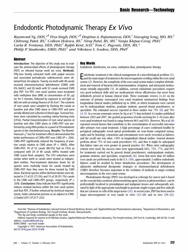
in the CMD group was aseptically mounted on a rubber dam attachedto a rack. The contents of root canals were sampled by flushing theroot canals with a coronal application of 1 mL of sterile phosphate-buffered saline (PBS) with a Pro Rinse 30-gauge irrigation needle(Dentsply Maillefer) (Fig. 2). The bacterial suspension was collectedin a 1.5-mL microcentrifuge tube positioned below the apical foramen,and bacterial yielding was measured spectrophotometrically for eachsample. After vortexing for 20 seconds, serial dilutions were prepared,and 100-mL aliquots were inoculated onto blood agar and incubatedanaerobically for 7 days.
PDTAfter CMD, specimens in the CMD+PDT group were treated byMB-
mediated PDT. Methylene blue (Sigma, St Louis, MO) was dissolved insterile PBS and filter-sterilized immediately before use. The finalconcentration of MB was 50 mg/mL (134 mmol/L). The ultraviolet-visible absorption spectra of MB in PBS were recorded from200–800 nm by using quartz cuvettes with 1-cm path length ona diode-array spectrophotometer and were characterized by a long-wavelength maximum at 665 nm as shown previously (14).
All individual specimens were aseptically mounted on a rubberdam, with the rubber dam frame attached to a rack. Then the canalswere filled to the level of the access cavity with MB solution by usinga Pro Rinse 30-gauge irrigation needle for 5 minutes. After incubation,the canal was dried with a paper cone. Light was then applied in the rootcanal system of the specimens in appropriate groups for 2.5 minutes,followed by a break of 2.5 minutes and a second light exposure for2.5 minutes. The irradiation source was a diode laser (BWTEK Inc,Newark, DE) with an output power of 1 W and a central wavelengthof 665 nm. The system was coupled to a 250-mm diameter optical fiber(22) that was mechanically notched over a 1-cm length at approxi-mately 1-mm intervals (Schoelly Imaging Inc, Worcester, MA). The fiberwas able to uniformly distribute light at 360 degrees within the rootcanal. The power density was 100 mW/cm2, and the total energy fluencedose was 30 J/cm2. The fiberoptic was wiped with ethanol after thecompletion of each light exposure.
JOE — Volume 37, Number 2, February 2011
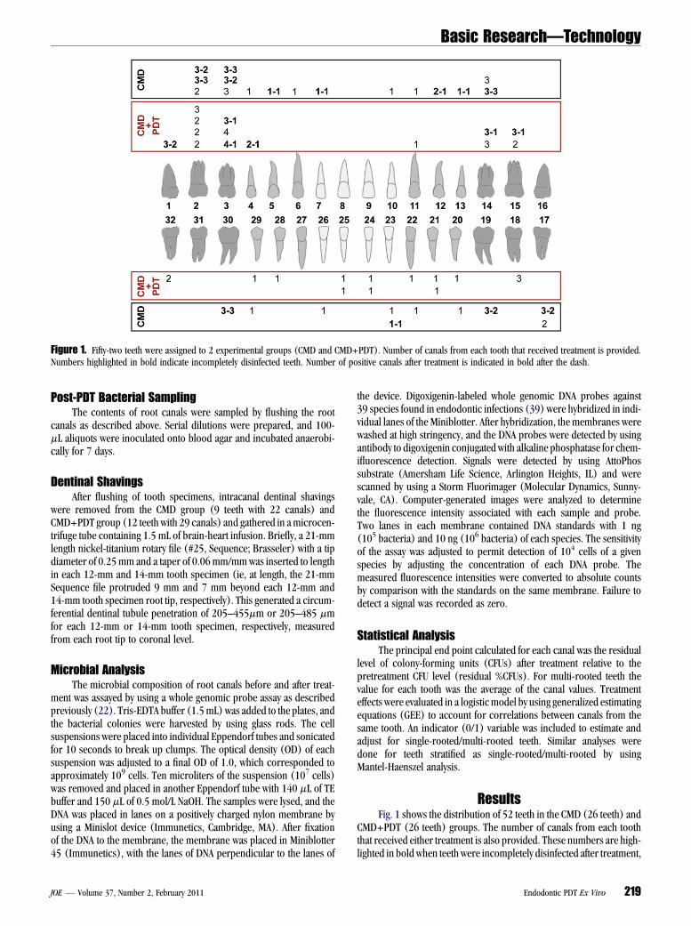
Figure 1. Fifty-two teeth were assigned to 2 experimental groups (CMD and CMD+PDT). Number of canals from each tooth that received treatment is provided.Numbers highlighted in bold indicate incompletely disinfected teeth. Number of positive canals after treatment is indicated in bold after the dash.
Basic Research—Technology
Post-PDT Bacterial SamplingThe contents of root canals were sampled by flushing the root
canals as described above. Serial dilutions were prepared, and 100-mL aliquots were inoculated onto blood agar and incubated anaerobi-cally for 7 days.
Dentinal ShavingsAfter flushing of tooth specimens, intracanal dentinal shavings
were removed from the CMD group (9 teeth with 22 canals) andCMD+PDT group (12 teeth with 29 canals) and gathered in amicrocen-trifuge tube containing 1.5 mL of brain-heart infusion. Briefly, a 21-mmlength nickel-titanium rotary file (#25, Sequence; Brasseler) with a tipdiameter of 0.25 mm and a taper of 0.06 mm/mmwas inserted to lengthin each 12-mm and 14-mm tooth specimen (ie, at length, the 21-mmSequence file protruded 9 mm and 7 mm beyond each 12-mm and14-mm tooth specimen root tip, respectively). This generated a circum-ferential dentinal tubule penetration of 205–455mm or 205–485 mmfor each 12-mm or 14-mm tooth specimen, respectively, measuredfrom each root tip to coronal level.
Microbial AnalysisThe microbial composition of root canals before and after treat-
ment was assayed by using a whole genomic probe assay as describedpreviously (22). Tris-EDTA buffer (1.5mL) was added to the plates, andthe bacterial colonies were harvested by using glass rods. The cellsuspensions were placed into individual Eppendorf tubes and sonicatedfor 10 seconds to break up clumps. The optical density (OD) of eachsuspension was adjusted to a final OD of 1.0, which corresponded toapproximately 109 cells. Ten microliters of the suspension (107 cells)was removed and placed in another Eppendorf tube with 140 mL of TEbuffer and 150 mL of 0.5 mol/L NaOH. The samples were lysed, and theDNA was placed in lanes on a positively charged nylon membrane byusing a Minislot device (Immunetics, Cambridge, MA). After fixationof the DNA to the membrane, the membrane was placed in Miniblotter45 (Immunetics), with the lanes of DNA perpendicular to the lanes of
JOE — Volume 37, Number 2, February 2011
the device. Digoxigenin-labeled whole genomic DNA probes against39 species found in endodontic infections (39) were hybridized in indi-vidual lanes of the Miniblotter. After hybridization, the membranes werewashed at high stringency, and the DNA probes were detected by usingantibody to digoxigenin conjugated with alkaline phosphatase for chem-ifluorescence detection. Signals were detected by using AttoPhossubstrate (Amersham Life Science, Arlington Heights, IL) and werescanned by using a Storm Fluorimager (Molecular Dynamics, Sunny-vale, CA). Computer-generated images were analyzed to determinethe fluorescence intensity associated with each sample and probe.Two lanes in each membrane contained DNA standards with 1 ng(105 bacteria) and 10 ng (106 bacteria) of each species. The sensitivityof the assay was adjusted to permit detection of 104 cells of a givenspecies by adjusting the concentration of each DNA probe. Themeasured fluorescence intensities were converted to absolute countsby comparison with the standards on the same membrane. Failure todetect a signal was recorded as zero.
Statistical AnalysisThe principal end point calculated for each canal was the residual
level of colony-forming units (CFUs) after treatment relative to thepretreatment CFU level (residual %CFUs). For multi-rooted teeth thevalue for each tooth was the average of the canal values. Treatmenteffects were evaluated in a logisticmodel by using generalized estimatingequations (GEE) to account for correlations between canals from thesame tooth. An indicator (0/1) variable was included to estimate andadjust for single-rooted/multi-rooted teeth. Similar analyses weredone for teeth stratified as single-rooted/multi-rooted by usingMantel-Haenszel analysis.
ResultsFig. 1 shows the distribution of 52 teeth in the CMD (26 teeth) and
CMD+PDT (26 teeth) groups. The number of canals from each tooththat received either treatment is also provided. These numbers are high-lighted in bold when teeth were incompletely disinfected after treatment,
Endodontic PDT Ex Vivo 219

Figure 2. Post-flushing of tooth specimen under rubber dam.
TABLE 1. Frequency of Root Canal Infection after CMD or CMD+PDT Treatment
Survival after treatment
None <0.1% >0.1%Total no.of canals
CMD, no. 24 14 11 49CMD, % 49.0 28.6 22.4CMD+PDT, no. 45 7 0 52CMD+PDT, % 86.5 13.5 0.0
CMD, chemomechanical debridement; PDT, photodynamic therapy.
Basic Research—Technology
and the number of positive canals is given after the dash. The resultsclearly demonstrated the better performance of CMD+PDT overCMD. The summary Mantel-Haenszel c2 test for treatment effects wassignificant (P= .026). Overall, 13 of 26 teeth (50%) were positive afterCMD, whereas 6 of 20 teeth (30%) were positive after CMD+PDT.Among single-rooted teeth, 5 of 14 teeth (35.7%) were positive afterCMD, whereas only 1 of 12 teeth (8.3%) was positive after CMD+PDT.Among multi-rooted teeth, 8 of 12 teeth (66.7%) were positive afterCMD, and 5 of 14 teeth (35.7%) were positive after CMD+PDT.
CMD+PDT significantly reduced the frequency of positive canalsrelative to CMD alone (P = .0003) (Table 1). Canals from single-rooted teeth were less likely to be positive after treatment than canalsfrom multi-rooted teeth (P = .10). After CMD+PDT, 45 of 52 canals(86.5%) had no CFUs as compared with 24 of 49 canals (49%) treatedwith CMD (Table 1) (canal flush samples). Post-treatment microbiallevels were low as a percent of pretreatment levels (%CFU). ForCMD+PDT, only 7 of 52 canals (13.5%) were positive, and all had %CFU less than 0.1% of pretreatment CFU levels. However, after CMD,25 of 49 canals were positive (51%), and 22.4% of canals had post-treatment values greater than 0.1% of pretreatment levels (Table 1).
220 Ng et al.
The CFU reductions were similar when teeth or canals were treatedas independent entities. Analysis stratified by tooth type indicated thatpost-treatment %CFU values were more often positive and also at higherlevels of infection in canals that received CMD relative to canals that hadreceived CMD+PDT (P < .0001).
The microbial composition of canal biofilms (canal flushsamples) was studied by checkerboard DNA-DNA hybridization.Pretreatment and post-treatment frequencies (�) were obtained for39 species found in endodontic infections with whole genomic probesfor 45 canals that received CMD+PDT and 44 that received CMD alone(Fig. 3). The number of canals positive for each species pretreatmentwas quite high, and the pattern was similar for both treatments. Post-treatment detection levels for all species were systematically and mark-edly lower for canals treated by CMD+PDT than for those treated byCMD alone. Key endodontic pathogens resisting intracanal disinfectionprocedures (40) were dramatically reduced (Fig. 3, highlighted ingray).
The frequency of dentinal infection (after debridement up to 485mm) was also evaluated (Table 2). In the CMD group, tubules from17 of 22 canals (77.3%) were positive after treatment, whereas inthe CMD+PDT group, tubules from 15 of 29 (51.7%) canals werepositive (P = .034).
DiscussionThe present study was built on the interdependent foundations of
(1) developing an in vitro model for testing PDT (14, 20, 22); (2) theutilization of a Food and Drug Administration–approved drug, MB, asthe photosensitizer (14, 20, 22); (3) the development of a novel lightdelivery system that maximizes the distribution of light within theentire anatomy of the root canal system (22); (4) the ongoing refine-ment of light and drug dosimetry (14, 20, 22, 41); and (5) theassessment of PDT safety (41). The hypothesis of this study was thatnear-complete elimination of residual root canal bacteria could beachieved by using PDT as an adjunctive procedure to CMD in chroni-cally infected extracted human teeth ex vivo. Our findings showedthat MB-mediated PDT significantly enhanced the effect of CMD. Fourin vivo studies have also suggested the potential of PDT as an adjunctivetechnique to eliminate residual root canal bacteria after CMD (29–32).Toluidine blue–mediated PDT offered a means of destroyingmicroorganisms remaining after using sodium hypochlorite alone(29) or citric acid and sodium hypochlorite as co-irrigants (30).PDT significantly enhanced the effect of CMD in teeth with necrotic pulpsby using a conjugate between polyethyleneimine chlorine e6 conjugate(31) and toluidine blue (32).
The incomplete bacterial killing in dentinal tubules after PDT mightbe due to (1) incomplete MB penetration in the tubules that might berelated to binding interactions with dentin components; (2) failure ofMB to penetrate canal biofilms; and (3) insufficient oxygenation. Wehave proposed the encapsulation of MB within poly(D,L-lactide-co-glycolide) (PLGA) nanoparticles that might offer a novel nano-platform
JOE — Volume 37, Number 2, February 2011
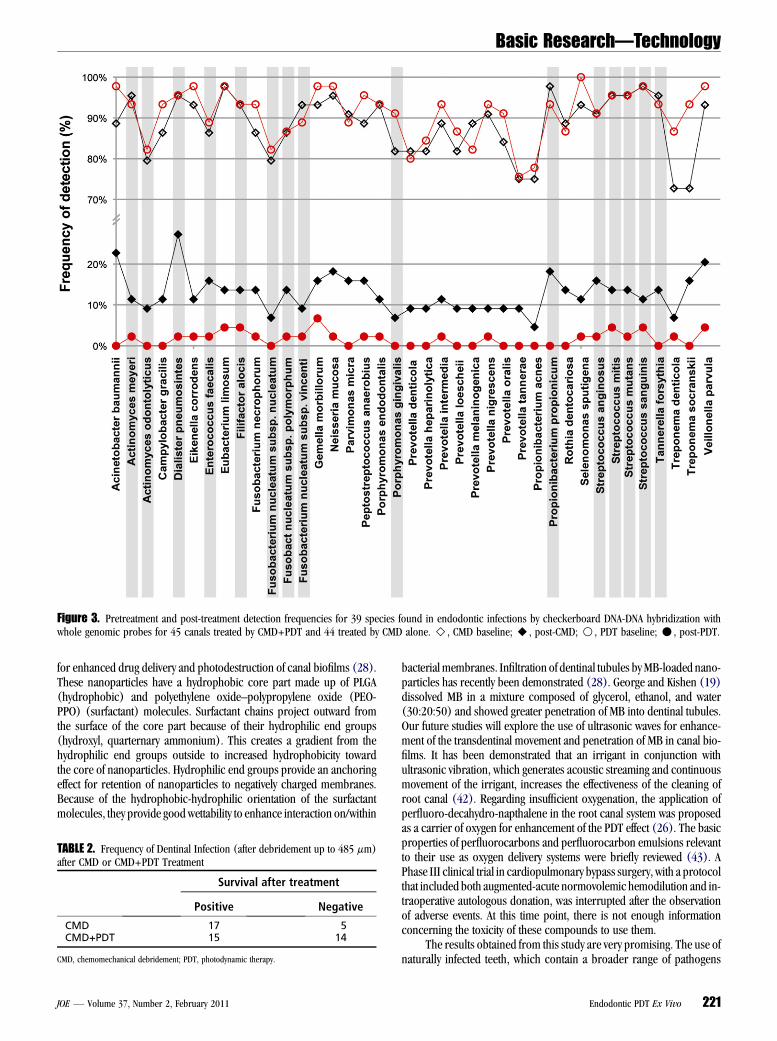
Figure 3. Pretreatment and post-treatment detection frequencies for 39 species found in endodontic infections by checkerboard DNA-DNA hybridization withwhole genomic probes for 45 canals treated by CMD+PDT and 44 treated by CMD alone. >, CMD baseline; A, post-CMD; B, PDT baseline; C, post-PDT.
Basic Research—Technology
for enhanced drug delivery and photodestruction of canal biofilms (28).These nanoparticles have a hydrophobic core part made up of PLGA(hydrophobic) and polyethylene oxide–polypropylene oxide (PEO-PPO) (surfactant) molecules. Surfactant chains project outward fromthe surface of the core part because of their hydrophilic end groups(hydroxyl, quarternary ammonium). This creates a gradient from thehydrophilic end groups outside to increased hydrophobicity towardthe core of nanoparticles. Hydrophilic end groups provide an anchoringeffect for retention of nanoparticles to negatively charged membranes.Because of the hydrophobic-hydrophilic orientation of the surfactantmolecules, they provide goodwettability to enhance interaction on/within
TABLE 2. Frequency of Dentinal Infection (after debridement up to 485 mm)after CMD or CMD+PDT Treatment
Survival after treatment
Positive Negative
CMD 17 5CMD+PDT 15 14
CMD, chemomechanical debridement; PDT, photodynamic therapy.
JOE — Volume 37, Number 2, February 2011
bacterial membranes. Infiltration of dentinal tubules byMB-loaded nano-particles has recently been demonstrated (28). George and Kishen (19)dissolved MB in a mixture composed of glycerol, ethanol, and water(30:20:50) and showed greater penetration of MB into dentinal tubules.Our future studies will explore the use of ultrasonic waves for enhance-ment of the transdentinal movement and penetration of MB in canal bio-films. It has been demonstrated that an irrigant in conjunction withultrasonic vibration, which generates acoustic streaming and continuousmovement of the irrigant, increases the effectiveness of the cleaning ofroot canal (42). Regarding insufficient oxygenation, the application ofperfluoro-decahydro-napthalene in the root canal system was proposedas a carrier of oxygen for enhancement of the PDT effect (26). The basicproperties of perfluorocarbons and perfluorocarbon emulsions relevantto their use as oxygen delivery systems were briefly reviewed (43). APhase III clinical trial in cardiopulmonary bypass surgery, with a protocolthat included both augmented-acute normovolemic hemodilution and in-traoperative autologous donation, was interrupted after the observationof adverse events. At this time point, there is not enough informationconcerning the toxicity of these compounds to use them.
The results obtained from this study are very promising. The use ofnaturally infected teeth, which contain a broader range of pathogens
Endodontic PDT Ex Vivo 221
Basic Research—Technology
than in vitromodel systems, provides an excellent test of the potential ofPDT in achieving root canal disinfection. However, because some livingbacteria were still present in dentinal tubules after PDT, further refine-ment and enhancement of the PDT procedure might be necessary. Theeffect of biophysical means and surface tension-reducing agents on thetransdentinal penetration of MB as well as the effect of supplementalhyperoxygenation should be evaluated.AcknowledgmentsThis work was supported by NIDCR grant RO1-DE-16922.The authors deny any conflicts of interest related to this study.
References1. Figdor D. Apical periodontitis: a very prevalent problem. Oral Surg Oral Med Oral
Pathol Oral Radiol Endod 2002;94:651–2.2. Nair PN. Pathogenesis of apical periodontitis and the causes of endodontic failures.
Crit Rev Oral Biol Med 2004;15:348–81.3. Siqueira JF Jr, Rocas IN, Paiva SS, Magalh~aes KM, Guimar~aes-Pinto T. Cultivable
bacteria in infected root canals as identified by 16S rRNA gene sequencing. OralMicrobiol Immunol 2007;22:266–71.
4. Lewsey JD, Gilthorpe MS, Gulabivala K. An introduction to meta-analysis within theframework of multilevel modelling using the probability of success of root canaltreatment as an illustration. Community Dent Health 2001;18:131–7.
5. Basmadjian-Charles CL, Farge P, Bourgeois DM, Lebrun T. Factors influencing thelong-term results of endodontic treatment: a review of the literature. Int Dent J 2002;52:81–6.
6. Ng Y-L, Mann V, Rahbaran S, Lewsey J, Gulabivala K. Outcome of primary root canaltreatment: systematic review of the literature—part 1:effects of study characteristicson probability of success. Int Endod J 2007;40:921–39.
7. Ng Y-L, Mann V, Gulabivala K. Tooth survival following non-surgical root canal treat-ment: a systematic review in the literature. Int Endod J 2010;43:171–89.
8. Wu M-K, Shemesh H, Wesselink PR. Limitations of previously published systematicreviews evaluating the outcome of endodontic treatment. Int Endod J 2009;42:656–66.
9. Nash KD, Brown LJ, Hicks ML. Private practicing endodontists: production ofendodontic services and implications for workforce policy. J Endod 2002;28:699–705.
10. Brown LJ, Nash KD, Johns BA, Warren M. The economics of endodontics. ADA Healthpolicy Resources Center dental health policy analysis series. Chicago, IL, 2003.
11. Dougherty TJ, Gomer CJ, Henderson BW, et al. Photodynamic therapy. J Natl CancerInst 1998;90:889–905.
12. Silbert T, Bird PS, Milburn GJ, Walsh L. Disinfection of root canals by laser dyephotosensitization. J Dent Res 2000;79:569.
13. Seal GJ, Ng YL, Spratt D, Bhatti M, Gulabivala K. An in vitro comparison of the bacte-ricidal efficacy of lethal photosensitization or sodium hyphochlorite irrigation onStreptococcus intermedius biofilms in root canals. Int Endod J 2002;35:268–74.
14. Soukos NS, Chen PS, Morris JT, et al. Photodynamic therapy for endodontic disin-fection. J Endod 2006;32:979–84.
15. Garcez AS, N�u~nez SC, Lage-Marques JL, Jorge AO, Ribeiro MS. Efficiency of NaOCland laser-assisted photosensitization on the reduction of Enterococcus faecalisin vitro. Oral Surg Oral Med Oral Pathol Oral Radiol Endod 2006;102:e93–8.
16. Williams JA, Pearson GJ, Colles MJ. Antibacterial action of photoactivated disinfec-tion {PAD} used on endodontic bacteria in planktonic suspension and in artificialand human root canals. J Dent 2006;34:363–71.
17. Garcez AS, Ribeiro MS, Tegos GP, N�u~nez SC, Jorge AO, Hamblin MR. Antimicrobialphotodynamic therapy combined with conventional endodontic treatment to elimi-nate root canal biofilm infection. Lasers Surg Med 2007;39:59–66.
18. George S, Kishen A. Advanced noninvasive light-activated disinfection: assessment ofcytotoxicity on fibroblast versus antimicrobial activity against Enterococcus faeca-lis. J Endod 2007;33:599–602.
222 Ng et al.
19. George S, Kishen A. Photophysical, photochemical, and photobiological character-ization of methylene blue formulations for light-activated root canal disinfection.J Biomed Opt 2007;12:034029-1–034029-10.
20. Foschi F, Fontana CR, Ruggiero K, et al. Photodynamic inactivation of Enterococcusfaecalis in dental root canals in vitro. Lasers Surg Med 2007;39:782–7.
21. Bergmans L, Moisiadis P, Huybrechts B, Van Meerbeek B, Quirynen M,Lambrechts P. Effect of photo-activated disinfection on endodontic pathogens exvivo. Int Endod J 2008;41:227–39.
22. Fimple JL, Fontana CR, Foschi F, et al. Photodynamic treatment of endodontic poly-microbial infection in vitro. J Endod 2008;34:728–34.
23. Fonseca MB, J�unior PO, Pallota RC, et al. Photodynamic therapy for root canalsinfected with Enterococcus faecalis. Photomed Laser Surg 2008;26:209–13.
24. George S, Kishen A. Augmenting the antibiofilm efficacy of advanced noninvasivelight activated disinfection with emulsified oxidizer and oxygen carrier. J Endod2008;34:1119–23.
25. George S, Kishen A. Influence of photosensitizer solvent on the mechanisms of pho-toactivated killing of Enterococcus faecalis. Photochem Photobiol 2008;84:734–40.
26. Lim Z, Cheng JL, Lim TW, et al. Light activated disinfection: an alternative endodonticdisinfection strategy. Aust Dent J 2009;54:108–14.
27. Souza LC, Brito PRR, Machado de Oliveira JC, et al. Photodynamic therapy with twodifferent photosensitizers as a supplement to instrumentation/irrigation proceduresin promoting intracanal reduction of Enterococcus Faecalis. J Endod 2010;36:292–6.
28. Pagonis TC, Chen J, Fontana CR, et al. Nanoparticle-based endodontic antimicrobialphotodynamic therapy. J Endod 2010;36:322–8.
29. Bonsor SJ, Nichol R, Reid TM, Pearson GJ. An alternative regimen for root canaldisinfection. Br Dent J 2006;22:101–5.
30. Bonsor SJ, Nichol R, Reid TM, Pearson GJ. Microbiological evaluation of photo-activated disinfection in endodontics (an in vivo study). Br Dent J 2006;25:337–41.
31. Garcez AS, N�u~nez SC, Hamblin MR, Ribeiro MS. Antimicrobial effects of photody-namic therapy on patients with necrotic pulps and periapical lesion. J Endod2008;34:138–42.
32. Pinheiro SL, Schenka AA, Neto AA, de Souza CP, Rodriguez HM, Ribeiro MC. Photo-dynamic therapy in endodontic treatment of deciduous teeth. Lasers Med Sci 2009;24:521–6.
33. Harris F, Chatfield LK, Phoenix DA. Phenothiazinium based photosensitisers-photodynamic agents with a multiplicity of cellular targets and clinical applications.Curr Drug Targets 2005;6:615–27.
34. Wainwright M, Mohr H, Walker WH. Phenothiazinium derivatives for pathogen inac-tivation in blood products. J Photochem Photobiol B 2007;86:45–58.
35. Ojetti V, Persiani R, Nista EC, et al. A case-control study comparing methylene bluedirected biopsies and random biopsies for detecting pre-cancerous lesions in thefollow-up of gastric cancer patients. Eur Rev Med Pharmacol Sci 2007;11:291–6.
36. Creagh TA, Gleeson M, Travis D, Grainger R, McDermott TE, Butler MR. Is therea role for in vivomethylene blue staining in the prediction of bladder tumour recur-rence? Br J Urol 1995;75:477–9.
37. Wainwright M, Phoenix DA, Marland J, Wareing DR, Bolton FJ. A study of photobac-tericidal activity in the phenothiazinium series. FEMS Immunol Med Microbiol 1997;19:75–80.
38. Usacheva MN, Teichert MC, Biel MA. The interaction of lipopolysaccharides withphenothiazine dyes. Lasers Surg Med 2003;33:311–9.
39. Brito LC, Teles FR, Teles RP, et al. Use of multiple-displacement amplification andcheckerboard DNA-DNA hybridization to examine the microbiota of endodonticinfections. J Clin Microb 2007;45:3039–49.
40. Siqueira JF Jr, Rocas I. Clinical implications and microbiology of bacterial persis-tence after treatment procedures. J Endod 2008;34:1291–301.
41. Xu Y, Young MJ, Battaglino RA, et al. Endodontic antimicrobial photodynamictherapy: safety assessment in mammalian cell cultures. J Endod 2009;35:1567–72.
42. Gutarts R, Nusstein J, Reader A, Beck M. In vivo debridement efficacy of ultrasonicirrigation following hand-rotary instrumentation in human mandibular molars.J Endod 2005;31:166–70.
43. Riess JG. Perfluorocarbon-based oxygen delivery. Artif Cells Blood Substit ImmobilBiotechnol 2006;34:567–80.
JOE — Volume 37, Number 2, February 2011