Embed Size (px)
Citation preview

Emocolture o Biologia Molecolare?
Dr Andrea Rocchetti


• Affermazione 1
• Per emocoltura si intende la coltura di campioni di sangue prelevati in condizioni di asepsi.
• Argomentazione
• Si tratta di un metodo diagnostico fondamentale per la diagnosi microbiologica di batteriemia in quanto consente di confermare il sospetto clinico di sepsi, di accertarne l’agente eziologico e di studiarne la sensibilità in vitro agli antibiotici.
• Bibliografia essenziale
• CLSI – Principles and Procedures for Blood Cultures; Approved Guideline. – CLSI document M47-A. Wayne, PA; Clinical and Laboratory Standards Institute; 2007.
•

Focalizzare velocemente la terapia antibiotica può portare a:
Usare responsabilmente l’antibiotico, evitando il rischio di sviluppo diceppi microbici multiresistenti
Aumentare la sopravvivenza del paziente settico:
MIGLIOR OUTCOME CLINICO
Abbassare i costi ospedalieri, riducendo la spesa associata ad un lungotrattamento antibiotico e alla lunga degenza del paziente


Blood
Draw
Gram
StainPositiveApprox. 1 Hr.
2Hrs.
Standard Testing
Blood
Culture
32 Hrs. 1,40 min. 65,32Hrs.
35 Hrs
ORA DI CAMBIO DELLA TERAPIA
?
Anno 2013
Avvio incubatore emocolture decentrato all’ Ospedale Infantile e Urgenze Laboratorio Analisi o area ritenuta strategica.
Il tempo al gram rappresenta il momento cardine del passaggio dalla terapia antibiotica empirica alla terapia mirata.
Inoltre questo tempo rappresenta il punto nevralgico delle analisi di “tecnology improvement” e di valorizzazione delle risorse organizzative.
COLLOCAZIONE IN
PRONTO SOCCORSO DI
UN MODULO PER
EMOCOLTURA
COLLEGATO IN REMOTO
CON IL LABORATORIO


© 2016 BD. BD and the BD Logo are trademarks of Becton, Dickinson and Company.11
Blood Culture Satellite Solutions

MALDI TOF
< 20-30 minuti
BatteriFunghiMicobatteri
< 20-30 minuti< 20-30 minuti
15 minuti

Verigene
8 batteri G-CTX-MKPCNDMVIMIMPOXA12 atteri G+Mec AVanAVanB
< 5 minuti
< 2,5 – 3 ore
8 batteri G-CTX-MKPCNDMVIMIMPOXA12 batteri G+Mec AVanAVanB

E PLEX
5 < minuti
< 2 ore
33 batteri G-CTX-MKPCNDMVIMIMPOXA24 Batteri G+Mec AMec CVanAVanB

BioFire FilmArray
13batteri G-KPC8 batteri G+Mec AVanA5 Candide spp
< 5 minuti
< 65 minuti

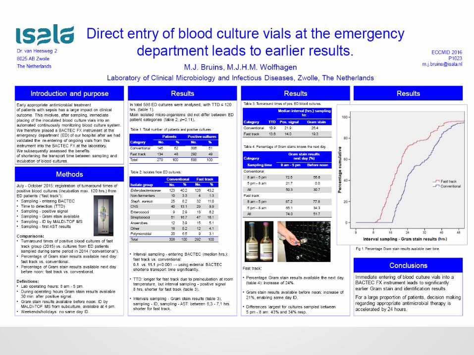
Sample Extraction, Amplification, and Detection: It’s All in the Pouch
The FilmArray pouch is loaded into the FilmArray instrument
8. The FilmArray performs a melt to confirm the presence or absence of assay-specific temperature signatures of the second stage PCR product for each well in the array
2. Nucleic acids bound by magnetic beads move from the lysis chamber to the purification chamber. A wash buffer removes cellular and pathogen debris
3. An elution buffer removes purified nucleic acids from the magnetic beads
4. Nucleic acids move to the first-stage PCR chamber. Reverse transcriptase converts target RNA to DNA, followed by a high-order multiplex PCR
5. Products from the first-stage PCR are diluted to remove any remaining PCR primers
6. First-stage PCR products are added to fresh master mix and are aliquotedinto each well of the array
7. Each well is pre-spotted with a single pair of second-stage PCR primers, resulting in specific amplification of target DNA only. A fluorescent double-stranded DNA binding dye monitors each reaction
1. Sample moves into lysis chamber. Cells and pathogens are lysed by bead beating, releasing nucleic acids
65:00
Sample extraction, amplification, and detection: It’s all in the pouch
65 minutes
run-time
64:0063:0062:0061:0060:0059:0058:0057:0056:0055:0054:0053:0052:0051:0050:0049:0048:0047:0046:0045:0044:0043:0042:0041:0040:0039:0038:0037:0036:0035:0034:0033:0032:0031:0030:0029:0028:0027:0026:0025:0024:0023:0022:0021:0020:0019:0018:0017:0016:0015:0014:0013:0012:0011:0010:0009:0008:0007:0006:0005:0004:0003:0002:0001:0000:0065:00

Automated Results Analysis
102 individual 2nd stage PCR wells
Each well contains one reaction
All targets tested in triplicate
Melt curves generated for each well

ACCELERATE
< 2 minuti
• 90 minuti per una identificazione
• 5 ore per un antibiogramma con MIC
• 7 ore il tempo totale

FISH IDENTIFICATION
FUNGALC. albicans
C. glabrata
AO - Acridine Orange
S. lugdunensis
S. aureusCoNS spp.
E. faecalis
E. faeciumS. pneumoniae
S. agalactiaeStrep spp.
GRAM POSITIVEEnterobacter spp. E. coli
Klebsiella spp. Proteus spp.
Citrobacter spp S. marcescens
P. aeruginosa A. baumannii
GRAM NEGATIVE
Fluorescence In-Situ Hybridization
Universal Bacterial Probes in all ID channels Note: Visual demonstrates proportional usage of flow channels Actual channel usage is alternate/balanced

Automated Sample Preparation Identification& Quantitation
Sample Filtration
(GEF)
Cell Immobilization
(EKC)
Target ID, Universal and
Polymicrobial Detection
Quantitation of
individual cells
Susceptibility Analysisand Reporting
Image analysis via
proprietary algorithmsMIC determination and
SIR interpretation
TECHNOLOGY OVERVIEW
Proprietary Supercomputing

MORPHOKINETIC CELLULAR ANALYSIS
Rapid Direct From Specimen MIC Reporting
Identification results guide selection of antimicrobials for susceptibility testing. Bacteria are grown in the presence of a single concentration of each antimicrobial. Growth response is analyzed using time-lapse imaging of individual bacteria.

Microscopy Images Computed into Many Individual Growth Curves
Founder cells Clonal growth Growth
Resistant
Susceptible
Time
Log(
mas
s)
Founder cells Clonal growth Lysis
TRACK GROWTH/LYSIS OF LIVE INDIVIDUAL BACTERIAL CELLS
Time
Log(
mas
s)

DID T2
< 10 minuti
Candida albicansCandida glabrataCandida tropicalisCandida KruseiCandida parapsilosis

Average Time to Identification
-
Molecular
Direct from patient blood sample
4.3 hours*Dependent on Blood Culture Positivity
49 hrs.†
51 hrs.†
72 hrs.+
129 hrs.+
Time Savings: 2-7 Days
19
Automated ID/AST
MALDI-TOF
Nanosphere
BioFire
T2
Susceptibility
4-48 hours
Species ID
1 hour – 2 days
Blood Culture
1-5 Days
BC+ andGram Stain
~1 hour


n = 617
n = 207
n = 198
n = 212
Pathogens were:
Gram+ 54.8%
Gram- 32.6%
Candida sp. 2%
Polymicrobial 10.5%
Among subjects with PCR testing, 81% of organisms isolated were detectable by FA BCID panel
* For all groups, MALDI-TOF MS or pathogen identification of colonies
isolated from positive BCBs and rapid testing for MRSA colonies were used

Median time from Gram stain result to organism identification was
shorter in both intervention groups (both 1.3 hours) versus the
control group (22 hours) (P < .0001)
INT1
INT2


Scienza, tecnologia, economia e …coraggio