Embed Size (px)
Citation preview

SHOCK
Emergency pediatric – PICU division
Pediatric Department
Medical Faculty, University of Sumatera Utara – H. Adam Malik Hospital
1

Definition
Shock is an acute, complex state of
circulatory dysfunction that results in
failure to deliver sufficient amounts of
2
failure to deliver sufficient amounts of
oxygen and other nutrients to meet tissue
metabolic demands

Pathophysiology
Delivery of Oxygen (DO2):
DO2 = Cardiac output (CO) x Arterial oxygen content (CaO2)
CO = Heart Rate (HR) x Stroke Volume (SV)
CaO2= Hb x SaO2 x 1,39
3

Blood CO
SV
Preload
Myocard
Contractility
Blood
PressureAfterloadHR
SVR
4
CO = Cardiac Output
SVR = Systemic Vascular resistance
SV = Stroke Volume
HR = Heart Rate

Clinical Manifestation
Clinical Sign Compensated Uncompensated Irreversible
Heart rate
Systolic BP
Pulse volume
Capillary refill
Tachycardia +
Normal
Normal/reduced
Normal/increased
Tachycardia ++
Normal or falling
Reduced +
Increased +
Tachycardia
/bradicardia
Plummeting
Reduced ++
Three phases: compensated, uncompensated, irreversible
5
Capillary refill
Skin
Respiratory rate
Mental state
Normal/increased
Cool,pale
Tachypnoea +
Mild agitation
Increased +
Cool,mottled
Tachypnoea ++
Lethargic
Uncooperative
Reduced ++
Increased ++
Cold,deathly pale
Sighing respiration
React only to pain or
unresponsive

Management
• Intubation & mechanical ventilation
• Fluid resuscitation
• Vasoactive infusion
6
• Vasoactive infusion

FUNCTIONAL CLASSIFICATION
• Hypovolemia
• Cardiogenic
• Obstructive
7
• Distributive
• Septic
• Endocrine

HYPOVOLEMIC SHOCK
• A decrease in intra vascular blood volume to such an extent thateffective tissue perfusion can not be maintain
• Most common cause of shock in infants & children
• Etiology:
– Hemorrhage
– Plasma loss
8
– Plasma loss
– Fluid & electrolyte loss
• Hypovolemia � ↓ preload � ↓ SV � ↓ CO

CLINICAL MANIFESTATION:
• Tachycardia
• Skin mottling
• Prolonged capillary refill
• Cool extremities
• ↓ UOP
9
• ↓ UOP
• Hypotensive
• Lethargy / comatose

THERAPY
• Adequate oxygenation and ventilation
• Rapid volume replacement � reestablish circulation:– Crystalloid: 20 ml/kg � shock persist � 20 ml/kg
– Hemorrhagic: transfusion
10Continuous monitoring of HR, arterial BP, CVP, UOP Continuous monitoring of HR, arterial BP, CVP, UOP
Shock (+)Shock (+)

CVP:
– < 10 mmHg ���� ↑ fluid infusion until preload is reach
– >10 mmHg ���� indication: flow-direct thermo dilution
pulmonary artery catheter and/or echocardiogram
Ventricular filling pressure rises without evidence of improvement
11
Ventricular filling pressure rises without evidence of improvement
in cardiovascular performance
Discontinue fluid resuscitation
Inotropic agent (+)

REFRACTORY SHOCK:
– Unrecognized pneumothorax / pericardial effusion
– Intestinal ischemia
– Sepsis
– Myocardial dysfunction
12
– Adrenal cortical insufficiency
– Pulmonary hypertension

CARDIOGENIC SHOCK
• The pathophysiologic state in which abnormality of cardiac
function is responsible for the failure of the cardiovascular
system to meet the metabolic needs of tissue
� Depressed CO
13
� Depressed CO
• Etiology: Heart rate abnormalities, Cardiomyopathies/carditis,
Congenital heart disease, Trauma
• Myocardial dysfunction is frequently a late manifestation of
shock of any etiology

CLINICAL MANIFESTATION
• Tachycardia
• Hypotensive
• Diaphoretic
• Oliguria
• Acidotic
• Cool extremities
14
• Cool extremities
• Altered mental status
• Hepatomegaly
• Jugular venous distension
• Rales
• Peripheral edema

THERAPY
• ↑ Tissue oxygen supply
• ↓ Tissue oxygen requirements
• Correct metabolic abnormalities
• Preload should be optimized
15
• Preload should be optimized
• Myocardial contractility: inotropic agent ���� cathecholamine:
norepinephrine, epinephrine, dopamine & dobutamine

OBSTRUCTIVE SHOCK
• Caused by inability to produce adequate CO despite normal
intravascular volume & myocardial function
• Causative factor:
– Acute pericardial tamponade
16
– Tension pneumothorax
– Pulmonary / systemic hypertension
– Congenital / acquired outflow obstruction
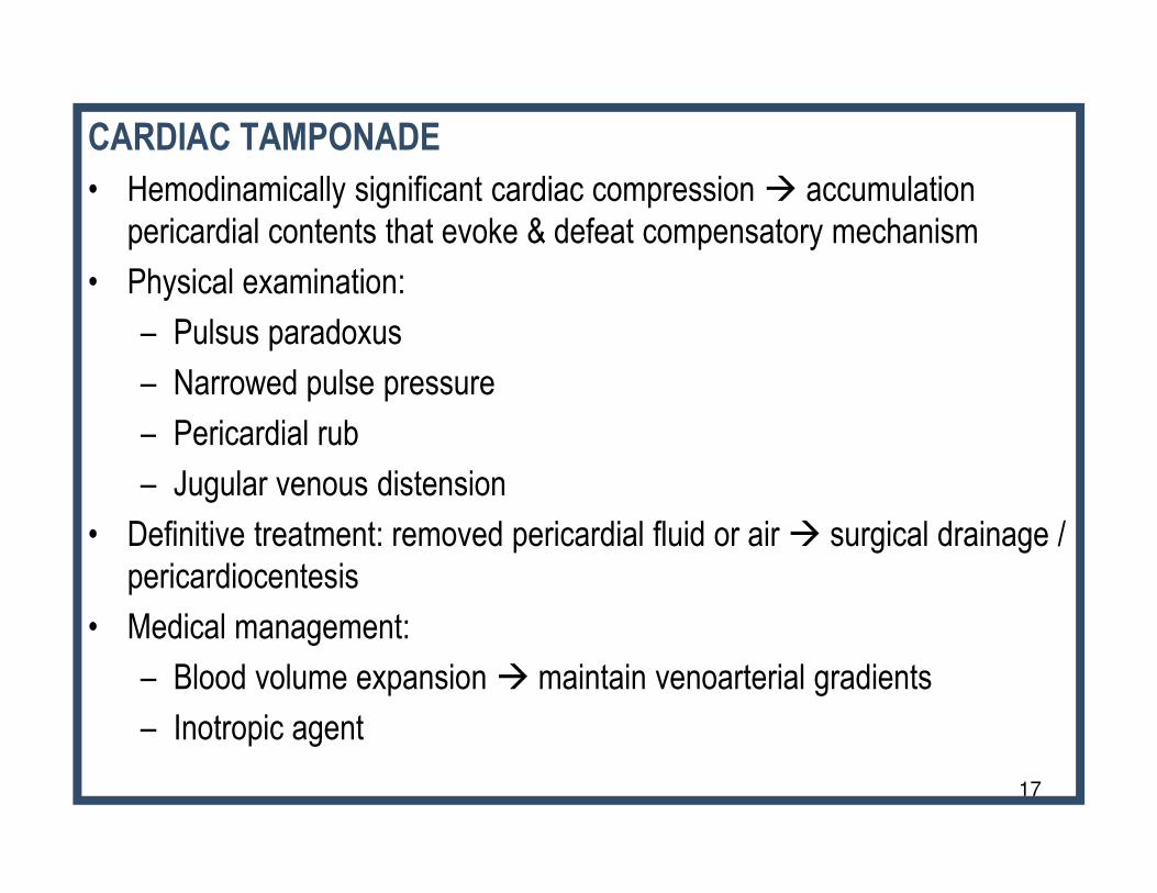
CARDIAC TAMPONADE
• Hemodinamically significant cardiac compression � accumulation
pericardial contents that evoke & defeat compensatory mechanism
• Physical examination:
– Pulsus paradoxus
– Narrowed pulse pressure
– Pericardial rub
17
– Pericardial rub
– Jugular venous distension
• Definitive treatment: removed pericardial fluid or air � surgical drainage /
pericardiocentesis
• Medical management:
– Blood volume expansion � maintain venoarterial gradients
– Inotropic agent

DISTRIBUTIVE SHOCK
• Results from maldistribution of blood flow to the tissue
• May be seen with anaphylaxis, spinal / epidural
anesthesia, disruption of spinal cord, inappropriate
administration vasodilatory medication
18
• Treatment:
– Reversal underlying etiology
– Vigorous fluid administration
– Vasopressor infusion

SEPTIC SHOCK
• Contains many elements of the other types of shock discussed
previously (hypovolemic, cardiogenic, and distributive shock)
• SIRS (Systemic Inflammatory Response Syndrome): non specific
inflammatory response
• Modified criteria for SIRS:
– Temp. >38,5 C or < 36 C
19
– Temp. >38,5 C or < 36 C
– Tachycardia
– Tachypnea
– WBC ↑ / ↓ or >10% immature neutrophils

• Sepsis: SIRS + documented infection
• Severe sepsis: Sepsis + end organ dysfunction
• Septic shock: Sepsis with hypotension despite adequate fluid
20
• Septic shock: Sepsis with hypotension despite adequate fluid
resuscitation

MANAGEMENT:
• Early recognition
• Antibiotics appropriate with microbiological examination
• Initial fluid resuscitation 20 ml/kg boluses over 5-10 minutes up to 40-60 ml/kg in the first hour
• Inotropic / vasopressor ���� refractory to fluids
21
• Inotropic / vasopressor ���� refractory to fluids
• Mechanical ventilation ���� refractory shock
• Hydrocortisone
• Glycemic control
• Blood transfusion

Catecholamine-resistant shock resistant
Observe in PICUTitrate epinephrine for cold shock, norepinephrine for warm shock to
Normal MAP-CVP difference for age and SVCO2 saturation > 70%
Establish central venous access, begin dopamine orDobutamine therapy and establish arterial monitoring
Push 20 cc/kg isotonic saline or colloid boluses up to and Over 60 cc/kg correct hypoglycemia and hypocalcemia
Fluid responsive*
15 min
Recognize decreased mental status and perfusion.Maintain airway and establish acces according to PALS guidelines
0 min5 min
Fluid refractory-dopamine/dobutamine resistant shock
Fluid refractory shock**
ECMORefractory shockStart cardiac output measurement and direct fluid, inotrope, vasopressor, vasosilator,
and hormonal therapies to attain normal MAP-CBP and CI > 3.3 and < 6.0 L/min/m2
Persistent Catecholamine-resistant shock
Add vasodilator or type III PDE
inhibitor with volume loading
Normal Blood Pressure Cold ShockSVC O2 Sat < 70%
Low Blood Pressure Cold ShockSVC O2 Sat < 70%
Titrater volume resuscitation
and epinephrine
Low Blood Pressure Warm ShockSVC O2 Sat < 70%
Titrater volume and
norepinephrine
60 minDraw baseline cortisol level
Then give hydrocortisone
Draw baseline cortisol level or perform
ACTH stim test. Do not give hydrocortisone
Not at risk ?At risk of adrenal insufficiency ?

THANK YOUTHANK YOUTHANK YOUTHANK YOU
23