Embed Size (px)
Citation preview

1
Emergency diagnosis and management of acute
heart failure
By Zoltán Szabó MD, PhD
31-12-2014
This e-book was sponsored by the TÁMOP 4.1.1.C-13/1/KONV-2014-0001 project. This project
is implemented through the New Hungary Development
Plan, co-financed by the European Union and the
European Social Fund.

2

3
Table of contents
1. Objective .............................................................................. Hiba! A könyvjelző nem létezik.
2. Terminology..............................................................................................................................5
3. Necessary devices .....................................................................................................................4
4. Personal conditions .............................................................. Hiba! A könyvjelző nem létezik.
5. Methods ............................................................................... Hiba! A könyvjelző nem létezik.
6. Dangers, consequences .............................................................................................................5
7. Drugs .........................................................................................................................................5
8. Documentation ..................................................................... Hiba! A könyvjelző nem létezik.
9. Ethical and legal aspects ...................................................... Hiba! A könyvjelző nem létezik.
10. Tasks of the instructor and the students ....................................................................................6
10.1. Tasks of the instructor ................................................................................................................ 7
10.2. Tasks of the students .................................................................................................................. 7
11. Heart failure: definition and onset ....................................... Hiba! A könyvjelző nem létezik.
12. Epidemiology ............................................................................................................................7
13. Systolic and diastolic heart failure ............................................................................................7
14. Pathopysiology..........................................................................................................................8
15. Clinical manifestations of acute heart failure ...........................................................................9
16. Prognosis for acute heart failure .............................................................................................10
17. Diagnosis of acute heart failure ........................................... Hiba! A könyvjelző nem létezik.
17.1. Anamnézis ................................................................................. Hiba! A könyvjelző nem létezik.
17.2. Physical examination ................................................................ Hiba! A könyvjelző nem létezik.
17.2.1. Symptoms observed in inspection ......................... Hiba! A könyvjelző nem létezik.
17.2.1.1. Cyanosis ..................................................................... Hiba! A könyvjelző nem létezik.
17.2.1.2. Jugular venous distension ........................................ Hiba! A könyvjelző nem létezik.
17.3.. Symptoms observed during percussion ................. Hiba! A könyvjelző nem létezik.
17.4.. Symptoms observed during auscultation .............. Hiba! A könyvjelző nem létezik.
17.4.1. Mitral insufficiency ...................................................... Hiba! A könyvjelző nem létezik.
17.4.2. Aortic insufficiency .................................................. Hiba! A könyvjelző nem létezik.
17.5. Auscultation- accessory heart sounds .................................................................... 17
17.6. Electrocariography ..........................................................................................................17
17.7. Chest X-ray ......................................................................................................................19

4
17.8. Blood gas analysis ...........................................................................................................19
17.9. Laboratory tests ...............................................................................................................20
17.10. Echocardiography ............................................................................................................20
17.11. Multiparametric monitoring in heart failure .......................................................................... 22
18. The treatment of acute heart failure ..................................... Hiba! A könyvjelző nem létezik.
18.1. Oxygen therapy ......................................................................... Hiba! A könyvjelző nem létezik.
18.2. Respiratory treatment ......................................................................................................25
18.3. Medicinal treatment of acute heart failure ............................................................................. 26
18.3.1. Morphine ..................................................................................................................26
18.3.2. Diuretics ...................................................................................................................27
18.3.3. Vasodilator compunds ..............................................................................................27
18.3.4. Inotropic agents ........................................................................................................28
18.3.4.1. Dopamine and dobutamine ...................................................................................... 28
18.3.4.2. Calcium sensitizing ................................................................................................... 29
18.3.4.3. Phosphodiesterase inhibitors ................................................................................... 29
18.3.4.4. Vasopressin antagonists ........................................................................................... 29
18.3.4.5. Vasopressor compunds ............................................................................................. 30
18.3.4.6. Cardiac glycosides .................................................................................................... 30
19. Special considerations in the treatment of acute cardiac failure ................................................. 31
20. References ...............................................................................................................................33
21. Self-controlling questions .................................................... Hiba! A könyvjelző nem létezik.
22. Case reports.......................................................................... Hiba! A könyvjelző nem létezik.
23. Tests ........................................................................................................................................38

5
1. Objective
The aim of this chapter is to define the different pathophysiological backgrounds of acute
and chronic heart failure, to show the clinical symptoms, to collect current data about cyanosis
and to learn about arterial blood gas analysis. Furthermore it is aimed to show the invasive and
non-invasive therapeutic strategies of heart failure.
2. Terminology
Hypoxia The decrease of tissue oxygenation due to the worsening of oxygen supply or
the increase in oxygen need.
Oxygen saturation The value which represents the oxygen ventilation/saturation of the
tissue
Asthma cardiale (cardiac asthma) Pulmonary edema due to a sudden left ventricular
failure
Cardiogenic shock Severe type of herat failure based on the acute worsening of
ventricular function and tissue hypoperfusion
Ejection fraction It describes left ventricular systolic function. It has a predictive value
with regard to cardiovascular mortality.
Blood gas analysis Determination of the oxygen and CO2 pressure (saturation) of the
arterial or capillary blood by means of a special device.
Cyanosis Bluish discoloration of the skin or mucus membrane due to hypocia. It can be
based on heart failure.
Afterload Right or left ventricular pressure load
3. Necessary devices
12-lead ECG
arterial cannula
infusion set
physiological saline solution (500 ml)

6
pulsoxymeter
blood-pressure meter
stethoscope
ECG monitor
skin disinfectant
oxygen mask
laryngoscope
nasolaryngeal mask
4. Personal conditions
A minimum of two students and one instructor must take part in the practical.
5. Methods
Students will join groups of five. Each group has to solve one case. The setting imitates the
Intensive Care Unit, where a patient with heart failure arrives. The students start to examine the
patient. According to the questions and answers of the instructor, they continue the examination
and treatment of the patient.
6. Dangers, consequences
During the practice of intravenous injection technique a damage may occur. To avoid this
consequence, the leader will strictly check the activity.
7. Drugs
Nitromint sublingual tablet
Tensiomin 25 mg tablet

7
Controloc 40 mg tablet, injection
Algopyrin tablet, injection
Aspirin tablet
Furosemid tablet and injection
Lanicor injection
8. Documentation
The appearance in the practical has to be proven by the instructor’s signature.
9. Ethical and legal aspects
It is obligatory to keep all ethical and legal rules during the practicals, which is supervised by the
instructor.
10. Tasks of the instructor and the students
10.1. Tasks of the instructor
The instructor and the students discuss the important topics like the characteristic
clinical features of heart failure. It is important for the students to be familiar with the theoretical
knowledge so that they can improve their manual skills during the practical
10.2. Tasks of the students
It is obligatory for the students to arrive at the practical on time. It is advisable for the
students to be familiar with the theoretical knowledge so that they can do the manual tasks during
the practicals. They have to be able to perform and to know the diagnosis and differential
diagnosis of heart failure.

8
11 Heart failure: definition and onset
Heart failure is a clinical syndrome comprised of dyspnoea or fatigue at rest or under physical
load, as well as symptoms resulting from fluid retention. The word ‘acute’ can refer to a fast rate
of deterioration in clinical status but also to the brief or transient appearance or reappearance of
the symptoms. In fact, 80% of the patients are admitted to hospital because of an acute
deterioration in the symptoms of a long standing chronic heart failure in the background.
12 Epidemiology
In the 51 countries surveyed by the European Society of Cardiology, approximately 15 million
people suffer from heart failure The estimated number of patients with asymptomatic left
ventricular dysfunction is about equal to that; consequently, about 4% of the European
population have some kind of ventricular dysfunction. Although with male dominance in the
younger generations, the difference between the sexes becomes negligible in older age. In
developed countries the average age of patients suffering from heart failure is 75 years. In Europe
5% of hospital admissions is related to heart failure, and about 10% of the patients hospitalized
(originally for other reasons) also have the symptoms of heart failure. The treatment of heart
failure is a significant burden on European economy, covering approximately 2% of the national
expenditure. In spite of the significant efforts made, however, about half of the patients are lost
within 4 years of the diagnosis and mortality is even worse with patients requiring
hospitalization.
13 Systolic and diastolic heart failure
Systolic function is defined as the contraction action potential of the left ventricle. Contractility is
measured by the left ventricular ejection fraction, which is determined primarily by means of
echocardiography. Ejection fraction is the ratio of the stroke volume (SV) and the end-diastolic
volume (EDV). Its value depends significantly on the end-diastolic volume of the ventricle,
which in turn is closely related to the diastolic function of the ventricle (i.e. its potential to
expand). The latter term only refers to an anomalous mechanical Thirty to 50% of the patients
with heart failure have isolated diastolic heart failure, with a retained systolic function of the left

9
ventricle. In some cases diastolic heart failure with clinical symptoms can be confirmed, in others
the diagnosis is asymptomatic diastolic dysfunction. Function of the ventricular muscle bundle
and cardiac ultrasound tests have an important role in its diagnostics. The significance of
diastolic dysfunction is enhanced by the fact that both the symptomatic and asymptomatic
versions are independent risk factors for cardiovascular mortality.
An ejection fraction below 40% generally signifies a reduced left ventricular systolic function,
but there is no definite consensus on its physiological value. Nevertheless, a left ventricular
ejection fraction of either below or above 40% can be accompanied by an abnormally high or
physiological end-diastolic volume of the left ventricle, which underscores the evident fact that
diastolic and systolic heart failure are often manifest together and are not clearly separable.
14 Pathophysiology
Acute ventricular insufficiency is most frequently caused by a sudden increase of afterload in the
right or left ventricle. A sudden increase of afterload in the left ventricle is most frequently
caused by a fast increase in arterial blood pressure. An increase in acute pulmonary pressure
(resistance) (e.g. in pulmonary embolism) can bring about acute decompensation symptoms in
systemic circulation by means of an acute afterload in the right ventricle.
A sudden onset of heart failure can be caused by an increase in ventricular preload, e.g. as a
result of acute volume load or severe fluid retention.
Acute heart failure can be caused by hyperkinetic circulation and associated decreasing diastolic
filling capacity. A condition involving increased cardiac output can be brought about by a
number of conditions: infections, anaemia, hyperthyreosis, quitting a medication (e.g. the
rebound effect subsequent to withdrawal of a beta blocker), or as the side effect of non-steroidal
anti-inflammatory drugs.
As a result of acute impaired tissue perfusion, Multi Organ Failure (MOF) can also develop.

10
Factors leading to an acute deterioration of ventricular function
● Heart rhythm disorders or bradycardia, paroxysmal atrioventricular block
● Acute coronary syndrome
● Mechanical complications of acute coronary syndrome (septum rupture, mitral valve cord
rupture, right ventricular infarction)
● Acute pulmonary embolism
● Hypertensive crisis
● Heart tamponade
● Aorta dissection
● Surgical procedures, perioperative period
● Peripartum cardiomyopathy
● Infection
● Chronic Obstructive Pulmonary Disease (COPD), or acute exacerbation of bronchial asthma
● Anaemia
● Liver failure
● Side effects of drugs, interaction (corticosteroids, NSAID)
● Thyroid dysfunction
● Toxic effect of alcohol and medication
Table 1. Frequent diseases contributing to acute heart failure
15 Clinical manifestations of acute heart failure
Six kinds of clinical manifestations of acute heart failure can be distinguished.
1. In aggravation of chronic heart failure peripheral edema formation and an increase in
pulmonary stasis is observed. Hypotension is a poor prognostic marker.
2. In pulmonary edema transudate appears in the alveoli, interfering with gas exchange,
which leads to respiratory distress. Respiratory dysfunction is indicated by decreasing
11
oxygen saturation of the blood (generally below 90% without oxygenation). Its clinical
manifestation is tachypnea, orthopnea (the patient tries to utilize the function of the
auxiliary respiratory muscles in a sitting position). In ausculation crackles or ronchi can
be heard over the lungs.
3. Hypertensive acute heart failure: this suddenly appearing /acute left ventricular
dysfunction is caused by increased ventricular afterload emerging as a result of increased
arterial pressure. Pulmonary stasis or edema are frequent but are not necessarily
accompanied by decompensation of the systemic circulation. The symptoms are quickly
alleviated with reduction of blood pressure, moderation of ventricular afterload, so with
adequate treatment its mortality is low.
4. Cardiogenic shock: a serious form of acute heart failure characterized by tissue
hypoperfusion as a result of the acute deterioration of ventricular function. The systolic
value of the blood is low, generally below 90 mm Hg and, as a result of kidney
hypoperfusion, glomerular filtration rate is also decreased, leading to oliguria, anuria, and
prerenal renal failure. Myocardial perfusion stress is a factor increasing
arrhythmogenicity.
5. In isolated right heart failure cardiac output decreases in the absence of pulmonary
hypertension or congestion. Owing to the increased tension in the systemic circulation
jugular venous distention (the jugular vein is clearly observable and palpable), and liver
stagnation with consequent hepatomegalia occur. Left ventricular end-diastolic pressure
(estimable via echocardiography) remains low.
6. Heart failure associated with acute coronary syndrome: In this syndrome the typical
symptoms of acute heart failure manifest themselves (in approx. 15% of the cases) in
combination with the clinical symptoms associated with myocardial oxygen failure
(angina pectoris syndrome, heart rhythm disorders such as paroxysmal atrial fibrillation,
ventricular tachycardia, bradycardia, etc.). As a result of myocardial damage, cardiac
necroenzyme levels are also elevated. In assessing the severity of heart failure associated
with acute coronary syndrome the Killip classification is used, while the severity of heart
failure resulting from myocardial damage is assessed by means of the Forrester scale.
16 Prognosis for acute heart failure

12
Mortality in acute heart failure is influenced by the severity of clinical status. In cardiogenic
shock mortality can rise to 40-60%. In contrast, hypertensive heart failure shows fast
improvement with adequate treatment and related mortality is not significant. The severity of this
disease is nevertheless indicated by the data of various heart failure registries which show that
half of the patients hospitalized for acute heart failure will require repeated hospitalization at
least once within 6 months.
17 Diagnosis of acute heart failure
17.1 Anamnesis
In establishing the diagnosis of acute heart failure it is essential to register the exact data
of case history. Questions will always have to include details of previous illnesses, since the
onset of acute heart failure can be interrelated with a number of factors.
17.2 Physical examination
17.2.1. Symptoms observed in inspection
17.2.1.1. Cyanosis
Cyanosis is the bluish discoloration of the skin and the mucous membranes as a consequence of
hypoxia and the resulting increased concentration of reduced hemoglobin. The degree of
cyanosis can depend significantly on pigmentation or thickness and color of the skin.
Consequently, the exact evaluation of cyanosis in physical examination may not be easy.
Cyanosis has two types: central and peripheral.
In central cyanosis, the oxygen saturation of the blood is decreased (arterial oxygen saturation
below 85%), or abnormal hemoglobin may be present, the oxygen binding and transporting
capacity of which is not equal to that of physiological hemoglobin. In the physical examination,
the bluish discoloration of the mucous membranes, lips and skin can be observed.

13
Figure 1. During the examination of the lips, a bluish discoloration (cyanosis) can be observed,
suggesting hypoxia
The phenomenon becomes manifest with the reduced hemoglobin content of the blood reaching
40 g/l or 4 g/dl. This also means that in some cases (e.g. in polycythaemia) cyanosis may also
appear in the absence of abnormal oxygenation. Hypoxia, the basic factor in developing
cyanosis, can be the consequence of various factors. In addition to the most frequent causes
(anemia, pulmonary diseases, CO intoxication resulting in the generation of carboxyhemoglobin
etc.), congenital heart disease (Fallot tetralogy or transposition of the large vessels) may also be
present in the background.
In atrial or ventricular septal defect, the direction of the shunt is initially left to right owing to the
higher pressure in the left heart, but later it may reverse or lead to cyanosis with the development
of pulmonary hypertension.
Central cyanosis Peripheral cyanosis
Congenital heart disease
Heart failure
Pulmonary or respiratory causes
- alveolar hypoventilation
- disproportion in ventilation and
perfusion
- decreased oxygen diffusion
- intrapulmonary shunts
decreased cardiac-output (CO)
altered temperature (cool)

14
- abnormal hemoglobins
o methemoglobin
o sulphemoglobin
o carboxihemoglobin
Reduced arterial oxygen saturation
Decrease in air pressure
arterial obstruction
venous obstruction
Table 2. Types and causes of cyanosis
17.2.1.2Jugular venous distention
Increased pressure in systemic circulation as a result of right atrium failure leads to
jugular venous distention, which can be observed in physical examination.
Figure 2. Right ventricular failure and consequent increased pressure in systemic circulation can
result in jugular venous distention.

15
17.3. Symptoms observed during percussion
Acute right ventricular failure can result in the accumulation of intrapleural fluid.
In this particular case transudate is formed between the visceral and parietal pleura. The affected
half of the thorax protrudes slightly and might fall behind in respiration. During percussion
blunting may be observed, which can sometimes be massive. The upper limit of the fluid column
is usually not horizontal: it rises from the spine to the posterior axillary line and then it turns
downwards. This characteristic curved boundary is called the Ellis-Damoiseau line.
Above the lower part of the blunted area, where the layer of fluid is thicker, pectoral
fremitus and bronchophony are weakened or unobservable. A large volume of intrapleural fluid
may push the mediastinum towards the opposite side. This dislocation may result in a triangular
blunted area in the lower part of the unaffected half of the thorax, close to the spine, called the
Korányi-Grocco-Rauchfuss triangle.
Figure 3. Direction of percussion and the position of the examiner’s hand during the topographic
examination of the lung.

16
Figure 4. In accumulation of intrapleural fluid, blunting is observed in the marked area. The
upper limit of the fluid is a characteristic curve called the Ellis-Damoiseau line.
When lung tissue is compressed by the fluid accumulated in the thoracic cavity and the region
behind it is blocked, atelectasis of the lung occurs. In addition to fluid accumulation, atelectasis
can be brought about by other conditions as well (e.g. foreign body blocking the airway, tumor,
etc.). In this case the air content of the affected region may decrease considerably or disappear
entirely, so percussion will yield a flat or sometimes hypersonorous sound resulting from the
relaxed state of the airless lung.
17.4. Symptoms observed during auscultation
Pulmonary stasis resulting from acute left ventricular insufficiency can be observed in
auscultation over the lungs. The most frequent form is bilateral basal stasis, often appearing in
the form of vesicular bearthing asounds and ronchi which becomes manifest during inhalation.
As a result of secondary peripheral airway obstruction, expiratory stridor may also occur.

17
Figure 5. In acute left ventricular insufficiency the most frequent consequence is intraalveolar
edema, a result of transudation. In diagnosis auscultation is an essential procedure to be applied
in physical examination
17.4.1. Mitral insufficiency
In the acute phase mitral insufficiency is very common. Mitral insufficiency is the condition
when the leaflets of the bicuspid valve do not close properly and part of the blood flows
backwards into the left atrium. Part of the blood will therefore regurgitate between the left atrium
and the left ventricle. In this case a holosystolic murmur will be heard on the apex / left
precordium, intensified when the patient lies on the left side, and radiates towards the axilla.
Regurgitation murmur is ribbon-like, with sometimes a pathologic third sound (ventricular
gallop) also present. Owing to the consequently elevated pulmonary pressure the pulmonary
second sound is split.
Figure 6. In mitral insufficiency a holosystolic ribbon-like murmur is produced, heard on the
apex / left precordium and radiating towards the axilla. In some cases ventricular gallop caused
by ventricular filling (makred by arrow) is also present.
18
17.4.2. Aortic insufficiency
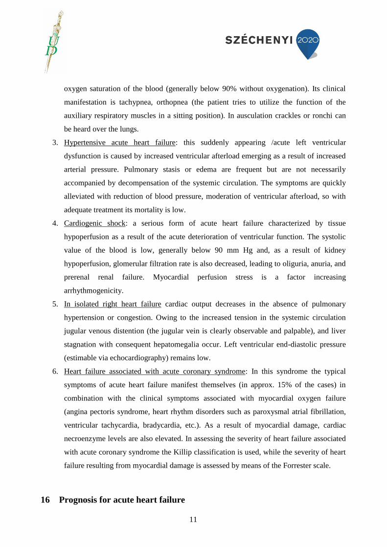
Aortic insufficiency is defined as the insufficient closing of the aortic valve, most frequently
caused by its stenosis (as a result of endocarditis or rheumatic carditis), but sometimes by aortic
root dilation, the pathological dilation of the structure supporting the valve (aorta ectasia). The
latter case is referred to as relative aortic insufficiency. It may also become manifest or
aggravated in acute heart failure. In aortic insufficiency a decrescendo murmur appears in the
diastole caused by the turbulance created by the blood flowing backwards through the
insufficiently closing valve.
Figure 7. Decrescendo diastolic murmur caused by aortic valve insufficiency, heard in 2R2.
If the anterior leaflet of the mitral valve is compressed by the blood, functional mitral
stenosis will occur, with the onset of a mesodiastolic-presystolic murmur known as the Austin-
Flint murmur.

19
Figure 8. Onset time (marked by arrow) of the Austin-Flint murmur caused by functional mitral
stenosis
In aortic insufficiency a number of further physical symptoms are observable, which can
facilitate a proper diagnosis. One is Corrigan’s pulse (celer et altus), another is the Musset-sign, a
rhythmic nodding of the head owing to a significant volume of regurgitating blood. In Quincke’s
capillary flow disorder alternating pulsation becomes visible after pressing and releasing the nail
on any of the fingers. A double sound, Traube double tone can be observed in auscultation over
the femoral artery. On pressing the phonendoscope, this may turn into a double murmur referred
to as the Duroziez murmur.
17.5. Ausculation – accessory heart sounds
The third extra heart sound is the ventricular filling sound, caused by the vibrations of the
ventricular wall in rapid filling. It is auscultated at the cardial apex and the IVth costal interspace
on the left side. If it appears at older age and does not recede in sitting position, it is termed
ventricular gallop or protodiastolic gallop.
The fourth extra heart sound is termed the atrial sound emerging from atrial systole, auscultated
at the cardial apex and the IVth costal interspace on the left side. It is also physiological in
childhood but pathological in adulthood because it appears primarily because of the pathological
expansion of the cardiac cavity. In acute decompensation auscultation of the systolic and
diastolic murmurs as well as of the 3rd
and 4th
heart sounds (S3 and S4) is necessary.

20
Figure 9. Sounds observable in auscultation of the heart: S1 systole, S2 diastole, S3 ventricular
filling sound, S4 sound emerging from atrial systole
17.6. Electrocardiography
Electrocardiography provides important information on heart frequency, heart rhythm, on the
presence of disturbances in the impulse generation and conduction system, and sometimes also
on their origin. ECG is suitable for identifying ischemic ST segment changes and for a
differential diagnosis of ST elevation myocardial infarction (STEMI) and non-STEMI.
It is important in the search for indications of hypertrophy, bundle branch block, electric
asynchrony, prolonged repolarization (QT interval, QT dispersion, U wave), arrhythmia or
pericarditis and myocarditis.
• Disturbances in impulse generation
• Disturbances in impulse conduction
• Depolarization
• Repolarization
• Rhythm / arrhythmia
• Ischaemia
• Ventricular hypertrophy

21
• Left and right heart load
• Pulmonary embolism
• Ionic disturbances
• Myocarditis/pericarditis
• Ion channel disorders
• Congenital heart defects
Table 3. Major types of information provided by electrocardiography
17.7. Chest X-ray
Chest-X ray is applicable in assessing the severity of pulmonary stasis in acute heart failure. It
can also provide information on the size of the heart or the presence of accompanying pulmonary
diseases (e.g. pneumonia, neoplasm, etc.). With patients in serious clinical condition the
information content of the X-ray scan can be of limited value.
17.8. Blood gas analysis
In arterial blood gas analysis oxygenation (pO2), respiratory function (PCO2) and the state
of the acid-base balance (pH) can be examined. In type 1 respiratory dysfunction hypoxia is
observed, while type 2 respiratory dysfunction is characterized by hypoxia and hypercapnia.

22
Figure 10. The results of blood gas analysis provide the partial pressure of oxygen and carbon
dioxide, the pH value of the blood, its lactate concentration, bicarbonate level, base excess (BE)
and oxygen saturation.
17.9. Laboratory tests
In acute heart failure the values of electrolytes, kidney function, cardiac necroenzymes,
blood count, liver enzymes and liver function can be of diagnostic and prognostic significance.
Hyponatremia, impaired kidney function (increase in urea and creatinine levels) mark the
severity and poor outcome of the disease. A slight increase in troponin values is nevertheless a
frequent phenomenon owing to increased intracardial pressure and consequent myocardial wall
stress and of the more frequent incidence of heart rhythm disturbances.
B type natriuretic peptides have a negative predictive value in acute heart failure and are suitable
for ruling out heart failure. Although there is no consensus regarding their normal value, an
increase in their level from admission to discharge is of prognostic significance.
17.10. Echocardiography
Cardiac ultrasound (M mode, 2D, Doppler) is an indispensable method of examination in
assessing the structural and functional status of the heart. The emergency treatment of patients
suffering from acute heart failure is essentially determined by the results of echocardiography, so
its urgent performance is fully justified. The test gives information on the size of the heart, size
of the cardiac chambers, the thickness of the ventricular wall, the state of the valves and the
anatomic properties of the pericardial space, the atrial and ventricular septum, the large vessels
(aorta, vena cava inferior, pulmonary tract). It can also give information on the global and
regional functional status of the left ventricle, on systolic and diastolic ventricular function
(Figure 11 and 12), and one can also check on the presence of asynchronous ventricular function,
a disturbance in wall motion affecting certain segments of the left ventricle. By determining flow
parameters, left ventricular end-diastolic pressure, right ventricular systolic pressure and
pulmonary artery pressure can be assessed. By determining average and peak gradients over the
valves, valve stenosis can also be quantitatively determined. This non-invasive method makes
hemodynamic monitoring possible, which can be of help in constantly checking the patient’s
state and in planning the therapy.
23
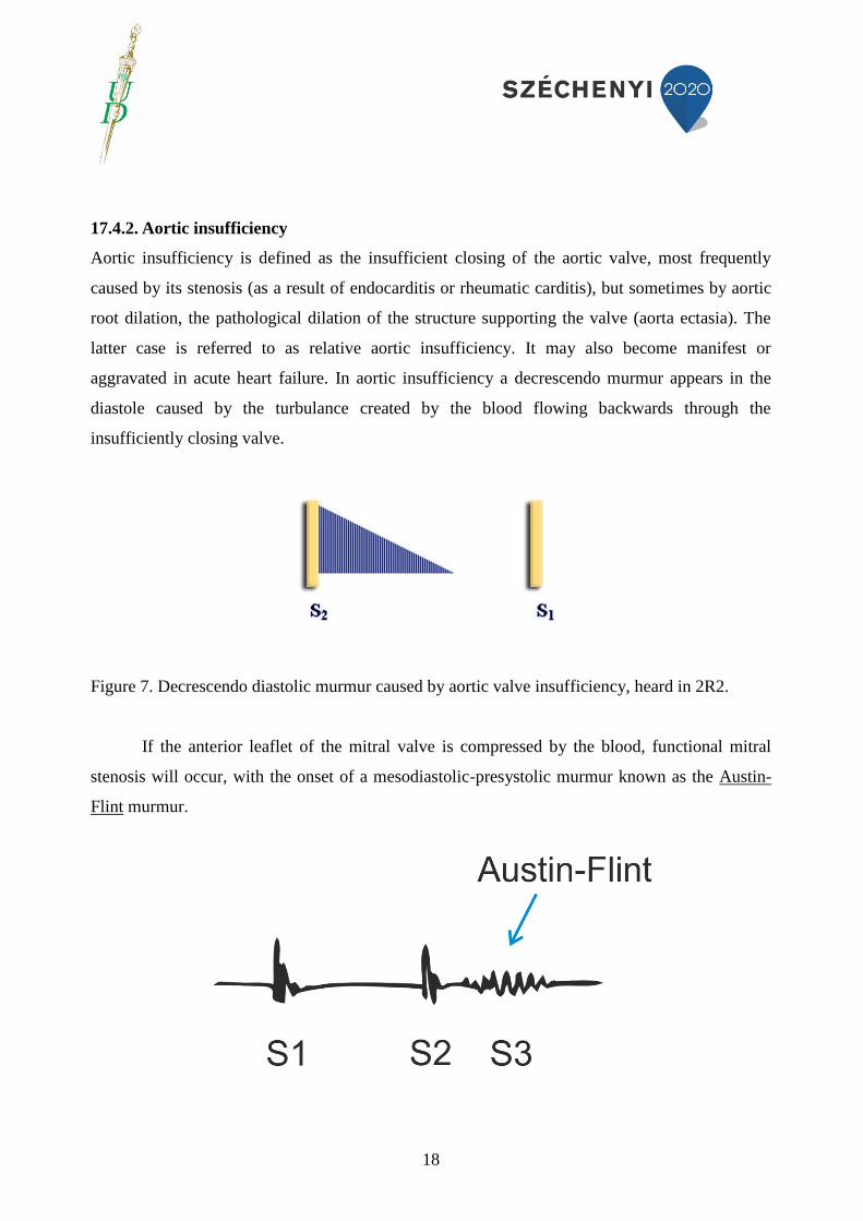
Figure 11. Left ventricular systolic function can be determined in different ways. With regard to
the volumetric method, systolic and diastolic ventricular volumes are determined from the apical
four chamber view. In the knowledge of these two readings the ventricular ejection fraction can
be calculated (Simpson method), which characterizes the systolic function and is a predictor of
cardiac mortality. In the Figure left ventricular diastolic volume (92ml) is shown.

24
Figure 12. In echocardiography left ventricle diastolic function is also determined. With the
pulsatile waveform Doppler method we can determine early diastolic mitral inflow velocity (E),
the maximum of flow velocity measured in atrial contraction in late diastole (A), as well as the
time between the peak and the end of the gradient of the E wave, termed deceleration time (DT,
marked in yellow in the Figure). In physiological cases E/A > 1, 0.8-1.5 depending on age, DT =
160-200msec. Criteria for 1st degree (mild) diastolic dysfunction (DD, relaxation disturbance):
E/A < 0.8, DT > 200 msec. Criteria for 2nd
degree (moderate) DD (pseudonormalization):
0.8 < EA < 1.5, DT = 160-200 msec. Criteria for 3rd
degree (severe) DD (restrictive function
disturbance): E/A < 2, DT <160 ms.
17.11. Multiparametric monitoring in heart failure
Monitoring of a patient with acute heart failure must be started immediately after
admission, or monitoring begun by the ambulance service must be continued in extended mode.
There are two types of monitoring. In non-invasive monitoring body temperature, oxygenation
(cf. pulsoxymetria and arterial bood gas analysis, cf. above), heart frequency, blood pressure,
electrocardiogram and volume of urination are monitored. In invasive monitoring manometry is
begun after the insertion of the arterial cannula, which provides continuous data of arterial blood
pressure. This is particularly justified if the patient displays symptoms suggesting hemodynamic
instability. The arterial cannula will also facilitate taking blood samples for the purposes of blood
gas analysis.
Figure 13. In arterial blood gas analysis the most frequent vessel punctured is the radial artery. In
frequent sampling, an arterial cannula is placed in the artery, through which blood samples can
be obtained several times a day. If the puncture or cannulation of the radial artery is not possible,

25
the brachial or femoral arteries can be used for the purpose. To prevent clogging, arterial
cannulae require frequent rinsing with heparin, which necessitates the use of a disposable
transducer to ensure overpressure.
A central venous catheter is applied when the need for constant volume arises, intravenous
medication is required, or when monitoring of central venous pressure or central venous
saturation is necessary. In some cases (therapy resistant edema formation), when
hemodiafiltration is necessary to reduce volume overload, the insertion of a double or triple
lumen catheter may be necessary in a great vein. The internal jugular vein, the subclavian vein or
the femoral vein can be used for the purpose.
Figure 14. To facilitate the effective therapy and monitoring of a patient with acute heart failure,
the insertion of a central venous catheter may become necessary. The subclavian vein is one of
the big veins that can be cannulated for that purpose. The end of the catheter is in the superior
vena cava or, in some cases, in the right atrium.
A pulmonary artery catheter is not frequently inserted in acute heart failure. Nevertheless, in
cases when both a pulmonary or cardiac primary disease needs to be considered as the cause

26
behind deteriorating condition, and the background mechanisms cannot be clarified by
echocardiography, this invasive monitoring technique can be clinically useful. Pulmonary
capillary wedge pressure is informative of left ventricle end-diastolic pressure, but in the case of
mitral valve stenosis, aortic insufficiency, pulmonary veno-occlusive state or elevated pulmonary
resistance and respiratory therapy the value may be affected.
18. The treatment of acute heart failure
18.1. Oxygen therapy
Improvement of arterial oxygenation is a primary aim in the therapy of a patient with acute heart
failure. The requirement is to attain 95% arterial oxygen saturation. The only exception from this
is the patient with confirmed chronic obstructive pulmonary disease, in which case a target value
of above 90% is acceptable to avoid hypercapnia.
Figure 15. To improve oxygenation, the use of a non-rebreather reservoir mask is recommended.
This device of oxygen therapy provides near 100% oxygen concentration on intake if the flow of
oxygen (10-15 liter/min.) is higher than the patient’s respiratory minute volume, the reservoir
balloon is full and the mask fits the patient’s face tight.
18.2. Respiratory treatment

27
There are two basically different procedures of ventilation. In non invasive ventilation (NIV) no
endotracheal intubation is performed. Instead, the patient’s breathing is assisted by means of a
special tight fitting mask secured on the face.
Figure 16. The special mask used in non invasive ventilation, secured on the patient’s face.
Positive end expiratory pressure ventilation (PEEP) is one of the cornerstones in the treatment of
pulmonary edema resulting from left ventricular insufficiency. In cardiogenic shock and right
ventricle insufficiency non invasive ventilation should be applied with caution since right
ventricular function can deteriorate. NIV improves clinical parameters and symptoms but
according to the findings of the 3CPO study it is neutral in terms of mortality. At the start of the
NIV therapy PEEP pressure is set to about 5-7.5 centimeters of water, which can be raised to 10
centimeters of water depending on clinical status. Invasive ventilation becomes necessary when
adequate oxygenation cannot be maintained in NIV therapy, or when the patient becomes
exhausted, hypercapnia develops or the general status of the patient deteriorates. In this case,
following anesthesia and relaxation, endotracheal intubation is performed and assisted or
controlled ventilation is applied depending on clinical status, with continuous monitoring of the
patient’s parameters.

28
Figure 17. In invasive ventilation an endotracheal tube is applied, which is passed into the
trachea of the anesthetized and relaxed patient with the help of a laryngoscope.
18.3. Medicinal treatment of acute heart failure
18.3.1. Morphine
Morphine (2.5-5 mg iv. bolus) can be administered if heart failure is accompanied by anxiety,
thoracic pain or dyspnoea since it can alleviate symptoms and improve the patient’s compliance.
Possible side effects are falling blood pressure, apnea, nausea, vomiting or disturbances in
impulse conduction (atrioventricular block, bradycardia), therefore close monitoring of the
patient is necessary.
18.3.2. Diuretics
One of the most important steps in treating congestive heart failure is the administration of
diuretics. A number of compounds belong in this group. The strongest diuretics are loop
diuretics. The most frequently used drug of this kind is furosemid, the initial intravenous dose of
which is 20-40 mg. Its overall dose must not exceed 240 mg in the first 2 hours. Mention can be
made of bumetanide (initial dose 0.5-1 mg) and torasemide (10-20 mg) as therapeutic
alternatives applicable in acute treatment. The efficacy of diuretics can be checked by measuring
the quantity of diuresis. For this reason bladder catheterisation and the precise collection of urine
may be necessary. In some cases of inadequate therapeutic effect loop diuretics can be combined

29
with diuretics of a different mechanism of action. The combined use of thiazide diuretics
(hydrochlorothiazide 25 mg) and loop diuretics is one of the possibilities. Aldosterone
antagonists (spironolactone and eplerenone) are very important in the treatment of heart failure
because, in addition to the diuretic properties, they are certified to have a beneficial effect on
cardiovasular outcome.
In the use of diuretics careful monitoring is important, since falling blood pressure, electrolyte
disturbances (frequently hyponatremia, hypokalemia), deteriorating kidney function (prerenal
kidney insufficiency) or in some cases hyperuricemia or neurohormonal activation can develop
as side effects.
18.3.3. Vasodilator compounds
The use of compounds of this type in the acute phase of heart failure is considered when the
patient’s systolic pressure is not lower than 90 mm Hg and/or obstructive valve insufficiency can
be ruled out (e.g. aortic stenosis), since these drugs reduce systolic pressure (without any
significant effect on diastolic pressure) and peripheral vascular resistance and thus reduce
intracardial end-diastolic pressure as well. With the reduction of left and right ventricular preload
and afterload ventricular function improves and pulmonary stasis is reduced without an increase
of oxygen demand in the myocardium.
The most significant representatives of this group of drugs are nitrates (e.g. nitroglycerin,
isosorbide mononitrate ISMN, dinitrate ISDN). Nitroglycerin can be repeated every 5 to 10
minutes in the form of spray (400 µg, 2 insufflations) or sublingually (0.25-0.5 mg) depending on
clinical status and blood pressure. ISDN can be administered buccally (1 or 3 mg). Intravenous
nitroglycerin (dosage: 10-20 µg/min., which can be increased by 5-10 µg/min. every 3-5 min. if
necessary) is an effective vasodilator and is very useful in the treatment of acute heart failure.
Intravenous nitroprusside and nesiritide (human recombinant form of B-type natriuretic peptide)
are also effective vasodilators. The initial dose of iv. nitroprusside infusion is 0.3 µg/kg/min.,
which can be increased to 0.5 µg/kg/min. The initial dose of iv. nesiritide is 0. 015 µg/kg/min.,
which can be increased to 0.03 µg/kg/min. In the administration of these drugs close monitoring
of blood pressure is necessary.
18.3.4. Inotropic agents
18.3.4.1. Dopamine and dobutamine

30
If the clinical status of the patient does not improve in spite of the application of diuretics
and vasodilators, the administration of inotropic agents becomes necessary. The use of these
drugs may be justified in persisting hypotension and low cardiac index, therapy refractory
congestion in the pulmonary or systemic circulation and clinical onset of tissue hypoperfusion.
Their application is restricted by the fact that they increase the hazard of atrial and ventricular
rhythm disorders, therefore in the administration of these compounds close monitoring of the
patient is necessary. It may be necessary to perform echocardiography when this kind of therapy
is initiated, since these drugs will primarly bring significant clinical benefit in patients with
hypokinetic, dilated left ventricles resulting in decreased ventricular functions. In cardiogenic
shock positive inotropic agents may stabilize the circulation of a critical patient and the
therapeutist can gain time for further invasive therapy (intraaortic balloon pump, ventricular
assist device, heart transplant surgery). Dobutamine increases ventricular contractility by
activating beta 1 receptors. Its doses can only be increased gradually: initially 2-3 µg/kg/min.
which can be titrated to 15 µg/kg/min., although along with beta blocker treatment the therapy
rate can be increased up to 20 µg/kg/min. It must be emphasized that the action of dobutamine on
ventricular function highly depends on the dosage applied. In finishing the therapy a slow
reduction of the dose is necessary. Dopamine leads to an increase in myocardial contractility via
stimulating beta adrenergic receptors. Its effect is dose dependent, diuretic in small doses. In
higher doses it increases systolic pressure, while in the case of increased doses a tendency for
arrhythmia and results in vasoconstriction owing to alpha adrenergic stimulation may appear.
Low doses of dopamine are often administered in combination with high doses of dobutamine.
18.3.4.2. Calcium sensitizing
Levosimendan, which belongs to the group of drugs defined as calcium sensitizers, binds
to troponin C and increases the contraction potential of the heart muscle. By means of
mechanisms mediated by ATP sensitive potassium channels it is an effective vasodilator and also
has a phosphodiesterase inhibitor function. In intravenous application levosimendan decreases
pulmonary pressure and peripheral vascular resistance, reducing preload and afterload. This
mechanism also contributes to its effect of increasing cardiac output and stroke volume.
Administration normally begins with a bolus loading dose (3-12 µg/kg in 10 minutes) and
continued a further 24 hours at a rate of 0.05-0.2 µg/kg/min. in a perfusor. Possible side effects
are hypotension and tachycardia, appearing most frequently subsequent to the loading dose. If the

31
patient’s initial systolic pressure is lower than 100 mm Hg, the administration of levosimendan
must be started without the initial loading dose to avoid or alleviate the side effects. The
hemodynamic effect of levosimendan is retained for several days after the end of the treatment.
18.3.4.3. Phosphodiesterase inhibitors
Milrinone and enoximone are type III phosphodiesterase inhibitors (PDEI), which prevent
decomposition of cyclic AMP and thus lead to an increase in inotropy and peripheral
vasodilation. By decreasing pulmonary arterial pressure and peripheral vascular resistance, they
decrease ventricular preload and afterload, thus decreasing the load exerted on the ventricles.
Cardiac output consequently increases and stroke volume improves as well. After an initial bolus
they are administered in a perfusor. Since in coronary disease they may increase mortality, they
should only be used with caution in this syndrome.
18.3.4.4. Vasopressin antagonists
Among vasopressin receptors, V1a receptors are responsible for vasoconstriction, while the V2
receptors found in the kidneys are responsible for the reabsorption of water. The most important
representatives of this group of drugs are conivaptan, a double action V1a/V2 receptor inhibitor,
and tolvaptan, a selectie V2 receptor inhibitor. Although by the results of the EVEREST study
tolvaptan had a beneficial effect on the symptoms of heart failure, it did not have a significant
effect on mortality.
18.3.4.5. Vasopressor compounds
The most important and most widely used member of this group is norepinephrine, to be used in
cardiogenic shock. It can be administered when the inotropic agents discussed above and volume
compensation (250 ml/10mins.) are no longer capable of stabilizing the patient’s circulation and
systolic pressure cannot be maintained above 90 mm Hg. In joint administration with dopamine
the vasopressor action of the two drugs can be combined, which may induce tissue perfusion
disorders. The other important member of this group is epinephrine, but its administration is only
justified in cardiopulmonary resuscitation, with strict observation of the regulations concerned.
18.3.4.6. Cardiac glycosides

32
These are glycosides with a similar structure which occur in some plants and in the skin of some
of the Anura. The best known are digoxin and digitoxin. Their therapeutic action is based on the
inhibition of the Na+-K+-ATPase present in the membranes of the myocardial cells. Owing to
blockage of the ion pump, intracellular Na+ concentration increases and as a result the Na+-Ca2+
exchange mechanism in the sarcolemma also increases. This mechanism changes intracellular
Na+ to Ca2+ and indirectly increases intracellular Ca concentration. As a result of these
processes contractility and the membrane potential of the cell increases. In addition, these agents
increase parasympathetic tone, have a negative chronotropic and dromotropic effect and also
decrease sympathetic tone. In acute heart failure they increase cardiac output and decrease
ventricular end-diastoloic pressure. According to the findings of the DIG survey, however, they
do not have an effect on long term mortality. Their clinical significance is enhanced by their
effect of decreasing ventricular frequency in atrial fibrillation. In kidney function disorder it may
be necessary to reduce the dose of digoxin, a drug with renal metabolism, or to dismiss it
altogether. In these cases digitoxin, which decomposes in the liver, can be a therapeutic
alternative.
Figure 18. In the action of digitalis, intracellular Na+ concentration increases owing to inhibition
of the Na+-K+-ATPase in the membranes of myocardial cells. As a result, the Na+-Ca2+

33
exchange in the sarcolemma also increases, leading to an increase in intracellular Ca
concentration.
19. Special considerations in the treatment of acute cardiac failure
In the first phase of the patient’s emergency treatment oxygenation is indispensible. For this
purpose oxygen must be administered nasally, through a mask, or by non invasive ventilation
(NIV). In acute decompensation of chronic heart failure a combination of vasodilators and loop
diuretics is necessary. Positive inotropic agents are only used if symptoms of persisting
hypotension and tissue hypoperfusion are observed. It may become necessary to reduce the doses
of or temporarily discontinue previously applied beta blockers. ACE blockers and ARBs should
be applied as early as possible. In pulmonary edema administration of morphine may be
necessary, especially if the patient has anxiety, dyspnoea or is in pain. If oxygenation cannot be
maintained by non invasive ventilation, endotracheal intubation and mechanical ventilation is
necessary. In hypertension the use of vasodilators and early application of diuretics is necessary.
In hypotension and tissue hypoperfusion positive inotropic agents are necessary. If the patient is
in cardiogenic shock, volume compensation (250 ml/10 mins.) and positive inotropic agents are
necessary until systolic pressure exceeds 90 mm Hg. If medicinal therapy remains unsuccessful,
mechanical circulatory support (e.g. intra aortic balloon pump (IABP) or ventricular assist device
(VAD)) may become necessary. In right ventricular insufficiency volume compensation and
positive inotropic agents are necessary. Mechanical ventilation can impair the patient’s state. In
acute coronary syndrome, subsequent to emergency echocardiography, primary coronary
intervention is recommended. If this cannot be performed in time, on diagnosis of STEMI
systemic thrombolysis is to be applied. In cardiogenic shock appearing as a result of acute
coronary syndrome, primary coronary intervention is necessary. If medicinal and cathether
therapy is unsuccessful, mechanical circulatory support (e.g. IABP) may become necessary.

34
Table 4. Emergency treatment protocol for a patient with acute heart failure.

35
20. References
ESC Guidelines for the diagnosis and treatment of acute and chronic heart failure 2012.
European Heart Journal (2012) 33, 1787–1847 doi:10.1093/eurheartj/ehs104
Joseph SM, Cedars AM, Ewald GA, Geltman EM, and Mann DL. Acute decompensated
heart failure: contemporary medical management.Tex Heart Inst J. 2009;36(6):510-20
Nyolczas Noémi: A Magyar Kardiológusok Társasága ajánlása „Az akut és krónikus
szívelégtelenség diagnózisa és kezelése 2008” címû ESC guideline-hoz. Cardiologia
Hungarica 2010; 40 :C1–C2
Kanu Chatterjee, Mark Anderson, Donald Heistad, Richard E Kerber: Manual of Heart
Failure. Jaypee Brothers Medical Publishers Ltd, 2014
Mark H Beers, Robert Berkow (Eds) MSD Orvosi Kézikönyv, Melánia Kiadó Kft, 1999,
pp 1682-1710
Tomcsányi János (szerk): Klinikai Kadiológia, Medintel Könyvkiadó, 1997, pp. 167-187.
Michael H. Crawford (Editor) Current Diagnosis and Treatment, Cardiology, 3rd
International Edition McGraw Hill, 2009, pp 204-232.
Lengyel Mária, Asbóth Richárd: Echocardiographia, Medicina Könyvkiadó, 2012, pp.
69-108.

36
21. Self-controlling questions
1. What is the definition of cyanosis?
Cyanosis is the bluish discoloration of the skin and mucus mebranes, caused by hypoxia
and the consequently elevated reduced hemoglobin concentration.
2. What are the most important causes of central cyanosis?
Congenital heart diseases
heart failure
Impaired breathing function
Decreased arterial oxygen saturation
Decrease in air pressure
3. What are the most frequent causes of sudden left ventricular failure?
Hypertension
Arrhythmias (e.g. paroxysmal supraventricular tachycardia, atrial fibrillation)
Acute coronary syndrome
4. What are the target levels in the case of blood gas analysis?
Oxygen saturation above 90 %
Oxygen pressure above 60 mmHg
5. Ho can left ventricular ejection fraction be measured?
With the determination of systolic and diastolic left ventricaular diameters during
echocardiography
Volumetric method by means of echocariography
6. Which are the effective drugs in the case of acute heart failure?

37
• Morphine
• Nitrate
• Diuretics
• Digitalis
• Levosimendan
• Saline infusion and norepinephrine in cardiogenic shock
Dopamine
• Dobutamine
22. Case reports
Case 1.
A middle-aged male patient was carried to the emergency by the ambulance due to rest dyspnea,
prolonged expiration, cyanosis, sweating, and paleness. Initial blood pressure was measured to be
80/60 mmHg, furthermore pulse oximetry proved a decrease in oxygen saturation (75%). The
latter improved to 95 % after the administration of oxygen. Body temperature was found to be
36.2 celsius. During the physical examination of the lungs bibasal crackles were audible,
expirium was prolonged. Moreover, peripheral pulse was measured to be 170/min, although it
was regular, but compressible.
Question: What is the possible diagnosis?
Answer: Acute left ventricular failure caused by arrhythmia.
Question: Which examinations would you perform to clarify the diagnosis?
Answer: Physical examination, ECG, arterial blood gas analysis, chest X-ray, laboratory tests
(cardiac necroenzymes), continuous pulse oximetry.
Question: Which type of arrhythmia can cause such symptoms?
Answer: Regular and rapid ventricular response can be caused by atrial tachycardia, atrial flutter,
atrioventricular node dependent reentry tachycardia (PSVT) and ventricular tachycardia.

38
Question: ECG reveals regular and narrow QRS complexes and “f” waves in every lead. What
can be the underlying arrhythmia?
Answer: Atrial flutter
Question: What is the most effective and recommended therapy in the case of atrial flutter with
hemodynamic deterioration?
Answer: Acute electrical cardioversion.
Case 2.
A 56 year-old male patient has been smoking for 20 years. He has been diagnosed with type 2
diabetes mellitus 10 years ago. He was carried to the emergency due to compressing chest pain,
tingling of the left arm, paleness, progressive dyspnea and cyanosis which have appeared 2 hours
ago.
Question: What is the most appropriate diagnosis?
Answer: Acute left ventricular failure sue to acute coronary syndrome.
Question: How would you clarify the diagnosis?
Answer: By means of ECG and laboratory tests.
Question: You find a significant elevation in troponin and CK levels, furthermore 3 mm ST
segment elevations are observed in the ECG leads V2-6. . What would be the most important to
be done?
Answer: Percutaneous coronary intervention. As a primary care: oxygen, morphine, nitrate,
clopidogrel, aspirin, heparin and due to pulmonary congestion diuretics should be administered.
Case 3.
39
An 80 year-old female patient is transported to the emergency due to progressive dyspnea,
headache, vertigo and tinnitus provoked by psychogenic stress. She has not been taking her drugs
regularly lately.
Question: What is the most appropriate cause of these symtomps??
Answer: Left ventricular dysfunction and encephalopathy due to hypertension..
Question: How would you clarify the diagnosis?
Answer: I would measeure blood pressure and complete physical examination..
Question: What are the main therapeutic possibilities?
Answer: The asministration of ACE inhibitors, diuretics, nitrate, oxygen, and if it is necessary
intravenous urapidil. The sudden decrease in blood pressure should be avoided.
Case 4.
A 70 year-old female patient is carried to the emergency due to edema in the legs, rest dyspnea,
and tachycardia. Anamnestic data include hypertension and ischemic heart disease. During the
physical examination of the patient cyanosis can be ebavulated. Moreover, during the percussion
of the lungs a significant dullness is found on the right basis.
Question: Whar can be the cause of these symptoms?
Answer: Heart failure with intrapleural effusion on the right side, edema formation and
concomitant hypoxemia.
Question: How would you clarify the diagnosis?
Answer: I would evaluate the structural and functional status of the heart by means of
echocardiography. Furthermore, blood gas analysis could reveal the level of hypoxia and
laboratory tests could exclude acute myocardial infarction (myocardial cell necrosis). The
severity of pleural effusion should be clarified with chest X-ray.

40
Case 5.
A middle-aged male patient is carried to the emergency due to hypokalemia. The electrolyte
imbalance was caused by diarrhea. During his observation suddenly a regular, wide QRS
tachycardia occurs with the ventricular frequency of 180/min. It is accompanied by hypotension
(RR 90/70 mmHg), cyanosis, increasing rest dyspnea and strong compressing chest pain.
Question: What can cause these symptoms?
Answer: It is possibly caused by ventricular tachycardia with hemodynamic deterioration (acute
heart failure, coronary perfusion defect, myocardial ischemia).
Question: What therapy would you chose?
Answer: Acute electrical cardioversion should be chosen.
41
23. Tests
23.3. True or false questions
Please underline the letter T if the answer is true and letter F if it is false. More true answers are
accepted!
1. TF Cyanosis is caused by the improvement in oxygenation
2. TF Central cyanosis is always caused by a peripheral circulatory insufficiency
3. TF Rcuced hemoglobin concentration is above 40 g/L in the case of cyanosis
4. TF Acute heart failure may lead to acute cyanosis
1. TF Cannulation of the radial artery is suitable for arterial blood gas investigation
2. TF The femoral artery is not suitable for blood gas analysis
3. TF Blood gas analysis is not suitable for determining PH
4. TF Hemoglobin concentration is calculated during pulse oximetry
1. TF Perfipheral cyanosis occurs only in heart failure
2. TF The rupture of the interventicular septum may cause acute heart failure
3. TF Clubbing of the nails can appear only in heart failure
4. TF Arrhytmia cannot cause heart failure
1. TF Hypertension does not cause ventricular strain
2. TF Myocardial infarction always leads to acute heart failure
3. TF Pulmonary edema is the leading symptom of right ventricular failure
4. TF Pulmonary edema is caused by left ventricular failure
1. TF Central cyanosis never occurs in heart failure
2. TF Cyanosis does not appear in congenital herta diseases

42
3. TF Cyanosis can apperar in the case of intrapulmonary shunts
4. TF Abnormal hemoglobins cause peripheral cyanosis
23.4. Multiple-choice questions
Please underline the correct answers! More answers may be true.
Congenital heart diseases
Impaired pulmonary or breathing function
Normal arterial oxygen saturation
Cold weather
Please underline the correct answers!
Pulse oximetry is often used during the postoperative period in the intensive care and
emergency units
If oxygen saturation is below 90 %, there is no more therapy indicated
Oxygen saturation above 90 % is a therapeutic goal
Movement does not affect pulse oximetry
Please underline the correct answers!!
Heart failure does not cause cyanosis
Peripheral and central cyanosis may occur together in severe heart failure
Hypotension leasdt to a sudden increase in ventricular afterload
Paroxysmal atrial fibrillation can provoke heart failure
Underline the correct answers!
Diuretics are first line therapeutic tools in heart failure
The administration of levosimendan is not recommended in acute heart failure
Verapamil improves ventricular function

43
The administration of digitalis is advantegous in atrila fibrillation with high ventricular response
Which of these do not cause cyanosis?
Obstructive pulmonary disease
External otitis
Reiter’s disease
Acute heart failure
23.5. Single-choice questions
Please underline the correct answer!
1. ACE inhibitors never cause dry cough
2. In the case of hypertensive heart failure the lowering of blood pressure is indicated
3. Diuretics do not cause electrolyte imbalance
4. Beta blockers worsen mortality in patients with heart failure
1. Abnormal hemoglobins do not affect tissue oxygenation
2. Intrapulmonary shunts do not cause abnormal oxygenation
3. Digitalis can cause proarrhythmia
4. Central cyanosis appears only in the case of pulmonary disease
1. Furosemide is an aldosterone antagonist drug
2. During pulse oximetry oxygen saturation of 80 % has to be achieved
3. Oxygen pressure below 60 mmHg means oxygenation abnormality
4. Loop diuretics have weak diuretic effect
1. In the case of liver dysfunction the administration of digitoxin is recommended

44
2. In the case of renal dysfunction digoxin may be cumulated even in therapeutic doses
3. Oxygen saturation above 90% indicates an immediate respiratory treatment
4. During NIV endotracheal intubation is performed
1. Before repiratory treatment endotracheal intubation may be needed
2. Cannulation of a big vein is not indicated in acute heart failure
3. Central cyanosis commonly occurs in peripheral arterial disease
4. The edge of a vena subclavia cannula has to be put to the left atrium
23.6. Relational analysis
A: Both statement and justification are true and the latter well explains the statement.
B: Both are true, but the justification do not explain the statement.
C: Statement is true, but justification is false.
D: Statement is false, but justification is true.
E: Both are false.
Ejection fraction is suitable for the evaluation of ventricular function, thus
echocardiography can help in the diagnosis of heart failure (A)
Furosemide has a weak diuretic effect, so its use is optional in acute heart failure (E)
Acute myocardial infarction can cause acute heart failure, thus everey patient with heart
failure should be treated in a coronary unit (C)
Digitalis decreases inotropy and improves life expectancy (E)
Arrhythmia never causes heart failure, thus ECG should not be performed during the
emergency evaluation of a patient (E)