Embed Size (px)
Citation preview
Medical Hypotheses 81 (2013) 643–649
Contents lists available at ScienceDirect
Medical Hypotheses
journal homepage: www.elsevier .com/locate /mehy
Embryogenesis, morphogens and cancer stem cells: Putting the puzzletogether
0306-9877/$ - see front matter � 2013 Elsevier Ltd. All rights reserved.http://dx.doi.org/10.1016/j.mehy.2013.07.021
⇑ Tel.: +48 61 829 58 55.E-mail addresses: [email protected] (A. Fontana), [email protected]
(B. Wróbel).
Alessandro Fontana ⇑, Borys Wróbel *
Evolving Systems Laboratory, Faculty of Biology, Adam Mickiewicz University, ul. Umultowska 89, 61-614 Poznan, Poland
a r t i c l e i n f o a b s t r a c t
Article history:Received 20 May 2013Accepted 6 July 2013
This paper describes a model which puts together three key elements of cancer theory: the analogiesbetween embryogenesis and carcinogenesis, the role played in both processes by morphogens andrelated pathways, and the recently emerged paradigm of cancer stem cells. The model is called EpigeneticTracking. Originally conceived as a model of embryonic development, it was later extended to interpretother aspects of biology, such as the presence of junk DNA, the phenomenon of ageing and the process ofcancer formation. In this work we deepen our vision of carcinogenesis, and propose a novel hypothesis onthe role of morphogen-processing pathways. According to the hypothesis, the interplay of these path-ways leads in stem cells to the production of new transcription factors, which act as drivers of cellulardifferentiation. The disruption of these pathways, caused by mutations in specific genes, would representthe first and most distinctive event in the carcinogenic process. Our hypothesis allows us to make testablepredictions on patterns of gene mutations involved in carcinogenesis. Our hypothesis also suggests thatcancer stem cells can stay dormant until they are activated in a process that resembles activation of stemcells during tissue repair or at a specific time during development.
� 2013 Elsevier Ltd. All rights reserved.
1. Introduction
Embryogenesis and cancer share many features: growth andmovement of cells, morphogenesis and cellular differentiation.These macroscopic behaviours appear to be mediated by commonmechanisms at the cellular level. Morphogens (e.g., Hedgehog, No-dal, Notch, Wnt) are a class of compounds known to play a role indevelopment, and involved in cancer formation. During develop-ment, they spread from localised sources and form concentrationgradients, which can be used by cells to induce the expression ofspecific genes. Distinct combinations of morphogen concentrationsin different regions of the embryo induce the emergence of distinctcell types through a characteristic ‘‘morphogenetic code’’ [12].Each morphogen is processed by a dedicated pathway, which startswhen a ligand binds a receptor on the cell surface, proceeds withthe generation of a cytoplasmic signal, and ends in the nucleus,where the induced signal participates in the regulation of targetgenes. The same pathways appear disrupted or abnormally acti-vated in cancer [5,13–15,18–20].
Stem cells are pluripotent cells able to give rise to all cell typesof the body during embryonic development, and to coordinate therepair of damaged tissues. Stem cells reside in ‘‘niches’’, where they
require specific signals for the maintenance of the stem state [23].A recently emerged theory [1] considers cancer stem cells (CSC)responsible for tumour formation and growth. CSC share character-istics with normal stem cells, specifically the ability to give rise tomultiple cell types, through the processes of self-renewal and dif-ferentiation. In recent years, new CSC models have been proposed,e.g., the ‘‘complex system model’’ [17], the ‘‘dynamic stemnessmodel’’ [22], and the ‘‘stemness phenotype model’’ [3]. These mod-els share the ideas that all cells in a tumour are potentially tumor-igenic, and that stemness is a dynamic property, both in tumoursand in healthy tissues. The stem-non stem conversion would occurin both directions, triggered by genetic and epigenetic factors, andinfluenced by the cellular microenvironment.
Epigenetic Tracking (ET) is the name of a cellular model, firstdescribed in [6]. Originally conceived as a model of embryonicdevelopment, it was later used to interpret other key biologicalphenomena, such as ageing, cancer and the hypothetical transferof genetic elements from soma to germline [7–10]. The modelwas recently extended to include the action of morphogens diffus-ing in the embryo [11]. While the study of life sciences often relieson a bottom–up method, that tries to infer general rules startingfrom genes and proteins, our work is informed by a top–down ap-proach, that proceeds from high-level abstractions (validatedthrough computer simulations) towards a proposal for biological,low-level molecular processes. Following this philosophy, in thiswork we continue the investigation on the topic of cancer, whichwas initiated in [8], and propose a vision of biology which inte-

644 A. Fontana, B. Wróbel / Medical Hypotheses 81 (2013) 643–649
grates embryogenesis, cancer, morphogens and stem cells into acoherent theoretical framework.
2. The model
In ET artificial bodies are composed of two categories of cells:stem cells (called driver cells in our previous papers) and normalcells. An identical genome is associated to all cells. The genome iscomposed of developmental genes having a left part, which containsthe conditions for gene activation, and a right part, which encodesthe actions performed upon gene activation. Cells have also a var-iable called mobile code (MOC), which can take different values indifferent cells and represents the main source of differentiationduring development. A given MOC value can be shared by manynormal cells, but each stem cell has a distinct MOC.
Development starts from a single cell and unfolds in a prede-fined number of developmental stages, counted by a variable calledglobal clock, shared by all cells. Stem cells direct the developmentalprocess. We hypothesise that, if a stem cell reaches a particularstate at a particular time, a specific change event is orchestrated.As an abstraction of this process, we use the match between theMOC and another variable called regulatory set (RES), which be-longs to the left part of developmental genes, and between theclock and the timer, also a field of the left part (Fig. 1).
The change event that takes place once the match occurs is en-coded in the right part of the developmental gene. Stem cells cancause either a large-scale apoptosis (death of a large number of cellsin the volume around a stem cell), or a proliferation event, whichcorresponds to several rounds of asymmetric cell divisions (i.e. thestem cell produces another identical stem cell and a normal cell).An apoptosis event may result in the deletion of cells of both catego-ries, while a proliferation event initially produces a large number ofnormal, plastic cells in the volume (Fig. 1, panel A). All normal cellsgenerated inherit the genome and the MOC of the stem cell whichgave rise to the proliferation (called mother cell).
Stem cells in our model correspond to biological stem cells, andmay play a role similar to that of biological ‘‘organiser’’ (such as theSpemann’s organiser [4]). The clock and the MOC can be inter-preted in biology as the set of all regulatory factors present inthe cell: for simplicity, we can think of them as a set of proteinsacting as transcription factors. The timer and the RES correspondto the gene regulatory sequences to which the factors can bind(Fig. 2). In our software implementation the clock and the timerare represented as numbers, while the MOC and the RES are repre-sented as sequences of numbers (each number in the sequence canbe interpreted as a transcription factor or a regulatory locus).
Fig. 1. Change events in ET. The figure shows an example of a proliferation event triggereclock value is 6).
While most normal cells generated during a proliferation eventbecome terminally differentiated cells, there are some cells which,instead of proceeding on their differentiation path, revert to stemcells (Fig. 1, panel B). Hence, in ET the stem-normal transition isa two-way street and may correspond to a fundamental principlepresent also in nature, that induces cellular systems to self-orga-nise in a hierarchical fashion. We think of this biological hierarchicalprinciple as a pervasive mechanism underlying all manifestationsof cellular life, in both physiological and pathological conditions.
The new stem cells generated from normal cells receive a newand unique MOC, to allow for differentiation. If in the genome adevelopment gene exists whose RES matches the MOC of a newstem cell, this cell can become the centre of another event in a sub-sequent stage, and development can move ahead. Thus, in ET,development is the outcome of a sequence of change events, eachorchestrated from a certain stem cell, by a given gene, at a precisemoment.
The set of stem cells and MOCs generated during developmenthas a hierarchical, or tree, structure (Fig. 3). This descends from thefact that each stem cell originates from the conversion of a normalcell, which has a single mother. This property, which is true for sin-gle body parts and for the whole body, appears to be coherent withthe information we have on the set of stem cells involved in thegeneration of particular organs or systems, such as the hematopoi-etic system [21]. Our model provides also a means to bridge theconceptual gap between embryonic and adult stem cells: embry-onic stem cells correspond to elements in the tree near the root,while adult stem cells correspond to the leaves of the tree. It alsoexplains how adult stem cells are generated, with the mechanismof conversion from normal cells.
The model of development described, coupled with a GeneticAlgorithm able to simulate Darwinian evolution, becomes an evo-devo process, which has been extensively validated through com-puter simulations. Our in silico experiments showed how this pro-cess is able to generate 3-dimensional structures with a complexity(measured in terms of number of cells – reaching millions – and le-vel of fine details) unmatched by any other model of embryogene-sis [11]. These experiments cannot replace traditional in vitro orin vivo experiments, yet they can provide an indirect support forour ideas.
3. The role of morphogens
In ET the process of cellular differentiation is achieved throughthe presence of simulated morphogens [11], acting with the fol-lowing mechanism. Some stem cells (Fig. 4) become sources of
d by the stem cell with the MOC labelled with A at developmental stage 6 (when the

Fig. 2. Biological MOC, RES, clock and timer. The figure shows the components of a biological MOC and a biological clock (transcription factors) binding to the components ofa biological RES and a biological timer (regulatory sequences of a gene). As a result, the gene is activated. In this example, only one biological gene is shown for simplicity. Ingeneral, a biological MOC comprises transcription factors (as a biological RES and timer comprise regulatory loci) relevant to several biological genes.
Fig. 3. Set of stem cells created during development. Stem cells marked in green become active during development. As a result, they create normal cells and induce theirdifferentiation. In parallel, other stem cells are created. (For interpretation of the references to colour in this figure legend, the reader is referred to the web version of thisarticle.)
A. Fontana, B. Wróbel / Medical Hypotheses 81 (2013) 643–649 645
specific morphogens (of k distinct types: M1, . . . Mk). These signalspartition the space into regions, each characterised by a distinctcombination of concentration values. For example, all cells in a gi-ven region ‘‘sense’’ morphogen of type M1 with concentration 7,morphogen of type M2 with concentration 2, etc. (concentrationvalues are integer numbers which represent categories or rangesof actual concentration values, expressed in mg/ml). In each regiona normal cell is selected and turned into a stem cell.
To each new stem cell a new and unique MOC must be assigned.When the stem cell is created, it has the same MOC as the normalcell from which it was obtained, which coincides with the MOC ofthe normal cell’s mother. The assignment of a new MOC is achievedthrough the processing of the signals received by a device calledMOC generator. The result is a new number which is appended atthe end of the existing MOC, to produce the new MOC for the stemcell (Fig. 5). In biological terms, this corresponds to producing anew transcription factor, which is added to the repertoire of factorsalready present in the cell. Fig. 6 shows an example of develop-
Fig. 4. Morphogens. The panel on the left shows a hypothetical distribution of stem celltypes, which diffuse across the space. On the right, the different combinations of the conwith a different colour). In each such region a new stem cell is created from a normal cellreader is referred to the web version of this article.)
ment for a hypothetical ‘‘organ’’, which alternates the occurrenceof change events and the creation of new stem cells based on themechanism described.
We hypothesise that the MOC generator, which supervises thecreation of the new element for the MOC, is implemented in natureby all pathways dedicated to the processing of biological morpho-gens. According to this hypothesis, each pathway senses the con-centration of a morphogen, and sends a signal into the nucleus.Here the interplay of signals conveyed by all morphogen pathwaysresults in the production of a new transcription factor (or morethan one factor). For example, if the combination of morphogenconcentrations is [2130] (e.g. Hedgehog is received with concen-tration 2, Nodal with concentration 1, Notch with concentration3 and Wnt with concentration 0), transcription factor X isproduced. If the combinations of the same concentrations is[3110], transcription factors Y is produced. The new transcriptionfactor is added to the repertoire of factors already present in thecell, and concur to determine the cellular state. The particular
s. Some of these stem cells (dashed circles) emit morphogens, of a finite number ofcentration values of the morphogens partition the space into regions (marked each(red circles). (For interpretation of the references to colour in this figure legend, the

Fig. 5. Generation of a new MOC for a newly formed stem cell in ET. Morphogens, released by some stem cells (indicated with dashed circles in the corners), reach the MOCgenerator. The output of this device is a new number (corresponding to a new transcription factor), which is added to the repertoire of numbers already present in the cell.The MOC generator is hypothesised to be implemented in nature by the genetic pathways dedicated to the processing of morphogens.
Fig. 6. Stem cells drive organogenesis in ET. The figure shows the development of a hypothetical ‘‘organ’’ in two stages, carried out by five developmental genes (stem cellsare marked in yellow, normal cells just created -plastic- in orange, normal differentiated cells are shown with different colours, each corresponding to a particulardifferentiation state). The right part of the figure reports the MOCs generated during development, which are organised in a tree-like structure. Each stage is shown in twopanels, A and B. Panel A shows the occurrence of one or more change events, panel B shows the creation of new stem cells. This process is based on morphogens emitted by asubset of stem cells, which partition the space in regions. One normal cell in each region is turned into a stem cell, and obtains a distinct MOC. (For interpretation of thereferences to colour in this figure legend, the reader is referred to the web version of this article.)
646 A. Fontana, B. Wróbel / Medical Hypotheses 81 (2013) 643–649
combinations of morphogen concentrations necessary for MOCgeneration may correspond to the set of signals required to main-tain stem cells in niches [23].
Once a new stem cell is created and obtains a new MOC, it canbecome the origin of another change event, if in the genome a geneexists, whose RES can match the stem’s MOC. Stem cells generatedin an organism’s lifetime can be divided in three categories. Somestem cells are activated during development, defined as the periodbefore reproduction, and contribute to influence the organism’sreproductive fitness. After activation, they persist in the body ina dormant condition, and can be reactivated to regenerate dam-aged tissues or worn-out body parts. Other stem cells are activatedafter the end of development, producing change events in what wecalled the ‘‘ageing’’ period (i.e. after reproduction) [7]. Finally, thereare stem cells (the vast majority) which are never activated.
The stage for a dangerous scenario is set if a fault arises in astem cell destined to be activated in the ageing period, affectingthe mechanism used by the cell to generate new MOCs. Let us as-
sume that in a stem cell (which we call mother cell) the MOC-RESmatch results in a proliferation event that creates new normalcells, some of which are later turned into stem cells (which wemay call daughter cells). The MOC generation device used by thedaughters is inherited from their mother: if it is damaged, thedamage will be present also in the daughters, affecting their capac-ity to generate new MOCs.
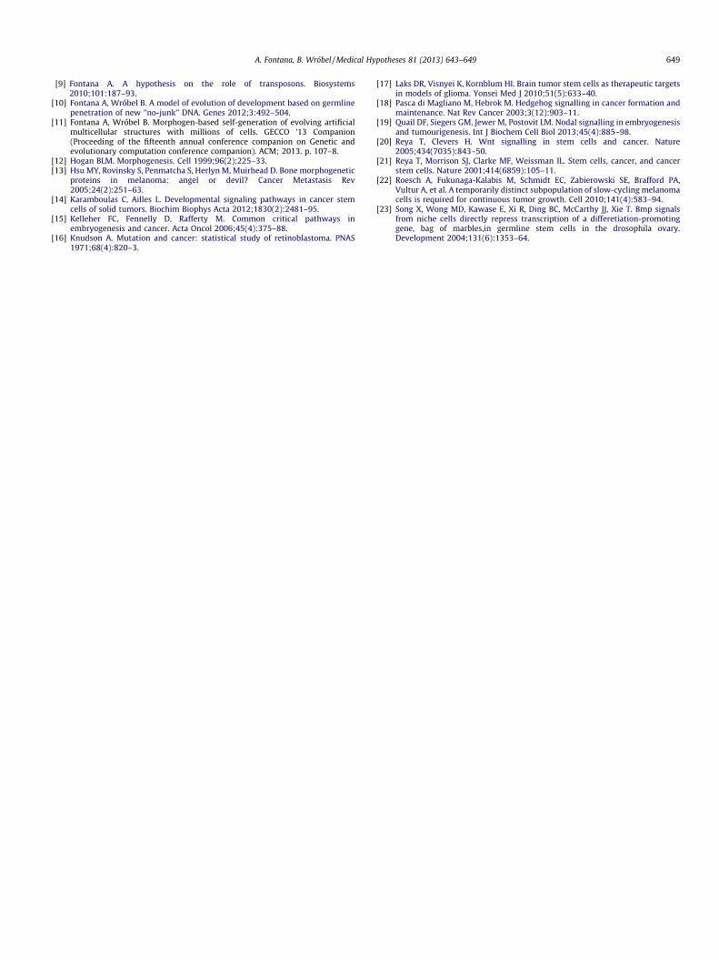
The damage may impede the creation of the new MOC elementwhich is responsible for differentiation (Fig. 7): therefore one ormore daughter stem cells end up having the same MOC as themother (Fig. 8). Since these stem cells also share the genome withtheir mother, the same MOC-RES match that triggered a prolifera-tion in the mother is bound to occur also in the daughters, givingrise to an identical proliferation. These proliferations triggered inthe daughters are destined to produce granddaughter cells withthe same fault in the MOC generator. The result of this scenariois a chain of proliferation events, the mark of carcinogenesis. Theengine of the process is a set of ‘‘cancer stem cells’’, which are
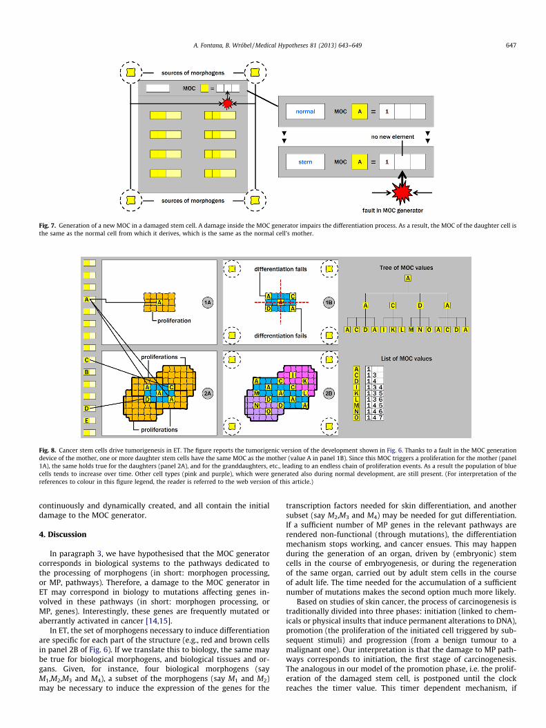
Fig. 7. Generation of a new MOC in a damaged stem cell. A damage inside the MOC generator impairs the differentiation process. As a result, the MOC of the daughter cell isthe same as the normal cell from which it derives, which is the same as the normal cell’s mother.
Fig. 8. Cancer stem cells drive tumorigenesis in ET. The figure reports the tumorigenic version of the development shown in Fig. 6. Thanks to a fault in the MOC generationdevice of the mother, one or more daughter stem cells have the same MOC as the mother (value A in panel 1B). Since this MOC triggers a proliferation for the mother (panel1A), the same holds true for the daughters (panel 2A), and for the granddaughters, etc., leading to an endless chain of proliferation events. As a result the population of bluecells tends to increase over time. Other cell types (pink and purple), which were generated also during normal development, are still present. (For interpretation of thereferences to colour in this figure legend, the reader is referred to the web version of this article.)
A. Fontana, B. Wróbel / Medical Hypotheses 81 (2013) 643–649 647
continuously and dynamically created, and all contain the initialdamage to the MOC generator.
4. Discussion
In paragraph 3, we have hypothesised that the MOC generatorcorresponds in biological systems to the pathways dedicated tothe processing of morphogens (in short: morphogen processing,or MP, pathways). Therefore, a damage to the MOC generator inET may correspond in biology to mutations affecting genes in-volved in these pathways (in short: morphogen processing, orMP, genes). Interestingly, these genes are frequently mutated oraberrantly activated in cancer [14,15].
In ET, the set of morphogens necessary to induce differentiationare specific for each part of the structure (e.g., red and brown cellsin panel 2B of Fig. 6). If we translate this to biology, the same maybe true for biological morphogens, and biological tissues and or-gans. Given, for instance, four biological morphogens (sayM1,M2,M3 and M4), a subset of the morphogens (say M1 and M2)may be necessary to induce the expression of the genes for the
transcription factors needed for skin differentiation, and anothersubset (say M2,M3 and M4) may be needed for gut differentiation.If a sufficient number of MP genes in the relevant pathways arerendered non-functional (through mutations), the differentiationmechanism stops working, and cancer ensues. This may happenduring the generation of an organ, driven by (embryonic) stemcells in the course of embryogenesis, or during the regenerationof the same organ, carried out by adult stem cells in the courseof adult life. The time needed for the accumulation of a sufficientnumber of mutations makes the second option much more likely.
Based on studies of skin cancer, the process of carcinogenesis istraditionally divided into three phases: initiation (linked to chem-icals or physical insults that induce permanent alterations to DNA),promotion (the proliferation of the initiated cell triggered by sub-sequent stimuli) and progression (from a benign tumour to amalignant one). Our interpretation is that the damage to MP path-ways corresponds to initiation, the first stage of carcinogenesis.The analogous in our model of the promotion phase, i.e. the prolif-eration of the damaged stem cell, is postponed until the clockreaches the timer value. This timer dependent mechanism, if

648 A. Fontana, B. Wróbel / Medical Hypotheses 81 (2013) 643–649
present in biology, may explain the long latency period observedbetween the exposure to carcinogenic substances (e.g. tobaccosmoke) and the appearance of a tumour (e.g. lung cancer). Finally,progression, the third and last phase of carcinogenesis, may belinked to further mutations which confer additional properties tothe already transformed cells, i.e. the capacity to infiltrate tissuesand to produce distant metastases.
Our proposal is fully consistent with the CSC model, with the re-cent paradigm of dynamic stemness, and with the idea that eachcell in a tumour has tumorigenic potential: in ET this potential isassociated to a damage to MP genes, which is present initially ina single stem cell, and subsequently spreads to all normal and stemcells derived from it. The ET contribution to the field mainly lies intwo ideas. The first idea consists in the notion of stem cell differen-tiation, which explains the emergence of a hierarchy of cell types,and brings together embryonic, adult and cancer stem cells under aunified framework. The second idea is a proposal for a low-leveldifferentiation mechanism, through the actions of morphogensand the division of space into regions, which may correspond tobiological niches.
The attempt to link cancer or specific cancers to patterns ofgene mutations, consistently found in all tumour samples, has sofar eluded the efforts of biologists. Some cancer-related genes, suchas p53, are indeed mutated in the majority of tumours, but manyother cancer genes are changed in only a minority of patients, asmall fraction of cancer types, or a subset of cells within a tumour.In our interpretation, morphogens and MP pathways are organ-specific, and a fully working differentiation machinery requiresthe functioning of all MP pathways involved in the generation ofthe organ. On the other hand, a reduced form of differentiationcan be obtained in many different ways, corresponding to all pos-sible different subsets of non-functional MP pathways.
Table 1 gives an overview of a hypothetical involvement of fourMP pathways (a, b, c, d) in four organs/cancer types (bladder, brain,breast, colon). For each organ three MP pathways are hypothesisedto be necessary for differentiation. The row labelled ‘‘combinationsof damaged pathways’’ lists all possible combinations of damagedpathways which lead to failed differentiation (and hence tumourformation) in the relevant organ. In the case of breast, for instance,pathways a, c and d have to be functional for full differentiation tooccur. Different combinations (sets) of damaged pathways ({a}, {c},{d}, {a,c}, {a,d}, {c,d}, {a,c,d}) cause impaired differentiation andmay correspond to tumours of different grades. Considering thata damage in each MP pathway can be caused by mutations to anumber of MP genes, this scheme can help to explain theheterogeneity of the mutational patterns observed, and thepresence in tumours of cells of different types, having different de-grees of differentiation and organised in a hierarchical fashion. Thesuggested scheme of gene mutations could be verifiedbioinformatically.
Cancer is mostly a disease of the old age: it is a rare occurrencein young people and becomes more and more common with ageprogression. The standard explanation of this phenomenon is
Table 1A hypothetical involvement of damage of four imagined morphogen processingpathways in different types of cancer.
Cancer type Bladder Brain Breast Colon
MP pathways involved a,b,c a,b,d a,c,d b,c,dCombinations of damaged
pathways{a} {a} {a} {b}
{b} {b} {c} {c}{c} {d} {d} {d}{a,b} {a,b} {a,c} {b,c}{a,c} {a,d} {a,d} {b,d}{b,c} {b,d} {c,d} {c,d}{a,b,c} {a,b,d} {a,c,d} {b,c,d}
based on the ‘‘multi-hit’’ hypothesis [16]: the late onset of cancerwould simply reflect the time necessary for a cell to accumulatethe number of mutations necessary for transformation. In ET, can-cer initiation is linked to a damage to the MOC generator, and pro-gression may require the match between the clock and the timer.The damage to the MOC generator can occur either (1) in a stemcell activated during embryogenesis and reactivated in the courseof adult life to repair damaged tissues, or (2) in a stem cell acti-vated during adult life for the first time. In ET reactivation for tis-sue repair is triggered by the occurrence of damage, and does notdepend on the timer: therefore, in case 1, the temporal dynamicsis expected to be analogous to the one predicted by the ‘‘multi-hit’’ hypothesis. In case 2, the same event is hypothesised to con-tribute to ageing (if the MOC generator is intact) or cause a tumour(if the MOC generator is damaged): as a result, in this case, thetemporal patterns of ageing and cancer are coincident. The factthat measures known to delay ageing, such as dietary restriction,correspondingly delay the onset of several cancers, seems to sug-gest the presence of this dynamics also in biological systems [2].
5. Conclusions
Epigenetic Tracking is a model of systems of biological cells,originally conceived as a model of embryonic development and la-ter extended to interpret other aspects of biology, such as the pres-ence of ‘‘junk DNA’’, the phenomenon of ageing and the process ofcarcinogenesis. In this work we have explored more deeply theinterpretation of carcinogenesis, trying to bridge the gap betweenabstract concepts and molecular pathways. We have argued thatmorphogen-processing pathways are crucial for the process of cel-lular differentiation, which plays a key role in development andfails in carcinogenesis. According to our hypothesis the disruptionof these pathways represents the first and most distinctive event inthe carcinogenic process. Future work will be aimed at furtherclosing the gap between ET and molecular biology, mapping themodel variables to individual biological elements.
6. Conflict of interest
None.
Acknowledgement
This work was supported by the Polish National Science Centre(project BIOMERGE, 2011/03/B/ST6/00399)
References
[1] Bonnet D, Dick JE. Human acute myeloid leukemia is organized as a hierarchythat originates from a primitive hematopoietic cell. Nat Med 1997;3(7):730–7.
[2] Colman RJ, Anderson RM, Johnson SC, Kastman EK, Kosmatka KJ, Beasley TM,et al. Caloric restriction delays disease onset and mortality in rhesus monkeys.Science 2009;325(5937):201–4.
[3] Cruz MH, Sidén A, Calaf GM, Delwar ZM, Yakisich JS. The stemness phenotypemodel. ISRN Oncol 2012.
[4] De Robertis EM. Spemann’s organizer and self-regulation in amphibianembryos. Nat Rev Mol Cell Biol 2006;7(4):296–302.
[5] Fogarty MP, Kessler JD, Wechsler-Reya RJ. Morphing into cancer: the role ofdevelopmental signaling pathways in brain tumor formation. J Neurobiol2005;64(4):458–75.
[6] Fontana A. Epigenetic tracking, a method to generate arbitrary shapes by usingevo-devo techniques. In: Schlesinger M, Berthouze L, Balkenius C, editors.Proceedings of the 8th international conference on Epigenetic Robotics(EPIROB 08), 2008. (published online only)
[7] Fontana A. Epigenetic tracking: biological implications. In: Proceedings of 10thEuropean conference on Artificial Life (ECAL 2009), vol. 5777 of LNCS, Springer.2009. p. 10–7.
[8] Fontana A. An artificial life model for carcinogenesis. In: Proceedings of the12th international conference on the simulation and synthesis of livingsystems (ALIFE XII), The MIT press, 2010. p. 101–8.
A. Fontana, B. Wróbel / Medical Hypotheses 81 (2013) 643–649 649
[9] Fontana A. A hypothesis on the role of transposons. Biosystems2010;101:187–93.
[10] Fontana A, Wróbel B. A model of evolution of development based on germlinepenetration of new ’’no-junk’’ DNA. Genes 2012;3:492–504.
[11] Fontana A, Wróbel B. Morphogen-based self-generation of evolving artificialmulticellular structures with millions of cells. GECCO ’13 Companion(Proceeding of the fifteenth annual conference companion on Genetic andevolutionary computation conference companion). ACM; 2013. p. 107–8.
[12] Hogan BLM. Morphogenesis. Cell 1999;96(2):225–33.[13] Hsu MY, Rovinsky S, Penmatcha S, Herlyn M, Muirhead D. Bone morphogenetic
proteins in melanoma: angel or devil? Cancer Metastasis Rev2005;24(2):251–63.
[14] Karamboulas C, Ailles L. Developmental signaling pathways in cancer stemcells of solid tumors. Biochim Biophys Acta 2012;1830(2):2481–95.
[15] Kelleher FC, Fennelly D, Rafferty M. Common critical pathways inembryogenesis and cancer. Acta Oncol 2006;45(4):375–88.
[16] Knudson A. Mutation and cancer: statistical study of retinoblastoma. PNAS1971;68(4):820–3.
[17] Laks DR, Visnyei K, Kornblum HI. Brain tumor stem cells as therapeutic targetsin models of glioma. Yonsei Med J 2010;51(5):633–40.
[18] Pasca di Magliano M, Hebrok M. Hedgehog signalling in cancer formation andmaintenance. Nat Rev Cancer 2003;3(12):903–11.
[19] Quail DF, Siegers GM, Jewer M, Postovit LM. Nodal signalling in embryogenesisand tumourigenesis. Int J Biochem Cell Biol 2013;45(4):885–98.
[20] Reya T, Clevers H. Wnt signalling in stem cells and cancer. Nature2005;434(7035):843–50.
[21] Reya T, Morrison SJ, Clarke MF, Weissman IL. Stem cells, cancer, and cancerstem cells. Nature 2001;414(6859):105–11.
[22] Roesch A, Fukunaga-Kalabis M, Schmidt EC, Zabierowski SE, Brafford PA,Vultur A, et al. A temporarily distinct subpopulation of slow-cycling melanomacells is required for continuous tumor growth. Cell 2010;141(4):583–94.
[23] Song X, Wong MD, Kawase E, Xi R, Ding BC, McCarthy JJ, Xie T. Bmp signalsfrom niche cells directly repress transcription of a differetiation-promotinggene, bag of marbles,in germline stem cells in the drosophila ovary.Development 2004;131(6):1353–64.