Embed Size (px)
Citation preview


EMBO Practical Course on
X-ray crystal structure determination of macromolecules
September 14-20th, 2008, Synchrotron SOLEIL
L’Orme des Merisiers, Saint-Aubin, France
Summary
‚ Introduction ‚ Programme
‚ Oral Presentations and Posters
‚ List of Speakers
‚ List of Participants ‚ Practical Information

Dear participants, It is a great pleasure to welcome you to the Max-Inf2 EMBO Practical course “X-ray determination of macromolecules” here at SOLEIL. As scientists in a synchrotron facility, we know how important it is for you to gain independence in the pursuit of your research and we are aware that we must provide you up to date methodological expertise and practical experience to use beamlines in an optimal way. This course is meant to provide and strengthen instruction in the background, operation and evaluation of the X-ray data that you obtain from a synchrotron facility. We have invited tutors that are experts in every important aspect of X-ray structure determination. They will give lectures and practicals. In addition to these overviews, the Max-Inf2 EMBO Practical course is also meant to favor informal exchanges and we encourage you to use this opportunity to approach these tutors and experts with questions about your own research.
A synchrotron is a high energy place, where experiments are done around the clock. People travel from far away to do critical measurements in a limited amount of time. I would consider this course to be a success if some of this intensity bounces off, and if all of you return home with the confidence that you may succeed on your own and make the right decisions next time you visit any synchrotron beamline. Jean-Pierre Samama Scientific Director Synchrotron SOLEIL

EMBO Practical Course on X-ray crystal structure determination of macromolecules
September 14-20th, 2008
Synchrotron SOLEIL L’Orme des Merisiers, Saint-Aubin, France
Programme
Sunday, September 14th
16:00 Welcome and student poster presentation (Amphitheatre)
Microsymposium on Membrane Protein Crystallography
18:00 Daniel Picot
Membrane protein crystallography
18:45 Martin Pos The structure of the AcrB antibiotic efflux transporter
19:15 Alexey Amunts The structure of the Photosystem I supercomplex
20:00 Buffet
Monday, September 15
th
9:00 Louise Bird
Construct design
9:45 Herman Van Tilbeurgh Protein preps for difficult projects
10:30 Coffee Break
11:00 Paloma Fernandez-Varela Purification protocols
11:45 Enrico Stura Crystallization techniques
12:30 Lunch
14:00 Jim Pflugrath Diffraction Theory
14:45 Andy Thompson Synchrotron data collection
15:30 Coffee Break
16:00 Chris Nielsen X-ray detectors
16:45 Jim Pflugrath Data collection strategies
19:00 20:00
Dinner
Rapid Coot tutorial for Beginners (Kevin Cowtan)

Tuesday, September 16
th
9:00 Thomas Schneider
Theoretical basis for SAD and MAD
9:45 Thomas Schneider Case studies of finding substructures with SAD and MAD
10:30 Coffee Break
11:00 Clemens Vonrhein Phase improvement with SHARP
11:45 Kevin Cowtan Density modification techniques
12:30 Lunch
14:00 Practicals
19:00 Dinner
Wednesday, September 17
th
9:00 Randy Read
Theoretical basis for Molecular Replacement
9:45 Randy Read MR and SAD with Phaser
10:30 Coffee Break
11:00 Peter Zwart Structure refinement (Phenix related)
11:45 Tassos Perrakis Do's and don'ts in refinement
12:30 Lunch
14:00
Practicals
19:00 Dinner
20:00 Student presentations (15 minutes each): Burcu Kaplan Türköz Biophysical Characterization of A. thaliana Heterotrimeric G-protein Subunits Mugdha Bhati Exploring the LIM code for motor neuron specificity

Thursday, September 18
th
9:00 Tassos Perrakis
Automatic tracing with Arpwarp
9:45 Kevin Cowtan Tracing with Pirate/Buccaneer
10:30 Coffee Break
11:00 Piotr Sliz Low resolution structure phasing
11:45 Jane Richardson Validation: Molprobity
12:30 Lunch
14:00 Practicals
19:00 Dinner
Friday, September 19
th
9:00 Peter Zwart
Data stats: what can go wrong (Xtriage)
9:45 Peter Zwart Radiation damage
10:30 Coffee Break
11:00 Kevin Cowtan Molecular graphics: Coot
11:45 Piotr Sliz Low resolution structure refinement
12:30 Lunch
14:00 Practicals
19:00 Dinner
Saturday, September 20
th
9:00 Beamline available, 'Open tutorials'
12:30 Lunch
14:00 Beamline available, 'Open tutorials'
19:00 Dinner

ORAL PRESENTATIONS
AND POSTERS

EMBO COURSE 2008
Oral presentations & Posters
ORAL PRESENTATIONS O1 Markus Seeger, Thomas Eicher, Lorenz Brandstaetter, Andre Schiefner,
Kay Diederichs, Klaas Pos The AcrB Efflux Pump: Conformational cycling and peristalsis lead to multidrug resistance
O2 Alexey Amunts, Omri Drory, Nathan Nelson The 3.4 Å Resolution X-ray Crystal Structure of a Plant Photosystem I Supercomplex
O3 Burcu Kaplan Türköz, Sedef 1skit, Zehra Sayers Biophysical Characterization of A. thaliana Heterotrimeric G-protein Subunits
O4 M. Bhati, M. Lee, A. Nancarrow, V. Craig, J. M. Guss, J. M. Matthews Exploring the LIM code for motor neuron specificity
POSTERS
P1
Yara Al-Tall, Christina de Matteis Structural Studies on a Novel Nitroreductase; Molecular Activation of the Anti-Cancer Prodrug CB1954
P2 Sébastien Dandois, Georgios Moutzourelis, Frédéric Kerff, Raphaël Hermann, Eric Sauvage, Paulette Charlier, Bernard Joris Study of the BlaB protein involved in the Streptomyces cacaoi BlaL/BlaU beta-lactamases induction
P3 Maria Dimarogona, Evangelos Topakas, Paul Christakopoulos, Evangelia Chrysina Structural studies of new xylanolytic enzymes
P4 Jerome Gouge, Marc Delarue The structural study of Pyrococcus abyssi DNA polymerase B
P5 Nives Ivic, Marija Luic Structural characterization of Streptomyces coelicolor single-stranded DNA-binding proteins and their complexes
P6 Silvia Jansen, Maria Andries, Mathieu Bollen Crystallization of the NPP family
P7 Macarena Marín, Birgit Hofmann, Dietmar Pieper Structural characterization of 4-methylmuconolactone methyl isomerase from Pseudomonas reinekei MT1
P8 María Moreno Morcillo, Cameron Mackereth, Marta Martínez Júlvez, Sébastien Fribourg. Structural Determination of the Proteins Involved in the Messenger RNA 3' End Processing

EMBO COURSE 2008
Oral presentations & Posters
P9 Elisa Pasqualetto, Rosa Aiello, Valentina Rosan, Anke Seydel, Alberto Pellini, Roberto Battistutta Structural studies of the SLC26 anion transporter prestin
P10 Diorge Souza, Roberto Salinas, Chuck Farah Structural studies of Type III and IV Secretion System Proteins from Xanthomonas axonopodis pv. Citri
P11 Michael Raba, Adriana Rycovska, Hartmut Michel, Heinrich Jung X-ray crystallography of a member of the sodium solute symporter family
P12 Serena Sirigu, Sato Yoshiteru, Arnaud Poterszmann, Natacha Rochel, Dino Moras Structural study of nuclear receptors transcription regulation through phosphorylation.
P13 Monika Sokolowska, Magdalena Kaus-Drobek, Honorata Czapinska, Gintautas Tamulaitis, Roman H. Szczepanowski, Virginijus Siksnys, Matthias Bochtler Restriction endonuclease BcnI resembles a component of the bacterial DNA repair machinery
P14 Henrik Spahr, Guillermo Calero, Roger D. Kornberg Structure of an RNA Polymerase II/TFIIF early elongation complex
P15 Goran Stjepanovic, Richard Parlitz, Bianca Schrul, Astrid Hendricks, Irmi Sinning Characterization of the E. coli SRP receptor FtsY: the association with the inner membrane
P16 Ekaterina Sviridova, Eva Csefalvay, Ivana Kuta Smatanova Purification and crystallization of the HsdM subunit of the EcoR124I endonuclease from Escherichia coli
P17 Eva Torreira, Sudhakar Jha, José R. López-Blanco, Ernesto Arias-Palomo, Pablo Chacón, Anindya Dutta, Óscar Llorca 3D structure of the Rvb1p/Rvb2p complex, an essential component of several chromatin-remodelling complexes
P18 Aldino Viegas, Natércia Brás, Nuno Cerqueira, Pedro Fernandes, José Prates, Carlos Fontes, Marta Bruix, Maria Romão, Ana Carvalho, Maria Ramos, Anjos Macedo, Eurico Cabrita Molecular Determinants of Ligand Specificity in Family 11 Carbohydrate-Binding Modules (CBM11)
P19 Qilu Ye, Hailong Wang, Qun Wei, Zongchao Jia Structural Characterization of Calcineurin and Calmodulin Interaction
P20 Jean Christophe Zeeh, Mahel Zeghouf, valerie Biou, Bernard Guibert, Jacqueline Cherfils Biochemical characterization of inhibitors of GTPase activation by their GEFs.

O1- The AcrB Efflux Pump: Conformational cycling and peristalsis lead to multidrug resistance Markus Seeger, Thomas Eicher, Lorenz Brandstaetter, Andre Schiefner, Kay Diederichs, Klaas Pos Institute of Physiology, University of Zurich, CH-8057 Zurich, Switzerland Antimicrobial resistance of human pathogenic bacteria is an emerging problem for global public health. This resistance is often associated with the overproduction of membrane transport proteins that are capable to pump chemotherapeutics, antibiotics, detergents, dyes and organic solvents out of the cell. In Gram-negative bacteria such as Escherichia coli and Pseudomonas aeruginosa, tripartite multidrug efflux systems extrude a large variety of cytotoxic substances from the cell membrane directly into the medium bypassing the periplasm and the outer membrane. In E. coli, the tripartite efflux system AcrA/AcrB/TolC is the pump in charge of the efflux of multiple antibiotics, dyes, bile salts and detergents. The trimeric outer membrane factor (OMF) TolC forms a b-barrel pore in the outer membrane and exhibits a long periplasmic g-helical conduit. The periplasmic membrane fusion protein (MFP) AcrA serves as a linker between TolC and the trimeric resistance nodulation cell division (RND) pump AcrB, located in the inner membrane acting as a proton/drug antiporter. The newly elucidated asymmetric structure of trimeric AcrB reveals three different monomer conformations representing consecutive states in a transport cycle. The monomers show tunnels with occlusions at different sites leading from the lateral side through the periplasmic porter domains towards the funnel of the trimer and TolC. The structural changes create a hydrophobic binding pocket in one monomer, which is not present in the other two monomers. From the asymmetric structure a possible peristaltic pump transport mechanism based on a functional rotation of the AcrB trimer has been postulated. The novel transport model merges Jardetzky’s alternate access pump mechanism with the rotating site catalysis of F1Fo ATPase and suggests a working hypothesis for the transport mechanism of RND transporters in general.
O2- The 3.4 Å Resolution X-ray Crystal Structure of a Plant Photosystem I Supercomplex Alexey Amunts, Omri Drory, Nathan Nelson Biochemistry, Tel Aviv University, 69978 Tel Aviv, Israel A plant Photosystem I (PSI) is exquisitely organised, multi-subunit membrane super-complex of protein and non-protein components that drive the photosynthesis process. PSI captures sunlight through sophisticated pigment network and uses the energy to perform transmembrane electron transfer. It consists of the Reaction Center complex (RC), where the charge separation reaction takes place and the Light Harvesting complex (LHCI), which serves as an additional antenna system. PSI performs a photochemical activity with the unprecedented quantum yield of close to 1.0, being the most efficient light capturing and energy conversion device. We determined the X-ray crystal structure of the intact PSI at 3.4 Å resolution [1]. The current crystal structure provides a picture at near atomic detail of 17 protein subunits including an additional subunit (PsaN) being identified for the first time at the lumenal side of the supercomplex. Positions of 3038 amino acids were assigned, as well as 168 chlorophylls, 2 phyloquinones, 3 Fe4S4 clusters and 5 carotenoids. The remarkable feature of PSI structure is the unprecedented extremely high content of non-protein components – approximately one third of the total mass of about 650 KDa consists of different co-factors. The structure reveals intriguing insights regarding unique interactions between the RC and the LHCI complexes and provides a structural basis for the state transitions phenomenon. In addition, putative docking sites of the soluble electron carriers are described for the first time. [1] Amunts, A., Drory, O. & Nelson, N. (2007) Nature, 447, 58-63.

O3- Biophysical Characterization of A. thaliana Heterotrimeric G-protein Subunits Burcu Kaplan Türköz, Sedef 1skit, Zehra Sayers Biological Sciences and Bioengineering, Sabanci University, 34956 Istanbul, Turkey Heterotrimeric G proteins, composed of alpha, beta and gamma subunits, are a major group of signaling molecules in eukaryotic organisms. Plant heterotrimeric G proteins are involved in various important processes including growth, development and stress response. The structure of the plant heterotrimer or the individual subunits are not defined, unlike the mammalian heterotrimer which is both biochemically and structurally well characterized. Structural information would provide insight into the specific mechanisms for the activity of the plant G-proteins and help to develop a better understanding of their functional roles. For characterization of the individual subunits and the reconstituted heterotrimer we have cloned the A.thaliana G protein alpha (GPA1), beta (AGB1) and gamma (AGG1/2) subunits and used different expression systems (P.pastoris and E. coli) for production of recombinant proteins. GPA1 was produced with 90% purity in a monomeric form. The optimum purification conditions are being investigated for various constructs of AGB1 and AGG1/2 subunits. We have been working on recombinant protein production mainly for SAXS analysis which is extremely dependent on protein quality. Similarly the importance of protein quality in order to generate well diffracting high quality crystals is well known. Besides protein expression and purification steps performed, we will be presenting our results on how protein quality is affected by several factors, including the ionic strength and the pH of the environment, the stability of the buffer system used, storage conditions and freeze-thaw cycles, and the presence of ligands or other proteins. Protein characterizations are / will be conducted using circular dichroism spectropolarimetry, dynamic light scattering and uv-vis spectrophotometer. O4- Exploring the LIM code for motor neuron specificity M. Bhati, M. Lee, A. Nancarrow, V. Craig, J. M. Guss, J. M. Matthews School of Molecular and Microbial Biosciences, University of Sydney, Sydney, NSW, Australia LIM-HD (LIM homeodomain) proteins are essential for defining cell fate, especially in the developing central nervous system. Isl-1 and Lhx-3 are two LIM-HD proteins implicated in neuronal development that form the basis of regulatory complexes in two adjacent cell types in the ventral spinal chord, V2 interneurons and motor neurons. Both cell types express Lhx-3 and the nuclear adaptor protein Ldb1, however, Isl-1 is only expressed in postmitotic motor neurons. In the two complexes, the two LIM domains of Lhx-3 mediate different protein:protein interactions that appear to be critical for the regulation of the two different cell types. In V2 interneurons, this involves a direct interaction with the LIM interaction domain (LID) of Ldb1, whereas in motorneurons Isl-1 interacts directly with Ldb1-LID and Lhx-3 binds instead to Isl-1. We are interested in characterising these interactions with the overall goal of understanding their role in neuronal development. Deletion mutagenesis analyses have revealed a 30-residue region of Isl-1 that binds the Lhx-3 LIM domains (LIMs). We have solved a crystal structure of a protein complex between Lhx3 and Isl1 to show that, despite low sequence homology, the LIDs from Ldb1 and Isl1 bind Lhx3 in an essentially identical manner. Binding and stability studies of these different complexes suggest that a ternary Lhx3:Isl1:Ldb1 complex can only form if the complex binds to DNA that contains Lhx3 and Isl1 recognition sequences. This work highlights a general mode of interaction between different LIM-HD proteins involved with LIM codes.

P1- Structural Studies on a Novel Nitroreductase; Molecular Activation of the Anti-Cancer Prodrug CB1954 Yara Al-Tall, Christina de Matteis Centre for Biomolecular Sciences, School of Pharmacy, University of Nothingham, NG7 2RD Nothingham, United Kingdom (NTR-A) is a novel flavoprotein that shows promising activity in the reduction of the prodrug CB1954 to its active cytotoxic form. It functions as a nitroreductase, reducing the 4-nitro group of CB1954, and hence has potential in directed-enzyme prodrug therapy (DEPT). This protein was expressed, purified and crystallized, and crystals of the NTR-A/CB1954 complex were obtained by both crystal soaking and co-crystallization. The structure was solved by molecular replacement, followed by model building with ARP/wARP. Rigid body followed by restrained refinement and manual rebuilding were used to optimize the fit of the molecular coordinates into the electron density map at 1.7 Å and 1.2 Å. However, the structure of the protein-ligand complex is still in progress. We aim to study the complexed structure and to investigate the structure-function relationships through comparisons with other nitroreductases. During the course of these studies it has become apparent that the pro-drug is likely to be degraded by the acidic crystallisation conditions and analytical studies are underway to explore this chemistry. P2- Study of the BlaB protein involved in the Streptomyces cacaoi BlaL/BlaU beta-lactamases induction Sébastien Dandois, Georgios Moutzourelis, Frédéric Kerff, Raphaël Hermann, Eric Sauvage, Paulette Charlier, Bernard Joris Centre d'Ingénierie des Protéines (CIP), Université de Liège, Faculté des Sciences de la Vie, Belgium, 4000 Sart-Tilman, Liège, Belgium Among the bacterial kingdom, the production of beta-lactamases is one of the most common and most effective resistance mechanisms against beta-lactam compounds such as penicillins and cephalosporins. Streptomyces cacaoi is one of the few bacterial species that exhibits an inducible production of beta-lactamases in presence of beta-lactam antibiotics [1]. So far, two regulatory proteins, BlaA and BlaB, are necessary in the induction mechanism of the two S. cacaoi beta-lactamases, BlaL and BlaU [2]. BlaA belongs to the LysR activator-repressor family and recognizes a cis-acting element upstream of blaL and blaU genes. BlaB possesses conserved residues involved in the catalytic activity of serine penicillin recognizing enzymes (PREs) but although being able to interact with beta-lactam compounds; BlaB does not possess any beta-lactamase activity [3]. Here, we describe the new strategy for production and purification of BlaA and BlaB proteins. The protein BlaB was crystallised and its structure, obtained by molecular replacement method using the structure of SME1 (PDB code 1DY6) was refined to a resolution of 1.8 Å. The structure of BlaB has been compared with those of PREs combined with western blotting and protein labelling To identify the ligand of BlaB, possible compounds were tested with BlaB and a strategy developed to purify BlaB protein under stress conditions combined with its ligand. These preliminary data allow a first model for the induction mechanism of the beta-lactamases in S. cacaoi. BlaB would be at the same time the sensor of the cytoplasmic stress generated by the antibiotics outside the cell and the relay for transferring the information to BlaA, the transcriptional regulator that controls the expression of the beta-lactamase genes. The emergence of full genome sequences and alignment analysis revealed that this resistance mechanism would be more widespread than first estimated as blaB-like and blaA-like genes have been identified in several other actinomycetes including marine organisms, industrial strains and pathogenic germ. These findings indicate that BlaB could be the first element of a new kind of PREs used by actinomycetes, neither for peptidoglycan synthesis as the penicillin binding proteins nor for antibiotic degradation as beta-lactamases, but for stress control and signalisation. References 1) Lenzini VM, Magdalena J, Fraipont C, Joris B, Matagne A, Dusart J. Induction of a Streptomyces cacaoi beta-lactamase gene cloned in S. lividans. Mol Gen Genet. 1992 Oct 235(1):41-8. 2) Magdalena J, Gerard C, Joris B, Forsman M, Dusart J. The two beta-lactamase genes of Streptomyces cacaoi, blaL and blaU, are under the control of the same regulatory system. Mol Gen Genet. 1997 Jun 255(2):187-93. 3) Raskin C, Gerard C, Donfut S, Giannotta E, Van Driessche G, Van Beeumen J, Dusart J. BlaB, a protein involved in the regulation of Streptomyces cacaoi beta-lactamases, is a penicillin-binding protein. Cell Mol Life Sci. 2003 Jul 60(7):1460-9.

P3- Structural studies of new xylanolytic enzymes Maria Dimarogona, Evangelos Topakas, Paul Christakopoulos, Evangelia Chrysina Institute of Organic & Pharmaceutical Chemistry (IOPC), The National Hellenic Research Foundation (NHRF), 11471 Athens, Greece Xylanolytic enzymes are employed as molecular targets for industrial process optimization because of their pivotal role in catalysis either alone or synergistically with other enzymes. Xylan, the basic constituent of hemicellulose, is a linear polymer of く-D-xylopyranosyl units linked by (1-4) glycosidic bonds. These bonds are cleaved by endo-く-1,4-xylanases. Xylanases are organized into two major families of glycosyl hydrolases: Family 10 (F) and Family 11 (G) according to their sequence similarity. Our work is focused on molecular and structural studies of these enzymes with the aim to elucidate their catalytic mechanism in the presence of oligosaccharides (substrate). Here we report the results obtained from our structural studies performed on an alkaline endo-beta-1,4-xylanase FoXyn10a, from Fusarium oxysporum F3. We have been successful in growing single FoXyn10a crystals(16 0C) using the sitting drop vapor diffusion method. Preliminary characterization showed that the crystals belong to space group P4212 with unit cell dimensions a=b=123.4 Å, c=117.3 Å, and two molecules in the asymmetric unit. X-ray crystallographic data were collected under cryogenic conditions at SRS-Daresbury Laboratory-UK (station PX 10.1). FoXyn10a crystals diffract to 2.03 Å resolution and a complete diffraction data set was collected from a single crystal. Structure determination of FoXyn10a is currently in progress using the isomorphous molecular replacement method employing the three dimensional structures of previously determined F/10 homologues as starting model. Cocrystallisation efforts are also underway to obtain diffracting crystals of FoXyn10a in complex with linear or branched oligosaccharides (e.g. feruloyl arabinoxylodisaccharide, FAX2). We are also trying to clone and express a hypothetical protein of Fusarium oxysporum genome that was previously prepared by fermentation in our laboratory. This protein appears to exhibit potential xylanolytic activity, a property that no other enzyme of the same glycosil hydrolase family is known to have. To corroborate this assumption, we are currently working on the sample preparation using recombinant DNA techniques followed by standard purification protocols to achieve high purity. Our ultimate aim is crystallize the new enzyme for structural studies in our laboratory. P4- The structural study of Pyrococcus abyssi DNA polymerase B Jerome Gouge, Marc Delarue Structural Dynamics of Macromolecules, Pasteur Institute, 25 Rue du Docteur Roux, 75015 Paris, France DNA polymerases can be classified into seven main groups. The B family contains replicative polymerases: they are processive and error-free. Combined with thermostability, these enzymes become powerful tools for cloning and mutation diagnostic. Understanding the structural basis of these features is important to generate DNA polymerases more efficient during PCR. The Pyrococcus abyssi polymerase B (PabPolB) is commonly known as Isis DNA polymerase (Qbiogen Molecular Biology). It can be divided into 5 separated domains: the NH2, 3’-5’ proof reading, fingers, palm and thumb domains. This enzyme is 40 times more accurate than Taq DNA polymerase and it is able to amplify up to 6 kb on genomic DNA (3 kb for Taq). Moreover, its half-life at 100°C is 5 hours. This work presents the first steps of the structural study of PabPolB. The goal is to resolve the structure of the apo protein, then in complex with DNA and an incoming nucleotide. The purification is achieved in 3 steps. The overall yield is 15mg of purified protein for 2 liters of culture. Crystallization trials of PabPolB have been set up using a robot (576 drops). Only one condition led to crystals. A manual optimization allowed them to grow faster and bigger. The diffraction pattern indicated that the crystals were salt. Meanwhile a sequence-based approach was used to set up crystallization trials. Needle clusters were obtained, but manual optimizations didn’t lead to any improvement of the morphological aspect. Other genetic constructions are currently studied to achieve this structural work.

P5- Structural characterization of Streptomyces coelicolor single-stranded DNA-binding proteins and their complexes Nives Ivic, Marija Luic Laboratory for Chemical and Biological Crystallography, Rudjer Boskovic Institute, 10 000 Zagreb, Croatia Single-stranded DNA-binding proteins (SSBs) are required in DNA replication, recombination and repair, in all organisms, from bacteria to human. Their ability to bind single-stranded DNA (ssDNA) with high affinity makes them important for protection of ssDNA from chemical and nuclease attacks and from forming aberrant secondary structures. Bacterial SSBs have two distinct domains: N-terminal domain responsible for DNA binding and C-terminal tail that mediates protein-protein interactions (1,2). Bacteria Streptomyces coelicolor encodes two SSBs (ScSSB) that differ in size: larger contains 199 amino acids (3) and shorter 156 amino acids (data not published). Recently, the crystal structure of larger ScSSB has been solved (data not published), and first crystals of short ScSSB have been obtained. Because of low diffraction quality of the crystals, new crystallization conditions should be found. Our goal is also to prepare ScSSB-ssDNA complexes, crystallize them and solve their structures. Currently, we are trying to characterize SSB-ssDNA complex in order to determine binding properties (intrinsic binding constant (K), nucleotide binding site-size (n) and cooperativity parameter (の)). Quenching of the intrinsic tryptophan fluorescence of the SSB by the nucleic acid is used to monitor binding. This characterization will help in generating homogeneus complexes necessary for preparing crystals for structure determination. References: 1. Lohman, T.M., Ferrari, M.E. (1994) Escherichia coli single-stranded DNA-binding protein: Multiple DNA binding modes and cooperativities. Annu. Rev. Biochem., 63:527-570. 2. Raghunathan, S., Kozlov, A.G., Lohman, T.M., Waksman, G. (2000) Structure of the DNA binding domain of E. coli SSB bound to ssDNA. Nature Struct. Biol., 7:648-652. 3. Mijakovic, I., Petranovic, D., Macek, B., Cepo, T., Mann, M., Davies, J., Jensen, P.R., Vujaklija, D. (2006) Bacterial single-stranded DNA-binding proteins are phosphorylated on tyrosine. Nucleic Acids Res., 34: 1588-1596
P6- Crystallization of the NPP family Silvia Jansen, Maria Andries, Mathieu Bollen Laboratory of Biosignaling & Therapeutics, Katholic University Leuven, 3000 Leuven, Belgium The seven members of the family of the nucleotide pyrophosphatases/phosphodiesterases (NPPs) are all characterized by a conserved catalytic domain. Despite their similar catalytic mechanism, these proteins show a distinct substrate-specificity and hence, are all implicated in varying (patho)physiological processes (including cancer, bone mineralization, etc.). Via crystallization of the two best characterized NPPs, namely NPP1 and NPP2/autotaxin, we want to delineate the residues responsible for the distinct substrate-specificity of these proteins. For this purpose we set up a cooperation to crystallize NPP2. However, major difficulties were encountered due to the differential glycosylation of this protein. By enzymatic deglycosylation and mutagenesis of the predicted glycosylation sites, we demonstrated that only the glycan moiety at N524 is essential for both the expression of enzymatic activity and the motility-stimulation of NPP2. In addition, sequencing of this sugar moiety revealed that it corresponded to a basic oligomannosidic glycan chain without heterogenic complex glycosylation. Using this information we generated a fully active NPP2 protein with minimal glycosylation which is more suitable for obtaining high-resolution crystals. Our next goal is to crystallize NPP1, which will enable us to understand the distinct substrate-specificity of these NPP isoforms. Moreover, NPP1 plays a role in bone formation via hydrolysis of ATP to AMP and inorganic pyrophosphate (PPi), an inhibitor of mineralization. With aging the expression of NPP1 is increased, which can result in pathologic calcification such as chondrocalcinosis. Hence, NPP1 is an attractive therapeutic target for the treatment of calcification diseases. We are currently screening a library of over 25,000 compounds for inhibitors of NPP1. We plan to co-crystallize NPP1 with the most promising lead compounds. The obtained structural data should be instrumental for the design of more potent and more specific NPP1 inhibitors.

P7- Structural characterization of 4-methylmuconolactone methyl isomerase from Pseudomonas reinekei MT1 Macarena Marín, Birgit Hofmann, Dietmar Pieper Microbiology, Helmoltz Zentrum für Infektionsforschung (HZI), 38124 Braunschweig, Germany Pseudomonas reinekei MT1 is able to degrade 4-methylsalicylate via 4-chlorocatechol and an ortho cleavage pathway. This metabolic route of intermediate 4-methylmuconolactone has been partially described in a Cupriavidus and a Rhodococcus strain, but genetic information is restricted o Cupriavidus and only some crucial genes/enzymes have been identified. One key enzyme of the pathway is 4-methylmuconolactone methyl isomerase. which catalyzes the transformation of 4-methylmuconolactone to 3-methylmuconolactone by an unknown enzyme mechanism. The gene encoding for this activity in strain MT1 was localized and cloned. The protein was expressed and purified from E. coli and characterized for its kinetic properties. Even though the enzyme was inhibited by p-chloromercurybenzoate, site directed mutants in which either of the two cysteine residues was replaced by a serine, remained active. Crystallization experiments were initiated and crystals were obtained in the absence and presence of substrate, product or a substrate analogue, which should give indications on the substrate binding pocket and residues critical for enzyme activity. P8- Structural Determination of the Proteins Involved in the Messenger RNA 3' End Processing. María Moreno Morcillo, Cameron Mackereth, Marta Martínez Júlvez, Sébastien Fribourg Institut Europeen de Chimie et Biologie, 2 Rue Robert Escarpit, 33607 Pessac, France Cleavage stimulation factor (CstF) is one of the multiple factors required for the mRNA 3' end processing. This complex is composed of distinct subunits called CstF-77, CstF-64, and CstF-50 (named by molecular weight). CstF-77 associates with the other two subunits, and its structure was solved in the Fribourg group. We try to solved the structure of the two others proteins. CstF-64 binds to a conserved sequence of the RNA (AAUAAA), and CstF-50 binds to both RNA polymerase II and BARD1. In this study I present the structure of CstF-50 region which interact with the CTD of the RNA polymerase II. We observed that the protein is organized in a dimer. It is necessary to determine the structure of this complex in order to progress in the understanding of the overall molecular mechanism of the mRNA 3' maturation.

P9- Structural studies of the SLC26 anion transporter prestin Elisa Pasqualetto, Rosa Aiello, Valentina Rosan, Anke Seydel, Alberto Pellini, Roberto Battistutta Department of Chemical Sciences , University of Padova, 35131 Padova, Italy The membrane protein prestin (member of the SLC26 family of anion transporter) is the voltage-sensitive molecular motor underlying somatic electromotility of the outer hair cells of the cochlea. Due to the low abundance of the protein from natural sources, the unavoidable choice is the production of recombinant material. The full-length protein has been recently produced in small amount by cell-free system and different variants of the cytosolic C-terminal STAS domain were cloned and expressed in bacterial systems. The two longer fragments of the C-terminal domain were also characterized by circular dichroism, fluorescence spectroscopy and dynamic light scattering. The two polypeptides possess a three-dimensional structure and form mainly dimers and tetramers in solution. These data correlate with that of full-length prestin that forms stable tetramers, suggesting that the C-terminal domain may play an important role in modulating the properties of the entire prestin [*]. Three chimeric constructs of the C-terminal domain, devoid of a variable loop, show a better profile in size-exclusion chromatography. We are performing crystallization trials on these chimeric constructs, using the Oryx8 protein crystallization robot. We have just obtained small crystals and now we are optimizing crystallization conditions to get better quality crystals. [*] E. Pasqualetto, A. Seydel, A. Pellini and R. Battistutta (2008). Expression, purification and characterization of the C-terminal STAS domain of the SLC26 anion transporter prestin. Protein Expr. Purif. 58(2):249-56.
P10- Structural studies of Type III and IV Secretion System Proteins from Xanthomonas axonopodis pv. citri Diorge Souza, Roberto Salinas, Chuck Farah Department of Biochemistry, University of São Paulo / Chemistry Institute, 05508-000 São Paulo, Brazil Xanthomonas axonopodis pv. citri (Xac) is a gram-negative bacterial phytopathogen that infects citrus. One possible virulence determinant is a chromosomally encoded Type IV Secretion System (T4SS), a multiprotein complex that spans the bacterial periplasm and both inner and outer membranes. The T4SS is used by some bacteria to secrete proteins and/or DNA to the extracellular milieu or the host interior. The model Agrobacterium tumefaciens T4SS is made up of twelve structural proteins: VirB1-VirB11 and VirD4. The aim of this project is the study of structure-function relationships in the T4SS. Fourteen T4SS protein-coding genes, including whole proteins or domains, were cloned and the proteins were produced. Nine proteins were purified and submitted to crystallization trials using the sitting-drop vapour-diffusion method. Two crystals have been obtained, but they are too small to collect good quality diffraction data. Further refinements are in progress to improve the crystals size and diffraction quality. One T4SS protein has been studied using NMR spectroscopy, and its assignment and structure were obtained. Its oligomerization, dynamics and interactions with other T4SS components are in progress.

P11- X-ray crystallography of a member of the sodium solute symporter family Michael Raba, Adriana Rycovska, Hartmut Michel, Heinrich Jung Department of Mintcrobiology, Ludiwg-Maximilins-Universität Muenchen, 85638 Munich, Germany Secondary transporters play a crucial role in the selective translocation of solutes across the cytoplasmic membrane of cells. We use PutP of E. coli and homologues, members of the Na /solute symporter family (SSSF), to obtain insights into the three dimensional structure by single crystal x-ray crystallography. Even though some insights into the structure of PutP of E. coli could be gained by electron paramagnetic resonance (EPR) spectroscopy, no high resolution structure of a member of this family is available yet. Since secondary transporters are embedded in the mainly apolar environment of a lipid bilayer the protein surface is strongly hydrophobic. Due to this chemical nature, membrane proteins, especially g-helical polytopic membrane proteins, tend to form aggregates in polar solutions. In addition to the relatively small hydrophilic domains, the high flexibility of transport proteins, is the major bottelneck on the way of building rigid crystal contacts and well-ordered crystals. In order to overcome these basic obstacles we pursue different strategies. On the one hand we are working with fusion proteins to increase the polar surface and the stability of the target proteins. On the other hand we are searching for protein mutants which are arrested in one conformation to reduce protein dynamics. We are also exploiting the fact that hyperthermophilic prokaryotes also harbour SSSF homologues. These proteins show a much lesser tendency to aggregation and are “frozen” in one conformation at room temperature. P12- Structural study of nuclear receptors transcription regulation through phosphorylation. Serena Sirigu, Sato Yoshiteru, Arnaud Poterszmann, Natacha Rochel, Dino Moras Structural Biology, IGBMC, 67100 Strasbourg, France Recent studies highlighted that nuclear receptors (NRs) mediated transcription is regulated not only by the ligand but also through post-translational modification events such as phosphorylation. NRs are phoshorylated at their N-terminal (AF-1) domain by the TFIIH associated form of CAK and at their ligand binding domain (AF-2) by the cyclic AMP dependent proteine kinase (c-PKA). Phosphorylation at the ligand binding domain (AF-2) by PKA enhances the binding of cyclinH to the receptor and therefore facilitates the phophorylation of RARけ (AF-1) domain by CAK. This project aims at providing new structural insights to understand at atomic level how phosphorylation by PKA propagates cAMP signalling. We want to describe the process of nuclear receptor recognition by TFIIH and obtain the first structural evidences for an allosteric control of the molecular communication pathway between a general transcription factor and components of the activated transcription machinery. To decipher the structure-function of nuclear receptors transcription regulation through phosphorylation we need high resolution crystal structures. So far we expressed, purified and crystallized a point mutant of RARけ ligand binding domain in which the mutation is well known to mimic a phopshorylation event. We need now to solve the structure of the protein. The active cyclic AMP dependent proteine kinase (c-PKA) was expressed and purified in order to phosphorylate in vitro and subsequently crystallize the phosphoryalated form of RARけ ligand binding domain. The comprehension of the role of the phosphorylation in the cross-talk between nuclear receptors and the basal transcription machinery will provide structural explanations of the effects of mutations found in Xeroderma Pigmentosum patients consisting in the drop of ability of certain nuclear receptors to be phosphorylated by TFIIH and mediate transcription.

P13- Restriction endonuclease BcnI resembles a component of the bacterial DNA repair machinery Monika Sokolowska, Magdalena Kaus-Drobek, Honorata Czapinska, Gintautas Tamulaitis, Roman H. Szczepanowski, Virginijus Siksnys, Matthias Bochtler Laboratory of Structural Biology, International Institute of Molecular and Cell Biology, 02-109 Warsaw, Poland The Type II restriction endonuclease BcnI recognizes the pseudopalindromic target sequence CC/SGG (S stands for C or G) and cleaves both strands downstream of the second specifically recognized cytosine residue. Although the enzyme catalyzes two scission events, our biochemical data indicate that it is a monomer in solution and interacts with target DNA in 1:1 molar ratio. In order to understand the unusual properties of the BcnI restriction endonuclease, we have crystallized BcnI in the apo-form and in the complex with DNA. Diffraction data were collected using synchrotron radiation (EMBL Hamburg, beamline BW6) and extend to 1.6 and 1.45 Å resolution, respectively. The structures were solved by the multiwavelength anomalous diffraction (MAD) method, using the selenomethionine variant of the protein and a sodium bromide soak. The refined structures confirm that BcnI is a monomer in the absence and presence of DNA. They show that the enzyme consists of a catalytic lobe, which is similar to the catalytic core of other restriction endonucleases, and a recognition lobe, which mediates the major grove interactions with DNA and is characteristic for BcnI and closely related enzymes. A comparison of the BcnI structure with the structures of other proteins in the Protein Data Bank (PDB) confirms the expected similarity to the restriction endonuclease MvaI, which recognizes the sequence CC/WGG (W stands for A or T) and further reveals an interesting similarity to the DNA repair protein MutH, which nicks the unmethylated DNA strand in the hemimethylated duplex (/GATC). P14- Structure of an RNA Polymerase II/TFIIF early elongation complex Henrik Spåhr, Guillermo Calero, Roger D. Kornberg Metabolic diseases, Karolinska Institute, SE-141 86 Stockholm, Sweden An important aspect of transcription is the transition between initiation and elongation. Transcription factor IIF (TFIIF) is the only general transcription factor that has a role in transcription initiation and then remains bound to RNAP II to aid elongation. Some light was shed on the mechanism when we, during my time as a postdoc in Roger Kornbergs laboratory at Stanford, obtained X-ray structures of two different forms of early elongation complexes from S. cerevisiae. They consisted of RNAP II, Tfg2 or TFIIF, a 53mer DNA with a 15mer open bubble, and a 9mer RNA that diffracted to 4.3 and 3.6 Angstrom resolution respectively. Upstream duplex DNA, the non-template strand, and the RNA exit path not previously shown could be revealed in these structures. To build on the information we obtained and get a complete mechanistic picture, a limited proteolysis approach will be taken to gain higher resolution. In addition we intend to do structural analysis of an early elongation complex from S. pombe to shed more light on the transition mechanism.

P15- Characterization of the E. coli SRP receptor FtsY: the association with the inner membrane Goran Stjepanovic, Richard Parlitz, Bianca Schrul, Astrid Hendricks, Irmi Sinning BZH Structural Biology, University of Heidelberg, D-69120 Heidelberg, Germany The SRP system of protein transport is universally conserved. It is primarily involved in the transport and insertion of integral membrane proteins. In E. coli SRP consists only of one protein (Ffh, SRP54 homolog) and a 4.5S RNA. The SRP receptor protein FtsY is the functional homolog of eukaryotic SRg and consists of three domains: the highly negatively charged A domain, the N and the G domain. The NG-domains form the SRP GTPase. FtsY shows an unusual intracellular distribution: 30 % is stably associated with inner membrane and about 60 % is soluble. However, FtsY does not contain any transmembrane helices which could explain its affinity for the inner membrane. Moreover a membrane receptor, in homology to SRく in eukaryotic system, has not been indentified until now. Despite of that, FtsY is capable to sense the membrane lipid composition and to associate transiently with lipid vesicles. FtsY interacts directly with E. coli phospholipids with a preference for anionic phospholipids. We investigated the lipid interaction of FtsY and identifed an essential lipid binding motif. Functional studies of the FtsY GTPase activity have been performed using artificial vesicles of different lipid compositions. We provide evidence for a specific lipid mediated stimulation of the FtsY GTPase. In addition we provide structural insights into the A domain.
P16- Purification and crystallization of the HsdM subunit of the EcoR124I endonuclease from Escherichia coli Ekaterina Sviridova, Eva Csefalvay, Ivana Kuta Smatanova Lab. of Crystallogenesis and Biomolecular Crystallography, Institute of Physical Biology, Nove Hrady, CZ, 373 33 Nove Hrady, Czech Republic EcoR124I is a multicomplex enzyme belonging to the Type I restriction modification system from Escherihia coli. HsdM is the subunit that is responsible for forming a functional DNA methyltransferase which protects the host genome from cleavage by methylation of the adenine residues in the recognition sites in one or both stands. Recombinant HsdM subunit was expressed in BL21(DE3) E.coli from the plasmid pJS4M. HsdM protein purification has been carried out on the DEAE Sepharose column using Fast Protein Liquid Chromatography technique. Finally crystallization conditions for HsdM are discussed.

P17- 3D structure of the Rvb1p/Rvb2p complex, an essential component of several chromatin-remodelling complexes Eva Torreira, Sudhakar Jha, José R. López-Blanco, Ernesto Arias-Palomo, Pablo Chacón, Anindya Dutta, Óscar Llorca Protein Science: Electron Microscopy and Three Dimensional , Biologic Reseach Center (CIB, CSIC), 29040 Madrid, Spain Rvb1p and Rvb2p proteins belong to the AAA family and they are essential for the structural integrity and catalytic activity of chromatin remodelling complexes such as Ino80, SNF2-related CBP activator protein (SRCAP), and Tip60/NuA4. Its precise role in chromatin-remodelling is far from clear but it has been proposed that they can act as scaffolding proteins. That is, they could regulate the recruitment of different components in every complex by inducing conformational changes coupled to ATP hydrolysis. In the present work we describe the co-infection of insect cells with recombinant baculoviruses to in vivo assemble yeast Ruvbl1/Ruvbl2 complexes. These complexes have been purified and their oligomerization analysed by size-exclusion chromatography. A purified fraction containing dodecameric Ruvbl1/Ruvbl2 complexes was further investigated by studying their ATP activity and their three-dimensional (3D) structure was determined using cryo-electron microscopy. This 3D structure was analysed by comparison with the atomic structure of domains in human Ruvbl1. We propose a model of the overall organization of the Rvb1/Rvb2 complex and reveal conformational differences with respect to the human Ruvbl1 crystal with functional implications. P18- Molecular Determinants of Ligand Specificity in Family 11 Carbohydrate-Binding Modules (CBM11) Aldino Viegas, Natércia Brás, Nuno Cerqueira, Pedro Fernandes, José Prates, Carlos Fontes, Marta Bruix, Maria Romão, Ana Carvalho, Maria Ramos, Anjos Macedo, Eurico Cabrita Chemistry, Faculdade de Ciências e Tecnologia - UNL, 2829-516 Caparica, Portugal The plant cell wall, composed mainly of cellulose and xylan, is one of the largest sources of renewable energy on earth. The anaerobic bacterium Clostridium thermocellum produces a high molecular weight extra-cellular complex, termed the cellulosome, which efficiently hydrolyses plant cell wall polysaccharides. Nuclear Magnetic Resonance, X-ray Crystallography and Molecular Modelling / Molecular Dynamics calculations are being used to determine the molecular determinants of ligand specifity of a CBM11 (a sub-unit of the cellulosome from C. thermocellum)1. Cellohexaose is being used as a model compound to study the interaction between the soluble protein and cellulose. Interaction studies by NMR (STD-NMR and line broadening) showed that CtCBM11 binds to cellohexaose and allowed to epitope map the interaction in the ligand structure. The binding cleft of CtCBM11 interacts more strongly with the central glucose units of cellotetraose, mainly through interactions with the sugar units at positions 2 and 6. These results are in agreement with the average structures obtained with MD simulations. The crystal structure of C. thermocellum CBM11 has been resolved to 1.98 A ˚ in the apo form and co-crystallization experiments are under way. These studies revealed a specificity area at the CtCBM11 binding cleft, which is lined with several aspartate residues. In addition, a cluster of aromatic residues was found to be important for guiding and packing of the polysaccharide2. 1. A.L.Carvalho, A. Goyal, J.A.M.Prates, D.N. Bolam, H.J. Gilbert, V.M.R. Pires, L.M.A. Ferreira, A. Planas, M.J. Romão, and C.M.G.A. Fontes, J. Biol. Chem. 2004, 279, 34785-34793. 2. Viegas A, Brás N, Cerqueira N, Fernandes PA, Prates JAM, Fontes CMGA, Bruix M, Romão MJ, Carvalho AL, Ramos MJ, Macedo AL and Cabrita EJ, FEBS Journal 2008, 275, 2524–2535
The authors would like to thank the research network REQUIMTE (Project Reqmol), as well as the Portuguese Science and Technology Foundation (FCTMCTES), for financial support through project PTDC⁄QUI⁄ 68286 ⁄ 2006 and scholarships SFRH⁄BPD⁄ 27237 ⁄ 2006 and SFRH⁄BD⁄ 31359 ⁄ 2006.

P19- Structural Characterization of Calcineurin and Calmodulin Interaction Qilu Ye, Hailong Wang, Qun Wei, Zongchao Jia Dept. of Biochemistry, Queen's University, K7L 3N6 Kingston, Canada Abstract Calcineurin (CN) is a member of the serine/threonine protein phosphatase family. It is the only phosphatase whose activity is directly regulated by calcium and calmodulin (CaM). CN plays a critical role in the regulation of many cellular processes that are sensitive to changes in the concentration of intracellular Ca2 in response to external signals. Many neuromuscular diseases result from pathological defects affecting cellular processes under CN control. As a heterodimeric protein, CN consists of catalytic subunit A (CnA) and regulatory subunit B (CnB). CnA is further subdivided into four functional domains: a catalytic domain, a CnB binding domain (BBH), a calmodulin-binding domain (CBD), and an auto-inhibitory domain (AID). Up to date, the complex structure of CnA with CaM represents a major missing piece of the CN regulation puzzle. Recently, we have solved crystal structures of CaM in complex and fused with a 25-residue CBD peptide. Both structures display identical novel structure of CaM binding to the a-helical peptide, where the CaM forms an intimate dimer with a native-like, extended conformation. Through our further biochemical study, we have found CBD of CnA is considerably larger than that previously defined 25-residue fragment, and residues close to AID significantly contribute to CaM binding which results in AID displacement. To understand the structural interaction of CaM with extended CBD segment and to gain an insight into the novel CaM binding site of CnA, we have designed a fusion construct which, through a glycine linker, connects CaM and a 66-residue fragment of CBD. Our previous work has proved that the short linker between CaM and the peptide does not affect complex structure conformation. Currently, we have obtained good quality selenium incorporated protein crystals (diffracted up to 2.3 Å). This structure will reveal the complete CaM-binding site of CnA, the structural basis of CN-CaM interaction and its implication in CN regulation. P20- Biochemical characterization of inhibitors of GTPase activation by their GEFs. Jean Christophe ZEEH, Mahel ZEGHOUF, valerie BIOU, Bernard GUIBERT, Jacqueline CHERFILS Laboratoire d Enzymologie et Biochimie Strucuturale LEBS, CNRS, 911198 Gif sur yvette cedex, France Guanine nucleotide exchange factors (GEFs) are essential regulators of the spatio-temporal conditions of small GTP-binding proteins (SMG) activation. Their cellular activities combine the biochemical stimulation of GDP/GTP exchange, which leads to the active conformation of the SMG, to the detection of upstream signals. Inhibition of GEF activities by small molecules has become recently a very active field, both for understanding biology with the tools of chemistry and because GEFs are emerging as therapeutic targets (1). All GEFs function according to a multistep mechanism, which offers many opportunities for blockage. Our laboratory characterized the crystal structure of the first known GEF inhibitor, Brefeldin A (BFA) bound to its target, a complex between the SMG Arf and the catalytic Sec7 domain of one of its GEF (2). This lead us to define a new concept for the inhibition of protein-protein interactions, which we called “interfacial inhibition” (3). In my thesis, I have characterized 4 different GEF inhibitors by biochemical and structural approaches, which I will present during this conference. This work allowed us to get extensive insight into the exchange reaction mechanism and how it can be blocked by inhibitors. I first characterized the molecular basis of BFA inhibition by determining its specificity towards various Arf/Sec7 pairs in vitro using fluorescence kinetics. I demonstrated that BFA has a dual specificity for both the Sec7 domain and for class 1 and 2 Arfs, a remarkable property which provides an important potential to interfacial inhibitors (4). We thus applied the interfacial concept to discover new inhibitors by in silico screening using the crystal structure of a complex between Arf-GDP and the Sec7 domain of Cytohesin2 previously solved in the laboratory (2). This lead us to

discover a new noncompetitive inhibitor called LM11, which is specific to Arf and Cytohesin2 in vitro and in cells (5). This is a proof of principle that activation of SMG/GEF can be inhibited by targeting transient protein-protein complexes. Our laboratory also collaborates with Dr. Famulok and colleagues (Bonn University, Germany) for the characterization of another Cytohesin inhibitor, SecinH3, which they have discovered using an aptamer displacement method (6). Using Cytohesin mutants, I have found that SecinH3 is not sensitive to key residues essential for BFA or LM11 inhibition, suggesting that it targets another region of the GEF. I am currently continuing the structural and biochemical characterization of this inhibitor. Finally, I extended my work on GEF inhibitors to the first known inhibitor of a RhoGEF, a peptidic inhibitor called TRIPg discovered by our collaborator A. Debant (CNRS, Montpellier). TRIPg inhibits the activation of Rho by its GEF Trio (7). This system is of major interest, since a splicing variant of Trio is an oncogene. I could use the approach devised for the ArfGEF inhibitors to characterize the inhibitory parameters of TRIPg and related peptides in vitro and compared its activity between Trio and its oncogenic form (8). In conclusion, combining biochemical and structural approaches to discover and characterize GEF inhibitors provided us a better understanding of GEF catalysis exchange mechanism. These inhibitors should be instrumental in deciphering cellular pathways and elucidating their specificity, with the potential for development into new therapeutics compounds. References: 1. Zeeh J.C., Antonny B., Cherfils J. and Zeghouf M. (2007). “In vitro assays to characterize BFA and other ArfGEFs inhibitors.” Methods in Enzymology. volume « GTPases in disease », editor W. Balch. 2. Renault, L., B. Guibert and J. Cherfils (2003). "Structural snapshots of the mechanism and inhibition of a guanine nucleotide exchange factor." Nature 426(6966): 525-30. 3. Pommier, Y. and J. Cherfils (2005). "Interfacial inhibition of macromolecular interactions: nature's paradigm for drug discovery." Trends Pharmacol Sci 26(3): 138-45. 4. Zeeh, J. C., M. Zeghouf, C. Grauffel, B. Guibert, E. Martin, A. Dejaegere and J. Cherfils (2006). "Dual specificity of the interfacial inhibitor brefeldin a for arf proteins and sec7 domains." J Biol Chem 281(17): 11805-14. 5. Viaud, J., M. Zeghouf, H. Barelli, J. C. Zeeh, A. Padilla, B. Guibert, P. Chardin, C. A. Royer, J. Cherfils and A. Chavanieu (2007). "Structure-based discovery of an inhibitor of Arf activation by Sec7 domains through targeting of protein-protein complexes." Proc Natl Acad Sci U S A 104(25): 10370-5. 6. Hafner, M., A. Schmitz, I. Grune, S. G. Srivatsan, B. Paul, W. Kolanus, T. Quast, E. Kremmer, I. Bauer and M. Famulok (2006). "Inhibition of cytohesins by SecinH3 leads to hepatic insulin resistance." Nature 444(7121): 941-4. 7. Schmidt, S., S. Diriong, J. Mery, E. Fabbrizio and A. Debant (2002). "Identification of the first Rho-GEF inhibitor, TRIPalpha, which targets the RhoA-specific GEF domain of Trio." FEBS Lett 523(1-3): 35-42. 8. Bouquier N., Fromont S., Zeeh J.C., Auziol C., Larrousse P.,Robert B., Zeghouf M., Cherfils J., Debant A. and Schmidt S (2008). "Inhibition of Tgat Rho-GEF activity anf oncogenicity by aptamer-derived peptides. " J Biol Chem. Submitted.

LIST OF SPEAKERS
LIST OF PARTICIPANTS

List of speakers
EMBO Practical Course 2008
List of Speakers
Dr. BIRD Louise OPPF Wellcome Trust Centre for Human Genetics OXFORD - UNITED KINGDOM [email protected] Dr. COWTAN Kevin Structural Biology Laboratory YORK - UNITED KINGDOM [email protected]
Dr. NIELSEN Chris ADSC POWAY - UNITED STATES [email protected] Dr. PERRAKIS Anastassis Netherlands Cancer Institute Dept. Molecular Carcinogenesis AMSTERDAM - THE NETHERLANDS [email protected] Dr. PFLUGRATH James Rigaku/MSC, Inc. THE WOODLANDS - UNITED STATES [email protected] Dr. PICOT Daniel Institut de Biologie Physico-Chimique (IBPC) Lab.de Physico-Chimie Moléculaire des Membranes Biologiques (UMR7099) PARIS - FRANCE [email protected] Prof. Dr. READ Randy Cambridge Institute for Medical Research Univ. of Cambridge – Dept.of Haematology CAMBRIDGE - UNITED KINGDOM [email protected] Prof. Dr. RICHARDSON Jane Richardson Lab DURHAM - UNITED STATES [email protected]

List of speakers
Dr. SCHNEIDER Thomas R. EMBL c/o DESY HAMBURG - GERMANY [email protected] Assistant Prof. Dr. SLIZ Piotr Harvard Medical School, BCMP BOSTON - UNITED STATES [email protected] Dr. STURA Enrico A. CEA Saclay LTMB / SIMOPRO / IBiTec-S GIF-SUR-YVETTE - FRANCE [email protected] Prof. Dr. VAN TILBEURGH Herman Institut de Biochimie et de Biophysique Moléculaire et Cellulaire Université Paris-Sud ORSAY - FRANCE [email protected] Dr. VONRHEIN Clemens Global Phasing Ltd. CAMBRIDGE - UNITED KINGDOM [email protected] Dr. ZWART Peter H. Berkeley Center for Structural Biology Ernest Orlando Lawrence Berkeley National Laboratory BERKLEY - UNITED STATES [email protected]

List of Participants
EMBO Practical Course 2008
List of Participants
Miss AL-TALL Yara Centre for Biomolecular Sciences, School of Pharmacy - University of Nottingham NOTTINGHAM - UNITED KINGDOM Tel. 441 158 468 009 [email protected]
Mr AMUNTS Alexey Faculty of Life Sciences - Tel Aviv University - Biochemistry Department TEL AVIV - ISRAEL Tel. 972-3-6406020 [email protected] Ms BHATI Mugdha University of Sydney - School of Molecular and Microbial Biosciences CAMPERDOWN, NSW - AUSTRALIA Tel. 61-2-9036-7890 [email protected] Mr DANDOIS Sébastien Université de Liège - Faculté des Sciences de la Vie - Centre d'Ingénierie des Protéines (CIP) LIEGE - BELGIUM Tel. (32) 04 366 9830 [email protected]
Mr DE SOUZA Diorge Paulo University of São Paulo - Chemistry Institute - Dept. of Biochemistry SÃO PAULO - BRAZIL Tel. (+55 11) 3091-3312 ext 110 [email protected] Ms DIMAROGONA Maria The National Hellenic Research Foundation (NHRF) - Institute of Organic & Pharmaceutical Chemistry (IOPC) ATHENS - GREECE [email protected] Mr GOUGE Jérôme Pasteur Institute - Structural Dynamics of Macromolecules PARIS - FRANCE Tel. 33 (0)1 40 61 35 65 [email protected] Miss IVI4 Nives Rudjer Boskovic Institute - Laboratory for Chemical and Biological Crystallography ZAGREB - CROATIA Tel. 38514561111 (ext. 1205) [email protected]

List of Participants
Dr JANSEN Silvia Catholic University Leuven - Laboratory of Biosignaling & Therapeutics LEUVEN - BELGIUM Tel. 0032-16330251 [email protected] Mrs KAPLAN TÜRKÖZ Burcu Sabancす University - Biological Sciences and Bioengineering, FENS ISTANBUL - TURKEY Tel. 00902164839000-2327 [email protected] Ms MARIN Macarena Helmholtz Zentrum für Infektionsforschung (HZI) BRAUNSCHWEIG - GERMANY Tel. 4 953 161 814 228 [email protected] Miss MORENO MORCILLO Maria Institut Européen de Chimie et Biologie (IECB) U869 PESSAC - FRANCE Tel. 33 (0)5 40 00 30 38 [email protected]
Miss PASQUALETTO Elisa University of Padova - Dept. of Chemical Sciences PADOVA - ITALY Tel. 39.049.7923236 [email protected] Dr POS Martin University of Zurich - Institute of Physiology ZURICH - SWITZERLAND Tel. 41 44 635 5046 [email protected] Mr RABA Michael Ludwig-Maximilians-Universität Mひnchen - Dept. of Microbiology MUNICH - GERMANY Tel. 498921806139 [email protected]
Miss SIRIGU Serena Institut de Génétique et de Biologie Moléculaire et Cellulaire (IGBMC) - Structural Biology STRASBOURG - FRANCE Tel. 03 88 65 57 73 [email protected] Miss SOKOLOWSKA Monika International Institute of Molecular and Cell Biology - Laboratory of Structural Biology WARSAW - POLAND Tel. 48 22 5970731 [email protected]

List of Participants
Dr SP┭HR Henrik Karolinska Institute - Division of Metabolic Diseases STOCKHOLM - SWEDEN Tel. 46 8 58583671 [email protected] Mrs STJEPANOVIC Goran University of Heidelberg - BZH Structural Biology HEIDELBERG - GERMANY Tel. 49 (0)6221-54 4787 [email protected] Mrs SVIRIDOVA Ekaterina Institute of Physical Biology - Lab. of Crystallogenesis and Biomolecular Crystallography NOVÉ HRADY - CZECH REPUPLIC [email protected] Ms TORREIRA Eva María Biologic Reseach Center (CIB, CSIC) - Centro de Investigaciones Biológicas MADRID - SPAIN Tel. 31918373112 (ext. 4436) [email protected]
Mr VIEGAS Aldino Faculdade de Ciências e Tecnologia - UNL CAPARICA - PORTUGAL Tel. 351212948300 [email protected] Dr YE Qilu Queen's University - Dept. of Biochemistry KINGSTON - CANADA Tel. 613-533-6392 [email protected] Mr ZEEH Jean-Christophe CNRS Laboratoire d Enzymologie et Biochimie Structurale (LEBS) GIF-SUR-YVETTE - FRANCE Tel. 33 (0)1 69 82 35 00 [email protected]
PRACTICAL INFORMATION

SOLEIL General instructions
SOLEIL Building map
Please, bring your passport or your identity card with you. You need to present identification at the reception to obtain a badge that will allow you to enter SOLEIL. Please, keep your badge with you at all times during the course at SOLEIL
It will enable you to enter without leaving your ID card or your passport each morning.
MEETING POINT: Next to the Auditorium in the Central building, in the cafeteria space. This is also the assembly point for the tutorials each day
SOLEIL beamlines map
Guest House
Canteen
Synchrotron building Central building
Reception building
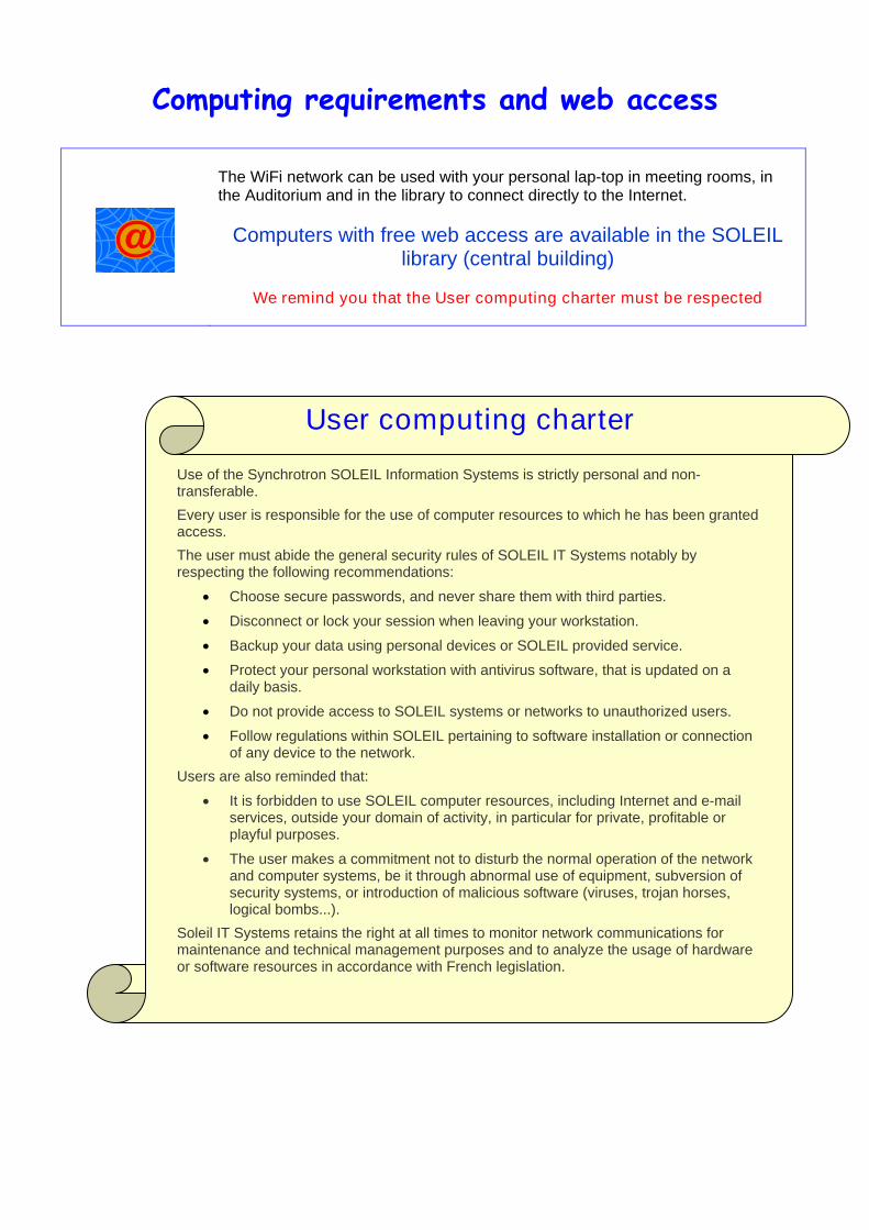
Computing requirements and web access
The WiFi network can be used with your personal lap-top in meeting rooms, in the Auditorium and in the library to connect directly to the Internet.
Computers with free web access are available in the SOLEIL
library (central building)
We remind you that the User computing charter must be respected
User computing charter
Use of the Synchrotron SOLEIL Information Systems is strictly personal and non-transferable.
Every user is responsible for the use of computer resources to which he has been granted access.
The user must abide the general security rules of SOLEIL IT Systems notably by respecting the following recommendations:
‚ Choose secure passwords, and never share them with third parties.
‚ Disconnect or lock your session when leaving your workstation.
‚ Backup your data using personal devices or SOLEIL provided service.
‚ Protect your personal workstation with antivirus software, that is updated on a daily basis.
‚ Do not provide access to SOLEIL systems or networks to unauthorized users.
‚ Follow regulations within SOLEIL pertaining to software installation or connection of any device to the network.
Users are also reminded that:
‚ It is forbidden to use SOLEIL computer resources, including Internet and e-mail services, outside your domain of activity, in particular for private, profitable or playful purposes.
‚ The user makes a commitment not to disturb the normal operation of the network and computer systems, be it through abnormal use of equipment, subversion of security systems, or introduction of malicious software (viruses, trojan horses, logical bombs...).
Soleil IT Systems retains the right at all times to monitor network communications for maintenance and technical management purposes and to analyze the usage of hardware or software resources in accordance with French legislation.

Useful phone numbers
SOLEIL contacts Rob MEIJERS È(office): 01 69 35 97 12
Andrew THOMPSON È(office): 01 69 35 96 08 -Ç06 08 25 90 02
William SHEPARD È(office): 01 69 35 96 36 -Ç06 78 41 79 25
Pierre LEGRAND È(office): 01 69 35 96 60
Paloma FERNANDEZ-VARELA È(office): 01 69 35 97 28
PROXIMA1 beamline phone numbers
È 01 69 35 97 38 or 01 69 35 97 39
SOLEIL Guard
È 01 69 35 91 22 or Ç 06 77 20 39 83
SAMU (Emergency services)* È 15
Police* È 17
Pompiers (Fire brigade)* È 18
Appel d’urgence européen (European emergency call)* È 112
Centre Anti-Poison (Poisons unit) È 01 40 05 48 48
Pharmacy Delmas Pharmacy - 48 rue Henri Amodru - 91190 Gif-sur-Yvette
È 01 69 07 50 53
Doctor during working hours Dr Yves PUSCHE - 2 route de Saint-Aubin - 91190 Villiers-le-Bâcle
È 01 69 85 33 13
Doctor at night During the week: from 20:00 to 8:00 the day after
During the week-end: from Saturday 12:00 to Monday 8:00 È 01 69 25 91 91
Orsay hospital Standard: È 01 69 29 75 75
Emergencies: È 01 69 29 75 63 or 01 69 29 75 62
* Free phone number
- For SOLEIL inside calls, you can just dial the four last numbers.
- For external calls using a SOLEIL phone, you have to add “ 0” before dialling the number (for example to call the Guard cell phone dial: 0 06 77 20 39 83)

Accommodation near SOLEIL The 24 participants are accommodated in twin rooms at:
Les Chevaliers Des Balances Hotel Place de la Mairie - 91190 SAINT-AUBIN Tel.: +33 (0)1 69 41 20 55 - Mobile : +33 (0)6 81 20 14 70 Fax : +33 (0)1 69 85 50 75
Receptionnist : - from 6:00 to 22:00 from Monday to Thursday, - from 6:00 to 15:00 on Friday
You can check in from 13:00 and you will have to check out before12:00.
according the distribution below:
Name First name Jansen Silvia Twin
room Torreira Eva Sokoloswka Monika Twin
room Sviridova Ekatarina Ivic Nives Twin
room Dimaragona Maria Al-Tall Yara Twin
room Kaplan Turkoz Burcu Sirigu Serena Twin
room Pasquellato Elisa Morcillo Maria Twin
room Marin Macarena
Name First name
Ye Qilu Twin room Bhati Mugdha
Pos Martin Twin room Amunts Alexey
Spahr Henrik Twin room Zeeh Jean Christophe
Raba Michael Twin room Stjepanovic Goran
Dandois Sebastien Twin room Gouge Jerome
Viegas Aldino Twin room Paulo de Souza Diorge
ATTENTION: Breakfast will be taken at the SOLEIL canteen.
SOLEIL canteen scheduling Week-days Week-ends Breakfast 7:30-9:00 7:30-9:00
Lunch 11:45-13:45 12:30-13:30
Les Chevaliers Des Balances Hotel is about 5 minutes walking distance from SOLEIL.
Hotel

Information about means of transportation
From PARIS and CHARLES-DE-GAULLE AIRPORT
RER B southbound to SAINT-RÉMY-LÈS-CHEVREUSE, stop at "LE GUICHET" station and walk to the bus station, take bus n°269-02 to SACLAY, stop at "L’ORME DES MERISIERS".
Or
RER B southbound to Massy-Palaiseau station then take bus n°91-06 to SAINT-QUENTIN GARE, stop at “L’ORME DES MERISIERS”.
From ORLY AIRPORT Take the ORLYVAL train, stop at ANTONY, and get on the RER B southbound (same as above).
Bus N° Line Company Schedules
269-02 Les Ulis (Essouriau) – Saclay (Mairie-Val d'Albian) T.I.P.S.
http://www.transport-idf.com/frontal?controller=modules.horaires.RechercheFichesHorairesPlans
91-06
Massy (Gare RER) – Direction Montigny le Bretonneux (St Quentin Gare)
ALBATRANS http://www.albatrans.net/
BE CAREFUL ! There are no regular buses on Sunday. We will provide a shuttle service from the hotel to the Massy RER B station upon arrival (at 14:00 on the 14th of September) to SOLEIL, and from the hotel (at 9:00) back to Massy on Sunday the 21st of September. IF YOU NEED TO TAKE THE BUS, PLEASE CHECK IT BEFORE YOUR ARRIVAL (see SCHEDULES). Paris Metro and RER network plans will be given at your arrival. For more information: http://www.ratp.fr