Embed Size (px)
Citation preview

589E.M. Davison et al., Two new species of Amanita from Western Australia
Nuytsia The journal of the Western Australian Herbarium
23: 589–606 Published online 21 November 2013
© Department of Parks and Wildlife 2013 ISSN 2200-2790 (Online)http://florabase.dpaw.wa.gov.au/nuytsia/ ISSN0085-4417(Print)
Amanita lesueurii and A. wadjukiorum (Basidiomycota), two new species from Western Australia, and an expanded description of A. fibrillopes
Elaine M. Davison1,2,5, Laurton E. McGurk1,3, Neale L. Bougher2, Katrina Syme2,4 and Elizabeth L.J. Watkin3
1Department of Environment and Agriculture, Curtin University, GPO Box U1987, Perth, Western Australia 68452Western Australian Herbarium, Department of Parks and Wildlife, Locked Bag 104, Bentley Delivery Centre, Western Australia 6983
3School of Biomedical Sciences, Curtin University, GPO Box U1987, Perth, Western Australia 684541874 South Coast Highway, Denmark, Western Australia 6333
5Corresponding author, email: [email protected]
Abstract
Davison, E.M., McGurk, L.M., Bougher, N.L., Syme, K. & Watkin, E.L.J. Amanita lesueurii and A. wadjukiorum (Basidiomycota), two new species from Western Australia, and an expanded description of A. fibrillopes. Nuytsia 23: 589–606 (2013). Three species of Amanita Pers. are documented from Western Australia. Amanita lesueurii E.M.Davison is described from the mid-west region. It is distinguished by its small to medium fruiting bodies with a white pileus and white universal veil (both of which become vinaceous-buff or grey with age), white gills, short white stipe with a small obconic or turbinate bulb, white partial veil, amyloid, elongate to cylindrical spores, and no clamp connections. Amanita wadjukiorum E.M.Davison is described from the Perth metropolitan area. It has medium to large fruiting bodies with a cream pileus that ages milky coffee to snuff brown, a pale grey or buff universal veil that ages hazel to drab, cream gills, grey to buff stipe with a napiform or fusiform bulb, white to cream to vinaceous-buff partial veil that disappears with age, amyloid, ellipsoid to elongate spores and no clamp connections. Amanita fibrillopes O.K.Mill., which was previously only known from the type locality, is a widespread but misidentified species. It has small to large fruiting bodies with a pale peach to pale salmon pileus that rapidly ages cream, a white universal veil that rapidly ages buff or milky coffee, white gills that age buff, white or pale pink stipe with a spherical or obconic or tapered bulb, white or buff apical partial veil that disappears with age, inamyloid, ellipsoid to elongate spores and no clamp connections. A BLASTn search has shown that there are no exact matches of the nuclear ribosomal internal transcribed spacer (ITS) region of each species with those in GenBank.
Introduction
The genus Amanita Pers. (Agaricales: Amanitaceae) is a large, cosmopolitan and conspicuous part of the fungal flora of many areas. The majority of species are difficult to identify using macroscopic features alone, and microscopic characters such as spore amyloidy, presence of clamp connections, and the microanatomy of the subhymenium and universal veil are very important for identification and constructing diagnostic keys (Bas 1969; Reid 1980; Tulloss 1994; Wood 1997). The genus is divided into two subgenera: subg. Amanita sensu Corner & Bas emend. Bas and subg. Lepidella (E.-J.Gilbert) Veselý emend. Corner & Bas, that are separated by the amyloid reaction of the spores.

590 Nuytsia Vol. 23 (2013)
Many species of Amanita occur in Western Australia (Gentilli 1953; Bas 1969; Reid 1980; Miller 1991, 1992). Of these, some have probably not been discovered and several are yet to be formally named. In this paper we describe two new species from Western Australia, one from the mid-west region and another from the Perth metropolitan area.
Some named species are difficult to identify with certainty because they were described from a small number of collections or localities, so that the range of variation within each species has not been established. Amanita fibrillopes O.K.Mill. (MycoBank number MB 358174), for example, was described from a single collection (Miller 1991), however it appears to be a widespread but misidentified species. Its protologue describes a small species with inamyloid spores and an aerolate pileus covered with dense, pointed brown warts. Whilst reviewing the Miller types, one of us (EMD) was struck by similarities between A. fibrillopes and a common and widespread species Amanita sp. ‘persicina’ ined. (Bougher 2009), concluding that they were the same. They have similar microanatomy and are similar macroscopically apart from the protologue’s apparent incomplete description of the colours of the pileus and universal veil. Amanita sp. ‘persicina’ has a pale peach or salmon pileus when young, but the colour fades rapidly to buff, and the universal veil darkens with age from white to buff to brown. It appears that the type described by Miller (1991) was a collection of small, old Amanita sp. ‘persicina’. In this paper we provide an expanded description of A. fibrillopes based on a larger number of collections than those studied by Miller (1991).
DNA barcodes have become an important identification tool for all organisms. The nuclear ribosomal internal transcribed spacer (ITS) region has been proposed for adoption as the primary fungal barcode marker (Schoch et al. 2012). It has been widely used by mycologists and more than 172,000 fungal ITS sequences are deposited in GenBank. We provide new ITS sequence data for the two new taxa and for A. fibrillopes. Other gene regions provide more resolution in some fungal lineages. The most recent molecular phylogeny of Amanita uses the nuclear and mitochondrial large and small subunits of rRNA gene regions (Wolfe et al. 2012). These regions will provide additional information on relationships in future studies.
Methods
Macroscopic and microscopic observations. Methodology follows that of Tulloss (2008); stipe length is from the bottom of the pileus context to the top of the bulb, stipe width is at mid stipe. Where two dimensions are given, length comes before width. Colours, including the colour of spores in fresh deposit and other shades of white to cream (designated by letters A–G), are from the Royal Botanic Garden, Edinburgh (1969) and colour codes in the form of ‘3A2’ are from Kornerup and Wanscher (1978). Lamella spacing follows Largent (1986). Dried material was rehydrated in 10% NH4OH or 3% KOH and stained in 1% Congo Red. Biometric variables for spores follow Tulloss (2012), i.e. ‘L= the average spore length computed for one specimen examined and the range of such averages, L’ = the average spore length computed for all spores measured, W = the average spore width computed for one specimen examined and the range of such averages, W’ = the average spore length computed for all spores measured, Q = the ratio of length/breadth for a single spore and the range of the ratio of length/breadth for all spores measured, Q = the average value of Q computed for one specimen examined and the range of such averages; Q’ = average value of Q computed for all spores measured’. Authorities for fungal names follows Landcare Research and RBG Kew: Mycology (2013) and for plant names follows Western Australian Herbarium (1998–). Herbarium codes follow Thiers (continuously updated).
Molecular sequences. DNA was extracted from a 3 × 3 mm piece of dried lamella tissue from one sample from each species by a modification of the leaching method of Castalanelli et al. (2010).
591E.M. Davison et al., Two new species of Amanita from Western Australia
Co-extracted inhibitors were removed using chloroform. The ITS region was amplified with primers ITS4 and ITS5 (White et al. 1990) in a Corbett Research Palm-Cycler™ thermocycler (Mortlake, New South Wales, Australia) at 95º C for 2 min followed by 34 cycles of 95º C denaturation for 30 s, 54º C annealing for 1 min, 72º C elongation for 1.5 min, with a single final elongation at 72º C for 5 min. 10 μL of CES inhibitor sequestration mix (Ralser et al. 2006) was included in all PCR reactions. All PCR products were purified using the QIAquick® PCR Purification Kit (QUIAGEN, Pty Ltd, Doncaster, Victoria, Australia) then cloned into the pGEM®-T Easy Vector Systems (Promega Corporation, Sydney, New South Wales, Australia), in accordance with the manufacturers instructions. Ligations were transformed into JM109 High Efficiency Competent Cells (Promega Corporation, Sydney, New South Wales, Australia). The colonies were checked for the correct-sized inserts using SP6 and T7 Promoter Primers (Promega Corporation, Sydney, New South Wales, Australia), and these were screened using ApaL1 restriction enzyme to select for Amanita ITS4 ITS5 products. The PCR products of clones containing Amanita inserts were purified using the QIAquick® PCR Purification Kit according to the manufacturer’s instructions and sequenced using Big Dye® Terminator v3.0 Cycle Sequencing Kit in accordance with ABI protocols (Applied Biosystems, Foster City, California, USA). Sequence data was assembled with Geneious version 5.4 created by Biomatters (undated). At least one cloned sequence for each species has been deposited in GenBank; sequence identifiers and voucher information are given under each species in the paper. They were used as queries of NCBI nucleotide database using BLASTn (National Library of Medicine 2013).
Taxonomy
Amanita lesueurii E.M.Davison, sp. nov.
Type: Lesueur National Park, Shire of Dandaragan, Western Australia, 6 June 2010, E.M. & P.J.N. Davison EMD 14-2010 (holo: PERTH 08351325, sequence GenBank JX398315). (MB 800268).
Pileus 30–72 mm wide, up to 10 mm thick, initially white becoming clay-buff to vinaceous-buff (6B2–C3) with age, no surface staining or bruising, initially convex becoming plane, surface slightly tacky when moist, margin non-striate, appendiculate, becoming decurved with age. Universal veil on pileus as adnate, aerolate, floccose-felted warts or patches becoming adpressed with age, white at first aging pale vinaceous-buff to grey (6B–C2). Lamellae adnate to adnexed, close to subdistant, white and unchanging, 5–10 mm broad, margin concolorous, occasionally fimbriate; lamellulae shortest truncate, longest attenuate, scarce to plentiful, in several lengths. Stipe length 16–30 mm, width 8–21 mm, cylindric or narrowing upwards, solid, white, slightly floccose below partial veil. Partial veil inferior to median to superior, descendant, membranous, adpressed, white unchanging, persistent or disappearing with age. Bulb 5–20 × 10–22 mm, obconic or tapered or turbinate, encrusted with soil. Remains of universal veil at top of bulb not apparent, or as slight, fragile membrane or small, marginate rim, white. Pileus and stipe context white bruising pale vinaceous-buff (6B2). Smell none when young, strong when old. Spore deposit white. (Figure 1)
Basidiospores [200/10/5] (9–)10.5–13(–14) × 5–6.5(–7) µm, (L = 11.1–12.4 µm; L’ = 11.7 µm, W = 5.5–6.2 µm; W’ = 5.8 µm; Q = (1.62–)1.82–2.33(–2.60); Q = 1.90–2.26; Q’ = 2.03), hyaline, colourless, with wall slightly thickened, smooth, amyloid, elongate to cylindric; apiculus sublateral, short, cylindric to truncate-conic, up to 1 × 2 µm, truncate; contents monoguttulate. Pileipellis difficult to delimit in young specimens, up to 250 µm thick in older specimens, initially colourless, becoming pale brown in NH4OH; filamentous hyphae 3–10 µm wide with thick gelatinising walls, radially orientated, periclinal, occasionally interwoven; inflated cells not observed; vascular hyphae 5–10 µm wide, pale yellow, infrequent, occasionally sinuous; clamp connections not observed. Pileus context

592 Nuytsia Vol. 23 (2013)
filamentous, hyphae 3–30 µm wide, with widest constricted at septa, thin-walled, hyaline, dominant; acrophysalides up to 220 × 20 µm, thin-walled, cylindric or clavate, colourless; vascular hyphae 3–10 µm wide, occasionally branching, pale yellow, very infrequent to locally frequent, occasionally sinuous; clamp connections not observed. Lamella trama bilateral, divergent; central stratum when well rehydrated comprising 12–18 % of distance between bases of basidia on opposing hymenial surfaces, central stratum including undifferentiated filamentous hyphae 2–18 µm wide with widest constricted at septa, thin-walled, hyaline; inflated cells not observed; vascular hyphae 2–6 µm wide, rare, pale yellow, occasionally sinuous; clamp connections not observed; subhymenial base with initial angle of divergence from 15° to 30° from central stratum with filamentous hyphae following smooth broad curve to subhymenium, of dominant filamentous hyphae 4–25 µm wide, frequently branched, thin-walled, hyaline; inflated cells infrequent, colourless, up to 80 × 30 µm, clavate or fusiform or ovoid; vascular hyphae 3–5 µm wide, rare, pale yellow, occasionally sinuous; clamp connections not observed; subhymenium with basidia arising terminally from barely inflated to pyriform hyphal segments 5–12 µm wide; clamp connections not observed; lamella edge tissue sterile, with inflated cells pyriform or clavate or capitate, 18–50 × 10–20 µm, colourless, infrequent to frequent; clamp connections not observed. Basidia [100/5/5] (42–)44–63(–75) × (9–)10–13(–14) µm, thin-walled, colourless, about 86% 4-spored, about 6% 3-spored, about 4% 2-spored, about 3% 1-spored, sterigmata up to 20 × 3 µm wide, most less than 6 × 2 µm, clamp connections not observed. Universal veil on pileus is not layered, with elements somewhat erect or irregularly disposed; filamentous undifferentiated hyphae 3–10 µm wide, colourless or pale brown; inflated cells dominant, ellipsoid or ovoid or pyriform (up to 115 × 60 µm), singly or in terminal chains of up to 3 cells in basal part, ellipsoid or globose (up to 45 × 40 µm) in upper part with thin or slightly thickened gelatinising walls, contents colourless or light brown or darker in collapsed cells; vascular hyphae 3–10 µm wide, frequently branching, sinuous, colourless or pale yellow or brown, most frequent nearest pileipellis; clamp connections not observed. Universal veil on stipe base without clear orientation, pale yellow or pale brown in NH4OH; filamentous hyphae 3–12 µm wide hyaline, gelatinising; inflated cells plentiful to dominant, ellipsoid or globose or ovoid or pyriform or clavate or fusiform (up to 65 × 55 µm) in terminal chains of up to 4 cells, with thin or slightly thickened walls; vascular hyphae 3–5 µm wide, pale yellow, infrequent; clamp connections not observed. Stipe context longitudinally acrophysalidic; filamentous hyphae 3–15 µm wide, hyaline; acrophysalides dominant, up to 200 × 40 µm, mainly clavate, thin-walled, colourless; vascular hyphae 5–10 µm wide, occasionally branching, colourless or pale yellow, not particularly concentrated at stipe apex; clamp connections not observed. Partial veil filamentous, hyphae 3–12 µm wide, hyaline, radially orientated, dominant, disarticulating at septa; inflated cells infrequent, clavate, up to 25 × 10 µm, thin-walled colourless; vascular hyphae 2–8 µm wide, pale yellow, frequent; clamp connections not observed. (Figure 2)
A BFigure 1. Amanita lesueurii. General appearance. Images from E.M. Davison 9-2010, PERTH 08351317 (A) and K. Syme 2110, PERTH 8106266 (B). Photographs by E.M. Davison (A) and K. Syme (B).

593E.M. Davison et al., Two new species of Amanita from Western Australia
Diagnostic features. Small to medium fruiting bodies with a white pileus that is partly or completely covered with white, floccose or felted, flat-topped warts, that become vinaceous-buff to grey with age, white gills, a short, white stipe with a small, obconic or turbinate bulb, a white partial veil that is inferior to median to superior. The spores are amyloid and elongate to cylindric, the universal veil on the pileus has elements with a somewhat erect orientation and is composed of dominant inflated cells that are single or in short terminal chains, which have colourless or brown contents. There are no clamp connections.
Other collections examined. WESTERN AUSTRALIA: [localities withheld for conservation reasons] 5 June 2010, E.M. & P.J.N. Davison EMD 9-2010 (PERTH); 5 June 2010, E.M. & P.J.N. Davison EMD 10-2010 (PERTH); 6 June 2010, E.M. & P.J.N. Davison EMD 13-2010 (PERTH); 15 Sep. 2008, K. Syme KS 2110 (PERTH).
Fruiting period. June to September.
Figure 2. Amanita lesueurii. A – spores from spore print; B – squash of basidia and subhymenium; C – lamella edge cells; D –cells from the proximal part of the universal veil on the pileus, squash, vascular hypha is stippled; E – cells from the distal part of the universal veil on the pileus, squash; F – cells from the universal veil at the stipe base, squash; G – cells from the partial veil. Scale bar = 10 µm. Images from E.M. Davison 9-2010, PERTH 08351317 (A, B, G), E.M. Davison 10-2010, PERTH 08351295 (C, F) and E.M. Davison 14-2010, holotype, PERTH 08351325 (D, E).
B
A
C D
E
F
G

594 Nuytsia Vol. 23 (2013)
Distribution and habitat. Solitary to gregarious, in sandy soil in kwongan vegetation and low mulga woodland over scrub; nearby plants include Allocasuarina humilis, Corymbia calophylla, Acacia aneura, Daviesia sp., Eremophila sp. and Melaleuca sp. Occurs in the Geraldton Sandplains and Yalgoo bioregions (Department of the Environment 2013).
Conservation status. To be listed as Priority Two under Department of Parks and Wildlife (DPaW) Conservation Codes for Western Australian Flora (M. Smith pers. comm.). A poorly known taxon.
Etymology. The epithet celebrates Charles-Alexandre Lesueur (1778–1846) botanical artist and natural history painter on the Baudin expedition to Australia (1800–1804).
Suggested common name. Lesueur’s lepidella.
Affinities based on ITS sequence. The sequence JX398315 is 765 bases long. A BLASTn search showed that there are no exact matches in GenBank. The closest match is Amanita sp. LEM 13-2005 JX398328 (93% query coverage, 92% maximum identity).
Notes. The amyloid spores, non-striate, appendiculate margin and floccose universal veil place this species in subg. Lepidella sect. Lepidella sensu Bas (1969). The absence of rows of elongate, inflated cells and absence of a submembranous universal veil place this in Amanita subsect. Solitariae Bas; the absence of clamps, whitish universal veil which forms angular patches and warts on the pileus, together with spores longer than 10 µm place this species in stirps Strobiliformis (Bas 1969).
Within this stirps the spores are of similar length but narrower than A. strobiliformis (Paulet ex Vittad.) Bertill. of Europe and are longer, but of similar width to those of A. centunculus Corner & Bas of south-east Asia. Amanita cinereopannosa Bas of eastern North America has much larger basidiomes, and has a differently coloured pileus and universal veil. All of these species are considered endemic in their ranges (R.E. Tulloss pers. comm.) and have not been recorded in Australia. The ITS sequence does not assist with the placement of this species because the closest match is undescribed.
A number of small to medium white species in sect. Lepidella have been described from Australia. The clampless taxon with elongate to cylindric spores that lacks a clear assignment to stirps in the literature is A. clelandii E.-J.Gilbert. Amanita lesueurii may be similar to A. clelandii, a poorly known species from South Australia. The lectotype (ADW 9173) has been examined by Reid (1980), Grgurinovic (1997) and Wood (1997), and a comparison of their observations with A. lesueurii is given in Table 1. These authors differ in their interpretation of whether the spores of A. clelandii are amyloid. In Reid’s (1980) key the spores are treated as amyloid, however in one place in his description the spores are described as inamyloid, although in another place he describes them as amyloid. Grgurinovic (1997) describes the spores as inamyloid, whilst Wood (1997) describes them as amyloid, and suggests that Reid’s (1980) reference to them being inamyloid is a typographical error, and this seems to be the most likely explanation (Tulloss 2013a). Grgurinovic (1997) describes the type as not well preserved, so that additional collections are needed.
Amanita lesueurii is shorter and squatter than A. clelandii. There are also differences in the universal veil. Its colour is not mentioned in the original collector’s annotation for the lectotype of A. clelandii in which the cap is simply described as white, without reference to the universal veil (Reid 1980). Reid (1980) described the universal veil as being of ‘inconspicuous felty patches near the edge of the cap’ which is quite different from the universal veil of A. lesueurii which is of flat-topped, straight-sided,

595E.M. Davison et al., Two new species of Amanita from Western Australia
aerolate warts or patches. The microscopic structure of the universal veil also differs. Reid (1980) describes that of A. clelandii as ‘comprising a mixture of thin-walled, hyaline, branched, septate hyphae, 4–10 µm in diam., and swollen, ovate or elliptic elements, up to 55 µm long and 30 µm wide. These elements would seem to be irregularly disposed.’ This differs from the structure of the universal veil of A. lesueurii in which the filamentous hyphae are hyaline or pale brown, the inflated cells are dominant, many are in short terminal chains, and many have light brown contents.
On the basis of stature, and the microscopic structure of the universal veil A, lesueurii is considered to differ significantly from A. clelandii.
Amanita wadjukiorum E.M.Davison, sp. nov.
Typus: Kings Park and Botanic Garden, Perth, Western Australia, 22 June 2010, E.M. & P.J.N. Davison EMD 25-2010 (holo: PERTH 08403988, sequences GenBank KF258719, KF258720, KF258721, KF258722). (MB 804576).
Pileus 50–105 mm wide, up to 10 mm thick, initially cream D to clay buff (pale 4A2–6C3) becoming milky coffee to hazel to drab to snuff brown (5D4–6D3–4, 5E4–7) with age, occasionally paler at margin, no surface staining or bruising, initially convex becoming plane with depressed centre, surface slightly tacky when moist, margin non-striate, appendiculate, becoming decurved with age. Universal veil on pileus adnate, floccose, initially crustose breaking into low conical aerolate warts or patches over entire pileus or disc only, becoming adpressed with age, initially white or pale smoke-grey or pale vinaceous-buff or clay-buff (6B2–C1–3) aging hazel to drab (5D3–4, E3–6E4). Lamellae narrowly adnate to adnexed to free, close to subcrowded, cream B–C (pale 3B1–pale 4B1), unchanging, 4–12 mm broad, margin concolorous, fimbriate; lamellulae shortest truncate, longest sub-attenuate, plentiful, in several lengths. Stipe length 30–72 mm, width 8–20 mm, cylindric, flaring or occasionally narrowing upwards, solid, initially white becoming pale smoke-grey to vinaceous-buff (5B1–2), densely covered with white fibrils and floccose scales below partial veil that ages grey to clay-buff to sienna (5B1–5) in some collections. Partial veil apical to superior, descendant, soft, often adpressed, striate above, white to cream (B) to vinaceous-buff (3A2–6B2), disappearing with age. Bulb 10–35 × 25–55 mm, initially napiform or turbinate becoming fusiform, encrusted with soil. Remains of universal veil at top of bulb not apparent, or as small, soft warts or small, free limb, often remaining in soil, white aging pale buff (4A2). Pileus and stipe context white to cream (B) becoming vinaceous-buff to fawn (5B2–C4) in centre of pileus and vinaceous-buff to smoke-grey to mouse-grey (5B2–D1) at base of stipe. Smell none to strong. Spore deposit white to cream (B). (Figure 3)
Basidiospores [220/11/10] (8.0–)9.0–12.0(–14.0) × (5.5–)6.0–7.0(–8.0) µm, (L = 9.1–11.6 µm; L’ = 10.1 µm, W = 5.9–6.7 µm; W’ = 6.4 µm; Q = (1.33–)1.42–1.82(–2.00); Q = 1.49–1.77; Q’ = 1.58), hyaline, colourless, with wall slightly thickened, smooth, amyloid, ellipsoid to elongate; apiculus sublateral, short, cylindric, 0.5–1.5 × 1–1.5 µm, rounded to truncate; contents granular or monoguttulate. Pileipellis up to 400 µm thick in older specimens, with initially colourless gelatinised suprapellis to 150 µm thick becoming brown with age, and pale brown subpellis to 250 µm thick; filamentous hyphae 3–12 µm wide with thick gelatinising walls, radially orientated, periclinal, occasionally interwoven; inflated cells not seen; vascular hyphae 2–13 µm wide, occasionally branching, yellow-brown, very infrequent to frequent, occasionally sinuous; clamp connections not observed. Pileus context filamentous hyphae 3–25 µm wide, with widest constricted at septa, thin-walled, hyaline, dominant; acrophysalides up to 240 × 45 µm, thin-walled, clavate or ventricose, colourless; vascular hyphae 3–12 µm wide, occasionally branching, yellow-brown, infrequent, occasionally sinuous; one clamp connection seen in one collection. Lamella trama bilateral, divergent; central stratum when

596 Nuytsia Vol. 23 (2013)
Tabl
e 1.
Com
paris
on o
f Am
anita
cle
land
ii w
ith A
. les
ueur
ii, b
ased
on
desc
riptio
ns b
y th
ree
auth
ors.
Spec
ies
A. c
lela
ndii
A. l
esue
urii
Ref
eren
ceR
eid
(198
0)G
rgur
inov
ic (1
997)
Woo
d (1
997)
This
pap
er
Col
lect
ion(
s)Le
ctot
ype A
DW
917
3Le
ctot
ype A
DW
917
3A
DW
917
3, U
NSW
83/
203
PERT
H 0
8106
266;
PER
TH 0
8351
295;
PE
RTH
083
5130
9; P
ERTH
083
5131
7;
PERT
H 0
8351
325.
Mac
rosc
opic
cha
ract
ers
Pile
usto
25
mm
, slig
htly
con
vex,
whi
te,
smoo
thto
25
mm
, sl
ight
ly c
onve
x, w
hite
, sm
ooth
to 70
mm
, con
vex t
hen fl
at co
nvex
, sm
ooth
, pa
le d
rab-
buff
to p
ale g
rey-
brow
n, m
argi
n sl
ight
ly a
ppen
dicu
late
to 72
mm
, con
vex t
hen p
lane
, whi
te be
com
-in
g vi
nace
ous-
buff,
mar
gin
appe
ndic
ulat
e
Lam
ella
eju
st re
achi
ng th
e ste
m, w
hite
, clo
seju
st re
achi
ng th
e st
ipe,
clo
se, w
hite
free
, cro
wde
d, w
hite
to p
ale
crea
mad
nate
, whi
te, c
lose
Stip
esl
ende
r, 38
mm
, w
hite
, sl
ight
ly
mea
ly,
base
bul
bous
, ?s
light
ly
mar
gina
te, t
ende
ncy
to b
e ho
llow
to 3
8 m
m,
slen
der,
slig
htly
mea
ly,
base
bul
bous
, ?s
light
ly m
argi
nate
, te
nden
cy to
be
hollo
w, w
hite
to 1
20 m
m, t
hin,
whi
te to
off-
whi
te w
ith
age,
smoo
th, b
ase b
ulbo
us to
turb
inat
e with
sa
ccat
e mem
bran
ous v
olva
, with
clea
r fre
e up
per p
ortio
n
to 60
mm
, whi
te, t
hick
, whi
te, s
light
ly fl
oc-
cose
, bas
e con
ical
to tu
rbin
ate,
imm
argi
nate
or
with
a sl
ight
mar
gina
te ri
m, s
olid
Uni
vers
al v
eil
on
pile
usin
cons
picu
ous
felty
pat
ches
nea
r th
e ed
ge o
f the
cap
-no
ne o
n pi
leus
flat
topp
ed w
arts
or
patc
hes,
floc
cose
be
com
ing
felte
d w
ith a
ge,
whi
te a
ging
vi
nace
ous-
buff
Mic
rosc
opic
cha
ract
ers
Bas
idio
spor
es L
×W
(µm
)11
.2–1
6.8
× 5.
0–5.
6,
9.0–
16.0
× 4
.5–6
.0,
10.6
–16.
0 ×
5.2–
8.0,
Q=2
.0,
11.7
–12.
6(–1
3.5)
× 5
.8–7
.3,
Q=1
.79–
1.91
(9–)
10.5
–13(
–14)
× 5
–6.5
(–7)
, Q=2
.03
Am
yloi
dy1
amyl
oid/
inam
yloi
d in
amyl
oid
amyl
oid
amyl
oid
Bas
idia
L ×
W
(µm
)42
× 1
1, c
lava
te33
.6–4
4.0
× 11
.2–1
4.0
35–4
0 ×
16–2
0(4
2–)4
3–62
(–75
) × (9
–)10
–12(
–13)
Subh
ymen
ium
--
sub
basi
dial
cel
l a li
ttle
infla
ted
of in
flate
d py
rifor
m c
ells
Uni
vers
al v
eil
on
pile
us (s
izes
in µ
m)
mix
ture
of
thin
-wal
led,
hya
line,
br
anch
ed,
sept
ate
hyph
ae 4
–10,
an
d sw
olle
n ov
ate
or e
llipt
ic e
le-
men
ts u
p to
55
× 30
, irr
egul
arly
di
spos
ed
mix
ture
of
thin
-wal
led,
hya
line,
br
anch
ed se
ptat
e fila
men
tous
hyph
ae
and s
wol
len o
void
or el
lipso
idal
cells
mai
nly
fairl
y na
rrow
fila
men
tous
hyp
hae
6–10
, w
ith a
sm
all
quan
tity
of i
nflat
ed
cells
, 30–
50
filam
ento
us h
ypha
e 3–
10 h
yalin
e or
pal
e br
own,
som
ewha
t an
ticlin
al o
rient
atio
n,
with
dom
inan
t infl
ated
cells
to 90
× 50
, with
co
lour
less
or
brow
n co
nten
ts in
term
inal
ch
ains
of u
p to
3 c
ells
1 In R
eid’
s ke
y (R
eid
1980
) A. c
lela
ndii
is k
eyed
out
as
havi
ng a
myl
oid
spor
es, b
ut in
the
desc
riptio
n it
is s
tate
d th
at th
ey a
re n
on-a
myl
oid.
Thi
s is
inte
rpre
ted
by W
ood
(199
7) a
s a
typo
-gr
aphi
cal e
rror
, bec
ause
he
stat
es th
at th
e sp
ores
are
stro
ngly
am
yloi
d. In
cor
resp
onde
nce
from
C. B
as to
R.N
. Hilt
on (8
th M
arch
197
6) d
iscu
ssin
g sp
ecim
en U
WA
200
3 w
hich
, on
the
basi
s of
mac
rosc
opic
al c
hara
cter
s Bas
was
cer
tain
fitte
d in
to se
ct. L
epid
ella
, Bas
foun
d th
at th
e sp
ores
wer
e no
n-am
yloi
d; h
e fu
rther
com
men
ts ‘I
hav
e m
et o
ne si
mila
r cas
e be
fore
in In
dia
and
Gilb
ert d
escr
ibed
A. c
lela
ndii
from
Sou
th A
ustra
lia th
at fi
ts ra
ther
per
fect
ly in
sect
ion
Amid
ella
, but
als
o ha
s non
-am
yloi
d sp
ores
(muc
h sl
ende
rer t
han
in 2
003
how
ever
).’

597E.M. Davison et al., Two new species of Amanita from Western Australia
well-hydrated comprising 12–23% of the distance between bases of basidia on opposing hymenial surfaces, central stratum including undifferentiated filamentous hyphae 3–20 µm wide, with widest constricted at septa, thin-walled, hyaline; inflated cells not observed; vascular hyphae 3–15 µm wide, occasionally branching, colourless or pale yellow, infrequent, occasionally sinuous; clamp connections not observed; subhymenial base with initial angle of divergence from 12° to 30° from central stratum with filamentous hyphae following smooth broad curve to subhymenium, of dominant filamentous hyphae 3–20 µm wide, frequently branched, the widest constricted at the septa, thin-walled, hyaline; inflated cells infrequent, colourless, up to 150 × 25 µm, clavate or cylindric or ovoid; vascular hyphae 2–7 µm wide, occasionally branching, colourless or pale yellow, infrequent to frequent, occasionally sinuous; clamp connections not observed; subhymenium with basidia arising terminally from barely inflated to pyriform hyphal segments 7–14 µm wide; lamella edge tissue sterile, with inflated cells pyriform or ovoid 20–40 × 12–25 µm, colourless, abundant; clamp connections not observed. Basidia [120/6/6] (35–)38–54(–63) × (9–)10–13(–15) µm, thin-walled, colourless, about 85% 4-spored, about 6% 3-spored, about 8% 2-spored, about 1% 1-spored, sterigmata up to 6 × 2 µm wide at base; one clamp connection seen in one collection. Universal veil on pileus is not layered with elements somewhat erect; filamentous undifferentiated hyphae 3–15 µm wide, hyaline or pale brown; inflated cells dominant, ovoid or ellipsoid or pyriform or broadly clavate or spherical (up to 90 × 50 µm, smaller in upper part), singly or in terminal chains of up to 4 cells, with slightly thickened brown walls, contents pale brown; vascular hyphae 3–17 µm wide, occasionally branching, yellow brown, infrequent to frequent, occasionally sinuous; clamp connections not observed. Universal veil on stipe base without clear
A
B C DFigure 3. Amanita wadjukiorum. A – collection; B–D – surface of the pileus showing the change in colour and form of the universal veil with age. Images from E.M. Davison 39-2012, PERTH 08403910 (A), E.M. Davison 25-2010, PERTH 08403988 holotype (B, C) and E.M. Davison 17-2008, PERTH 08403961 (D). Photographs by E.M. Davison.

598 Nuytsia Vol. 23 (2013)
orientation; filamentous hyphae 3–12 µm wide, hyaline; inflated cells plentiful to dominant, clavate or fusiform or spherical or ovoid (up to 130 × 40 µm), with slightly thickened walls, contents pale brown or colourless; vascular hyphae 3–15 µm wide, occasionally branching, pale yellow or yellow-brown, frequent, occasionally sinuous; clamp connections not observed. Stipe context longitudinally acrophysalidic; filamentous hyphae 3–10 µm wide, hyaline; acrophysalides dominant up to 400 × 50 µm, clavate, thin-walled, colourless; vascular hyphae 2–15 µm wide, occasionally branching, pale yellow or yellow-brown, frequent, occasionally sinuous, not concentrated at stipe apex; one clamp connection seen in basal bulb in one collection. Ornamentation on stipe surface filamentous hyphae 3–8 µm wide, hyaline; inflated cells dominant, up to 135 × 35 µm, clavate, colourless or pale brown; clamp connections not observed. Partial veil filamentous hyphae 2–10 µm wide, hyaline; inflated cells dominant to equal, up to 90 × 25 µm, clavate or pyriform, colourless or pale brown; vascular hyphae 1–9 µm wide, branching, pale yellow or yellow-brown, frequent, occasionally sinuous; clamp connections not observed. (Figure 4)
Diagnostic features. Medium to large fruiting bodies with a pileus that is initially cream that becomes milky coffee to drab to snuff-brown with age, and a universal veil that is initially crustose, pale grey or buff that breaks up into patches and low conical warts that become hazel to drab with age, white to cream gills, the stipe is grey to buff with a napiform to fusiform base, and a white to cream to buff partial veil that disappears with age. The spores are amyloid and ellipsoid to elongate, the universal veil on the pileus has elements with a somewhat anticlinal orientation and is composed of dominant inflated cells that are single or in short terminal chains, with brown contents. Clamp connections are absent, or very rare.
Other collections examined. WESTERN AUSTRALIA: [localities withheld for conservation reasons] 4 Aug. 2002, E.M. & P.J.N. Davison EMD 14-2002 (PERTH); 5 July 2003, E.M. & P.J.N. Davison EMD 20-2003 (PERTH); 8 July 2008, E.M. & P.J.N. Davison EMD 14-2008 (PERTH); 21 July 2008, E.M. & P.J.N. Davison EMD 16-2008 (PERTH); 11 Aug. 2008, E.M. & P.J.N. Davison EMD 17-2008 (PERTH); 14 Aug. 2012, E.M. & P.J.N. Davison EMD 39-2012 (PERTH); 26 Aug. 2012, E.M. & P.J.N. Davison EMD 43-2012 (PERTH); 7 Sep. 2012, L.E. McGurk EMD 44-2012 (PERTH); 7 Sep. 2012, L.E. McGurk EMD 45-2012 (PERTH).
Fruiting period. June to September.
Distribution and habitat. Solitary to gregarious, in sandy soil in degraded native vegetation; nearby plants include Allocasuarina fraseriana, Corymbia calophylla, C. citriodora and Brachychiton sp. Occurs in the Swan Coastal Plain SWA2 Perth sub-bioregion (Department of the Environment 2013).
Conservation status. To be listed as Priority Three under DPaW Conservation Codes for Western Australian Flora (M. Smith pers. comm.). A poorly known taxon.
Etymology. Wadjuk is the name for the Nyoongar aboriginal tribal group that roamed within a 50 km radius of Perth, Western Australia. It means ‘the guardians of the link between the land and the sea’. This link is the Swan River that flows through the centre of Perth. Amanita wadjukiorum has not been found outside this region (R.M. Robinson pers. comm.).
Suggested common name. Wadjuk lepidella.

599E.M. Davison et al., Two new species of Amanita from Western Australia
Figure 4. Amanita wadjukiorum A – spores from spore print; B – basidia and subhymenium, gentle squash; C – lamella margin cells, gentle squash; D – universal veil on the pileus, section in the middle of a wart on the pileus, unsquashed; E – top of the same wart in D, unsquashed; F – universal veil at the stipe base, squash; G – ornamentation on the stipe surface; H – very young partial veil, squash. Scale bar = 10 µm. Images from E.M. Davison 17-2008, PERTH 08403961 (A), E.M. Davison 14-2008, PERTH 08403953 (B), E.M. Davison 25-2010, PERTH, 08403988 holotype (C, H), E.M. Davison 14-2002, PERTH 08403945 (D, E), E.M. Davison 16-2008, PERTH 08403899 (F) and E.M. Davison 39-2012, PERTH 08403910 (G).
B
A
C
E
F
G
H
D

600 Nuytsia Vol. 23 (2013)
Affinities based on ITS sequence. The sequences KF258719, KF258720, KF258721 and KF258722 are between 670 and 684 bases long. A BLASTn search showed that there are no exact matches in GenBank. The closest match is A. lesueurii JX398315 (100% query coverage, 82% maximum identity).
Notes. The amyloid spores, non-striate, appendiculate margin and floccose universal veil place this species in subg. Lepidella, sect. Lepidella. The absence of rows of elongate inflated cells and absence of a submembranous universal veil place this in Amanita subsect. Solitariae. The absence of clamps (although a single clamp was seen in three of the ten collections), the universal veil that is initially whitish and forms angular patches and warts on the pileus, together with spores that are about 10 µm long, places this species in stirps Strobiliformis (Bas 1969).
Several medium-sized brown species in sect. Lepidella have been described from Western Australia. The clampless taxon with ellipsoid spores that is most similar is A. walpolei O.K.Mill., which was described from a single collection. However, re-examination of the type has found that clamp connections are frequent in the lamellae, so that it is quite different from A. wadjukiorum.
Amanita wadjukiorum differs from A. lesueurii in its larger size, the colour of the pileus in age which becomes hazel to drab to snuff-brown, compared with vinaceous-buff. It is similar to A. lesueurii in the form and initial colour of the universal veil on the pileus, however this ages hazel to drab in A. wadjukiorum whilst it ages vinceous-buff to grey in A. lesueurii. The lamellae of A. wadjukiorum are cream, whilst those of A. lesueurii are white. The stipe of A. wadjukiorum is longer than that of A. lesueurii; in both species it is initially white, but becomes pale smoke-grey to vinaceous-buff in A. wadjukiorum and is densely covered with fibrils and floccose scales below the partial veil, that age grey to clay-buff to sienna in some collections. The shape of the bulb differs; it is initially napiform or turbinate, becoming fusiform in A. wadjukiorum, whilst it becomes obconic or tapered in A. lesueurii. The flesh of A. wadjukiorum is white to cream, and becomes vinaceous-buff to smoke-grey to mouse-grey at the base of the stipe whilst it is white, bruising pale vinaceous-buff in A. lesueurii. These two species differ in spore shape; spores of A. wadjukiorum are ellipsoid to elongate, whilst those of A. lesueurii are elongate to cylindric. Basidia are of similar size in both species and the structure of the universal veil on the pileus is similar. The partial veil differs however, with inflated cells being more abundant in A. wadjukiorum, compared with A. lesueurii.
On the basis of its larger size, colour of the pileus, universal veil, lamellae, stipe and stipe ornamentation, shape of the bulb, colour changes in the flesh, and spore shape, A. wadjukiorum differs significantly from A. lesueurii.
Gentilli (1953) described A. loricata Gentilli (not validly published) from Kings Park, Perth, Western Australia. The macroscopic description is somewhat similar to A. wadjukiorum in size, colour of the pileus, colour and form of the universal veil, colour, attachment and depth of the lamellae, ornamentation on the stipe surface and fugacious nature of the partial veil. Unfortunately, no microscopic details are given apart from stating that the spores are hyaline, and ellipsoid to ovate. This may represent an early collection of A. wadjukiorum, however no herbarium specimens are known (Reid 1980).
Expanded description of Amanita fibrillopes
Amanita fibrillopes O.K.Mill., Canad. J. Bot. 69: 2700–2703 (1991). Type: Walpole/Nornalup National Park, Western Australia [precise locality withheld for conservation reasons], 10 June 1989, O.K. & H.H. Miller OKM 23890 (holo: VPI: VTMH-152). (MB 358174).

601E.M. Davison et al., Two new species of Amanita from Western Australia
Pileus 32–130 mm wide, up to 13 mm thick, initially pale peach or pale salmon (4B4–C3, 6A3–5, 7B4) (Figure 5B, C) or white with a very pale pink flush at margin (8A2) becoming cream (D) to buff (2A2–C3, 5A2) with age (Figure 5D), no surface staining or bruising, initially convex becoming plano-convex to plane with depressed centre, surface dry, margin entire, non-striate, slightly appendiculate in some specimens, becoming slightly decurved with age. Universal veil on pileus adnate, initially crustose, as aerolate flat-topped or pointed floccose warts to 1.5 × 5 mm in centre and soft, felted scales towards margin, white at first, aging pale buff or clay-pink or vinaceous-buff or milky coffee (2A2, 5B2–D4, 6B3–D6). Lamellae free to narrowly adnate, close to subcrowded, white aging buff (4A2–B4–C3, 5D4), 5–10 mm broad, occasionally bifurcate, margin concolorous, slightly fimbriate; lamellulae truncate, sub-attenuate to attenuate, frequent or infrequent or absent. Stipe length 22–100 mm, width 5–27 mm, cylindric or clavate, flaring at apex in some, initially solid becoming hollow, white aging cream (B–D) to pale buff (4A2, 5A2), bruising vinaceous-buff (6C3) in some specimens, smooth or pulverulent or with mealy floccules below partial veil. Partial veil apical, descendant, soft to felted with upper surface sometimes striate, white, cream (B–D) to buff (3A3, 4A2–B3, 5A2) especially at ragged margin, easily detached. Bulb 15–50 × 12–38 mm, spherical, ovoid, obconic, tapering, occasionally marginate, sometimes stipe simply clavate, encrusted with sand. Remains of universal veil at top of bulb not apparent, or as thin, circumsessile soft ridges, scales or thick warts, white occasionally pale peach (6A2), often remaining in soil. Pileus and stipe context in pileus white, occasionally pale pink beneath pileipellis in young specimens, unchanging or becoming water-soaked with age; in stipe white, becoming cream (D) (4A3) becoming water-soaked in bulb in some specimens. Smell none to mushroom. Spore deposit white to pale cream. (Figure 5)
Basidiospores [240/12/10] (8.5–)9–12(–14) × (5-)6–7.5(–8) µm, (L = 10.1–11.1 µm; L′ = 10.9 µm; W = 6.2–7.0 µm; W′ = 6.6 µm; Q = (1.29–)1.42–1.85(–2.08); Q = 1.49–1.78; Q′ = 1.65), hyaline, colourless, thin-walled, smooth, inamyloid, ellipsoid to elongate, occasionally adaxially flattened; apiculus sub-lateral, truncate, 1 × 1.5 µm; contents monogluttulate. Pileipellis difficult to delimit in young specimens up to 300 µm thick in older specimens, suprapellis colourless up to 180 µm thick partially gelatinised with age, subpellis colourless up to 250 µm thick; filamentous hyphae 2–7 µm wide with slightly thickened walls, hyaline, radially orientated, periclinal, occasionally interwoven; inflated cells not observed; vascular hyphae 3–7 µm wide, occasionally branching, pale yellow, infrequent, sinuous; clamp connections not observed. Pileus context hyphae 3–15 µm wide, thin-walled, hyaline, dominant, mainly with radial orientation; acrophysalides up to 250 × 50 µm, thin-walled, clavate, colourless; vascular hyphae 2–10 µm wide, occasionally branching, colourless, infrequent, sinuous; clamp connections not observed. Lamella trama bilateral, divergent; difficult to rehydrate; central stratum comprising 10–25% of the distance between bases of basidia on opposing hymenial surfaces, central stratum including undifferentiated filamentous hyphae 3–15 µm wide, thin-walled, hyaline; inflated cells not observed; vascular hyphae 3–4 µm wide, branches not observed, pale yellow, very infrequent, some sinuous; clamp connections not observed; subhymenial base with initial angle of divergence about 20º from central stratum with hyphae bending in smooth curve to subhymenium, of dominant filamentous hyphae 3–15 µm wide, frequently branched, thin-walled, hyaline; inflated cells infrequent, colourless up to 100 × 40 µm clavate or pyriform or fusiform; vascular hyphae not observed; clamp connections not observed; subhymenium with basidia arising terminally at first from barely inflated hyphal segments (4–8 µm wide) with these becoming pyriform with age; clamp connections not observed; lamella edge sterile with inflated cells pyriform or clavate or ovoid 25–35 × 10–17 µm, thin-walled, colourless; clamp connections not observed. Basidia [160/9/9] (33–)43–62(–73) × 10–14(–16) µm, walls thin or slightly thickened, colourless, about 90% 4-spored about 5% 3-spored about 5% 2-spored, with sterigmata up to 7 × 2 µm; clamp connections not observed. Universal veil on pileus basal layer thin; filamentous hyphae 3–7 µm wide, hyaline, periclinal orientation; inflated cells dominant spherical or ellipsoid (up to 40 × 40 µm) colourless; vascular hyphae 4–5 µm wide, branches

602 Nuytsia Vol. 23 (2013)
not observed, pale yellow, very infrequent, sinuous; clamp connections not observed. Universal veil on pileus intermediate layer very wide with elements having erect orientation; filamentous hyphae 3–10 µm wide, hyaline, gelatinising; inflated cells dominant, spherical or pyriform or ovoid or ellipsoid or (occasionally) strangulate (up to 80 × 50 µm) in terminal chains of up to 7 cells, colourless or with pale brown contents; vascular hyphae 3–6 µm wide, branches not observed, pale yellow, very infrequent, some sinuous; clamp connections not observed. Universal veil on pileus superficial layer narrow, only found in some immature basidiomes; filamentous hyphae dominant 5–12 µm wide, hyaline, periclinal orientation, gelatinising rapidly; inflated cells not observed; vascular hyphae not observed; clamp connections not observed. Universal veil on stipe base without clear orientation; filamentous hyphae 2–8 µm wide, hyaline, many disarticulating at septa, gelatinising; inflated cells equal, spherical or pyriform (up to 50 × 50 µm, most less than 25 × 25 µm), terminal, singly or in chains of up to 3 cells, colourless; vascular hyphae not observed; clamp connections not observed. Stipe context longitudinally acrophysalidic; filamentous hyphae 2–15 µm wide, hyaline; acrophysalides dominant or equal, up to 275 × 50 µm, clavate or pyriform, colourless; vascular hyphae 2–15 µm wide, occasionally branching, colourless or pale yellow, infrequent to frequent, sinuous, not particularly concentrated at stipe apex; clamp connections not observed. Ornamentation on stipe surface filamentous hyphae 3–10 µm wide, many collapsed and gelatinising; inflated cells dominant, up to 100 × 35 µm, pyriform or clavate,
A
B D
C
Figure 5. Amanita fibrillopes. A – holotype; B – young specimens; C – surface of young pileus showing white universal veil; D – surface of old pileus showing brown universal veil. Images from O.K. Miller 23890, VPI: VTMH-152 , E623 (A), E.M. Davison 10-2008 PERTH 08347387 (B) and E.M. Davison 12-2008 PERTH 08347360 (C, D). Photographs by N.L. Bougher (A), and E.M. Davison (B–D).

603E.M. Davison et al., Two new species of Amanita from Western Australia
colourless; clamp connections not observed. Partial veil filamentous hyphae 3–8 µm wide, hyaline, thin-walled, radially orientated, most collapsed, many disarticulating at septa; inflated cells dominant to equal, pyriform or spherical or ellipsoid or clavate (up to 45–100 µm but most 25–35 × 50 µm) thin-walled, colourless; vascular hyphae not observed; clamp connections not observed. (Figure 6)
Diagnostic features. Small to large fruiting bodies with a pileus that is initially pale peach to pale salmon that rapidly ages cream, and a thick universal veil that is initially crustose, and which breaks up into straight-sided, polygonal flat-topped warts and scales, initially white but becomes buff to clay-pink or vinaceous-buff or milky coffee with age. The gills are white to buff, the stipe is white or pale clay-pink with a spherical, ovoid, obconic or tapered base, and apical white to buff partial veil that is easily detached. The spores are inamyloid, ellipsoid to elongate, the universal veil on the pileus is layered, and the thickest, intermediate layer has elements that have an anticlinal orientation. This layer is composed of dominant inflated cells that are in terminal chains of up to seven cells. Clamp connections are absent. This taxon is illustrated in Bougher (2009) as Amanita sp. ‘persicina’.
Collections examined. WESTERN AUSTRALIA: [localities withheld for conservation reasons] 21 May 2006, N.L. Bougher Bougher 187 (PERTH); 12 June 1988, E.M. & P.J.N. Davison EMD 9-1988 (PERTH); 25 June 2006, E.M. & P.J.N. Davison EMD 8-2006 (PERTH); 18 May 2008, E.M. & P.J.N. Davison EMD 9-2008 (PERTH, nrITS RNA sequence GenBank JX398314); 18 May 2008, E.M. & P.J.N. Davison EMD 10-2008 (PERTH, PERTH); 1 June 2008, E.M. & P.J.N. Davison EMD 12-2008 (PERTH); 28 May 2010, E.M. & P.J.N. Davison EMD 4-2010 (PERTH); 18 June 2006, J. Keeble & A. Francis EMD 6-2006 (PERTH); 20 May 2006, P. Robertson E 8315 (PERTH).
Fruiting period. April to July.
Distribution and habitat. Solitary or gregarious, in sandy or gravelly soil in dry sclerophyll forest and Banksia woodland, or in humus rich soil in seasonally wet eucalypt and paperbark woodland, often associated with Eucalyptus marginata, E. jacksonii, Allocasuariana fraseriana, Corymbia calophylla, Melaleuca preissiana and Agonis sp. Amanita fibrillopes is a distinctive species that is widely distributed and common. It occurs in the Swan Coastal Plain, Jarrah Forest and Warren bioregions (Department of the Environment 2013). It has not been recorded in South Australia (Grgurinovic 1997) or eastern Australia (Wood 1997).
Conservation status. To be listed as Priority Three under DPaW Conservation Codes for Western Australian Flora (M. Smith pers. comm.). A poorly known taxon..
Suggested common name. Peach amanita.
Affinities based on ITS sequence. The sequence JX398314 is 705 bases long. A BLASTn search showed that there are no exact matches in GenBank. The closest named match is A. multisquamosa Peck AB103329 (95% query coverage, 82% maximum identity) from sect. Amanita.
Notes. In 1975 R.N. Hilton (University of Western Australia) sent herbarium material of a salmon/cinnamon coloured Amanita (not seen by us) from the south-west of Western Australia to Bas at Leiden. Part of this collection (PERTH 7575386) was retained in Western Australia. Bas responded (letter dated 8 March 1976) that he believed it to be a new species, suggesting the informal name Amanita ‘persicina’ ined. to reflect its colour. It had amyloid spores, and Bas placed it in subg. Lepidella sect. Amidella E.-J.Gilbert. The name Amanita sp. ‘persicina’ has however also been used in Western
604 Nuytsia Vol. 23 (2013)
Australia for pink- or peach-coloured amanitas with inamyloid spores which are placed in subg. Amanita (Bougher 2009). Thus the name has been used for two species with similar colouration but different microanatomy (R.E. Tulloss pers. comm.). Amanita ‘persicina’ sensu Bas (PERTH 7575386) awaits description.
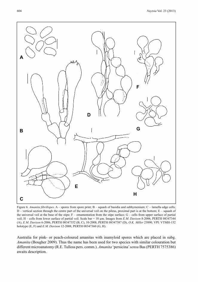
Figure 6. Amanita fibrillopes. A – spores from spore print; B – squash of basidia and subhymenium; C – lamella edge cells; D – vertical section through the centre part of the universal veil on the pileus, proximal part is at the bottom; E – squash of the universal veil at the base of the stipe; F – ornamentation from the stipe surface; G – cells from upper surface of partial veil; H – cells from lower surface of partial veil. Scale bar = 10 µm. Images from E.M. Davison 8-2006, PERTH 08347344 (A), E.M. Davison 6-2006, PERTH 08347352 (B, C), 10-2008, PERTH 08347387 (D), O.K. Miller 23890, VPI: VTMH-152 holotype (E, F) and E.M. Davison 12-2008, PERTH 08347360 (G, H).
B
A
C
E
F
G
H
D

605E.M. Davison et al., Two new species of Amanita from Western Australia
Amanita fibrillopes is placed within subg. Amanita because of the inamyloid spores, and in sect. Amanita because of the eccentric primordium, which results in a distinct bulb at the base of the stipe (Tulloss 2013b). This placement is supported by the nrITS RNA sequence. It is placed in the provisional subsect. Amanitella (Earle) Tulloss & Zhu L. Yang comb. prov. [ser. Farinosae Tulloss and Zhu L. Yang nom. prov.] stirps Roseitincta based on pigmentation, anatomy of the universal veil and pileipellis (R.E. Tulloss pers. comm.).
Acknowledgements
The Massey Herbarium, Virginia Polytechnic Institute and State University (VPI) is thanked for the loan of the type of Amanita fibrillopes (VPI: VTMH-152). R.E. Tulloss is thanked for helpful discussions concerning Amanita ‘persicina’ ined. sensu Bas, the abundance of clamp connections, for suggesting placement of A. fibrillopes within sect. Amanita, and numerous suggestions which improved this manuscript. C. Rodriguez is thanked for editing the description of A. lesueurii. R.M. Robinson kindly provided additional information about the distribution and fruiting period of A. fibrillopes. LEM was supported by a Curtin University Postgraduate scholarship. This work has been supported in part by Australian Biological Resources Study grant CN211-40. We thank the Nuytsia editors for assistance with the final form of the manuscript.
References
Bas, C. (1969). Morphology and subdivision of Amanita and a monograph of its section Lepidella. Persoonia 5: 285–579. Bougher, N.L. (2009). Fungi of the Perth region and beyond. (Western Australian Naturalists’ Club (Inc.): Perth.)Castalanelli, M.A., Severtson, D.L., Brumley, C.J., Szito, A., Foottit, R.G., Grimm, M., Munyard, K. & Groth, D.M. (2010).
A rapid non-destructive DNA extraction method for insects and other arthropods. Journal of Asia-Pacific Entomology 13: 243–249.
Department of the Environment (2013). Interim Biogeographic Regionalisation for Australia (IBRA) Version 7. http://www.environment.gov.au/topics/land/national-reserve-system/science-maps-and-data/australias-bioregions-ibra%C2%A0 [accessed 19 March 2013].
Geneious version 5.4 created by Biomatters. (undated). http://www.geneious.com/ [accessed 7 August 2012].Gentilli, J. (1953). Amanitas from King’s Park, Perth. The Western Australian Naturalist 4: 25–34.Grgurinovic, C.A. (1997). Larger fungi of South Australia. (The Botanic Gardens of Adelaide and State Herbarium: Adelaide.)Kornerup, A. & Wanscher, J.H. (1978). Methuen handbook of colour. (Methuen: London.)Landcare Research & RBG Kew: Mycology (2013). http://www.indexfungorum.org [accessed 30 March 2013].Largent, D.L. (1986). How to identify mushrooms to genus I: macroscopic features. (Mad River Press Inc.: Eureka, CA 95501.)Miller, O.K. (1991). New species of Amanita from Western Australia. Canadian Journal of Botany 69: 2692–2703.Miller, O.K. (1992). Three new species of Amanita from Western Australia. Mycologia 84: 679–686.National Library of Medicine (2013). http://www.ncbi.nlm.nih.gov/blast/Blast.cgi?CMD=Web&PAGE_TYPE=BlastHome
[accessed 4 April 2013]. Ralser, M., Querfurth, R., Warnatz, H.-J., Lehrach, H., Yaspo, M.-L. & Krobitsch S. (2006). An efficient and economic enhancer
mix for PCR. Biochemical and Biophysical Research Communications 347: 747–751.Reid, D.A. (1980). A monograph of the Australian species of Amanita Pers. ex Hook. (Fungi). Australian Journal of Botany,
Supplementary Series No. 8: 1–97.Royal Botanic Garden, Edinburgh (1969). Flora of British fungi: colour identification chart. (Her Majesty’s Stationery Office:
Edinburgh.)Schoch, C.L., Seifert, K.A., Huhndorf, S., Robert, V., Spouge, J.L., Levesque, C.A., Chen, W. & Fungal Barcoding Consortium
(2012). Nuclear ribosomal internal transcribed spacer (ITS) region as a universal DNA barcode marker for Fungi. Proceedings of the National Academy of Sciences 109: 6241–6246.
Theirs, B. (continuously updated). Index Herbariorum: A global directory of public herbaria and associated staff. New York Botanical Garden’s Virtual Herbarium. http://sweetgum.nybg.org/ih/ [accessed 30 March 2013].

606 Nuytsia Vol. 23 (2013)
Tulloss, R.E. (1994). Type studies in Amanita Section Vaginatae I: some taxa described in this Century (studies 1–23) with notes on description of spores and refractive hyphae in Amanita. Mycotaxon 52: 305–396.
Tulloss R.E. (2008). Notes on methodology for study of Amanita (Agaricales). In: Tulloss, R.E & Yang, Z.L. Studies in the genus Amanita Pers. (Agaricales, Fungi). http://pluto.njcc.com/~ret/amanita/mainaman.html [accessed 1 August 2009].
Tulloss R.E. (2012). Biometric variables. http://www.amanitaceae.org/?How To’s&howto=8 [accessed 26 March 2013].Tulloss, R.E. (2013a). Amanita clelandii. http://www.amanitaceae.org/?Amanita+clelandii [accessed 30 March 2013].Tulloss, R.E. (2013b). Sections of the genus Amanita. http://www.amanitaceae.org/?Sections+of+Amanita [accessed 3 April 2013].Western Australian Herbarium (1998–). FloraBase—the Western Australian Flora. Department of Parks and Wildlife. http://
florabase.dpaw.wa.gov.au [accessed 1 July 2013].White, T.J., Bruns, T.D., Lee, S. & Taylor, J.W. (1990). Amplification and direct sequencing of fungal ribosomal RNA genes
for phylogenetics. In: Innis, M.A., Gelfland, D.H., Sninsky, J.J. & White, T.J. (eds) PCR protocols: a guide to methods and applications. pp. 315–322. (Academic Press: New York.)
Wolfe, B.E., Tulloss, R.E. & Pringle, A. (2012). The irreversible loss of a decomposition pathway marks the single origin of an ectomycorrhizal symbiosis. PLoS ONE 7(7): e39597. doi: 10.137/journal.pone.0039597
Wood, A.E. (1997). Studies in the genus Amanita (Agaricales) in Australia. Australian Systematic Botany 10: 723–854.


![Nuytsia - FloraBase · 2016. 11. 17. · 80 Nuytsia Vol. 18 (2008) Typus: Porongurup Range [precise locality withheld for conservation reasons], 3 December 1988, G.J. Keighery 10416](https://img.dokumen.tips/doc/110x75/614a6cc212c9616cbc69682e/nuytsia-florabase-2016-11-17-80-nuytsia-vol-18-2008-typus-porongurup.jpg)

![aia usa Nuytsia - FloraBase—the Western Australian Flora · Type: c. 35 km east-northeast of Newman, Western Australia [precise locality withheld for conservation reasons], 26 April](https://img.dokumen.tips/doc/110x75/60b1611b00293b58fe114a3c/aia-usa-nuytsia-florabaseathe-western-australian-flora-type-c-35-km-east-northeast.jpg)