Embed Size (px)
Citation preview

ELISA Handbook
Principle, Troubleshooting, Sample Preparation
and Assay Protocols ……
https://www.bosterbio.com/ebooks

1
Table of Contents Page
1. Introduction ………………………………………………………………………... 2
2. General ELISA Procedure …………………………………………………………... 2
3. ELISA Types ………………………………………………………………………… 3
Direct ELISA ………………………………………………………………….… 4
Indirect ELISA …………………………………………………………………… 4
Sandwich ELISA ………………………………………………………………… 5
Competitive ELISA ……………………………………………………………… 5
4. ELISA Data Interpretation ……………………………………………………………. 6
5. Sample Preparation ……………………………………………………………………. 7
Cell Culture Supernatants …………………………………………………………. 7
Cell Extracts ……………………………………………………………………… 7
Conditioned Media ……………………………………………………………….. 7
Tissue Extract ……………………………………………………………………... 8
6. Recommended Protocols ………………………………………………………………. 9
Reagent Preparation ………………………………………………………………... 9
Sandwich ELISA …………………………………………………………………… 10
Indirect ELISA ……………………………………………………………………. 12
Direct ELISA ……………………………………………………………………… 14
Competitive ELISA ………………………………………………………………… 16
7. Troubleshooting Guide ………………………………………………………………….. 19
Weak or No Signal ………………………………………………………………….. 19
Saturated Signal …………………………………………………………………….. 20
High Background ………………………………………………………………….... 21
Low Sensitivity ……………………………………………………………………… 23
Poor Standard Curve ………………………………………………………………... 24
Poor Replicate Data …………………………………………………………………. 25
Inconsistent Assay-to-Assay Results …………………………………………………. 26
Slow Color Development ……………………………………………………………. 26
Plate Imaging Problem ……………………………………………………………… 27
8. FAQs …………………………………………………………………………………….. 28
9. Ordering Information ……………………………………………………………………. 30

2
Introduction
ELISA (enzyme-linked immunosorbent assay) is a plate-based assay technique designed for detecting
and quantifying peptides, proteins, antibodies and hormones. In an ELISA, an antigen must be
immobilized to a solid surface and then complexed with an antibody that is linked to an enzyme.
Detection is accomplished by assessing the conjugated enzyme activity via incubation with a substrate
to produce a measureable product. The most crucial element of the detection strategy is a highly
specific antibody-antigen interaction.
ELISAs are typically performed in 96-well (or 384-well) polystyrene plates, which will passively bind antibodies
and proteins. It is this binding and immobilization of reagents that makes ELISAs so easy to design and
perform. Having the reactants of the ELISA immobilized to the microplate surface makes it easy to separate
bound from non-bound material during the assay. This ability to wash away nonspecifically bound materials
makes the ELISA a powerful tool for measuring specific analytes within a crude preparation.
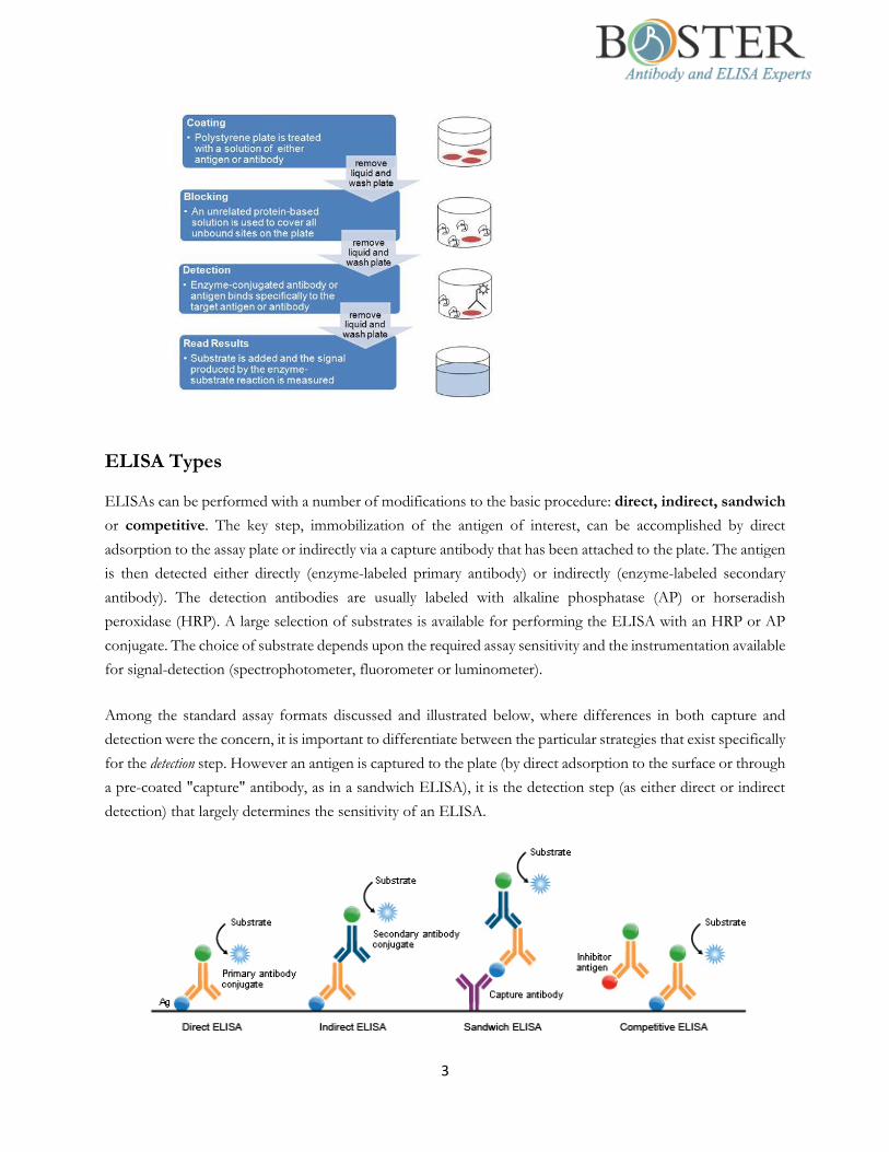
General ELISA Procedure
Unless you are using a kit with a plate that is pre-coated with antibody, an ELISA begins with a coating step,
in which the first layer, consisting of a target antigen or antibody, is adsorbed onto a 96-well polystyrene plate.
This is followed by a blocking step in which all unbound sites are coated with a blocking agent. Following a
series of washes, the plate is incubated with enzyme-conjugated antibody. Another series of washes
removes all unbound antibody. A substrate is then added, producing a calorimetric signal. Finally, the plate is
read.
Because the assay uses surface binding for separation, several washes are repeated in each ELISA step to remove
unbound material. During this process, it is essential that excess liquid is removed in order to prevent the
dilution of the solutions added in the next assay step. To ensure uniformity, specialized plate washers are often
used.
ELISAs can be quite complex and include multiple intervening steps, especially when measuring protein
concentration in heterogeneous samples such as blood. The most complex and varying step in the overall
process is detection, where multiple layers of antibodies can be used to amplify signal.
To be continued on next page
3
ELISA Types
ELISAs can be performed with a number of modifications to the basic procedure: direct, indirect, sandwich
or competitive. The key step, immobilization of the antigen of interest, can be accomplished by direct
adsorption to the assay plate or indirectly via a capture antibody that has been attached to the plate. The antigen
is then detected either directly (enzyme-labeled primary antibody) or indirectly (enzyme-labeled secondary
antibody). The detection antibodies are usually labeled with alkaline phosphatase (AP) or horseradish
peroxidase (HRP). A large selection of substrates is available for performing the ELISA with an HRP or AP
conjugate. The choice of substrate depends upon the required assay sensitivity and the instrumentation available
for signal-detection (spectrophotometer, fluorometer or luminometer).
Among the standard assay formats discussed and illustrated below, where differences in both capture and
detection were the concern, it is important to differentiate between the particular strategies that exist specifically
for the detection step. However an antigen is captured to the plate (by direct adsorption to the surface or through
a pre-coated "capture" antibody, as in a sandwich ELISA), it is the detection step (as either direct or indirect
detection) that largely determines the sensitivity of an ELISA.

4
1. Direct ELISA
For direct detection, an antigen coated to a multi-well plate is detected by an antibody that has been directly
conjugated to an enzyme. This detection method is a good option if there is no commercially available
ELISA kits for your target protein.
Advantages
Quick because only one antibody and fewer steps are used.
Cross-reactivity of secondary antibody is eliminated.
Disadvantages
Immunoreactivity of the primary antibody might be adversely affected by labeling with enzymes or
tags.
Labeling primary antibodies for each specific ELISA system is time-consuming and expensive.
No flexibility in choice of primary antibody label from one experiment to another.
Minimal signal amplification.
2. Indirect ELISA
For indirect detection, the antigen coated to a multi-well plate is detected in two stages or layers. First an
unlabeled primary antibody, which is specific for the antigen, is applied. Next, an enzyme-labeled secondary
antibody is bound to the first antibody. The secondary antibody is usually an anti-species antibody and is
often polyclonal. The indirect assay, the most popular format for ELISA, has the advantages and
disadvantages:
Advantages
A wide variety of labeled secondary antibodies are available commercially.
Versatile because many primary antibodies can be made in one species and the same labeled secondary
antibody can be used for detection.
Maximum immunoreactivity of the primary antibody is retained because it is not labeled.
Sensitivity is increased because each primary antibody contains several epitopes that can be bound by
the labeled secondary antibody, allowing for signal amplification.
Disadvantages
Cross-reactivity might occur with the secondary antibody, resulting in nonspecific signal.
An extra incubation step is required in the procedure.

5
3. Sandwich ELISA
Sandwich ELISAs typically require the use of matched antibody pairs, where each antibody is specific for a
different, non-overlapping part (epitope) of the antigen molecule. A first antibody (known as capture
antibody) is coated to the wells. The sample solution is then added to the well. A second antibody (known
as detection antibody) follows this step in order to measure the concentration of the sample. This type of
ELISA has the following advantages:
High specificity: the antigen/analyte is specifically captured and detected
Suitable for complex (or crude/impure) samples: the antigen does not require purification prior to
measurement
Flexibility and sensitivity: both direct or indirect detection methods can be used
4. Competitive ELISA
The key event of competitive ELISA (also known as inhibition ELISA) is the process of competitive
reaction between the sample antigen and antigen bound to the wells of a microtiter plate with the primary
antibody. First, the primary antibody is incubated with the sample antigen and the resulting antibody–antigen
complexes are added to wells that have been coated with the same antigen. After an incubation period, any
unbound antibody is washed off. The more antigen in the sample, the more primary antibody will be bound
to the sample antigen. Therefore, there will be a smaller amount of primary antibody available to bind to the
antigen coated on the well, resulting in a signal reduction. The main advantage of this type of ELISA arises
from its high sensitivity to compositional differences in complex antigen mixtures, even when the specific
detecting antibody is present in relatively small amounts.

6
Summary of Key Steps in Different ELISA Types
Indirect Direct Sandwich Competitive
Capture Ab Coating X X √ X
Antigen Coating √ √ X √
Blocking √ √ √ √
Sample (Antigen) Incubation X X √ √
Primary Ab Incubation √ √ √ √
Secondary Ab Incubation √ X √ √
Substrate Prep √ √ √ √
Signal Detection √ √ √ √
Data Analysis √ √ √ √
ELISA Data Interpretation
The ELISA assay yields three different types of data output:
1) Quantitative: ELISA data can be interpreted in comparison to a standard curve (a serial dilution of a known,
purified antigen) in order to precisely calculate the concentrations of antigen in various samples.
2) Qualitative: ELISAs can also be used to achieve a yes or no answer indicating whether a particular antigen
is present in a sample, as compared to a blank well containing no antigen or an unrelated control antigen.
3) Semi-Quantitative: ELISAs can be used to compare the relative levels of antigen in assay samples, since
the intensity of signal will vary directly with antigen concentration.
ELISA data is typically graphed with optical density vs log concentration to produce a sigmoidal curve as shown
below. Known concentrations of antigen are used to produce a standard curve and then this data is used to
measure the concentration of unknown samples by comparison to the linear portion of the standard curve. In
fact, it is the relatively long linear region of the curve that makes the ELISA results accurate and reproducible.
The unknown concentration can be determined directly on the graph or with curve fitting software which is
typically found on ELISA plate readers.

7
Sample Preparation
The procedure below provides a general guidance for the preparation of commonly tested samples for use in
ELISA assays. At Boster, we are working on our detailed sample preparation protocols that cover more than 20 sample types
and expecting to update this handbook in the near future. Please check with the literature for experiments similar to
yours for your new assay development. Generally:
Protein extract concentration is at least 1-2 mg/mL.
Cell and tissue extracts are diluted by 50% with binding buffer.
Samples are centrifuged at 10,000 rpm for 5 min at 4°C to remove any precipitate before use.
1. Cell Culture Supernatants
Centrifuge cell culture media at 1,500 rpm for 10 min at 4°C. Aliquot supernatant immediately and hold at
-80°C, avoiding freeze/thaw cycles.
2. Cell Extracts
Place tissue culture plates on ice. Remove the media and gently wash cells once with ice-cold PBS. Remove
the PBS and add 0.5 ml extraction buffer per 100 mm plate. Tilt the plate and scrape the cells into a pre-
chilled tube. Vortex briefly and incubate on ice for 15-30 min. Centrifuge at 13,000 rpm for 10 min at 4°C
(this creates a pellet from the insoluble content). Aliquot the supernatant into clean, chilled tubes (on ice)
and store samples at -80°C, avoiding freeze/thaw cycles.
3. Conditioned Media
Plate the cells in complete growth media (with serum) until the desired level of confluence is achieved.
Remove the growth media and gently wash cells using 2- 3 mL of warm PBS. Repeat the wash step. Remove
the PBS and gently add serum-free growth media. Incubate for 1-2 days. Remove the media into a centrifuge
tube. Centrifuge at 1,500 rpm for 10 min at 4°C. Aliquot the supernatant and keep samples at -80°C, avoiding
freeze/thaw cycles.
To be continued on next page

8
4. Tissue Extract
Mince tissue on ice in ice-cold buffer, preferably in the presence of protease inhibitors. Place the tissue in
micro-centrifuge tubes and dip into liquid nitrogen to snap freeze. Keep samples at -80°C for later use or
keep on ice for immediate homogenization.
For every 5 mg of tissue, add 300 µL of extraction buffer to the tube and homogenize:
100 mM Tris, pH 7.4
150 mM NaCl
1 mM EGTA
1 mM EDTA
1% Triton X-100 0.5%
0.5% sodium deoxycholate
(This portion of the buffer can be prepared ahead of time and stored at 4°C. Immediately before use, the
buffer must be supplemented with phosphatase inhibitor cocktail [as directed by manufacturer], protease
inhibitor cocktail [as directed by manufacturer] and PMSF to 1 mM to generate a complete extraction buffer
solution.)
Rinse the blade of the homogenizer twice with 300 µL extraction buffer. Place the sample on a shaker at
4°C for 2 hours.
Centrifuge the sample for 20 min at 13,000 rpm at 4°C. Aliquot the supernatant into pre-chilled tubes sitting
in ice. Keep the samples at -80°C, avoiding freeze/thaw cycles.
Note: Lysis buffer volume must be determined according to the amount of tissue present. Typical
concentration of final protein extract is at least 1 mg/mL.
To be continued on next page

9
Recommended Protocols
Reagent Preparation
1. Standard Solutions
10,000 pg/mL: Add 1 mL of sample diluent buffer into one tube of standard (10 ng per tube) and mix
thoroughly. Note: Store this solution at 4°C for up to 12 hours (or -20°C for 48 hours) and avoid freeze-
thaw cycles.
5,000 pg/mL: Mix 0.3 mL of 10,000 pg/mL with 0.3 mL of sample diluent buffer and mix thoroughly.
2,500 pg/mL: Mix 0.3 mL of 5,000 pg/mL with 0.3 mL of sample diluent buffer and mix thoroughly.
Perform similar dilutions until the standard solutions with these concentrations (pg/mL) are made:
1,250, 625, 312, 156 and 78.
Add 100 µL of each of the diluted standard solutions to the appropriate empty wells. Repeat in duplicate
or triplicate for accuracy.
Note: The standard solutions are best used within 2 hours.
2. Biotinylated Antibody
Calculate the total volume needed for the assay by multiplying 0.1 mL/well and the number of wells
required. Add 2-3 extra wells to the calculated number of wells to account for possible pipetting errors.
Generate the required volume of diluted antibody by performing a 1:100 dilution (For each 1 µL
concentrated antibody, add 99 µL antibody dilution buffer) and mixing thoroughly.
3. Avidin-Biotin-Peroxidase (ABC)
Calculate the total volume needed for the assay by multiplying 0.1 mL/well and the number of wells
required. Add 2-3 extra wells to the calculated number of wells to account for possible pipetting errors.
Generate the required volume of diluted ABC solution by performing a 1:100 dilution (For each 1 µL
concentrated ABC solution, add 99 µL ABC dilution buffer) and mixing thoroughly.
Note: The diluted ABC solution should not be prepared more than 1 hour prior to the experiment.
To be continued on next page

10
Sandwich ELISA
All of the ELISA kits from Boster use the sandwich format and avidin-biotin chemistry. Our ELISA assays
require the dilutions of standard solutions, biotinylated antibody (detection antibody) and avidin-biotin-
peroxidase.
1. Capture Antibody Coating
(These steps are not required if the pre-adsorbed Picokine ELISA kits from Boster are used)
Dilute the capture antibody to a final concentration of 1-10 μg/mL in bicarbonate/carbonate antigen-
coating buffer (100 mM NaHCO3 in deionized water; pH adjusted to 9.6).
Pipette 100 μL of diluted antibody to each well of a microtiter plate.
Cover the plate with adhesive plastic and incubate at 4°C overnight (or 37°C for 30 min).
Remove the coating solution and wash the plate 3X with 200 μL PBS (Phosphate Buffered Saline) buffer
(10 mM Na2HPO4 and 1.8 mM NaH2PO4 in deionized water with 0.2% Tween 20; pH Adjusted to 7.4)
with for 5 minutes each time. The coating/washing solutions can be removed by flicking the plate over
a sink. The remaining drops can be removed by patting the plate on a paper towel or by aspiration. Do
not allow the wells to dry out at any time.
2. Blocking
(These steps are not required if the pre-adsorbed Picokine ELISA kits from Boster are used)
Pipette 200 μL blocking buffer (5% w/v non-fat dry milk in PBS buffer) per well to block residual
protein-binding sites. Alternatively, BSA or BlockACE can be used to replace non-fat dry milk.
Cover the plate with adhesive plastic and incubate for 1-2 hour(s) at 37°C (or at 4°C overnight).
Remove the blocking solution and wash the plate 2X with 200 μL PBS for 5 minutes each time. Flick
the plate and pat the plate as described in the coating step.
3. Reagent Preparation
Prepare for the diluted standard solutions, biotinylated antibody and ABC solutions as shown on p.9.
4. Sample (Antigen) Incubation
Serially dilute the sample with blocking buffer immediately before use. The optimal dilution should be
determined by a titration assay according to the antibody manufacturer.
Pipette 100 μL of each of the diluted sample solutions and control to each empty well. Repeat in duplicate
or triplicate for accuracy. The negative control should be species- and isotype-matched as well as non-
specific immunoglobulin diluted in PBS buffer.
Cover the plate with adhesive plastic and incubate for 2 hours at room temperature.
Remove the content in the wells and wash them 3X with 200 μL PBS buffer for 5 minutes each time.
Flick the plate and pat the plate as described in the coating step.
5. Biotinylated Antibody Incubation
Pipette 100 μL of diluted antibody to the wells with control, standard solutions and diluted samples.

11
Cover the plate with adhesive plastic and incubate for 1 hour at 37°C (or 2 hours at room temperature).
These incubation times should be sufficient to receive a strong signal. However, if a weak signal is
observed, perform incubation overnight at 4°C for a stronger signal.
Remove the content in the wells and wash them 3X with 200 μL PBS for 5 min each time. Flick the plate
and pat the plate as described in the coating step.
6. ABC Incubation
Pipette 100 μL of diluted ABC solution to the wells with control, standard solutions and diluted samples.
Cover the plate with adhesive plastic and incubate for 0.5 hour at 37°C.
Remove the content in the wells and wash them 3X with 200 μL PBS buffer for 5 min each time. Flick
the plate and pat the plate as described in the coating step.
7. Substrate Preparation
Prepare the substrate solution immediately before use or bring the pre-made substrate to room temperature.
The two widely used enzymes for signal detection are horse radish peroxidase (HRP) and alkaline
phosphatase (AP), and their corresponding substrates, stopping solutions, detection absorbance
wavelengths and color developed are as follows:
Enzyme Substrate* Stop Solution Absorbance (nm) Color Developed
HRP TMB 2M H2SO4 450 Yellow
AP pNPP 0.75M NaOH 405 Yellow
* TMB: 3,3’,5,5’-tetramethylbenzidine; pNPP: p-nitrophenyl-phosphate
Note:
The TMB substrate must be kept at 37°C for 30 min before use.
Hydrogen peroxide can also act as a substrate for HRP.
Sodium azide is an inhibitor of HRP. Do not include the azide in buffers or wash solutions if HRP-labeled
conjugate is used for detection.
8. Signal Detection
Pipette 90 μL of substrate solution to the wells with the control, standard solutions and diluted samples.
Incubate the plate at 37°C in the dark. If TMB is used, shades of blue will be observed in the wells with
the most concentrated solutions. Other wells may show no obvious color.
Color should be developed in positive wells after 15 min. After sufficient color development, pipette 100
μL of stop solution to the appropriate wells (if necessary).
Read the absorbance (OD: Optical Density) of each well with a plate reader.
9. Data Analysis
Prepare a standard curve using the data produced from the diluted standard solutions. Use absorbance on
the Y-axis (linear) and concentration on the X-axis (log scale).
Interpret the sample concentration from the standard curve.

12
Indirect ELISA
This is a general protocol in which antigen coating and blocking may not be required if the wells from the
manufacturer have been pre-adsorbed with the antigen.
1. Antigen Coating
Dilute purified antigens to a final concentration of 1-10 μg/mL in bicarbonate/carbonate antigen-coating
buffer (100 mM NaHCO3 in deionized water; pH adjusted to 9.6).
Pipette 100 μL of diluted antigen to each well of a microtiter plate.
Cover the plate with adhesive plastic and incubate at 4°C overnight (or 37°C for 30 min).
Remove the coating solution and wash the plate 3X with 200 μL PBS (Phosphate Buffered Saline) buffer
(10 mM Na2HPO4 and 1.8 mM NaH2PO4 in deionized water with 0.2% Tween 20; pH Adjusted to 7.4)
with for 5 minutes each time. The coating/washing solutions can be removed by flicking the plate over
a sink. The remaining drops can be removed by patting the plate on a paper towel or by aspiration. Do
not allow the wells to dry out at any time.
2. Blocking
Pipette 200 μL blocking buffer (5% w/v non-fat dry milk in PBS buffer) per well to block residual
protein-binding sites. Alternatively, BSA or BlockACE can be used to replace non-fat dry milk.
Cover the plate with adhesive plastic and incubate for 1-2 hour(s) at 37°C (or at 4°C overnight).
Remove the blocking solution and wash the plate 2X with 200 μL PBS for 5 minutes each time. Flick
the plate and pat the plate as described in the coating step.
3. Reagent Preparation
Prepare for the diluted standard solutions as shown on p.9.
4. Primary Antibody Incubation
Serially dilute the primary antibody of choice with blocking buffer. The optimal dilution should be
determined by a titration assay according to the antibody manufacturer.
Pipette 100 μL of each diluted antibody per well. Repeat in duplicate or triplicate for accuracy. The
negative control should be species- and isotype-matched as well as non-specific immunoglobulin diluted
in PBS buffer.
Cover the plate with adhesive plastic and incubate for 1 hour at 37°C (or 2 hours at room temperature).
These incubation times should be sufficient to receive a strong signal. However, if a weak signal is
observed, perform incubation overnight at 4°C for a stronger signal.
Remove the diluted antibody solution and wash the wells 3X with 200 μL PBS for 5 min each time. Flick
the plate and pat the plate as described in the coating step.
5. Secondary Antibody Incubation
Serially dilute the conjugated secondary antibody with blocking buffer immediately before use. The
optimal dilution should be determined by a titration assay according to the antibody manufacturer.

13
Pipette 100 μL of diluted secondary antibody solution to each well.
Cover the plate with adhesive plastic and incubate for 2 hours at room temperature.
Remove the content in the wells and wash them 3X with 200 μL PBS buffer for 5 min each time. Flick
the plate and pat the plate as described in the coating step.
6. Substrate Preparation
Prepare the substrate solution immediately before use or bring the pre-made substrate to room temperature.
The two widely used enzymes for signal detection are horse radish peroxidase (HRP) and alkaline
phosphatase (AP), and their corresponding substrates, stopping solutions, detection absorbance
wavelengths and color developed are as follows:
Enzyme Substrate* Stop Solution Absorbance (nm) Color Developed
HRP TMB 2M H2SO4 450 Yellow
AP pNPP 0.75M NaOH 405 Yellow
* TMB: 3,3’,5,5’-tetramethylbenzidine; pNPP: p-nitrophenyl-phosphate
Note:
The TMB substrate must be kept at 37°C for 30 min before use.
Hydrogen peroxide can also act as a substrate for HRP.
Sodium azide is an inhibitor of HRP. Do not include the azide in buffers or wash solutions if HRP-labeled
conjugate is used for detection.
7. Signal Detection
Pipette 90 μL of substrate solution to the wells with the control and standard solutions.
Incubate the plate at 37°C in the dark. If TMB is used, shades of blue will be observed in the wells with
the most concentrated solutions. Other wells may show no obvious color.
Color should be developed in positive wells after 15 min. After sufficient color development, pipette 100
μL of stop solution to the wells (if necessary).
Read the absorbance (OD: Optical Density) of each well with a plate reader.
8. Data Analysis
Prepare a standard curve using the data produced from the diluted standard solutions. Use absorbance on
the Y-axis (linear) and concentration on the X-axis (log scale).
Interpret the sample concentration from the standard curve.
To be continued on next page
14
Direct ELISA
This is a general protocol in which antigen coating and blocking may not be required if the wells from the
manufacturer have been pre-adsorbed with the antigen.
1. Antigen Coating
Dilute purified antigens to a final concentration of 1-10 μg/ml in bicarbonate/carbonate antigen-coating
buffer (100 mM NaHCO3 in deionized water; pH adjusted to 9.6).
Pipette 100 μL of diluted antigen to each well of a microtiter plate.
Cover the plate with adhesive plastic and incubate at 4°C overnight (or 37C for 30 min).
Remove the coating solution and wash the plate 3X with 200 μL PBS (Phosphate Buffered Saline) buffer
(10 mM Na2HPO4 and 1.8 mM NaH2PO4 in deionized water with 0.2% Tween 20; pH Adjusted to 7.4)
with for 5 minutes each time. The coating/washing solutions can be removed by flicking the plate over
a sink. The remaining drops can be removed by patting the plate on a paper towel or by aspiration. Do
not allow the wells to dry out at any time.
2. Blocking
Pipette 200 μL blocking buffer (5% w/v non-fat dry milk in PBS buffer) per well to block residual
protein-binding sites. Alternatively, BSA or BlockACE can be used to replace non-fat dry milk.
Cover the plate with adhesive plastic and incubate for 1-2 hour(s) at 37°C (or at 4°C overnight).
Remove the blocking solution and wash the plate 2X with 200 μL PBS for 5 min each time. Flick the
plate and pat the plate as described in the coating step.
3. Reagent Preparation
Prepare for the diluted standard solutions as shown on p.9.
4. Primary Antibody Incubation
Serially dilute the conjugated primary antibody with blocking buffer immediately before use. The optimal
dilution should be determined by a titration assay according to the antibody manufacturer.
Pipette 100 μL of diluted secondary antibody solution to each well.
Cover the plate with adhesive plastic and incubate for 2 hours at room temperature.
Remove the content in the wells and wash them 3X with 200 μL PBS buffer for 5 min each time. Flick
the plate and pat the plate as described in the coating step.
5. Substrate Preparation
Prepare the substrate solution immediately before use or bring the pre-made substrate to room temperature.
The two widely used enzymes for signal detection are horse radish peroxidase (HRP) and alkaline
phosphatase (AP), and their corresponding substrates, stopping solutions, detection absorbance
wavelengths and color developed are as follows:

15
Enzyme Substrate* Stop Solution Absorbance (nm) Color Developed
HRP TMB 2M H2SO4 450 Yellow
AP pNPP 0.75M NaOH 405 Yellow
* TMB: 3,3’,5,5’-tetramethylbenzidine; pNPP: p-nitrophenyl-phosphate
Note:
The TMB substrate must be kept at 37°C for 30 min before use.
Hydrogen peroxide can also act as a substrate for HRP.
Sodium azide is an inhibitor of HRP. Do not include the azide in buffers or wash solutions if HRP-
labeled conjugate is used for detection.
6. Signal Detection
Pipette 90 μL of substrate solution to the wells with the control and standard solutions.
Incubate the plate at 37°C in the dark. If TMB is used, shades of blue will be observed in the wells with
the most concentrated solutions. Other wells may show no obvious color.
Color should be developed in positive wells after 15 min. After sufficient color development, pipette 100
μL of stopping solution to the wells (if necessary).
Read the absorbance (OD: Optical Density) of each well with a plate reader.
7. Data Analysis
Prepare a standard curve using the data produced from the diluted standard solutions. Use absorbance on
the Y-axis (linear) and concentration on the X-axis (log scale).
Interpret the sample concentration from the standard curve.
To be continued on next page

16
Competitive ELISA
This is a general protocol in which antigen coating and blocking may not be required if the wells from the
manufacturer have been pre-adsorbed with the antigen.
1. Antigen Coating
Dilute purified antigens to a final concentration of 20 μg/ml in bicarbonate/carbonate antigen-coating
buffer (100 mM NaHCO3 in deionized water; pH adjusted to 9.6).
Pipette 100 μL of diluted antigen to each well of a microtiter plate.
Cover the plate with adhesive plastic and incubate at 4°C overnight (or 37°C for 30 min).
Remove the coating solution and wash the plate 3X with 200 μL PBS (Phosphate Buffered Saline) buffer
(10 mM Na2HPO4 and 1.8 mM NaH2PO4 in deionized water with 0.2% Tween 20; pH Adjusted to 7.4)
with for 5 minutes each time. The coating/washing solutions can be removed by flicking the plate over a
sink. The remaining drops can be removed by patting the plate on a paper towel or by aspiration. Do not
allow the wells to dry out at any time.
2. Blocking
Pipette 200 μL blocking buffer (5% w/v non-fat dry milk in PBS buffer) per well to block residual protein-
binding sites. Alternatively, BSA or BlockACE can be used to replace non-fat dry milk.
Cover the plate with adhesive plastic and incubate for 1-2 hour(s) at 37°C (or at 4°C overnight).
Remove the blocking solution and wash the plate 2X with 200 μL PBS for 5 min each time. Flick the plate
and pat the plate as described in the coating step.
3. Reagent Preparation
Prepare for the diluted standard solutions as shown on p.9.
4. Sample (Antigen) Incubation
Serially dilute the sample with blocking buffer immediately before use. The optimal dilution should be
determined by a titration assay according to the antibody manufacturer.
Pipette 100 μL of diluted sample to each well.
Cover the plate with adhesive plastic and incubate for 2 hours at room temperature.
Remove the content in the wells and wash them 3X with 200 μL PBS buffer for 5 minutes each time.
Flick the plate and pat the plate as described in the coating step.
5. Primary Antibody Incubation
Serially dilute the primary antibody of choice with blocking buffer. The optimal dilution should be
determined by a titration assay according to the antibody manufacturer.
Pipette 100 μL of each diluted antibody per well. Repeat in duplicate or triplicate for accuracy. The
negative control should be species- and isotype-matched as well as non-specific immunoglobulin diluted
in PBS buffer.
17
Cover the plate with adhesive plastic and incubate for 1 hour at 37°C (or 2 hours at room temperature).
These incubation times should be sufficient to receive a strong signal. However, if a weak signal is
observed, perform incubation overnight at 4°C for a stronger signal.
Remove the diluted antibody solution and wash the wells 3X with 200 μL PBS for 5 min each time. Flick
the plate and pat the plate as described in the coating step.
6. Secondary Antibody Incubation
Serially dilute the conjugated secondary antibody with blocking buffer immediately before use. The
optimal dilution should be determined by a titration assay according to the antibody manufacturer.
Pipette 100 μL of diluted secondary antibody solution to each well.
Cover the plate with adhesive plastic and incubate for 2 hours at room temperature.
Remove the content in the wells and wash them 3X with 200 μL PBS buffer for 5 min each time. Flick
the plate and pat the plate as described in the coating step.
7. Substrate Preparation
Prepare the substrate solution immediately before use or bring the pre-made substrate to room temperature.
The two widely used enzymes for signal detection are horse radish peroxidase (HRP) and alkaline
phosphatase (AP), and their corresponding substrates, stopping solutions, detection absorbance
wavelengths and color developed are as follows:
Enzyme Substrate* Stop Solution Absorbance (nm) Color Developed
HRP TMB 2M H2SO4 450 Yellow
AP pNPP 0.75M NaOH 405 Yellow
* TMB: 3,3’,5,5’-tetramethylbenzidine; pNPP: p-nitrophenyl-phosphate
Note:
The TMB substrate must be kept at 37°C for 30 min before use.
Hydrogen peroxide can also act as a substrate for HRP.
Sodium azide is an inhibitor of HRP. Do not include the azide in buffers or wash solutions if HRP-labeled
conjugate is used for detection.
8. Signal Detection
Pipette 90 μL of substrate solution to the wells with the control, standard solutions and diluted samples.
Incubate the plate at 37C in the dark. If TMB is used, shades of blue will be observed in the wells with
the most concentrated solutions. Other wells may show no obvious color.
Color should be developed in positive wells after 15 minutes. After sufficient color development, pipette
100 μL of stopping solution to the wells (if necessary).
Read the absorbance (OD: Optical Density) of each well with a plate reader.

18
9. Data Analysis
Prepare a standard curve using the data produced from the diluted standard solutions. Use absorbance on
the Y-axis (linear) and concentration on the X-axis (log scale).
Competitive ELISA yields an inverse curve: Higher values of antigen in the samples yield a smaller amount
of color change.
Interpret the sample concentration from the standard curve.
To be continued on next page

19
Troubleshooting Guide
The following guide serves as a checklist for the possible causes and solutions with respect to some of the most
commonly encountered problems from the ELISA assays.
1. Weak or No Signal
Possible Cause Solution
1 Blocking protein in coating solution
Eliminate blocking protein from coating solution
2 Capture antibody (or antigen) does not bind to plate
Use ELISA plate, not tissue culture plate Try longer coating time Increase concentration of coating components
3 Problem with the standard Use new sample Check that the standard is appropriately handled
4 Incubation time too short Follow the manufacturer guideline (If the problem persists, try incubating samples at 4°C overnight)
5 Incubation temperature too low Ensure incubations are done at correct temperature Before proceeding, all reagents, including plate, should be at room temperature or as recommended by the manufacturer
6 Incompatible sample type Use sample that the assay is known to detect a positive control (Include such control in your experiment)
7 Incompatible assay buffer Ensure assay buffer is compatible with the target of interest
8 Target present below detection limit
Decrease dilution factor or concentrate samples
10 Incorrect/Insufficient/No substrate
Check the substrate identity Increase concentration or amount of substrate Follow manufacturer guidelines
11 Incorrect/Insufficient/No antibody
Check the antibody identity Repeat the assay with higher antibody concentrations to find the optimal one for your experiment
12 Antibody stored at 4°C for several weeks or subjected to repeated freeze-thaw cycles
Use fresh aliquot of antibody that has been stored at -20°C or below

20
13 Incorrect reagents added/ prepared; Missing reagents
Check protocol, ensure correct reagents are added in proper order and prepared to correct concentrations (e.g. TMB for HRP-labeled antibodies)
14 Expired/Contaminated reagents Make and use fresh/uncontaminated reagents
15 Enzyme inhibitor present Avoid sodium azide in HRP reactions Avoid phosphate in AP reactions
16 Incorrect storage of components Double check storage conditions on kit level (Most kits need to be stored at 4°C)
17 Ultra vigorous plate washing Gently pipette wash buffer (manual method) Ensure correct pressure (automatic wash system)
18 Wells dry out Cover the plate using sealing film or tape for all incubations
19 Wells scratched with pipette or pipette tips
Carefully dispense/aspirate solutions into and out of wells
20 Plate read at incorrect detection wavelength
Use recommended wavelength/filter Ensure plate reader is set correctly for type of substrate used
21 Slow color development Prepare substrate immediately before use Allow longer incubation Ensure stock solution is unexpired and uncontaminated
22 Epitope recognition impeded by adsorption to plate
Conjugate peptide to large carrier protein before coating onto plate
2. Saturated Signal
Possible Cause Solution
1 High sample concentration Use higher sample dilutions (Determine the optimal dilutions by titration assay)
2 Excessive substrate Decrease concentration or amount of substrate: Follow manufacturer guidelines (The substrate provided with the ELISA kit might require further dilution)
3 Substrate color changed before use Make substrate immediately before use
4 Non-specific antibody binding Try different formulations in coating solutions

21
Ensure wells are pre-processed to prevent non-specific binding Use affinity-purified antibody and preferably one that is pre-adsorbed. Use serum (5-10%) from same species as secondary antibody (bovine serum is also recommended)
5 Incubation time too long Follow the manufacturer guidelines (If the problem persists, try incubating samples at 4°C overnight)
6 Excess antibody Repeat the assay with lower antibody concentrations to find the optimal one for your experiment
7 Contaminated buffers with metals or HRP
Make and use fresh buffers
9 Insufficient washing Follow the manufacturer guidelines At the end of each washing step, flick the plate over a sink and pat the plate on a paper towel
10 Plate sealers not used or re-used During incubations, cover plates with plate sealers. Use a fresh sealer every time the used sealer is removed from the plate
11 Plate read at incorrect detection wavelength
Use recommended wavelength/filter Ensure plate reader is set correctly for type of substrate used
12 Excess time before plate reading Read your plate within 30 minutes after adding the substrate (If the reading is not performed within this time frame, add a stopping solution after sufficient color is developed in the plate)
3. High Background
Possible Cause Solution
1 Insufficient washing Follow the manufacturer guidelines At the end of each washing step, flick the plate over a sink and pat the plate on a paper towel
2 Ineffective/Contaminated blocking buffer
Try higher blocking protein concentration Increase blocking time
22
Use fresh buffer
3 Excess antibody Repeat the assay with lower antibody concentrations to find the optimal one for your experiment
4 Excess substrate Decrease concentration or amount of substrate Follow manufacturer guidelines (Note: The substrate provided with the ELISA kit might require further dilution)
5 Cross reactivity (Detection antibody reacts with coating antibody)
Run appropriate controls
6 Non-specific antibody binding Try different formulations in coating solutions Ensure wells are pre-processed to prevent non-specific binding Use affinity-purified antibody and preferably one that is pre-adsorbed Use serum (5-10%) from same species as secondary antibody (bovine serum is also recommended)
7 Insufficient Tween in buffers Use PBS containing 0.05% Tween
8 Suboptimal salt concentration in washing buffer
Optimize salt concentration as high concentration can reduce non-specific interactions
9 Incubation temperature too high Optimize incubation temperature for your assay (antibodies bind optimally at very specific temperature)
10 Reagents were not mixed properly Thoroughly mix all reagents and samples before pipetting solutions into wells
11 Blanks contaminated with samples
Change pipette tips when switching between blanks and samples Put a lid on pates to avoid any spilling between wells
12 Sample contaminated with enzymes
Test samples with substrate alone to check for contaminating enzymes
13 Contaminated TMB substrate Use a clean container to check that the substrate in not contaminated (TMB substrate should be clear and colorless before adding to wells)
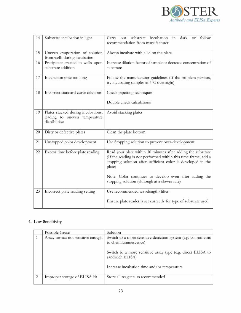
23
14 Substrate incubation in light Carry out substrate incubation in dark or follow recommendation from manufacturer
15 Uneven evaporation of solution from wells during incubation
Always incubate with a lid on the plate
16 Precipitate created in wells upon substrate addition
Increase dilution factor of sample or decrease concentration of substrate
17 Incubation time too long Follow the manufacturer guidelines (If the problem persists, try incubating samples at 4°C overnight)
18 Incorrect standard curve dilutions Check pipetting techniques Double check calculations
19 Plates stacked during incubations, leading to uneven temperature distribution
Avoid stacking plates
20 Dirty or defective plates Clean the plate bottom
21 Unstopped color development Use Stopping solution to prevent over-development
22 Excess time before plate reading Read your plate within 30 minutes after adding the substrate (If the reading is not performed within this time frame, add a stopping solution after sufficient color is developed in the plate) Note: Color continues to develop even after adding the stopping solution (although at a slower rate)
23 Incorrect plate reading setting Use recommended wavelength/filter Ensure plate reader is set correctly for type of substrate used
4. Low Sensitivity
Possible Cause Solution
1 Assay format not sensitive enough Switch to a more sensitive detection system (e.g. colorimetric to chemiluminescence) Switch to a more sensitive assay type (e.g. direct ELISA to sandwich ELISA) Increase incubation time and/or temperature
2 Improper storage of ELISA kit Store all reagents as recommended
24
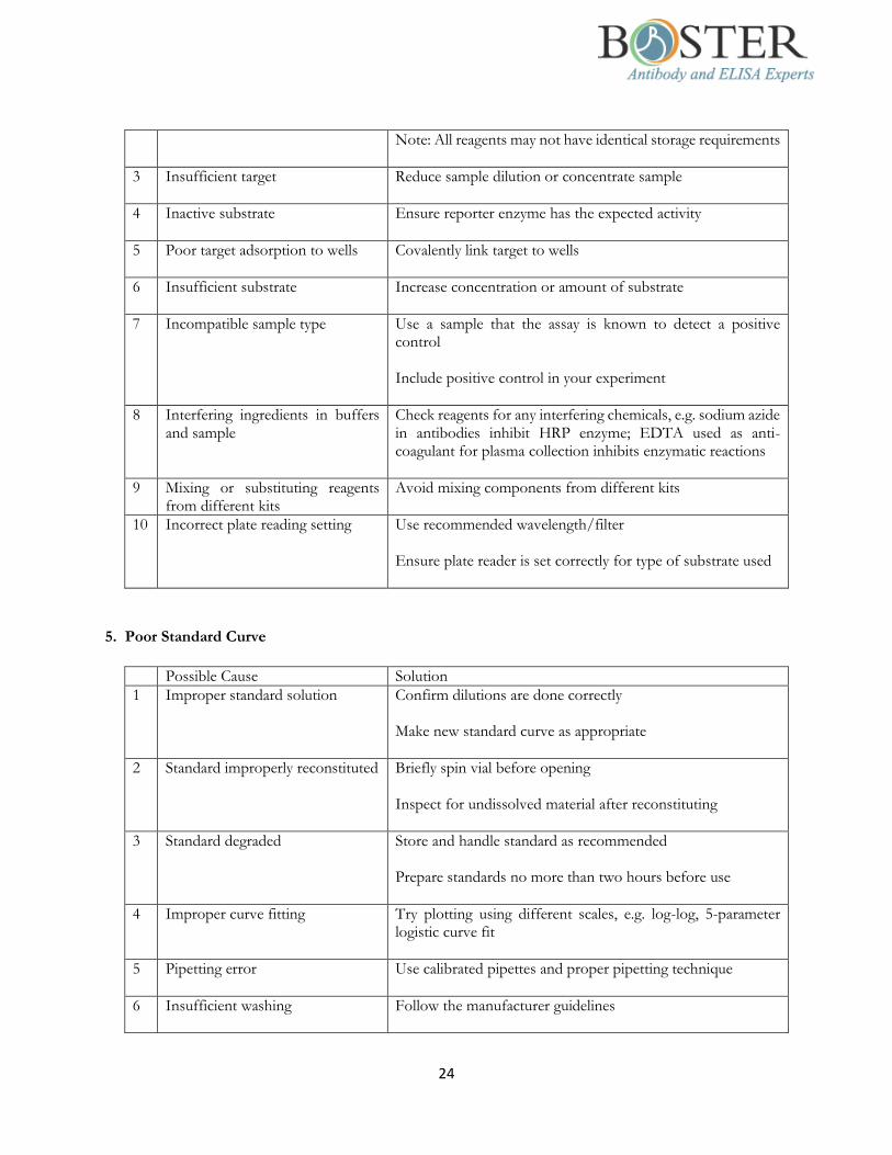
Note: All reagents may not have identical storage requirements
3 Insufficient target Reduce sample dilution or concentrate sample
4 Inactive substrate Ensure reporter enzyme has the expected activity
5 Poor target adsorption to wells Covalently link target to wells
6 Insufficient substrate Increase concentration or amount of substrate
7 Incompatible sample type Use a sample that the assay is known to detect a positive control Include positive control in your experiment
8 Interfering ingredients in buffers and sample
Check reagents for any interfering chemicals, e.g. sodium azide in antibodies inhibit HRP enzyme; EDTA used as anti-coagulant for plasma collection inhibits enzymatic reactions
9 Mixing or substituting reagents from different kits
Avoid mixing components from different kits
10 Incorrect plate reading setting Use recommended wavelength/filter Ensure plate reader is set correctly for type of substrate used
5. Poor Standard Curve
Possible Cause Solution
1 Improper standard solution Confirm dilutions are done correctly Make new standard curve as appropriate
2 Standard improperly reconstituted Briefly spin vial before opening Inspect for undissolved material after reconstituting
3 Standard degraded Store and handle standard as recommended Prepare standards no more than two hours before use
4 Improper curve fitting Try plotting using different scales, e.g. log-log, 5-parameter logistic curve fit
5 Pipetting error Use calibrated pipettes and proper pipetting technique
6 Insufficient washing Follow the manufacturer guidelines

25
At the end of each washing step, flick the plate over a sink and pat the plate on a paper towel
7 Poorly mixed reagents Thoroughly mix reagents
8 Poor/variable adsorption of reagents to plate
Extend incubation time Check coating buffer Use a different plate as appropriate Check homogeneity of samples
9 Plates stacked during incubation Keep plates separated if not using rotating plates
10 Dirty or defective plates Clean the plate bottom
6. Poor Replicate Data
Possible Cause Solution
1 Bubble in wells Ensure no bubbles are present prior to reading plate
2 Insufficient washing of wells Carefully wash wells Follow recommended protocols Check that all ports of the plate washer are unobstructed
3 Incomplete reagent mixing Ensure all reagents are mixed thoroughly
4 Inconsistent pipetting Use calibrated pipettes and proper pipetting techniques If a multi-channel pipette is used, ensure that all channels deliver the same volume
5 Inconsistent sample prep or storage
Ensure consistent sample prep and optimal sample storage conditions (e.g. minimize freeze/thaw cycles)
6 Particulates in samples Remove the particulates by centrifugation
7 Plate sealers not used or re-used During incubations, cover plates with plate sealers Use a fresh sealer every time the used sealer is removed from the plate
8 Cross-well contamination Ensure plate sealers and pipette tips are not contaminated with reagents

26
9 Edge effect (higher or lower OD in peripheral wells than in central wells)
Ensure plates and reagents are kept at room temperature before pipetting into wells unless otherwise instructed During incubation, seal the plate completely with a plate sealer and avoid stacking plates
7. Inconsistent Assay-to-Assay Results
Possible Cause Solution
1 Insufficient washing of wells Carefully wash wells Follow recommended protocols Check that all ports of the plate washer are unobstructed
2 Variation in incubation temperature
Adhere to recommended incubation temperature Avoid incubating plates in area where environmental conditions vary
3 Variation in protocol Adhere to the same protocol from run to run
4 Plate sealers not used or re-used During incubations, cover plates with plate sealers Use a fresh sealer every time the used sealer is removed from the plate
5 Incorrect dilutions Confirm dilutions are done correctly for standard solutions, etc Make new standard curve as appropriate
6 Contaminated buffers Make and use fresh buffers
7 Plates stacked during incubation Keep plates separated if not using rotating plates
8. Slow Color Development
Possible Cause Solution
1 Substrates too old, contaminated or used at incorrect pH
Make and use fresh substrates at correct pH: they should be prepared immediately before use
2 Expired/Contaminated solutions Make and use fresh reagents
3 Incorrect incubation temperature Ensure plates and reagents are kept at room temperature before pipetting into wells unless otherwise instructed

27
During incubation, seal the plate completely with a plate sealer and avoid stacking plates
4 Low antibody concentration Repeat the assay with higher antibody concentrations to find the optimal one for your experiment
5 Low substrate concentration Add more substrate to the wells Make substrate no more than one hour before use Note: Typical ELISA sensitivity is ~0.1 pg/mL with exact value depends on antibody used.
9. Plate Imaging Problem
Possible Cause Solution
1 Oversaturated image after acquisition
Use full resolution image to analyze results (Do not use jpeg or other compressed formats)
2 Blurry spots in images Re-focus your camera before taking a new image
3 Repeated pixel values or rectangular spots
Use lower bin size, higher image resolution and/or lossless file type
4 Flat standard in images Reduce acquisition time
28
FAQs
1. The ELISA protocols do not recommend shaking during incubations. Have you tested shaking
and decided against it or is it unnecessary?
We tested our protocols with and without shaking during incubations and determined that there is no
difference between the two approaches. Therefore, we believe that shaking is not necessary.
2. Is your ELISA kit suitable for use with tissue lysates? If so, what are the protocols?
Theoretically, our ELISA kit can work with tissue lysates. Our general sample preparation protocol for tissue
lysates is as follows:
Rinse the tissue with PBS to remove excess blood.
Chop the tissue into 1-2 mm pieces.
Using a tissue homogenizer, homogenize the samples in PBS or lysate solution such as the Mammal
Tissue Protein Extraction Reagent (Boster Bio Catalog Number AR0101) at a ratio of 10 mL lysate
solution to 1 g of tissue.
Centrifuge the homogenates at approximately 5000 x g for 5 min.
Assay immediately or store the homogenates at -20°C (avoid repeated freeze-thaw cycles).
3. My samples contain very low cytokine concentration. What is the minimum concentration that can
be measured with confidence using your ELISA kits?
Low cytokine concentrations are typical for many biological samples. When determining the minimum
concentration that can be reliably measured by ELISA, consider the following:
The standard curve: As the concentration range of ELISA is typically 0 - 1000 pg/mL, the data points
on the standard curve for this range correspond to 0, 15.6, 31.25, 62.5, 125, 250, 500, and 1000 pg/mL.
The assay sensitivity: Boster’s Picokine ELISA kits typically have a reported sensitivity of 10 pg/mL.
The concentration detected in many biological samples will fall between the 0 and 15.6 pg/mL data
points of the standard curve. As long as the value detected is above the statistical sensitivity of the
ELISA, (e.g., 5 pg/mL or greater), the value is statistically significant. Results below this detection limit
are of questionable validity.
4. Is your ELISA kit suitable for use with tissue homogenates?
For most cases, yes. If there is enough target protein present in the tissue of interest, the ELISA kit will
work. Also, if there are a known alternative processing of the protein in a specific tissue that results in
protein reactivity change to the kit, we will note it in our product datasheet.
5. Is your ELISA kit suitable for use with any non-validated sample types?
In order to use an ELISA kit with a non-validated sample type, it is necessary to perform a spike and recovery
study to determine if a non-validated sample type will work with a particular kit. To do this:
Divide the sample into two aliquots.
In one of the aliquots, you should “spike in” a known amount of the kit standard.
Perform a dilution series to compare the spiked to the unspiked sample.
Generally, samples with expected recovery and linearity between 80-120% are acceptable. This method can
be used to validate any sample type that has not been previously evaluated by Boster.

29
6. Can I extend the standard curve?
No one can guarantee the assay accuracy once the concentrations outside the specified range within the
curve are used. A specific range is generated to provide the statistical confidence for the assay accuracy.
7. What causes high variability between sample duplicates?
The two main reasons for high sample variability in an assay are inconsistent pipetting and washing. Thus,
it is important to perfect these techniques. However, some of this variability is unavoidable — this is the
rationale for calculating the average results from sample duplicates. Another possible culprit for high
variability is the “edge effect” in which the outermost wells of the plate are more vulnerable to drying out
due to evaporation. Plate stacking will also cause variability because temperature will be unevenly distributed
across the plates.
8. What are the differences between the sandwich ELISA and competitive ELISA?
Sandwich ELISAs typically require the use of matched antibody pairs, where each antibody is specific for a
different, non-overlapping part (epitope) of the antigen molecule. A first antibody (known as capture
antibody) is coated to the wells. The sample solution is then added to the well. A second antibody (known
as detection antibody) follows this step in order to measure the concentration of the sample. Higher signal
output reflects higher concentration of the target antigen in the sample.
The key event of competitive ELISA is the process of competitive reaction between the sample antigen and
antigen bound to the wells of a microtiter plate with the primary antibody. First, the primary antibody is
incubated with the sample antigen and the resulting antibody–antigen complexes are added to wells that
have been coated with the same antigen. After an incubation period, any unbound antibody is washed off.
The more antigen in the sample, the more primary antibody will be bound to the sample antigen. Therefore,
there will be a smaller amount of primary antibody available to bind to the antigen coated on the well,
resulting in a signal reduction.
9. Why do my wells turn green after I add the stop solution?
The green color is a result of incomplete mixing between the substrate and stop solution. After adding the
stop solution, gently tap the plate or place it on a shaker until the mixture in the wells turns yellow.
10. Why does a brown or orange-brown precipitate appear in my wells after adding the stop solution?
How can I resolve this issue?
The precipitate is a result from insufficient washing after incubation with the HRP- labeled detection
antibody. To resolve this issue, perform a 30-second soak during each wash step followed by a complete
removal of all liquid in the wells.
11. If I don’t use all the wells from a microtiter plate for my current ELISA assay, how can I preserve
the unused wells for future use?
The microtiter plate typically has removable strips of wells. Unused wells may be removed from the plate,
returned to the foil pouch containing the desiccant pack and stored at 2-8°C for up to one month.

30
Ordering Information
With more than 20 years of experience and trust from 10,000+ scientists, Boster is proud of offering more than
600 PicoKineTM ELISA kits that help accelerate scientific discovery in research areas including immunology,
neuroscience and cancer. Each of our ELISA kits has sufficient reagents for 96 tests per kit. The table below
shows some of the most commonly used ELISA kits.
Sample Types*
Target Species CCS Se P(h) P(e) P(c) U M CL T Cat. No.
Adiponectin Human √ √ √ √ √ √ EK0595
Angiopoietin-2 Mouse √ √ √ √ EK0938
BDNF Human √ √ √ √ √ EK0307
CRP Rat √ √ √ √ EK0978
CTLA4 Mouse √ √ √ EK0717
EGFR Human √ √ √ √ √ EK0327
FGF21 Human √ √ √ √ EK0994
IFN Gamma Human √ √ EK0373
IL-6 Human √ √ √ √ √ EK0410
IL-8 Human √ √ √ √ √ EK0413
IL-10 Human √ √ √ √ √ EK0416
Leptin Mouse √ √ √ √ EK0438
MCP-1 Rat √ √ EK0902
NGF Beta Rat √ √ EK0471
P53 Human √ EK0895
PD-1 Human √ EK0959
TGF Beta 1 Human √ √ √ √ EK0513
TNF Alpha Mouse √ √ √ √ EK0527
* Cell Culture Supernates [CCS], Serum [Se], Plasma: Heparin [P(h)], Plasma: EDTA [P(e)], Plasma: Citrate [P(c)],
Urine [U], Milk [M], Cell Lysate (CL), Tissue (T)
We also offer a variety of ELISA components that can be purchased separately from the kits:
Lyophilized recombinant standard (1 ng to 100 ng)
96-well plate (No antibody pre-coated)
Avidin-Biotin-Peroxidase Complex (ABC)
Buffers (Sample diluent, antibody diluent, ABC diluent, PBS, TBS)
TMB color developing agent and stop solution
Biotinylated antibody
Contact Information
Boster Biological Technology
3942 Valley Ave., Suite B, Pleasanton, CA 94566
Phone: (888) 466-3604
Fax: (925) 485-4560
Sales and Customer Service: [email protected]
Product and Technical Support: [email protected]
Business Development: [email protected]