Embed Size (px)
Citation preview

1 / 11
Electronic Supplementary Information
A Two-Photon Fluorescent Probe for Viscosity
Imaging in vivoPeng Ning, a Pengyu Dong, b Qian Geng, c Lei Bai, d Yaqi Ding, d Xiaohe Tian, c
Rong Shao, b* Lin Li d and Xiangming Meng *a
a School of Chemistry & Chemical Engineering, Anhui University, Hefei, 230601, P. R. China.
b Jiangsu Collaborative Innovation Center for Ecological Building Materials & Environmental
Protection Equipments, Yancheng Institute of Technology, Yancheng, 224051, P. R. China
c School of Life Science, Anhui University, Hefei, 230601, P. R. China
d Key Laboratory of Flexible Electronics (KLOFE) & Institute of Advanced Materials (IAM),
Jiangsu National Synergetic Innovation Center for Advanced Materials (SICAM), Nanjing Tech
University (NanjingTech), Nanjing, 211816, P. R. China
*Corresponding author. Fax: +86-551-63861467; Tel: +86-551-63861467
E-mail address: [email protected] (Xiangming Meng); [email protected] (Rong Shao).
Electronic Supplementary Material (ESI) for Journal of Materials Chemistry B.This journal is © The Royal Society of Chemistry 2017

2 / 11
List of Contents
1. UV-vis spectra of MCN in water and glycerol.
2. UV-vis and fluorescence emission spectra in different solvents.
3. pH stability.
4. Cytotoxicity assay.
5. Confocal co-localized images of MCN and PI.
6. Co-localized images of MCN and the nucleus dye.
7. Two-photon fluorescence imaging of MCN without etoposide.
8. Two-photon Fluorescence Lifetime Imaging (FLIM) in cells
9. Toxicity for zebrafish.
10. ESI-MS spectrum of MCN.
11. NMR spectra of MCN and its intermediates.

3 / 11
1. UV-vis spectra of MCN in water and glycerol.
Fig. S1 UV-vis spectra of MCN (10 μM) in water and glycerol, respectively.
2. UV-vis and fluorescence emission spectra in different solvents.
Fig. S2 (a) UV-vis spectra of MCN (10 μM) in different solvents; (b) fluorescence emission
spectra of MCN (10 μM) in different solvents, excited at 400 nm.
4 / 11
3. pH stability.
Fig. S3 Relative fluorescence emission intensity (I/I0, λem = 470 nm) of MCN (10 μM) at different
pH values in water/glycerol system (7:3, v/v, 10 mM PBS buffer, λex = 400 nm).
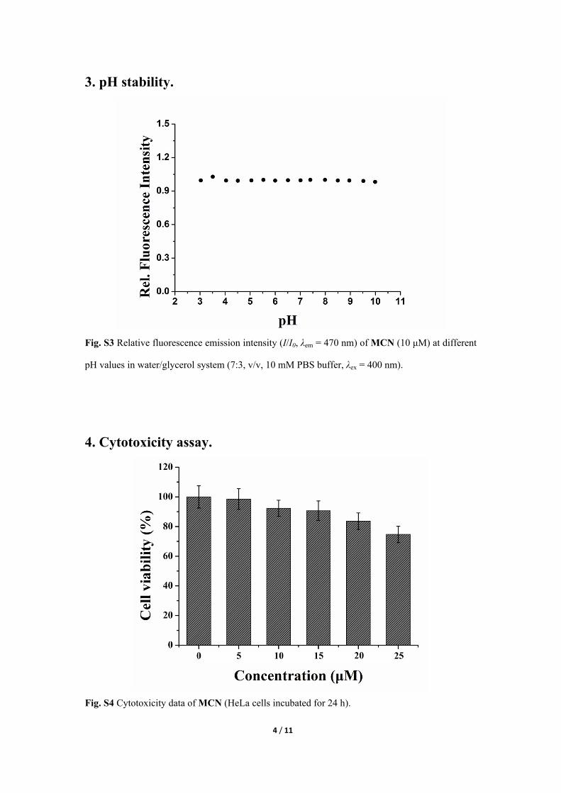
4. Cytotoxicity assay.
Fig. S4 Cytotoxicity data of MCN (HeLa cells incubated for 24 h).

5 / 11
5. Confocal co-localized images of MCN and PI.
Fig. S5 Confocal co-localized images of HeLa cells incubated with MCN (10 μM) for 30 min and
PI (3 μM) for 15 min. (a) Two-photon image of HeLa cells, λex = 800 nm, emission wavelength
from 450 nm to 490 nm; (b) one-photon image of HeLa cells, λex = 543 nm, emission wavelength
from 605 nm to 625 nm; (c) bright-field of HeLa cells; (d) the overlay of panels (a), (b) and (c).
Scale bar: 20 μm.
6. Co-localized images of MCN and the nucleus dye.
Fig. S6 Confocal co-localized images of HeLa cells incubated with MCN (10 μM) for 30 min and
NucRed® Live 647 ReadyProbes® Reagent (2 drops per mL of media) for 15 min. (a) Two-
photon image of HeLa cells, λex = 800 nm, emission wavelength from 450 nm to 490 nm; (b) one-
photon image of HeLa cells, λex = 633 nm, emission wavelength from 655 nm to 665 nm; (c)
bright-field of HeLa cells; (d) the overlay of panels (a), (b) and (c). Scale bar: 20 μm.

6 / 11
7. Two-photon fluorescence imaging of MCN without etoposide.
Fig. S7 (a) – (j) Two-photon confocal images of HeLa cells incubated with 10 μM MCN at different time points, λex = 800 nm, emission wavelength from 450 nm to 490 nm. Scale bars: 20 μm.
8. Two-photon Fluorescence Lifetime Imaging (FLIM) in cells
Fig. S8 (a) Fluorescence image obtained following 800 nm excitation and 470±20 nm detection
from HeLa cells incubated with 10 µM solution of MCN; (b) FLIM image obtained following 800
nm pulsed excitation of the same layer of cells; (c) Histogram of lifetimes.

7 / 11
9. Toxicity for zebrafish.
Fig. S9 Survival rate of larval zebrafish treated with MCN for up to 96 hours.
10. ESI-MS spectrum of MCN.
Fig. S10 ESI-MS spectrum of MCN.

8 / 11
11. NMR spectra of MCN and its intermediates.
Fig. S11 1H NMR of compound 1 in CDCl3.
Fig. S12 13C NMR of compound 1 in CDCl3.

9 / 11
Fig. S13 1H NMR of compound 2 in CDCl3.
Fig. S14 13C NMR of compound 2 in CDCl3.

10 / 11
Fig. S15 1H NMR of compound 3 in DMSO-d6.
Fig. S16 13C NMR of compound 3 in CDCl3.

11 / 11
Fig. S17 1H NMR of MCN in DMSO-d6.
Fig. S18 13C NMR of MCN in CDCl3.