Embed Size (px)
Citation preview

13
Electrochemical Sensors for Pharmaceutical and Environmental Analysis
Robert Săndulescu, Cecilia Cristea, Veronica Hârceagă and Ede Bodoki "Iuliu Haţieganu" University of Medicine and Pharmacy Cluj-Napoca,
Romania
1. Introduction
Improvement of life quality is one of the most important objectives of the global research efforts. Naturally, the quality of life is closely related to a better control of diseases, drug and food quality and safety, and last but not least, of the quality of our environment. In all these fields, a continuous, sensitive, fast and reliable monitoring is required to control key parameters (Castillo et al., 2004). In this context, the use of sensors and biosensors represent very promising tools. The interest in their application in the biomedical, pharmaceutical and environmental field increased lately as a result of their sensitivity, specificity and simple use, providing fast, cost-effective and repetitive measurements with miniaturized and portable devices.
2. How to construct and optimize an enzyme electrochemical biosensor?
A biosensor, according to the IUPAC definition, is a self-contained integrated device, which is capable of providing specific quantitative or semi-quantitative analytical information using a biological recognition element (biochemical receptor) which is retained in spatial contact with a physical transduction element (Thevenot et al., 2001). Actually they transform the reaction between the analyte and the biological component into a signal detected and monitored by the transducer that reflects the concentration of the analyte. Biosensors may be classified according to the bioactive component or the recognition event, the mode of signal transduction or according to the immobilization technique. In the construction of the biosensors the biocomponent is selected depending on the application and the performance criteria requested. The most commonly applied bioactive components are the enzymes. They rapidly and cleanly form selective bonds with the substrates and convert them into products. One of the major issues when working with enzymes is their stability. Enzymes are very sensitive to their environment. Deactivation, inhibition or unfolding upon adsorption and chemical or thermal inactivation are common (Grieshaber et al. 2008). There are several advantages of the enzyme biosensors. These include their availability and the ability to modify the catalytic properties or substrate specificity by means of genetic engineering and catalytic amplification of the biosensor response by modulation of the enzyme activity with respect to the target analyte (Rogers, 2006). Among the enzymes commercially available, the oxidases: the glucose oxidase (GOD) and horseradish peroxidase (HRP) are the most often used. This type of enzyme offers the
www.intechopen.com

Environmental Biosensors
278
advantages of being stable and in some situations does not require coenzymes or cofactors. Other, less commonly used enzymes comprise beta-lactamase, urease, tyrosinase and acetylcholinesterase or choline oxidase. Among different transducers employed in biosensors construction, the electrochemical transducers are the most frequently used. They occupy the first position as far as the disponibility on the market and they have already demonstrated their practical utility. The most well known enzyme electrochemical biosensors reported in the literature are the biosensors with glucose oxidase for the detection of glucose (Ammam et al., 2010), the biosensors with HRP for the detection of compounds which can be peroxidate like clozapine, (Yu et al., 2006a), acetaminophen, (Sima et al., 2008) and of the thiolic products (gluthation, N-acetilcysteine) (Yu et al., 2006b) and the biosensors with acetylcholine and choline oxidase for detection of drugs used in Alzheimer treatment (Lenigk et al., 2000) and the organophosphorus pesticides (Andreescu & Marty, 2006). The most important step in building a biosensor is the immobilization of the biomolecules. As a matter of fact the immobilization technique determines the biosensor performances. The selection of an appropriate immobilization method depends on the nature of the biological element, type of the transducer used, physicochemical properties of the analyte and operating conditions for the biosensor. The purpose of any immobilization method is to get high stability and efficiency from the biosensor. The biocomponent should be stably immobilized in a physical environment close to the natural enzyme’s environment in order to retain maximum functionality of its biological activity on the surface of the transducer. The immobilization might involve a number of disadvantages such as: worsening the performance of enzymatic system, lowering and even lost of activity, increase of Michaelis constant, modification of the optimal working temperature and pH of the enzyme in comparison with the free form. All these, however, may be reduced by applying an appropriate design. The system should assure also good diffusion properties for substrates and should establish satisfactory electrical communication between the active component and the electrode surface (Xu et al., 2006) in order to give the electrochemical output signal required. When the design of a biosensor is built, all the characteristics of an ideal analytical system: selectivity, limit of detection and limit of quantification, sensitivity, reliability, repetitively, robustness, throughput, reagentless, simplicity, costs, miniaturization and portability have to be taken into account and also the performances and the drawbacks of the systems already reported in the literature for the analyte of interest have to be deeply studied. Enzyme biosensors can be categorized into those that monitor the catalytic transformation of a target analyte by a specific enzyme or the enzyme biological activity by measuring the producing or consumption of a given analyte and those that monitor the inhibition of a biochemical reaction due to the presence of target analyte. The inhibition-based biosensors can be designed in two different ways: the inhibitor interacts directly with the enzyme, blocking its activity, or the inhibitor interacts with a product of the enzymatic reaction. Only specific enzyme inhibitors should be regarded as “true inhibitors”, which bind to the enzyme and inhibit its activity. However, the definition of “enzyme inhibitors” is sometimes misleading, for example in the case of tyrosinase based biosensors, the terminology being also used to refer to melanogenesis process inhibitors, whose action mainly reside in some interference in melanin formation, regardless of any direct inhibitor-enzyme interaction (Chang et al., 2009). “True tyrosinase inhibitors” can be considered kojic acid that links to the active site of tyrosinase like a copper chelator (Briganti et al., 1990), benzoic acid that
www.intechopen.com
Electrochemical Sensors for Pharmaceutical and Environmental Analysis
279
functions as a monophenol analog, binding to one copper ion of the binuclear site via its carboxylate group (Menon et al., 1990) and azelaic acid that blocks the access of the substrate to the enzyme active center (Briganti et al.,1990) but not ascorbic acid that interrupts melanin synthesis process by reducing the enzymatically generated dopaquinone intermediate back to L-dopa. When a biosensor applied to pharmaceutical or biomedical analysis is described the biological processes have to be studied in detail, taking in consideration their complexity, in order to clearly outline the field of application and the limitations of the device. For example the biosensor described by Sima et al. (2011) allowed the quantification and the characterization of inhibitors from the point of view of their interaction with tyrosinase and the enzymatic reaction in the presence of the natural substrate L-tyrosine. In the skin pigmentation process these compounds may interfere also in other biochemical steps having as final result a decrease in melanin formation. For example, azelaic acid inhibits melanin formation through other mechanism besides its interaction with the tyrosinase active site. It may also interfere with DNA synthesis and mitochondria activity in hyperactive and abnormal melanocytes (Briganti et al., 1990). Once the biocomponent, the transducer and the immobilization technique are chosen the operation mode of the device should be established. For example in the article published by Sima et al., (2011), the obtained configuration was used to monitor the reduction current of the enzymatically generated oxidized species of L-tyrosine, i.e. the dopaquinone, in the presence of molecular oxygen (Fig. 1).
Fig. 1. Biosensor’s mechanism of action (Sima et al., 2011)
The inhibition-based biosensors are preferred, if possible, in order to assure a good reproducibility of the results because in this case, when the analytes are evaluated, the biosensor inhibition signal is normalized to the initial signal of the substrate for each curve. Thus, the final results are not influenced by the variations of substrate’s signal which can occur due to the loss of the enzyme activity or due to random errors that can appear during biosensor’s preparation. Also blank experiments have to be performed in order to check if “the biosensor” without enzyme or the inhibition based biosensor operating without addition of the enzymatic substrate does not give any response under the selected experimental conditions and in the presence of the studied compounds.
www.intechopen.com
Environmental Biosensors
280
If the developed device is a novel analytical tool, in order to test its efficiency, it should be applied to the study of compounds that have already demonstrated their activity in clinical practice or by using other analytical methods, as it was the case in the article published by Sima et al. (2011). When testing different compounds, because there are many factors which contribute to the final results (type of enzyme, quantity of immobilized enzyme, immobilization method, type of substrate, substrate concentration, time of contact between the enzyme, substrate and inhibitor, pH, temperature, applied potential, rate of solution stirring), in order to obtain a correct comparison of the studied compounds, the experiments have to be performed under the same experimental conditions (Sima et al., 2011). First the working conditions for an optimal response to substrate of the enzyme based biosensor have to be tested. If the biosensor is designed for testing biologically active compounds it has to be taken into account that the device should operate as close as possible to the natural enzyme’s environment. The working applied potential, the type of enzyme substrate, the buffer pH, the quantity of immobilized enzyme, the substrate concentration in case of the inhibition studies, the presence of cofactors if necessary; all these have to be considered. In order to select the applied potential for the amperometric testing, first of all, the biological process that produces electroactive compounds has to be demonstrated under the experimental conditions, even though it is well know in its natural environment. Normally cyclic voltammetric experiments are performed for detecting the electrochemical reduction or oxidation potential of the enzymatically generated electroactive species (Yu et al., 2006). It may be sometimes the case that if low reduction or oxidation currents are generated they might not be detected in cyclic voltammetric experiments, so amperometry is recommended, thanks to its low background currents (Shan et al., 2008). The applied potential has to be considered taking into account the magnitude of the electrochemical signals, the ratio between signal and background current, the steady-state of the response plateau, the possible interfering species at that applied potential and also, the working potential of other similar biosensors described in the literature. When selecting the enzymatic substrate in case of inhibition based biosensors, this has to be the same as in the biological process, if possible, because it has been demonstrated that the potency of inhibitors varies considerably depending on the characteristics of the substrate (Shan et al., 2008). The sensitivity of the response has to be high enough and the steady-state of the response plateau has to be stable for assuring reliable results and the lag period of the enzymatic step sufficiently short for further readily implementation of the testing conditions. It is well known that enzyme activity is highly pH dependent and that the optimum pH for an enzymatic assay must be determined empirically (Amine et al., 2006). The buffer pH considered optimal has to be chosen taking in consideration the magnitude of the amperometric signal, the steady-state of the response, the enzyme stability pH, the normal biological pH of the process and other similar biosensors reported in the literature. The amount of enzyme immobilized at the electrode surface has to offer a good sensitivity and repeatability of the biosensor response taking in consideration also the costs of the assay, since some enzymes can be quite expensive. The opinions about the substrate concentration to be considered in inhibition assays are quite contradictory. Kok et al. (2002), concluded, when measuring the inhibition potency of a competitive inhibitor with an acetylcholinesterase and a choline oxidase biosensor, that the
www.intechopen.com

Electrochemical Sensors for Pharmaceutical and Environmental Analysis
281
inhibition percentage increased by raising the substrate concentration. Therefore they worked in enzyme saturated substrate conditions. Shan et al. (2008) demonstrated that for a tyrosinase biosensor for the determination of benzoic acid, the concentration of the catechol substrate did not affect the maximum inhibition percentage but it affected the sensitivity of the method. It must be noted, however, that the use of a high substrate concentration would not yield sensitive inhibition responses when the quantification of a competitive inhibitor is performed by simultaneous addition of the inhibitor and the substrate. Because in competitive inhibition the substrate competes with the inhibitor for the enzyme active site, and the inhibition, especially at low inhibitor concentrations, would likely not be detected (Kok et al., 2002). In the case of the biosensor made by Sima et al. (2011) as it was not designed for the quantification of inhibitors but for the screening and quantification of their inhibitory potency, it was considered that all the immobilized enzyme molecules must take part in the reaction and this could only be possible in substrate concentration corresponding to the saturation portion of the activity versus substrate curve. When the concentration of substrate is quite high, it has to be established if the concentration of other cofactors participating in the reaction is not a limiting factor and that the enzymatic process is saturated by the substrate.
-8.00E-09
-7.00E-09
-6.00E-09
-5.00E-09
-4.00E-09
-3.00E-09
-2.00E-09
-1.00E-09
0.00E+00
0 500 1000 1500 2000
time (s)
I (A
)
Average of 5 curves
y = 5E-08x + 0.0076
R2 = 0.9999
0.000
0.005
0.010
0.015
0.020
0.025
0.030
0.0E+00 2.0E+05 4.0E+05 6.0E+05
1/conc (mol-1
*L)
1/In
(%
)
Response 1 Kojic acid
Response 2 Kojic acid
Response 3 Kojic acid
Response 4 Kojic acid
Response 5 Kojic acid
Average
Linear (Average)
Fig. 2. Tyrosinase based biosensor amperometric response. L-tyrosine 3.33·10-4M, Kojic acid inhibitor between 2.50·10-6 and 1.24·10-5M, 0.1M phosphate buffer pH 6.5, Eapp -100mV, 10µL of a 1.25mg/mL tyrosinase-NMPS suspension spiked onto the mCPE. Insert: inhibition calibration curve (Sima et al., 2011)
In case of electrochemical biosensors parameters like apparent Michaelis-Menten constant (Kmapp), maximum current intensity, limit of detection and linear range for the analyte of interest have to be determined.
www.intechopen.com

Environmental Biosensors
282
In case of electrochemical inhibition based biosensors parameters like IC50, Kiapp (inhibition constant) and mechanism of inhibition of the studied compounds have to be determined. In order to determine IC50, the concentration of inhibitor which inhibits 50% of the substrate signal, the concentration of inhibitors can be correlated with the percentage of inhibition (In %) which can be calculated using the relationship: In(%)=[(I0 - I1)/ I0]x100; with I0 and I1
being the biosensor current signals before and after the addition of the inhibitor, respectively (Fig. 2). The mechanism of inhibition can be established from the relationship between the biosensor response to substrate, Kmapp and Imax, in the absence and in the presence of different concentrations of inhibitor added in the initial testing solution (Fig. 3).
y = -7915.9x - 8E+07
R2 = 1
y = -24977x - 9E+07
R2 = 0.9995
y = -44490x - 1E+08
R2 = 0.9994
-2.00E+10
-1.80E+10
-1.60E+10
-1.40E+10
-1.20E+10
-1.00E+10
-8.00E+09
-6.00E+09
-4.00E+09
-2.00E+09
0.00E+00
0.0E+00 5.0E+04 1.0E+05 1.5E+05 2.0E+05 2.5E+05 3.0E+05 3.5E+05 4.0E+05 4.5E+05
1/L-tyrosine (M-1)
1/I
(A
-1)
a
b
c
Fig. 3. Tyrosinase based biosensor amperometric data: 1/Signal vs 1/[L-tyrosine]. L-tyrosine between 2.48·10-6 - 4.39·10-4M a. without Kojic acid, b. in the presence of 2.09·10-6M Kojic acid, c. in the presence of 4.18·10-6M Kojic acid, 0.1M phosphate buffer pH 6.5, Eapp - 100mV, 10µL of a 1.25mg/mL tyrosinase-NMPS suspension deposited onto the mCPE (Sima et al., 2011)
From the primary plot of 1/I(A-1) versus 1/substrate concentration (M-1) (Lineweaver-Burk), secondary plots can be generated, by plotting the slopes from the primary plots versus inhibitor concentration in order to determine the apparent inhibition constant, Kiapp (Fig. 4). Finally the long-term stability of the immobilized enzyme has to be evaluated by measuring the biosensor response to substrate. Normally when not in use, the enzyme has to be maintained in hydrated conditions at around 4ºC. In case of inhibition based biosensors IC50 of the tested inhibitors has to be determined at different time periods following enzyme immobilization to see if the obtained results are not affected by the loss of enzyme activity. Biosensors have some major advantages over traditional analytical methods, which will certainly lead to their even more pronounced use in the pharmaceutical and biomedical field in the near future: they are prone to miniaturization which is of great importance because biological samples are available in small amounts and tissue damage must be minimized in case of in-vivo monitoring, the detection of the key substrate is very often made without prior separation, their sensitivity is usually in the order of ng/ml, they have a high selectivity, sometimes even specificity, they are characterized by short response times and quickness of data collection, they are easy to use (Castillo et al, 2004) and they have a high benefit/cost ratio.
www.intechopen.com

Electrochemical Sensors for Pharmaceutical and Environmental Analysis
283
y = -9E+09x - 7507.1
R2 = 0.9991
y = -4E+08x - 8085.5
R2 = 0.9978
y = -4E+08x - 15201
R2 = 0.9974-8.00E+04
-7.00E+04
-6.00E+04
-5.00E+04
-4.00E+04
-3.00E+04
-2.00E+04
-1.00E+04
0.00E+00
-1.0E-04 -5.0E-05 0.0E+00 5.0E-05 1.0E-04 1.5E-04 2.0E-04
conc inhibitor (M)S
lop
e
a
b
c
Fig. 4. Tyrosinase based biosensor data: slope of Fig. 3 data vs [In]. a. Kojic acid, b. Benzoic acid, c. Azelaic acid, 0.1M phosphate buffer pH 6.5, Eapp -100mV, 10µL of a 1.25mg/mL tyrosinase-NMPS suspension deposited onto the mCPE (Sima et al., 2011)
However, many enzyme-based amperometric biosensors described in the recent literature still display a few drawbacks when compared to other analytical methods. The most difficult problems to overcome for biosensors are: the low reproducibility of the response and the reduced stability due to the fact that the enzymes, removed from their natural environment, tend to rapidly loose their activity and thus limit the lifetime of the sensor, the electrochemical interferences in complex sample matrices and the biocompatibility and biofouling in case of in-vivo measurements (Castillo et al, 2004). Even if they do present some drawbacks these devices can be very efficacious used for adequate analysis. Moreover these disadvantages can be limited and the performances can be optimized by an adequate immobilization method of the biocomponent at the transducer surface. While the concept of biosensors is simple, their commercialization is far from being simple. A lot of such devices are at scientific stage and only a few of the innovative ideas described in the scientific literature have reached the marketplace and they are only limited applied in clinical practice, as for example the well known glucose portable analyzer for home diagnostic (Wang, 1999); so still more efforts need to be undertaken.
3. Electrochemical sensors for health applications
3.1 Pharmaceutical analysis The need for new sensitive devices allowing the simultaneous detection of several drugs showed a steadily increasing trend in the pharmaceutical research. Some examples of electroanalytical methods applied to ascorbic acid (Săndulescu et al, 2000; Marian et al., 2000), phenotiazines (Săndulescu et al, 2000; Blankert et al., 2005), vitamine B1 (Marian et al., 2001; Bonciocat et al., 2003), effervescent solid dosage forms (Săndulescu et al, 2000), benzodiazepines (Bănică et al., 2007), colchicine (Bodoki et al., 2007; Bodoki et al., 2007) and
www.intechopen.com

Environmental Biosensors
284
paracetamol (Yu et al., 2006; Sima et al., 2008; Sima et al., 2010) were published in the last decade. The electrochemical behaviour of some pharmaceuticals (caffeine, aminophylline, teophylline, Fig. 5) was investigated by cyclic voltammetry (CV), square-wave voltammetry (SWV) and differential pulse voltammetry (DPV), on different electrode materials: glassy carbon in native and electrochemically activated form, bare or 1,4-benzoquinone modified carbon paste. (Câmpean et al., 2011)
(A)
(B) (C)
Fig. 5. Cyclic voltammograms of 10-3 M aminophylline (red line), caffeine (green line) and theophylline (blue line) solutions in Britton-Robinson buffer at pH 4 (black line), on: (A) glassy carbon electrode; (B) carbon paste electrode and (C) 1,4-benzoquinine modified carbon paste electrode (Reference electrode Ag/AgCl,(3 M KCl); auxilliary electrode Pt wire; scan rate 0.1 V s-1; 20 °C)
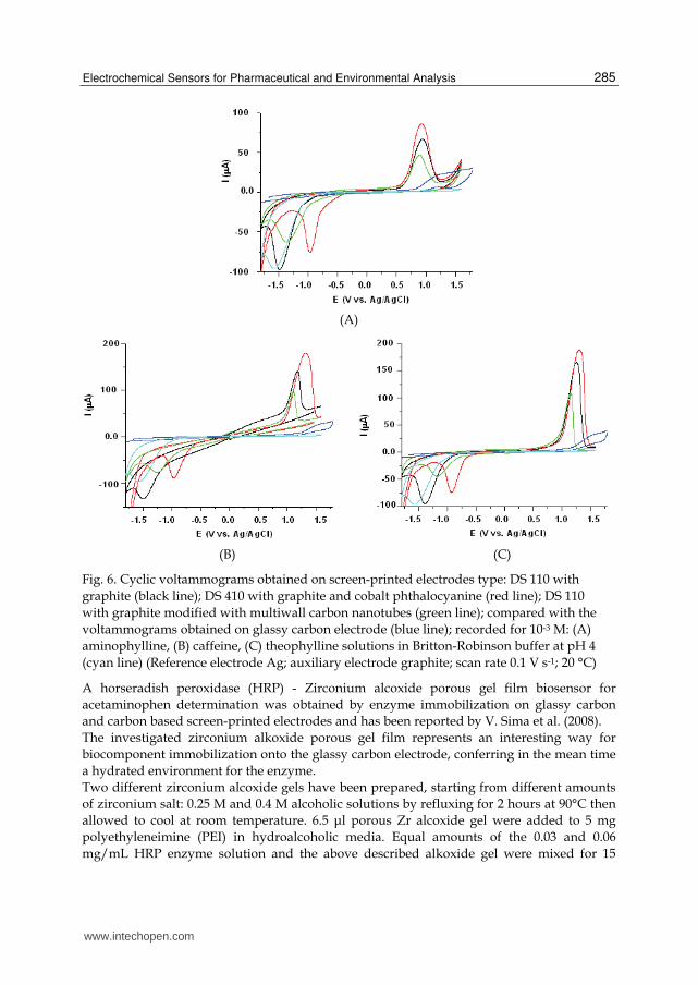
The preliminary studies performed on traditional electrodes were extended on carbon based screen-printed electrodes (Fig. 6), simple and modified with multi-wall carbon nanotubes or cobalt-phtalocyanine. (Câmpean et al., 2011)
www.intechopen.com
Electrochemical Sensors for Pharmaceutical and Environmental Analysis
285
(A)
(B) (C)
Fig. 6. Cyclic voltammograms obtained on screen-printed electrodes type: DS 110 with
graphite (black line); DS 410 with graphite and cobalt phthalocyanine (red line); DS 110
with graphite modified with multiwall carbon nanotubes (green line); compared with the
voltammograms obtained on glassy carbon electrode (blue line); recorded for 10-3 M: (A)
aminophylline, (B) caffeine, (C) theophylline solutions in Britton-Robinson buffer at pH 4
(cyan line) (Reference electrode Ag; auxiliary electrode graphite; scan rate 0.1 V s-1; 20 °C)
A horseradish peroxidase (HRP) - Zirconium alcoxide porous gel film biosensor for
acetaminophen determination was obtained by enzyme immobilization on glassy carbon
and carbon based screen-printed electrodes and has been reported by V. Sima et al. (2008).
The investigated zirconium alkoxide porous gel film represents an interesting way for
biocomponent immobilization onto the glassy carbon electrode, conferring in the mean time
a hydrated environment for the enzyme.
Two different zirconium alcoxide gels have been prepared, starting from different amounts
of zirconium salt: 0.25 M and 0.4 M alcoholic solutions by refluxing for 2 hours at 90°C then
allowed to cool at room temperature. 6.5 μl porous Zr alcoxide gel were added to 5 mg
polyethyleneimine (PEI) in hydroalcoholic media. Equal amounts of the 0.03 and 0.06
mg/mL HRP enzyme solution and the above described alkoxide gel were mixed for 15
www.intechopen.com
Environmental Biosensors
286
minutes and 20 μl of the resulting mixture was deposited on the glassy electrode surface and
left over night at 4°C for drying. For another 24 hours it was left at 4°C in 5 ml phosphate
buffer for hydratation.
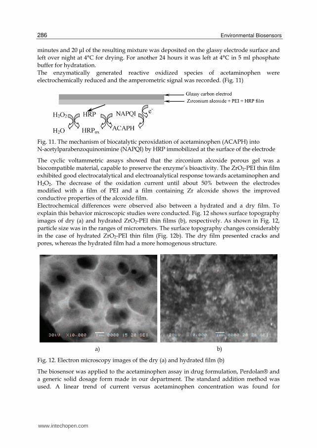
The enzymatically generated reactive oxidized species of acetaminophen were
electrochemically reduced and the amperometric signal was recorded. (Fig. 11)
Fig. 11. The mechanism of biocatalytic peroxidation of acetaminophen (ACAPH) into N-acetylparabenzoquinonimine (NAPQI) by HRP immobilized at the surface of the electrode
The cyclic voltammetric assays showed that the zirconium alcoxide porous gel was a
biocompatible material, capable to preserve the enzyme’s bioactivity. The ZrO2-PEI thin film
exhibited good electrocatalytical and electroanalytical response towards acetaminophen and
H2O2. The decrease of the oxidation current until about 50% between the electrodes
modified with a film of PEI and a film containing Zr alcoxide shows the improved
conductive properties of the alcoxide film.
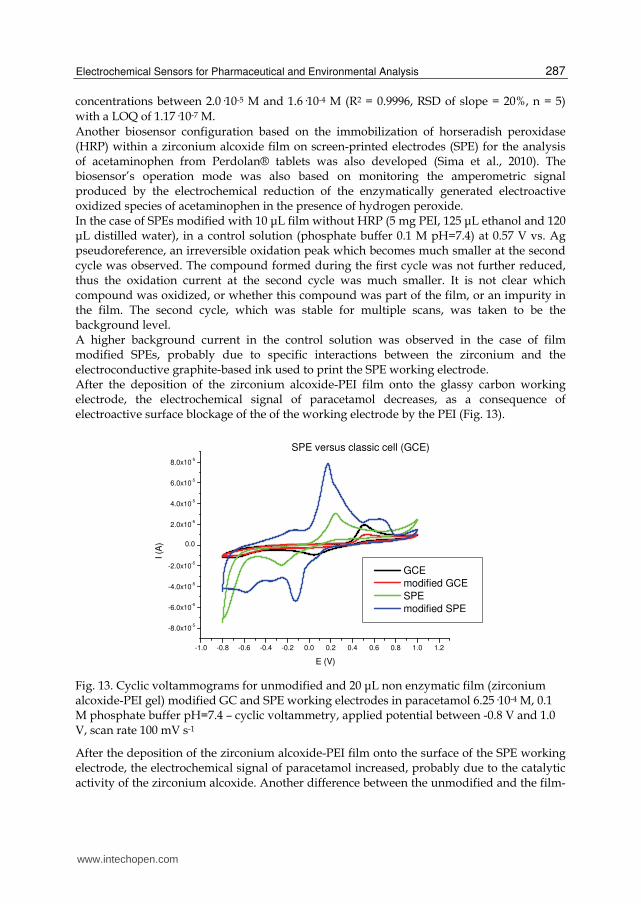
Electrochemical differences were observed also between a hydrated and a dry film. To
explain this behavior microscopic studies were conducted. Fig. 12 shows surface topography
images of dry (a) and hydrated ZrO2-PEI thin films (b), respectively. As shown in Fig. 12,
particle size was in the ranges of micrometers. The surface topography changes considerably
in the case of hydrated ZrO2-PEI thin film (Fig. 12b). The dry film presented cracks and
pores, whereas the hydrated film had a more homogenous structure.
a) b)
Fig. 12. Electron microscopy images of the dry (a) and hydrated film (b)
The biosensor was applied to the acetaminophen assay in drug formulation, Perdolan® and
a generic solid dosage form made in our department. The standard addition method was
used. A linear trend of current versus acetaminophen concentration was found for
www.intechopen.com
Electrochemical Sensors for Pharmaceutical and Environmental Analysis
287
concentrations between 2.0·10-5 M and 1.6·10-4 M (R2 = 0.9996, RSD of slope = 20%, n = 5)
with a LOQ of 1.17·10-7 M.
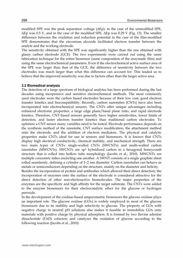
Another biosensor configuration based on the immobilization of horseradish peroxidase (HRP) within a zirconium alcoxide film on screen-printed electrodes (SPE) for the analysis of acetaminophen from Perdolan® tablets was also developed (Sima et al., 2010). The biosensor’s operation mode was also based on monitoring the amperometric signal produced by the electrochemical reduction of the enzymatically generated electroactive oxidized species of acetaminophen in the presence of hydrogen peroxide. In the case of SPEs modified with 10 μL film without HRP (5 mg PEI, 125 μL ethanol and 120 μL distilled water), in a control solution (phosphate buffer 0.1 M pH=7.4) at 0.57 V vs. Ag pseudoreference, an irreversible oxidation peak which becomes much smaller at the second cycle was observed. The compound formed during the first cycle was not further reduced, thus the oxidation current at the second cycle was much smaller. It is not clear which compound was oxidized, or whether this compound was part of the film, or an impurity in the film. The second cycle, which was stable for multiple scans, was taken to be the background level. A higher background current in the control solution was observed in the case of film modified SPEs, probably due to specific interactions between the zirconium and the electroconductive graphite-based ink used to print the SPE working electrode. After the deposition of the zirconium alcoxide-PEI film onto the glassy carbon working electrode, the electrochemical signal of paracetamol decreases, as a consequence of electroactive surface blockage of the of the working electrode by the PEI (Fig. 13).
-1.0 -0.8 -0.6 -0.4 -0.2 0.0 0.2 0.4 0.6 0.8 1.0 1.2
-8.0x10⁻⁵
-6.0x10⁻⁵
-4.0x10⁻⁵
-2.0x10⁻⁵
0.0
2.0x10⁻⁵
4.0x10⁻⁵
6.0x10⁻⁵
8.0x10⁻⁵
SPE versus classic cell (GCE)
I (A
)
E (V)
GCE
modified GCE
SPE
modified SPE
Fig. 13. Cyclic voltammograms for unmodified and 20 μL non enzymatic film (zirconium alcoxide-PEI gel) modified GC and SPE working electrodes in paracetamol 6.25·10-4 M, 0.1 M phosphate buffer pH=7.4 – cyclic voltammetry, applied potential between -0.8 V and 1.0 V, scan rate 100 mV s-1
After the deposition of the zirconium alcoxide-PEI film onto the surface of the SPE working electrode, the electrochemical signal of paracetamol increased, probably due to the catalytic activity of the zirconium alcoxide. Another difference between the unmodified and the film-
www.intechopen.com
Environmental Biosensors
288
modified SPE was the peak separation voltage (ΔEp): in the case of the unmodified SPE, ΔEp was 0.5 V, and in the case of the modified SPE, ΔEp was 0.29 V (Fig. 13). The smaller difference between the oxidation and reduction potential in the case of the film-modified SPE demonstrates that the zirconium alcoxide facilitated electron transfer between the analyte and the working electrode. The sensitivity obtained with the SPE was significantly higher than the one obtained with
glassy carbon electrode (GCE). The two experiments were carried out using the same
fabrication technique for the entire biosensor (same composition of the enzymatic film) and
using the same electrochemical parameters. Even if the electrochemical active surface area of
the SPE was larger than that of the GCE, the difference of sensitivity between the two
electrodes was much larger than what this difference can account for. This leaded us to
believe that the improved sensitivity was due to factors other than the larger active area.
3.2 Biomedical analysis The detection of a large spectrum of biological analytes has been performed during the last decades using inexpensive and sensitive electrochemical methods. The most commonly used electrodes were the carbon based electrodes because of their low cost, good electron transfer kinetics and biocompatibility. Recently, carbon nanotubes (CNTs) have also been incorporated into electrochemical sensors. The CNTs offer unique advantages including enhanced electronic properties, a large edge plane/basal plane ratio, and rapid electrode kinetics. Therefore, CNT-based sensors generally have higher sensitivities, lower limits of detection, and faster electron transfer kinetics than traditional carbon electrodes. To optimize a CNT sensor many variables need to be tested. Electrode performance depends on the synthesis method of the nanotube, CNT surface modification, the attachment method onto the electrode, and the addition of electron mediators. The physical and catalytic properties make CNTs ideal for use in sensors and biosensors. It is known that CNTs display high electrical conductivity, chemical stability, and mechanical strength. There are two main types of CNTs: single-walled CNTs (SWCNTs) and multi-walled carbon nanotubes (MWCNTs). SWCNTs are sp2 hybridized carbon in a hexagonal honeycomb structure that is rolled into hollow tube morphology (Jacobs et al., 2010). MWCNTs are multiple concentric tubes encircling one another. A SWNT consists of a single graphite sheet rolled seamlessly, defining a cylinder of 1–2 nm diameter. Carbon nanotubes can behave as metals or semiconductors depending on the structure, mainly on the diameter and helicity. Besides the incorporation of protein and antibodies which allowed their direct detection, the
incorporation of enzymes onto the surface of the electrode is considered attractive for the
direct detection of other non-electroactive biomolecules. The major properties of the
enzymes are the specificity and high affinity for the target substrate. The CNTs were added
to the enzyme biosensors for their electrocatalytic effect for the glucose or hydrogen
peroxide.
In the development of the oxidase-based amperometric biosensors the glucose oxidase plays
an important role. The glucose oxidase (GOx) is widely employed in most of the glucose
biosensors due to its stability and high selectivity to glucose. The property of GOx with
negative charge in neutral pH solutions also makes it feasible to immobilize GOx onto
materials with positive charge by physical adsorption. It is formed by two flavine adenine
dinucleotide (FAD) cofactors and catalyzes the oxidation of glucose according to the
following reaction (Jacobs et al., 2010):
www.intechopen.com

Electrochemical Sensors for Pharmaceutical and Environmental Analysis
289
Glucose + O2 → Gluconolactone + H2O2
The amount of glucose is proportional to that of the produced H2O2, by consequence the
glucose concentration can be directly determined through measuring the current derived
from the electrochemical reaction of H2O2 (Kong et al., 1009).
The ability of carbon nanotubes (CNT) to promote the electron-transfer reaction of hydrogen
peroxide suggests interesting applications of the CNTs for oxidase-based amperometric
biosensors (Laschi et al. 2008).
Amperometric biosensors, which combine the bioselectivity of redox enzymes with the
inherent sensitivity of amperometric transductions, have proven to be very useful for the
detection of glucose.
The glucose biosensor is one of the most studied devices and is widely used as a clinical
indicator of diabetes and in the food industry for quality control (Bakker, 2004; Prodromidis
& Karayannis, 2002; Yang et al., 2004; Kong, et al., 2009; Liu et al., 2008). The glucose
biosensors represent about 85% of the entire biosensor market. The considerable research
and innovative detection strategies associated with glucose biosensors are justified by the
interesting economic prospects. Amperometric glucose oxidase electrodes have played a
leading role in the simple one-step blood sugar testing and continuous real time glucose
monitoring. (Nenkova et al., 2010).
In the construction of an amperometric biosensor finding an appropriate matrix to
immobilize the enzyme and to keep its activity for long-term application is another key
issue. Many methods such as covalent binding (X.F. Yang et al., 2006) and cross-linking
method have been used to immobilize the GOx onto different supporting materials. Silica
gel (Jiang et al., 2006), polymers (Cosnier & Popescu, 1996), biomaterials (Wu et al., 2004)
and their composites have been extensively explored to encapsulate enzymes and
microorganism. Even that considerable progress has been made, some challenges still has to
be overcome, such as leakage of enzyme from the matrix, long and complicated procedures
for matrix preparation and short life time (Chang et al., 2010). Other examples of enzyme
immobilization is represented by their inclusion in gels, cross-linked polymers, conductive
salts or simply mixing them into carbon paste or carbon-organic polymer hosts. In general,
the main requirement of support materials for enzyme immobilization is their adsorption
ability and the preservation of the enzyme’s catalytic properties during and after the
immobilization process. Different polymeric materials have found wide application as
enzyme immobilization matrices. In the literature are reported glucose biosensors
developed by in situ electropolymerization of different monomers in the presence of glucose
oxidase (Cosnier et al., 1999; Cosnier, 1999; Olea et al., 2008; Rubio Retama et al., 2004; Pan et
al.; Ramanavicius et al., 2005).
The capture of CNTs in polymeric chains has been found useful for improving their
solubility without impairing their physical properties. This strategy, based on wrapping
water-soluble linear polymers around the tubes is robust and simple. Another advantage of
this noncovalent attachment is that the structure of the nanotube is not altered, thus its
mechanical properties remains unaltered. The advantages of dispersing multiwall carbon
nanotubes in polyethylenimine for the development of electrochemical sensors for the
detection of hydrogen peroxide, using screen-printed graphite-based electrodes as
transducers and glucoseoxidase as bioreceptor has been reported (Rubianes & Rivas, 2007;
Laschi et al., 2008).
www.intechopen.com

Environmental Biosensors
290
An interesting approach was used for the elaboration of a new disposable electrochemical
sensor for the detection of hydrogen peroxide, using screen-printed carbon-based electrodes
(SPCEs) modified with multi-walled carbon nanotubes (MWCNs) dispersed in a
polyethylenimine (PEI) mixture. The modified sensors showed an excellent electrocatalytic
activity towards the analyte, with respect to the high overvoltage characterizing unmodified
screen-printed sensors. The composition of the PEI/MWCNT dispersion was optimized in
order to improve the sensitivity and reproducibility.
Different compositions of PEI/MWCNT suspension were tested. The sensitivity of the sensor (estimated on the basis of the slope of the calibration curve obtained for hydrogen peroxide) was used as a parameter to compare the performance of sensors obtained with different coatings. Each composition was analyzed in triplicate with the same sensor and the average sensitivity values lay in the range of 3.33 - 101.23 μAmM−1. Not only the amounts of PEI and MWCNT were varied, but also the volume of the dispersion deposited onto the working electrode. The linear trend was always maintained, but an increase in sensitivity from 3.33 to 30.32 μAmM−1 was achieved by doubling the deposited volume from 5 to 10 μl. The optimized sensor showed good reproducibility (10% RSD calculated on three experiments repeated on the same electrode), whereas a reproducibility of 15% as RSD was calculated on electrodes from different preparations. The amperometric response of a PEI/MWCNT/GOD modified screen-printed electrode was
studied at +700mV in 0.05M phosphate buffer solution with pH 7.4. During successive
additions of 0.5 mM glucose, a well-defined current response was observed. For each
addition of glucose, a sharp rise in the current was observed, while no response was
recorded in analogous measurements at the bare PEI/MWCNT electrode. The calibration
plot was linear over a wide concentration range, 0.5–3.0 mM. The data interpolation in this
concentration range had a slope of 22.18 μAmM−1, with a correlation coefficient of 0.998.
Preliminary experiments carried out using glucose oxidase (GOD) as biorecognition element
gave rise to promising results indicating that these new devices may represent interesting
components for biosensor construction (Laschi et al., 2008).
3.3 Biorecognition and drug – receptor interaction studies Molecular recognition or biorecognition is the essence of all biological interactions. These interactions generally involve noncovalent bonding (i.e. ionic, hydrogen bonding, hydrophobic interactions), where additionally shape complementarity plays an important role. During the last few years screen printed electrodes proved to be valuable tools in the study and development of new bioselectors. Their great versatility lies in the wide range of ways in which the electrodes may be modified, either by altering the composition of the printing inks or by depositing thin films of purposely designed supramolecular structures on the manufactured electrodes (Dominguez Renedo et al., 2007). Films of biomolecules have been used to tailor the properties of interfaces, providing solid surfaces to catalyze enzyme reactions, enabling fundamental biochemical and biophysical studies and nonetheless, serve in biosensors as biorecognition and biomimetic elements (Rusling, 2010). The application field of such sensors is overwhelmingly broad, but the ones concerning clinical chemistry and analysis, drug and genetic testing stand out in particular. Electrochemical biosensors for the study of DNA interaction, hybridization and damage has become one of the most vibrant and dynamic areas of physical sciences and sensor engineering (Paleček et al., 1998; Teles & Fonseca, 2008). The molecule of DNA as carrier of
www.intechopen.com

Electrochemical Sensors for Pharmaceutical and Environmental Analysis
291
genetic information is a major target for drug interaction. This phenomenon may interfere with transcription (gene expression and protein synthesis) and DNA replication, representing a fundamental issue in medical science. The evaluation of any interaction with DNA using biosensors helps to predict unwanted toxic side-effects and prevent DNA damage caused by therapeutic drugs. Colchicine is a protoalkaloid, used as a specific antiinflammatory agent in acute attacks of gout by inhibiting the migration of leucocytes to inflammatory areas, thus interrupting the inflammatory response that sustains the acute attack. The drug also presents an antimitotic action, arresting dividing cells in metaphase by preventing normal function of mitotic spindle. By studying colchicine’s electrochemical signal obtained on graphite based screen printed
electrode the influence of several bioselectors (calf thymus DNA, ┚-tubulin and bovine
serum albumine) was investigated.
A disposable biosensor based on the immobilization of double – stranded calf thymus DNA
(type XV, Sigma, Milan, Italy) was used to assess its possible interaction with colchicine.
The detailed protocol of production and use of the DNA biosensor was described earlier
(Bagni et al., 2006), where the oxidation peak of guanine is used as the transduction signal to
recognize DNA interacting agents. As the result of a specific or non-specific interaction
(covalent binding, electrostatic interaction with the negatively charged nucleic acid sugar
phosphate, hydrogen and/or van der Waals bonds) of the double stranded calf thymus
DNA with a genotoxic agent, a decrease of the guanine peak (measured by square-wave
voltammetry - SWV) is detected. Measurement of the target analyte with the DNA modified
screen-printed electrode includes four main steps: (i) electrochemical conditioning of the
electrode surface in order to oxidize the graphite impurities and to obtain a more
hydrophilic surface to favor DNA immobilization, (ii) calf thymus ds-DNA immobilization,
(iii) interaction with the colchicine solution and (iiii) electrode surface interrogation. In
details, the experimental procedure was the following (Fig. 14) (Bodoki et al., 2009):
a. Electrode pre-treatment: applying potential of +1.6 V (vs. Ag-SPE pseudo-reference) for
120 s and +1.8 V (vs. Ag-SPE) for 60s; electrode in 5 mL of 0.25 M acetate buffer,
containing 10 mM KCl (pH=4.75), under stirred conditions;
b. DNA-immobilization: electrode dipped in a solution of 50 ppm calf thymus ds-DNA in
0.25 M acetate buffer containing 10 mM KCl, applying a potential of +0.5 V (vs. Ag-SPE)
for 5 minutes, under stirred conditions;
c. Blank or sample interaction: 10 uL of buffer or sample solution on the electrode’s
surface for 2 minutes;
d. Measurement: SWV scan in order to evaluate the oxidation of guanine residues on the
electrode surface. The height of the guanine peak (at +0.95 V vs. Ag-SPE) was measured
in 0.25 M acetate buffer, containing 10 mM KCl; SWV parameters: Ei +0.2V, Ef +1.45V,
υ = 200 Hz, Estep 15 mV, Eamplitude 40 mV.
Assessing the oxidation peak of guanine residues compared with the baseline, there is no
current decrease, nor any potential shift, after the exposure to colchicine of the DNA
biosensor, indicating an apparent lack of interaction.
However, at fairly higher concentrations of DNA (100 – 220 µg·L-1), based on the decrease of
colchicine’s peak current obtained on glassy carbon electrode by differential pulse
voltammetry, the formation of DNA-colchicine complex was demonstrated with a
combination ratio of 1:2 (Hui et al., 2011).
www.intechopen.com

Environmental Biosensors
292
Fig. 14. Square wave voltammograms using screen-printed DNA biosensor. (black) without DNA immobilization, blank of colchicine (2.6·10-5 M); (red) with DNA immobilization, no colchicine; (green) with DNA immobilization, with colchicine (2.6·10-5 M); (magenta) with DNA immobilization, with higher concentration of colchicine (2.6·10-4 M).
Colchicine acts as an antimitotic agent by inhibiting microtubule polymerization by binding
with high affinity to tubulin, a negatively charged protein (~120 kDa), formed by two
globular polipeptidic subunits (┙-tubulin and ┚-tubulin). Tubulin is one of the main
constituents of the microtubules of the division spindle. Inhibitors of tubulin polymerization
interacting at the colchicine binding site are potential anticancer agents, thus colchicine-
tubulin interaction was very extensively studied in the last twenty years.
It has been demonstrated that the binding to the tubulin dimmer (α- and ┚-subunits) is slow
and poorly reversible due to conformational changes in the protein’s structure, whereas
binding to ┚-tubulin monomers is reversible and 3-4 times faster (Banerjee et al., 1997).
Therefore, the screen-printed electrodes were modified applying 5 µL of 25 ng mL-1 ┚-
tubulin in 10 mM HEPES buffer on the electrode’s surface, followed by drying at room
temperature.
Due to the specific colchicine-┚-tubulin interaction, an anodic shift of colchicine’s oxidation
peak can be observed (+1.06 V vs. Ag-SPE) accompanied by a decrease of the anodic current
(Fig. 15). This decrease is not due to the electrode surface blockage by the deposited protein,
since the same potential shift and current decrease is observed when ┚-tubulin is directly
added in the voltammetric cell.
The literature describes also the formation of colchicine - bovine serum albumin complex,
stabilized by van der Waals interactions and hydrogen bonds (Hu et al., 2005). Therefore,
this interaction’s influence on the electrochemical behavior of colchicine was also
investigated using bovine serum albumin (BSA) surface modified SPE’s.
A similar potential shift and anodic current decrease as in the case of ┚-tubulin was
observed when the surface of screen-printed electrode was modified applying 7 µL of
0.025% m/v BSA followed by drying at room temperature or when the albumin was added
to the bulk solution of the voltammetric cell. These data might be explained by the strong
interaction of colchicine with the BSA determining a less favorable conformation of the
colchicine molecule for the heterogeneous electron transfer.
www.intechopen.com

Electrochemical Sensors for Pharmaceutical and Environmental Analysis
293
Fig. 15. Anodic differential pulse voltammograms using screen-printed electrodes modified with ┚-tubulin. (green) unmodified screen-printed electrode, 853.98 ng/mL colchicine in H3PO4/HClO4 0.01M; (black) ┚-tubulin modified screen-printed electrode, blank anodic DPV run in H3PO4/HClO4 0.01M; (red) ┚-tubulin modified screen-printed electrode, 853.98 ng/mL colchicine in H3PO4/HClO4 0.01M.
4. Electrochemical sensors for environmental applications
4.1 Heavy metal detection and removal The importance of sensor development for the detection of pollutants from environmental
samples lies in their large field of application. The detection and the removal of the heavy
metals show a great interest because they are very persistent in the environment, generally
without being decomposed by the bacteria, they are toxic even in traces and their
accumulation in the living cells can cause severe pathologies.
A flow electroanalytical system for trace analysis, using modified graphite felt as working
electrode was reported (Cristea et al., 2009). The graphite felt was chosen because of its
numerous advantages like great specific surface, lack of toxicity, possibility to be used in
flow systems, accessibility, low cost and ease of use (Cristea et al., 2005a; Cristea et al.,
2005b). The immobilization of 1,8 diamino 3,6 dioxaoctane as receptor has been studied in
order to increase the performances of the sensor in terms of selectivity (Geneste & Moinet,
2004; Geneste & Moinet, 2005; Nasraoui et al., 2009). The covalent attachment of the receptor
at the surface of the porous electrode has been made by anodic oxidation in a cross-flow cell.
The molecular structure of the receptor allows its attachment through either one or both
amino groups leading to the formation of a cycle.
The modification of the graphite felt electrode was made by using a ligand previously tested
in homogenous medium in reaction with some metallic ions (Loris et al., 1999). The
literature reports the stability constants of the complexes obtained with several metallic ions
(i.e. Pb(II), Cu(II), Ni(II), Cd(II), Zn(II), Hg(II)).
Graphite felt used as electrode in a flow cell was modified by anodic oxidation with 1,8-diamino-3,6-dioxaoctane in phosphate buffer at pH 7,2. Electrochemical and other experiments showed the double insertion of the compound by two amino groups giving a macrocycle at the surface of the graphite. In order to control the efficiency and the mode of
www.intechopen.com

Environmental Biosensors
294
grafting, part of the modified graphite was treated with a specific reagent for amino groups (4-O2NC6H4COCl) bearing an electroactive nitro group that can be electrochemically detected after reaction. Grafting according reaction 4 was not detected; unfortunately, grafting according 5 was not detected too. In this last case, a steric effect can be invoked due to the surface of graphite fibber for explaining the non reactivity of the acyl chloride with the secondary amino groups bonded on the graphite:
CH2N(CH2)2O(CH2)2O(CH2)2NH2
phosphate buffer pH = 7,2
E = 0,8 VSCE
C
C NH(CH2)2O(CH2)2O(CH2)2NH2
and (or)
NH
NH
O
O
4
5
Different techniques were developed during the study, which allowed the grafting of the compound of interest in three distinct ways: predominantly as a cycle, predominantly as a one-end-attached molecule and as a mixture of the first two. A formation mechanism of the macrocycle between the 1,8-diamino-3,6-dioxaoctane and the graphite felt was imagined. The proposed mechanism involves the two amino groups susceptible to oxidation at the same potential. It is probably a simultaneous radical attack of a double bond undergoing until the saturation of the surface sites. At this point, the oxidation driving force will disappear because of the lack of the radical coupling with the graphite and the oxidation of the two amino groups will become more difficult. It could be assumed that the macrocycle will be formed with two vicinal atoms of carbon:
C
C
H2N NH2
- 2e (E<1VECS)
C
C+ H2N NH2
.+ .+(- 2H+) C
C
NH
NH..
C
C
NH
NH
The detection of the heavy metals (especially lead) has been performed in two steps: the preconcentration by complexation of the analyte by percolating the sample solution through the modified electrode and its analysis by linear sweep stripping voltammetry. By studying the influence of different experimental parameters (receptor’s immobilization type, time of percolation, etc.) the best electrochemical signal for lead was obtained when the receptor was bonded to the surface of the electrode at only one end. However, the electroanalytical verification of grafting according reaction 5 was obtained by Pb(II) trapping by ultrasonication, from a solution of Pb(NO3)2 (5·10-2 M), with prior rinsing with water:
C
NH
NH
O
OC
NH
NH
O
O
Pb2+ (5x10⁻²M)
ultra sound (1.5h)Pb2+ rinsing(3x15mn)
ultra sound C
NH
NH
O
O
Pb2+
+ 2e(cathodic reduction)
- 2e(anodic disolution)
C
NH
NH
O
O
Pb
www.intechopen.com

Electrochemical Sensors for Pharmaceutical and Environmental Analysis
295
The cyclic voltammogram using an electrode of 1 cm diameter confirms the presence of
crypted Pb(II) which after the cathodic reduction shows a characteristic anodic peak
denoting the dissolution of lead (Fig. 16).
-0 ,8 -0 ,6 -0 ,4 -0 ,2 0 ,0
-8
-6
-4
-2
0
2
4
6
8
1 0
I (m
A)
E VE C S
Fig. 16. Cyclic voltamogram on a modified graphite felt porous electrode immersed for 20 h 5·10-2 M Pb(NO3)2; supporting electrolyte 0.1M LiClO4, scan rate 20mVs-1.
The reaction rate between the 1,8-diamino-3,6-dioxaoctane and the lead was also
investigated, which turns out to be strongly time dependent. It can be concluded that the
complexation reaction between the grafted ligand and the metal ion is relatively slow. The
results show that that the complete trapping of the metallic ion is done after 3 hours at a
flow rate of 0.6 mL/min of the metal ion solution. The signal decreases with the increase of
the flow rate to 1.2, 2.4 and 4.8 mL/min for a percolation time of 30 minutes.
The trapping of other five heavy metals ions were investigated using the grafted porous
graphite felt electrode. The obtained limits of detection expressed as concentration in
mol/cm3 of graphite felt are 1.6·10-7 (2.6·10-10) for Pb2+, 2·10-8 (3.3·10-11) for Ni2+, 1.2·10-8
(2·10-11) for Cd2+, 4.5·10-9 (7.4·10-12) for Zn2+ and 3.9·10-9 (6.4·10-12) for Cu2+.
Other ligands were studied in order to obtain new electrode materials for the removal of the
heavy metal ions from aqueous solutions. The covalent immobilization of cyclam on the
graphite felt was carried out via a previously attached amino acid linker and the resulting
sensor’s performance for heavy metals detection was studied. The cyclam-modified
electrode showed ability to detect Pb(II) ions without the interference of Cu(II) with a limit
of detection down to 5·10-8 mol L-1. Its behavior exhibits several differences compared with
the unmodified graphite felt: the influence of the flow rate on the complexation reaction and
an improved selectivity for Pb(II) over Zn(II) (Nasraoui et al., 2010).
Another modified graphite felt electrode was obtained by using the carbamoyl-armed
macrocycles which exhibit a good selectivity for Pb(II) ions. A flow-through electrochemical
sensor was prepared by covalent attachment of 1,4,8-tri(carbamoylmethyl) hydroiodide
(TETRAM) on a graphite felt electrode. This new sensor exhibits higher selectivity than the
cyclam-modified electrode for Pb(II) ions, and does not show interference from other
common metal ions. In the optimum analytical conditions a limit of detection of 2.5·10-8 mol
L-1 was assessed (Nasraoui et al., 2010).
www.intechopen.com

Environmental Biosensors
296
4.2 Polyphenols detection The phenolic compounds are widely used chemicals turning into toxic pollutants upon their release into the environment. Their environmental occurrence is a result of various production processes of man-made materials such as plastics, dyes, pesticides, paper and petrochemicals. Phenols are easily absorbed through the skin and mucous membranes of animals and humans, representing a serious environmental issue. The determination of phenolic compounds is of great importance due to their toxicity and persistency in the environment. Hence, the rapid determination of trace phenolic compounds is of great importance for evaluating the total toxicity of environmental or industrial samples. One of the most effective and simplest ways to detect the phenolic compounds was by
means of amperometric biosensors based on polyphenol oxidase (PPO) (Shan et al., 2007).
Polyphenol oxydase (PPO), also called tyrosinase, is a metalloenzyme that contains a
binuclear copper active site and catalyses, in the presence of dioxygen, the hydroxylation of
monophenols to catechols (monooxygenase activity), which in turn are oxidized to ortho-
quinone (catecholase activity) (Duckworth & Coleman, 1970). The phenol biosensor
transduction is thus based on the amperometric detection of the enzymatically generated o-
quinone.
The literature reports several configurations of amperometic biosensors for the detection of
polyphenols in environmental and industrial samples. One of this was using for the first
time a polycrystalline BiOx films for the entrapment of biomolecules. This procedure
constituted an inexpensive, fast and easy method for the elaboration of enzyme electrodes,
by the simple mixing of the colloidal suspension of bismuth oxide with proteins, followed
by their deposition on the electrode surface (Shan et al., 2009).
Since PPO catalyses the formation of ortho-quinone and subsequent consumption of
dioxygen, the majority of the PPO-based biosensors involves an amperometric transduction
based on the quinone reduction. Actually the quinoid products inhibit PPO by undergoing
an irreversible binding and passivate the electrode surface via the formation of insulating
polymer films. Thus, the majority of the biosensor configurations were based on a mediated
reduction of orhto-quinone. Different approaches involve conducting organic salt, metal
microparticles and redox mediators immobilization together with the enzyme in a polymer
matrix (Cosnier et al., 2001).
Recently, organic phase enzyme electrodes (OPEEs) have attracted considerable interest for
their application in environmental and clinical monitoring (Cosnier & Innocent, 1993;
Cosnier & Popescu, 1996; Mousty et al., 2001).
Electropolymerisation of functionalized polymers is an electrochemical method for the
immobilization of biomolecules where the attachment of proteins is performed directly at
the polymer–solution interface by chemical grafting or affinity. In comparison to the
physical entrapment in polymer films, this approach preserves the catalytic activity and the
molecular recognition properties of the immobilized biomolecules.
Several configurations of OPEE are reported in literature being recently reviewed (Lopez et
al., 2006).
The use of organic solvents facilitates the detection of non-soluble or poorly water soluble
compounds, prevents microbial contamination and may circumvent side reactions leading
to enzyme deactivation or electrode fouling.
www.intechopen.com

Electrochemical Sensors for Pharmaceutical and Environmental Analysis
297
The key component of biosensors is the enzyme that is responsible for the specific and sensitive recognition of the analyte. The literature data show that the organic phase enzyme electrodes have been prepared with some proteins that after immobilization preserve functional activity. Some of such biosensors have been successfully applied for real samples working in organic media. Besides tyrosinase other enzymes were also tested in monoenzymatic or in bienzymatic systems. As working electrodes, graphite, glassy carbon, and oxygen gaseous diffusion electrodes were generally used. As for the solvents, acetonitrile, chloroform, dioxane and hexane have been tested (Reviejo et al., 1994). The activity of enzymes in organic media is strongly dependent on their hydration layer, which is essential for their conformational flexibility and for the sensor’s stability. A novel organic phase enzyme electrode (OPEE) via polyphenol oxidase (PPO) entrapment within a hydrophilic polypyrrole film electrogenerated from a new bispyrrolic derivative containing a long hydrophilic spacer was obtained. The pyrrole derivative used in the construction of the biosensor bears two pyrrole groups linked by water solubilizing ethoxy chain:
Its electrochemical behavior was investigated by cyclic voltammetry at glassy carbon electrode (GC) in aqueous solution containing 0.1M LiClO4 as supporting electrolyte. During the oxidative potential scan up to 1.3V, the cyclic voltammogram of the monomer (5 mM) displays an irreversible anodic peak at 1.1V due to the oxidation of the pyrrole group into radical cation form. The repetitive potential scanning of the electrode between 0 and 0.8V induces the appearance and the growth of a quasi-reversible peak system, located around 0.4V. This evolution indicates the formation and growth of a polymer film on the glassy carbon surface. The polymerization can also be carried out by controlled potential electrolysis (Eapp = 0.8V versus Ag/AgCl). It proceeds via a radical cation coupling, involving the release of two electrons and protons per pyrrole unit. Owing to the partly lipophilic character of the long alkyl chain linking the two polymerizable pyrrole groups, the monomer can be adsorbed onto the glassy carbon electrode prior its electropolymerization. This original method, developed in Serge Cosnier’s laboratory (Cosnier et al. 2006) for amphiphilic polypyrrole films, allows the control of the monomer and enzyme amounts coated onto the electrode surface and, consequently, of the thickness of the polymer film. The immobilization of PPO was performed by electropolymerization of monomer /enzyme mixture deposited on the surface of glassy carbon electrode following the “adsorption step procedure”. For this purpose, a controlled potential of 0.8 V versus Ag/AgCl was applied at glassy carbon electrodes, modified by PPO-monomer coatings, with different ratios of enzyme/monomer of PPO. Since the polymer films were formed from an adsorbed layer, the enzyme loss during the polymerization step can be determined by measuring the enzymatic activity of the surrounding electrolyte. Regardless of the initial amount of PPO in the coating, the quantity of actually immobilized PPO in the polymeric matrix has been estimated at ~99%. The PPO–polymer biosensors were calibrated with catechol, using the o-quinone reduction at an applied potential of −0.2 V versus Ag/AgCl. The biosensor’s sensitivity to catechol (incorporating 150 nmol monomer and 0.40 mg PPO), measured in aqueous buffer solution (pH 6.5), was found to be 116 mAM−1 cm−2. In contact with anhydrous chloroform the biosensor still presents a rapid response (t90 ~30 s) to the
www.intechopen.com

Environmental Biosensors
298
changes in the catechol concentration. However, irrespective of the enzyme matrix composition, all sensitivities were significantly lower than that measured in the aqueous solution. It is worthy to mention that, the saturation of chloroform with water does not improve the biosensors sensitivity (Fig. 17).
1 min
0,05 μA
A.
1 min
0,05 μA
C.
0,01 μA
1 min
B.
Fig. 17. Amperometric response of the modified electrode with GC-polymer-PPO (150 mmoles monomer and 0.40 mg PPO) in: A. 0.1 M phosphate buffer pH=6.5, successive addition of catechol (10-6<c<10-5 M), B. CHCl3, 0.1 M TBAP and phosphate buffer pH=6.5 0.5% (v/v) to successive addition of cathecol (2·10-4<c<8·10-4 M), C. CHCl3 and 0.1 M TBAP to successive additions of catehcol (6·10-6<c<4·10-5).
Consequently, the strong decrease of the biosensor sensitivity observed in chloroform may be due to the decrease of modified polymer matrix permeability but also to the modification of the enzyme microenvironment within the cross-linked polymer. In order to optimize the biosensor’s construction, the effect of the composition of the PPO-monomer coatings on the biosensor’s sensitivity was investigated. For the same enzyme loading (0.20 mg), an increase in the monomer’s amount does not lead to an improvement of the biosensor sensitivity. As a matter of fact, an increasing polymer film thickness will increase the resistance to diffusion of the substrate from the polymer–solution interface to the enzyme located in proximity of the electrode surface. The optimum composition of the monomer–PPO coating was chosen as 150 nmol of monomer and 0.50 mg PPO. In order to examine the reproducibility of the biosensor’s construction, four electrodes were prepared using the same mixture of enzyme and monomer. A relative standard deviation (RSD) of 7.6% was recorded for the biosensor response to catechol (5μM), this substrate concentration corresponding to the linear part of the calibration curve.
5. Conclusion
Electrochemical sensors are very attractive tools in pharmaceutical, biomedical and environmental analysis, combining all the advantages of the electrochemical methods, especially their high sensitivity allowing the detection of very low concentrations of analytes, with the extreme specificity of bioelements (i.e. enzymes, antibodies or DNA). Biosensors offer an extreme flexibility brought on not only by the wide range of biocomponents and composite electrode materials, but also by the inherent conformational
www.intechopen.com

Electrochemical Sensors for Pharmaceutical and Environmental Analysis
299
and operational versatility that facilitates various configurations and geometrical shapes. Last but not least, their analytical value is further enhanced by the possibility of miniaturization (i.e. screen-printed electrodes and the so called „lab-on-a-chip” devices). Nevertheless there are still many drawbacks and problems more or less difficult to solve among which lack of reproducibility, very short service life (especially for enzyme-modified electrodes, either due to the immobilization or the loss of enzymatic activity), biocompatibility in the case of „in vivo” implanted sensors. However, all these issues represent an irresistible challenge for the scientists. It will be the task of this century to ensure the future development and discovery of new electrochemical sensors and their transfer from sophisticated laboratories to end-users from pharmaceutical industry, environmental agencies and hospitals.
6. References
Amine, A.; Mohammadi, H.; Bourais, I. & Palleschi, G. (2006). Enzyme inhibition-based biosensors for food safety and environmental monitoring. Biosensors and Bioelectronics, Vol. 21, No. 8, (February 2006), pp. 1405-1423
Ammam, M. & Fransaer, J. (2010). Glucose microbiosensor based on glucose oxidase immobilized by AC-EPD: Characteristics and performance in human serum and in blood of critically ill rabbits. Sensors and Actuators B, Vol. 145, No. 1, (March 2010), pp. 46-53
Andreescu, S. & Marty, J.L. (2006). Twenty years research in cholinesterase biosensors: From basic research to practical applications. Biomolecular Engineering, Vol. 23, No. 1, (March 2006), pp. 1-15
Bagni, G.; Osella, D.; Sturchio, E. & Mascini, M. (2006). Deoxyribonucleic acid (DNA) biosensors for environmental risk assessment and drug studies. Analytica Chimica Acta, Vol. 573-574, (2006), pp. 81-89
Bakker, E. (2004). Electrochemical sensors. Analytical Chemistry, Vol. 76, No. 12, (June, 2004), pp. 3285–3298
Banerjee, S.; Chakrabarti, G. & Bhattacharyya, B. (1997). Colchicine binding to tubulin monomers: A mechanistic study. Biochemistry, Vol. 36, No. 18, (1997), pp. 5600-5606
Bănică, F.; Bodoki, E.; Marian, E.; Vicaş, L. & Săndulescu, R. (2007). The construction and evaluation of an ion selective diazepam electrode for the potentiometric titration of diazepam. Farmacia, Vol. 55, No. 2, (March-April, 2007), pp. 207-216
Blankert, B.; Hayen, H.; van Leeuwen, S.M.; Karst, U.; Bodoki, E.; Lotrean, S.; Săndulescu, R.; Mora Diez, N.; Dominguez, O.; Arcos, J. & Kauffmann, J.-M. (2005). Electrochemical, chemical and enzymatic oxidations of phenothiazines. Electroanalysis, Vol. 17, No. 17, (September, 2005), pp. 1501-1510
Bodoki, E.; Laschi, S.; Palchetti, I.; Sandulescu, R. & Mascini, M. (2009). Electrochemical behavior of conchicine using graphite based screen-printed electrodes. Talanta, Vol. 76, No. 2, (2009), pp. 288-294
Bodoki, E.; Săndulescu, R. & Roman, L. (2007). Method validation in quantitative electrochemical analysis of colchicine using glassy carbon electrode. Central European Journal of Chemistry, Vol. 5, No. 3, (September, 2007), pp. 766-778
Bonciocat, N.; Marian, I.O.; Săndulescu, R.; Filip, C. & Lotrean, S. (2003). A new proposal for fast determination of vitamin B2 from aqueous pharmaceutical products. Journal of
www.intechopen.com
Environmental Biosensors
300
Pharmaceutical and Biomedical Analysis, Vol. 32, No. 4-5, (August, 2003), pp. 1093-1098
Briganti, S.; Camera, E. & Picardo, M. (2003). Chemical and instrumental approaches to treat hyperpigmentation. Pigment Cell Research, Vol. 16, No. 2, (April 2003), pp. 101-110
Castillo, J.; Gaspar, S.; Leth, S.; Niculescu, M.; Mortari, A.; Bontidean, I. et al. (2004). Biosensors for life quality design, development and applications. Sensors and Actuators B, Vol. 102, No.2, (September 2004), pp. 179-194
Câmpean, A.; Tertiş, M. & Săndulescu, R. (2011) Electrochemical behavior of some purine derivatives on carbon based electrodes. Central European Journal of Chemistry, (2011), accepted for publication
Chang, G.; Tatsu, Y.; Goto, T.; Imaishi, H. & Morigaki, K. (2010). Glucose concentration determination based on silica sol–gel encapsulated glucose oxidase optical biosensor arrays. Talanta, Vol. 83, No. 1, (November, 2010), pp. 61-65
Chang, T.S. (2009). An Updated Review of Tyrosinase Inhibitors. International Journal of Molecular Science, Vol. 10, No. 6, (May 2009), pp. 2440-2475
Cosnier, S. & Innocent, C. (1993). A new strategy for the construction of a tyrosinase-based amperometric phenol and o-diphenol sensor Bioelectrochemistty and Bioenergetics, Vol. 31, No. 2, (July, 1993), pp. 147-160
Cosnier, S. & Popescu, I.C. (1996). Poly( amphiphilic pyrrole)-tyrosinase-peroxidase electrode for amplified flow injection-amperometric detection of phenol. Analytica Chimica Acta, Vol. 319, No. 1-2, (January, 1996), pp. 145-151
Cosnier, S. (1999). Biomolecule immobilization on electrode surfaces by entrapment or attachment to electrochemically polymerized films. A review. Biosensors and Bioelectronics, Vol. 14, No. 5, (May 1999), pp. 443-456
Cosnier, S.; Mousty, C.; Gondran, G. & Lepellec, A. (2006). Entrapment of enzyme within organic and inorganic materials for biosensor applications: Comparative study. Materials Science and Engineering C , Vol. 26, No 2-3, ( March 2006), pp. 442–447
Cosnier, S.; Senillou, A.; Grätzel, M.; Comte, P.; Vlachopoulos, N.; Jaffrezic-Renault, N. & Martelet, C. (1999). A glucose biosensor based on enzyme entrapment within polypyrrole films electrodeposited on mesoporous titanium dioxide. Journal of Electroanalytical Chemistry, Vol. 469, No. 2, (July 1999), pp. 176-181
Cosnier, S.; Szunerits, S.; Marks, R.S.; Lellouche, J.P. & Perie, K. (2001). Mediated electrochemical detection of catechol by tyrosinase-based poly(dicarbazole) electrodes. Journal of Biochemical and Biophysical Methods, Vol. 50, No. 1, (December, 2001), pp. 65–77
Cristea, C.; Feier, B.; Geneste, F.; Săndulescu, R. & Moinet, C. (2009). New modified porous electrodes for the removal of heavy metal ions from aqueos solutions. Journal of Environmental Protection and Ecology, No. 3, (2009), pp. 633-640
Cristea, C.; Moinet, C.; Jitaru, M. & Dărăbanţu, M. (2005). Electroreduction of (1S,2S)-2-amino-1-(4-nitrophenyl)-propane-1,3-diol derivatives. Behaviour of electrogenerated species and applications to organic synthesis. Journal of Applied Electrochemistry, Vol. 35, No. 9, (2005), pp. 845-849
Cristea, C.; Moinet, C; Jitaru, M. & Popescu, I.C. (2005). Electrosynthesis of nitroso compounds from (1S, 2S)-2-amino-l-(4-nitrophenyl)-propane-1,3-diol derivatives. Journal of Applied Electrochemistry, Vol. 35, No.9, (2005), pp. 851-855
www.intechopen.com
Electrochemical Sensors for Pharmaceutical and Environmental Analysis
301
Dominguez Renedo, O.; Alonso-Lomillo, M.A.; & Arcos Martinez, M.J. (2007). Recent developments in the field of screen-printed electrodes and their related applications. Vol. 73, No. 2, (2007), pp.202-219
Duckworth, H. & Coleman, J.E. (1970). Determination of metals in 100 μl sample volumes by atomic absorption analysis using the “spike-height” method of absorbance measurement. Analytical Biochemistry, Vol. 34, No. 2, (April 1970), pp. 382-386
Geneste, F. & Moinet, C. (2004). Electrocatalytic activity of a polypyridyl ruthenium-oxo complex covalently attached to a graphite felt electrode. New Journal of Chemistry, Vol. 28, No. 6, (June, 2004), pp. 722-726
Geneste, F. & Moinet, C. (2005). Electrochemically linking TEMPO to carbon via amine bridges. New Journal of Chemistry, Vol. 29, No. 2, (February, 2005), pp. 269-271
Grieshaber, D.; MacKenzie, R.; Voros, J. & Reimhult, E. (2008). Electrochemical biosensors - sensor principles and architectures. Sensors, Vol. 8, No. 3, (April 2008), pp. 1400-1458
Hu, Y.J.; Liu, Y.; Zhang, L.X.; Zhao, R.M. & Qu, S.Sh. (2005). Studies of interaction between colchicine and bovine serum albumin by fluorescence quenching method. Journal of Molecular Structure, Vol. 750, No. 1-3, (2005), pp. 174-178
Jacobs, C.B.; Peairs, M.J. & Venton, B.J. (2010). Carbon nanotube based electrochemical sensors for biomolecules. Analytica Chimica Acta, Vol. 662, No. 2, (March, 2010), pp. 105-127
Jiang, Y.; Xiao, L.; Zhao, L.; Chen, X.; Wang, X. & Wong, K. (2006). Optical biosensor for the determination of BOD in seawater, Talanta Vol. 70, No. 1, (August, 2006), pp. 97-103
Kok, F.N.; Bozoglu, F. & Hasirci, V. (2002). Construction of an acetylcholinesterase–choline oxidase biosensor for aldicarb determination. Biosensors and Bioelectronics, Vol. 17, No. 6-7, (June 2002), pp. 531-539
Kong, T.; Chen, Y.; Ye, Y.; Zhang, K.; Wang, Z. & Wang, X. (2009). An amperometric glucose biosensor based on the immobilization of glucose oxidase on the ZnO nanotubes. Sensors and Actuators B, Vol. 138, No. 1, (April, 2009), pp. 344–350
Laschi, S.; Bulukin, E.; Palchetti, I,; Cristea, C. & Mascini, M. (2008). Disposable electrodes modified with multi-wall carbon nanotubes for biosensor applications. IRBM , Vol. 29, No. 2-3, (April-May 2008), pp. 202-207
Lenigk, R.; Lam, E.; Lai, A.; Wang, H.; Han, Y.; Carlier, P. & Renneberg, R. (2000). Enzyme biosensor for studying therapeutics of Alzheimer’s disease. Biosensors and Bioelectronics, Vol. 15, No. 9-10, (November 2000), pp. 541-547
Liu, X.; Shi, L.; Niu, W.; Li, H. & Xu, G. (2008). Amperometric glucose biosensor based on single-walled carbon nanohorns. Biosensors and Bioelectronics, Vol. 23, No. 12, (July, 2008), pp. 1887–1890
Loris, J.M.; Martinez-Manez, R.; Padilla-Tosta, M.E.; Pardo, T.; Soto, J.; Beer, P. D.; Cadman, J. & Smith, D. K. (1999). Cyclic and open-chain aza–oxa ferrocene-functionalized derivatives as receptors for the selective electrochemical sensing of toxic heavy metal ions in aqueous environments, Journal of the Chemical Society, Dalton Transactions, No. 14, (1999), pp. 2359-2370
Marian, I.O.; Bonciocat, N.; Săndulescu, R. & Filip, C. (2001). Direct voltammetry for vitamin B2 determination in aqueous solutions by using a glassy carbon electrode. Journal of Pharmaceutical and Biomedical Analysis, Vol. 24, No. 5-6, (March, 2001), pp. 1175-1179
www.intechopen.com
Environmental Biosensors
302
Marian, I.O.; Săndulescu, R. & Bonciocat, N. (2000). Diffusion coefficient and concentration determination of ascorbic acid in the Fredholm alternative using a carbon paste, Journal of Pharmaceutical and Biomedical Analysis, Vol. 23, No. 1, (August, 2000), pp. 227-230
Menon, S.; Fleck, R.W.; Yong, G. & Strothkamp, K.G. (1990). Benzoic acid inhibition of the alpha, beta and gama isozymes of Agaricus bisporus tyrosinase. Archives of Biochemistry and Biophysics, Vol. 280, No. 1, (July 1990), pp. 27-32
Mousty, C.; Lepellec, A.; Cosnier, S.; Novoa, A. & Marks, R. (2991). Fabrication of organic phase biosensors based on multilayered polyphenol oxidase protected by an alginate coating. Electrochemistry Communication, Vol. 3, No.12, (December 2001), pp. 727-732
Nasraoui, R.; Floner, D. & Geneste, F. (2009). Analytical performances of a flow electrochemical sensor for preconcentration and stripping voltammetry of metal ions. Journal of Electroanalytical Chemistry, Vol. 629, No. 1-2, (April, 2009), pp. 30–34
Nasraoui, R.; Floner, D. & Geneste, F. (2010). Improvement in performance of a flow electrochemical sensor by using carbamoyl-arms polyazamacrocycle for the preconcentration of lead ions onto the electrode. Electrochemistry Communications, Vol. 12, No. 1, (January, 2010), pp. 98–100
Nasraoui, R.; Floner, D.; Paul-Roth, C. & Geneste, F. (2010). Flow electroanalytical system based on cyclam-modified graphite felt electrodes for lead detection. Journal of Electroanalytical Chemistry, Vol. 638, No. 1, (January, 2010), pp. 9–14
Nenkova, R.; Ivanova, D.; Vladimirova, J. & Godjevargova, T. (2010). New amperometric glucose biosensor based on cross-linking of glucose oxidase on silica gel/multiwalled carbon nanotubes/polyacrylonitrile nanocomposite film. Sensors and Actuators B, Vol. 148, No. 1, (June, 2010), pp. 59–65
Olea, D.; Viratelle, O. & Faure, C. (2008). Polypyrrole-glucose oxidase biosensor: Effect of enzyme encapsulation in multilamellar vesicles on analytical properties. Biosensors and Bioelectronics, Vol. 23, No. 6, (January, 2008), pp. 788–794
Paleček, E.; Fojta, M.; Tomschik, M. & Wang, J. (1998). Electrochemical biosensors for DNA hybridization and DNA damage. Biosensors and Bioelectronics, Vol. 13, No. 6, (1998), pp. 621-628
Pan, D.; Chen, J.; Yao, Sh.; Nie, L.; Xia, J. & Tao, W. (2005). Amperometric glucose biosensor based on immobilization of glucose oxidase in electropolymerized o-aminophenol film at copper-modified gold electrode. Sensors and Actuators B, Vol. 104, No. 1, (January, 2005), pp. 68–74
Prodromidis, M.I. & Karayannis, M.I. (2002). Enzyme based amperometric biosensors for food analysis, Electroanalysis Vol. 14, No. 2 (2002) pp. 241–261
Ramanavicius, A.; Kausaite, A. & Ramanaviciene, A. (2005). Polypyrrole-coated glucose oxidase nanoparticles for biosensor design. Sensors and Actuators B, Vol. 111–112, (November, 2005), pp. 532–539
Reviejo, A.J.; Liu, F. & Pingarron, J.M. (1994). Amperometric biosensors in reversed micelles. Journal of Electroanalytical Chemistry, Vol. 374, No. 1-2, (August 1994), pp.133–139
Rogers, K.R. (2006). Recent advances in biosensor techniques for environmental monitoring. Analytica Chimica Acta, Vol. 568, No. 1-2, (May 2006), pp. 222-231
Rubianes, M.D. & Rivas, G.A. (2007). Dispersion of multi-wall carbon nanotubes in polyethylenimine: A new alternative for preparing electrochemical sensors. Electrochemistry Communications, Vol. 9, No. 3, (March, 2007), pp. 480–484
www.intechopen.com

Electrochemical Sensors for Pharmaceutical and Environmental Analysis
303
Rubio Retama, J.; Lopez Cabarcos, E.; Mecerreyesc, D. & Lopez-Ruiz, B. (2004). Design of an amperometric biosensor using polypyrrole-microgel composites containing glucose oxidase. Biosensors and Bioelectronics, Vol. 20, No. 6, (December, 2004), pp. 1111–1117
Rusling, J.F. (2010). Biomolecular Films. Design, Function, and Applications, Surfactant Science Series, Vol.111, Marcel Decker, Inc., 2003, New York; Pier Andrea Serra, Biosensors, InTech, 2010
Sanchez-Paniagua Lopez, M.; Lopez-Cabarcos, E. & Lopez-Ruiz, B. (2006). Organic phase enzyme electrodes. Biomolecular Engineering, Vol. 23, No. 4, (September 2006), pp. 135–147
Săndulescu, R.; Mirel, S. & Oprean, R. (2000). The development of spectrophotometric and electrochemical methods for ascorbic acid and acetaminophen determination and their applications in the analysis of effervescent dosage forms. Journal of Pharmaceutical and Biomedical Analysis, Vol. 23, No. 1, (August, 2000), pp. 77-87
Săndulescu, R.; Mirel, S.; Oprean, R. & Lotrean, S. (2000). Comparative electrochemical study of some phenothiazines with carbon paste, solid carbon paste and glassy carbon electrodes. Collection of Czechoslovak Chemical Communications, Vol. 65, No. 6, (June, 2000), pp. 1014-1028
Shan, D.; Li, Q.; Xue, H. & Cosnier, S. (2008). A highly reversible and sensitive tyrosinase inhibition-based amperometric biosensor for benzoic acid monitoring. Sensors and Actuators B, Vol. 134, No. 2, (September 2008), pp. 1016-1021
Shan, D.; Zhang, J.; Xue, H.; Zhang, Y.; Cosnier, S. & Ding, Sh. (2009). Polycrystalline bismuth oxide films for development of amperometric biosensor for phenolic compounds. Biosensors and Bioelectronics, Vol. 24, No. 12, (August, 2009), pp. 3671–3676
Shan, D.; Zhu, M.; Han, E., Xue, H. & Cosnier. (2007). Calcium carbonate nanoparticles: A host matrix for the construction of highly sensitive amperometric phenol biosensor. Biosensors and Bioelectronics, Vol. 23, No. 5, (December, 2007), pp. 648–654
Sima, V.; Cristea, C.; Bodoki, E.; Duţu, G. & Săndulescu, R. (2010). Screen-printed electrodes modified with HRP– zirconium alcoxide film for the development of a biosensor for acetaminophen detection. Central European Journal of Chemistry, Vol. 8, No. 5, (October 2010), pp. 1034-1040
Sima, V.; Cristea, C.; Lapadus, F.; Marian, I.O.; Marian, A. & Sandulescu, R. (2010). Electroanalytical properties of a novel biosensor modified with zirconium alcoxide porous gels for the detection of acetaminophen. Journal of Pharmaceutical and Biomedical Analysis, Vol. 48, No. 4, (December 2008), pp. 1195-1200
Sima, V.H.; Patris, S.; Aydogmus, Z.; Sarakbi, A.; Sandulescu, R. & Kauffmann, J.M. (2011). Tyrosinase immobilized magnetic nanobeads for the amperometric assay of enzyme inhibitors: Application to the skin whitening agents. Talanta, Vol. 83, No. 3, (January 2011), pp. 980-987
Teles, F.R.R. & Fonseca, L.P. Trends in DNA biosensors. Talanta, Vol.77, No. 2, (2008), pp. 606-623
Thevenot, D.R.; Toth, K.; Durst, R.A. & Wilson, G.S. (2001). Electrochemical biosensors: recommended definitions and classification. Biosensors and Bioelectronics, Vol. 16, No. 1-2, (January 2001), pp. 121-131
Wang, J. (1999). Amperometric biosensors for clinical and therapeutic drug monitoring: a review. Journal of Pharmaceutical and Biomedical Analysis, Vol. 19, No. 1-2, (February 1999), pp. 47-53
www.intechopen.com

Environmental Biosensors
304
Wu, B.; Zhang, G.; Shuang, Sh & Choi, M.M.F. (2004). Biosensors for determination of glucose with glucose oxidase immobilized on an eggshell membrane. Talanta, Vol. 64, No. 2, (Octomber, 2004), pp. 546-553
Xu, H.; Hou F. & Zhang, X. (2011). Electrochemical investigation on the interaction between colchicine and DNA. Current Physical Chemistry, Vol.1, No. 1, (2011), pp.11-15
Xu, Z.; Chen, X. & Dong, S. (2006). Electrochemical biosensors based on advanced bioimmobilization matrices. Trends in Analytical Chemistry, Vol. 25, No. 9, (October 2006), pp. 899-908
Yang, X.; Zhou, Z.; Xiao, D. & Choi, M.M.F. (2006). A fluorescent glucose biosensor based on immobilized glucose oxidase on bamboo inner shell membrane. Biosenosors and Bioelectronics, Vol. 21, No. 8, (February, 2006), pp. 1613-1620
Yang, Y.; Yang, H.; Yang, M; Liu, Y.; Shen, G. & Yu. R. (2004). Amperometric glucose biosensor based on a surface treated nanoporous ZrO2/Chitosan composite film as immobilization matrix, Analitica Chimica Acta, Vol. 525, No. 2, (November, 2004), pp. 213–220
Yu, D.; Blankert, B.; Bodoki, E.; Bollo, S.; Vire, J.C.; Sandulescu, R.; Nomura, A. & Kauffmann, J.M. (2006). Amperometric biosensor based on horseradish peroxidase-immobilised magnetic microparticles. Sensors and Actuators B, Vol. 113, No. 2, (February 2006), pp. 749-754
Yu, D.; Dominguez, R.O.; Blankert, B.; Sima, V.; Sandulescu, R.; Arcos, J. & Kauffmman, J.M. (2006). A peroxidase-based biosensor supported by nanoporous magnetic silica microparticles for acetaminophen biotransformation and inhibition studies. Electroanalysis, Vol. 18, No. 17, (September 2006), pp. 1637-1642
www.intechopen.com

Environmental BiosensorsEdited by Prof. Vernon Somerset
ISBN 978-953-307-486-3Hard cover, 356 pagesPublisher InTechPublished online 18, July, 2011Published in print edition July, 2011
InTech EuropeUniversity Campus STeP Ri Slavka Krautzeka 83/A 51000 Rijeka, Croatia Phone: +385 (51) 770 447 Fax: +385 (51) 686 166www.intechopen.com
InTech ChinaUnit 405, Office Block, Hotel Equatorial Shanghai No.65, Yan An Road (West), Shanghai, 200040, China
Phone: +86-21-62489820 Fax: +86-21-62489821
This book is a collection of contributions from leading specialists on the topic of biosensors for health,environment and biosecurity. It is divided into three sections with headings of current trends anddevelopments; materials design and developments; and detection and monitoring. In the section on currenttrends and developments, topics such as biosensor applications for environmental and water monitoring, agro-industry applications, and trends in the detection of nerve agents and pesticides are discussed. The section onmaterials design and developments deals with topics on new materials for biosensor construction, polymer-based microsystems, silicon and silicon-related surfaces for biosensor applications, including hybrid filmbiosensor systems. Finally, in the detection and monitoring section, the specific topics covered deal withenzyme-based biosensors for phenol detection, ultra-sensitive fluorescence sensors, the determination ofbiochemical oxygen demand, and sensors for pharmaceutical and environmental analysis.
How to referenceIn order to correctly reference this scholarly work, feel free to copy and paste the following:
Robert Sandulescu, Cecilia Cristea, Veronica Harceaga and Ede Bodoki (2011). Electrochemical sensors andbiosensors for the pharmaceutical and environmental analysis, Environmental Biosensors, Prof. VernonSomerset (Ed.), ISBN: 978-953-307-486-3, InTech, Available from:http://www.intechopen.com/books/environmental-biosensors/electrochemical-sensors-and-biosensors-for-the-pharmaceutical-and-environmental-analysis

© 2011 The Author(s). Licensee IntechOpen. This chapter is distributedunder the terms of the Creative Commons Attribution-NonCommercial-ShareAlike-3.0 License, which permits use, distribution and reproduction fornon-commercial purposes, provided the original is properly cited andderivative works building on this content are distributed under the samelicense.