Embed Size (px)
Citation preview
Citation :Egypt. Acad. J. Biolog. Sci. ( D-Histology and histochemistry) Vol.12(1)pp1-13(2020)
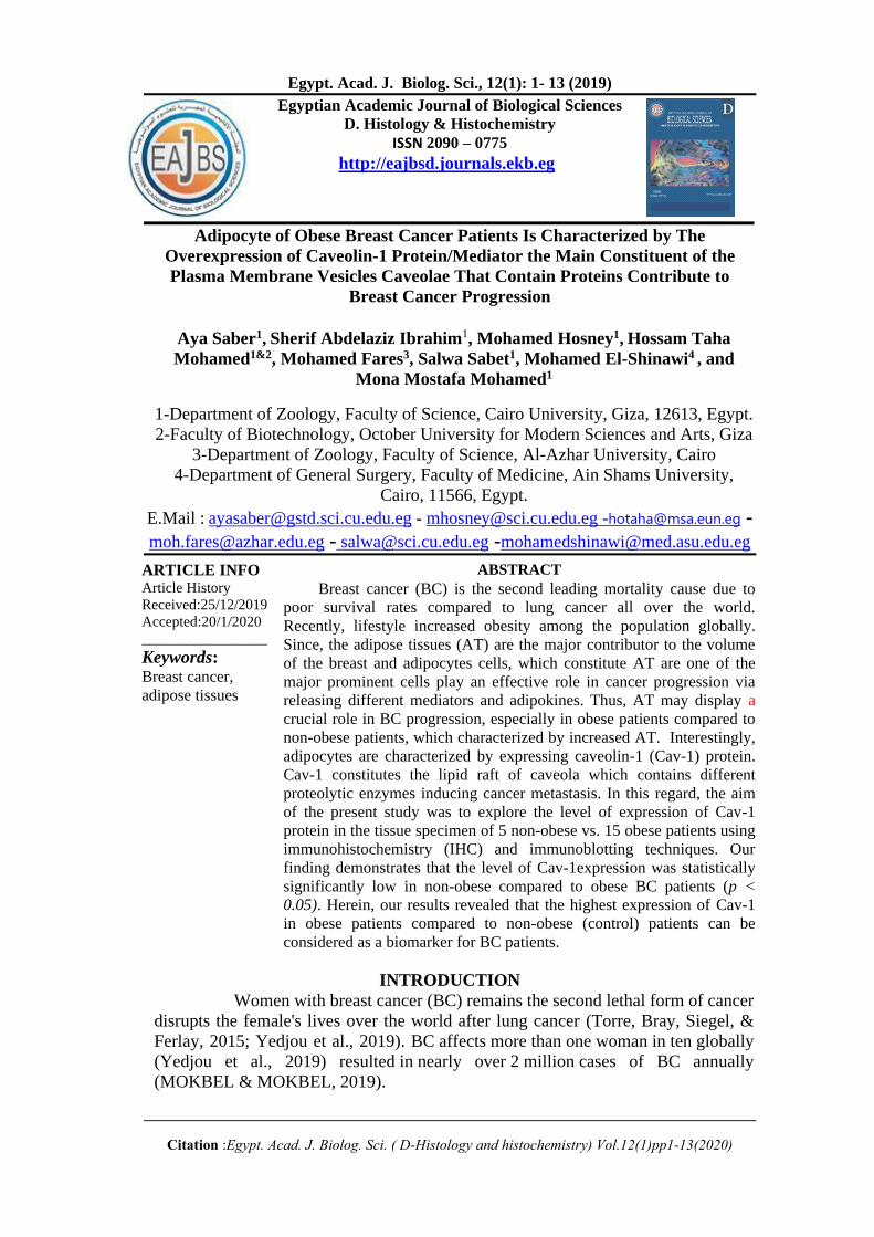
Egypt. Acad. J. Biolog. Sci., 12(1): 1- 13 (2019)
Egyptian Academic Journal of Biological Sciences
D. Histology & Histochemistry
ISSN 2090 – 0775
http://eajbsd.journals.ekb.eg
Adipocyte of Obese Breast Cancer Patients Is Characterized by The
Overexpression of Caveolin-1 Protein/Mediator the Main Constituent of the
Plasma Membrane Vesicles Caveolae That Contain Proteins Contribute to
Breast Cancer Progression
Aya Saber1, Sherif Abdelaziz Ibrahim1, Mohamed Hosney1, Hossam Taha
Mohamed1&2, Mohamed Fares3, Salwa Sabet1, Mohamed El-Shinawi4 , and
Mona Mostafa Mohamed1
1-Department of Zoology, Faculty of Science, Cairo University, Giza, 12613, Egypt.
2-Faculty of Biotechnology, October University for Modern Sciences and Arts, Giza
3-Department of Zoology, Faculty of Science, Al-Azhar University, Cairo
4-Department of General Surgery, Faculty of Medicine, Ain Shams University,
Cairo, 11566, Egypt.
E.Mail : [email protected] - [email protected] [email protected] -
[email protected] - [email protected] [email protected]
INTRODUCTION
Women with breast cancer (BC) remains the second lethal form of cancer
disrupts the female's lives over the world after lung cancer (Torre, Bray, Siegel, &
Ferlay, 2015; Yedjou et al., 2019). BC affects more than one woman in ten globally
(Yedjou et al., 2019) resulted in nearly over 2 million cases of BC annually
(MOKBEL & MOKBEL, 2019).
ARTICLE INFO ABSTRACT
Article History
Received:25/12/2019
Accepted:20/1/2020
_________________
Keywords: Breast cancer,
adipose tissues
Breast cancer (BC) is the second leading mortality cause due to
poor survival rates compared to lung cancer all over the world.
Recently, lifestyle increased obesity among the population globally.
Since, the adipose tissues (AT) are the major contributor to the volume
of the breast and adipocytes cells, which constitute AT are one of the
major prominent cells play an effective role in cancer progression via
releasing different mediators and adipokines. Thus, AT may display a
crucial role in BC progression, especially in obese patients compared to
non-obese patients, which characterized by increased AT. Interestingly,
adipocytes are characterized by expressing caveolin-1 (Cav-1) protein.
Cav-1 constitutes the lipid raft of caveola which contains different
proteolytic enzymes inducing cancer metastasis. In this regard, the aim
of the present study was to explore the level of expression of Cav-1
protein in the tissue specimen of 5 non-obese vs. 15 obese patients using
immunohistochemistry (IHC) and immunoblotting techniques. Our
finding demonstrates that the level of Cav-1expression was statistically
significantly low in non-obese compared to obese BC patients (p <
0.05). Herein, our results revealed that the highest expression of Cav-1
in obese patients compared to non-obese (control) patients can be
considered as a biomarker for BC patients.
Aya Saber et al. 2
In Egypt, 32% of all newly
diagnosed cancer among women in B
C with a high mortality rate
represents 15% of worldwide deaths
per year (Ibrahim, Khaled, Mikhail,
Baraka, & Kamel, 2014; Yehia
Ibrahim, 2019).
Molecular subtypes of BC are
essential for patient diagnosis,
treatment decision and patient
outcomes (Bouchal et al., 2019). BC
are categorized into four molecular
subtypes by three cell membrane
markers; estrogen receptor (ER),
progesterone receptor (PR), as well as
the human epidermal growth factor
receptor (HER2-) that is crucial for
overall survival (OS) prediction
(Nielsen et al., 2004; Shuch, Brian;
Linehan, B. W. M.L.; Srivasan,
2012). The molecular intrinsic
subtypes include luminal A (ER+
and/or PR+ and HER2-), luminal B
(ER+ and/or PR+ and HER2+), HER2+
(ER-, PR- and HER2+), and triple-
negative (ER-, PR-and HER2-).
The increase of fast food and
non-healthy diets are considered
threats for increasing obesity among
Egyptian women and worldwide
(Almuhanna, Alsaif, Alsaadi, &
Almajwal, 2014) that responsible for
developing various tumor forms,
including breast cancer and colon
cancer (Chu et al., 2019; Tiwari et al.,
2019). Several studies have found that
obesity can be considered as a
predictor for poor prognosis in pre-
and postmenopausal BC women
(Chan, Sc, Norat, & Ph, 2015; Tiwari
et al., 2019).
Obesity is identified as over
fat accumulation in the adipose tissues
(AT) which constitute mainly of cells
known as adipocytes (Engin, Engin, &
Gonul, 2019). Any dysfunction in AT
physiological role can promote excess
weight (Chu et al., 2019), which in
turn affects BC by releasing certain
bioactive proteins collectively termed
adipokines that contribute to
proliferation and migration of cancer
cells that influence the poor prognosis
in patients with BC (Guerro-Millo,
2004).
Caveolin‐1(Cav-1) protein is
the primary component of the caveola
(lipid raft) membrane (Chang et al.,
2017; Nouh et al., 2011). Cav-1
belongs to a group of Cav-2 and Cav-3
caveolins (Cohen, Hnasko, Schubert,
& Lisanti, 2004; Williams & Lisanti,
2004; Williams et al., 2006). It is
highly expressed by AT especially
adipocytes of fatty mammary pads
(Williams et al., 2006). Some studies
considered Cav-1 protein as adipokine
and exerts an effect on the role of the
adjacent cell in the microenvironment
(Chang et al., 2017). Cav‐1, 22 kDa
(Catalán et al., 2008) has been
demonstrated to be elevated by
numerous kinds of human cancers
including kidney, liver, breast, colon
(Nwosu, Ebert, Dooley, & Meyer,
2016). This elevated Cav-1 level built
caveola which contains different
proteases that stimulate extracellular
matrix (ECM) degradation and
promote cancer motility and invasion.
(Victor, Anbalagan, Mohamed, Sloane,
& Cavallo-medved, 2011). In addition,
secreted Cav‐1 proteins were increased
by obesity in mice model. The
objective of this study was to
determine the expression levels of
Cav-1 protein in AT of non-obese vs.
obese BC patients by IHC and
immunoblotting.
MATERIALS AND METHOD
:Patients’ Samples
For the purpose of patients’
enrolment in this study, Institutional
Review Board (IRB) approval was
obtained before the surgery and the
protocol was approved by the ethics
committee of Ain Shams University
(Cairo, Egypt). Each patient signed a
written consent form prior to
participation, including agreements
for enrolment in this study and
publication of data. Patients who
were pre-operatively diagnosed as BC
cases by clinical examination,
Adipocyte of Obese Breast Cancer Patients 3
mammography, ultrasound and Tru-
cut biopsy in breast clinic of Ain
Shams University hospitals have
participated in the present study. The
human AT was collected from 20
participants undergoing modified
radical mastectomy (MRM) or
conservative surgery. Patients
enrolled in our study were classified
into two subgroups; non-obese BC
patients (n=5) and obese BC patients
(n=15).
Sample Collection and Handling:
Within hr as soon as possible
after Dr. El-Shinawi's surgery AT
specimens were carried to the
laboratory from the operating room.
(Fig.1A)Each tissue specimen was
divided into 2 parts; one fixed at 10%
PBS-formalin buffered solution for
IHC diagnosis and the second part
was processed for further biochemical
and molecular studies.
Culture of Tumor-Associated
Adipose Tissue (TAAT) Isolated
During Breast Surgery:
The method of maintaining the
tissue specimens was performed under
an aseptic condition. ,TAAT provided
by the surgeon during breast cancer
surgery was transferred directly to the
lab (Fig 1A). AT specimens (Fig 1B)
were washed twice with Phosphate
Buffered Saline (PBS) and a total
amount of about 200-300 mg AT was
sliced into very small pieces. The
TAAT fragments were distributed
equally into a tissue culture dish
(Greiner bio-one, Frickenhausen,
Germany) supplemented with 2 ml
DMEM-F12 media containing 10%
FBS and incubated for 24 h in the
incubator. The media were discarded
was discarded on the next morning, 1
ml serum-free media were added and
incubated for 24 h in a humidified
atmosphere of 5% CO2 at 37 º.
Finally, TAAT conditioned media was
collected and stored at -80ºC for
cytokine profiling. Alternatively,
TAAT explants were kept at -80°C for
future experiments.
Profiling of the Secretions of TAAT-
CM Using Human Cytokine Array:
Ray-Bio cytokine antibody
array-C3 (RayBiotech, Inc. GA, USA)
was used to detect all different
adipokines in ATCM according to the
manufacturer’s instructions. Briefly,
the TAAT-CM was 5-fold
concentrated using Amicon
Ultracell10K filters (Millipore,
Billerica, MA). The cytokine array
membrane is incubated with 2 ml
blocking buffer for 1 h at RT followed
by incubation overnight at 4°C with
ATCM. Finally, the membrane was
probed with a biotinylated antibody
cocktail overnight at 4°C followed by
incubation with HRP-conjugated
streptavidin overnight at 4°C. ImageJ
software was used to evaluate the
adipokines signal intensity. Each
adipokine signal intensity value is
presented as mean ± SEM (n = 5).
Immunohistochemistry (IHC):
Immunohistochemical staining
was performed using 5 µm paraffin
adipose tissue sections sliced with a
microtome and mounted on positively
charged slides as described by the
author (Nouh et al., 2011). In brief, AT
sections were first de-waxed in xylene
clearing agent and hydrated through
descending serial dilutions of ethanol
(100%, 90%, 70%, and 50%)
(Sigma-Aldrich; Merck KGaA,
Darmstadt, Germany). For the heat-
induced antigen retrieval process,
slides were incubated in pre-heated
retrieval buffer in water steamer for 30
min, followed by cooling at room
temperature for 30 min. Endogenous
peroxidase activity was blocked for 10
min using Dako Dual Endogenous
Enzyme Block. After blocking,
adipose tissue sections were incubated
overnight at 4 °C with 1: 150 diluted
primary antibody Cav-1 (BD
Biosciences, San Diego, CA, USA).
After washing, the slides were
incubated with 100 µl horseradish
peroxidase (HRP) linked secondary
anti-mouse (EnVision + Dual Link
System, Dako, Denmark) for 45 min at
Aya Saber et al. 4
room temperature. IHC signal
development was carried out by adding
100 µL of the chromogen 3-3ʹ-
diaminobenzidine (DAB+) diluted
1:50 in substrate buffer for 10-15 min
depending on the appearance of a
brown color. Negative control slides
were run in parallel in which the
primary antibody was omitted. Finally,
tissue specimens were washed in
phosphate-buffered saline (PBS), the
nuclei were counterstained with
Mayer's haematoxylin for a minute,
washed in tap water following
dehydration through ascending ethanol
serial dilutions (70%, 90%, and 100%)
and clearing steps through xylene and
the specimens were mounted using
Permount® (Fisher Scientific,
Pittsburgh, PA, USA) and air-dried
overnight for examination by light
microscopy (Olympus, CX41, Japan).
The stained area fractions were
analyzed semi-quantitatively using
ImageJ software (National Institutes of
Health, MD, USA).
Sodium Dodecyl Sulfate-
Polyacrylamide Gel Electrophoresis
(SDS-PAGE) and Immunoblotting:
AT of non-obese and obese
fresh BC patients obtained by doctor
El-shinawi was lysed in radio-
immunoprecipitation assay buffer
(RIPA buffer) [25 mM Tris HCL pH
7.6, 150 mM NaCl, 1% Triton X-100,
1% Sodium deoxycholate and 0.1%
SDS (Sigma-Aldrich, St. Louis, MO,
USA)] and then centrifuged for 10
minutes at 10,000 × g at 4°C. The
protein content of adipose tissue
lysates was quantified using the
Bradford assay (BioRad Laboratories,
CA, USA) using Infinite®200 PRO
NanoQuant (Tecan, Zürich,
Switzerland). 25–50 μg of AT protein
lysates per lane were loaded and
resolved using SDS-PAGE (12%
acrylamide gel) using Mini-
PROTEAN®II Electrophoresis Cell
(BioRad Laboratories, CA,USA)
apparatus and transferred onto
polyvinylidene fluoride (PVDF)
membrane (Millipore, MA, USA).
After blocking for 1 hr with 5% non-
fat dry milk in TBS-0.5% Tween 20
(TBST), the membrane was probed
with primary antibody against 1:5000
dilution of cav-1 (BD Biosciences, San
Diego, CA, USA) overnight at 4 ˚C on
shaker and then washed and incubated
with secondary antibody conjugated
with 1:10,000 diluted horseradish
peroxidase (Santa Cruz Biotech, CA,
USA) for 1 hr at room temperature.
After washing, bound antibodies were
visualized by using Pierce Enhanced
Chemiluminescence immuno-blotting
substrate (ECL) (Thermo Scientific,
ON, Canada) according to the
manufacturer’s protocol, and analyzed
with ImageJ software (National
Institutes of Health, Bethesda, MA,
USA), which quantifies the density of
each band using β-actin (Santa Cruz
Biotech, CA, USA) as a loading
control.
Statistical Analysis:
The data are analyzed using
IBM SPSS version 15.0 (SPSS,
Chicago, IL, USA) and presented as
mean ± SEM. Differences among
patients were evaluated using Chi-
square and Student’s t-test. Pearson’s
Rank coefficient test was used to
assess the associations with patient
clinical data. Two-tailed p < 0.05 was
considered significant.
RESULTS
Clinical and Pathological
Characterization of BC Patients:
Clinical and pathological
characteristics of obese patients (n
=15) and non-obese (n =5) patients
who participated in the study are
presented in Table 1. The mean age of
obese patients was 52.5 ± 3.9 which
ranged from 34 to 71 years while the
mean age of non-obese patients was
52.6 ± 3.28 which ranged from 43 to
62 years. Tumor size measurements
revealed that 61.54% of obese patients
exhibited tumor masses less than or
equal 4 cm and 38.46% of them
exhibited tumor masses greater than 4
cm, whereas, in the non-obese group,
50% of the patients had a tumor size
Adipocyte of Obese Breast Cancer Patients 5
less than or equal 4 and 50% of them
having a tumor greater than 4. The
tumor grade analysis among obese
patients revealed that 78.57% were
classified as grade II (G2) and 21.43%
were grade III (G3), while 100%
classified as grade 2 (G2) in non-
obese. Among obese patients, 78.57%
were negative for lymph vascular
invasion and 21.43% were positive for
lymph vascular invasion. While 100%
of non-obese (control) patients were
negative for lymph vascular invasion.
All obese patients who underwent
surgery had positive metastatic lymph
nodes: 71.43% had had lymph nodes
less than or equal 4 positive lymph
nodes and 28.75% had greater than 4
positive metastatic lymph nodes while
in non-obese patients, 100% had ≤ 4
lymph nodes. ER, PR and HER-2
status were assessed as negative and
positive for all obese and non-obese
patients. Positive staining for ER, PR,
and HER-2 was detected in 53.33%,
60% and 33.33% obese patients,
respectively. Whereas, the positive
staining for ER, PR in non-obese
patients were 33.3% and 33.3%,
respectively.
Table 1. Clinical and pathological data of obese and non-obese breast cancer patients
Data are expressed as mean ± SEM
NA = not available
*significant P value calculated by ªStudent’s t-test or ᵇPearson Chi-Square
Aya Saber et al. 6
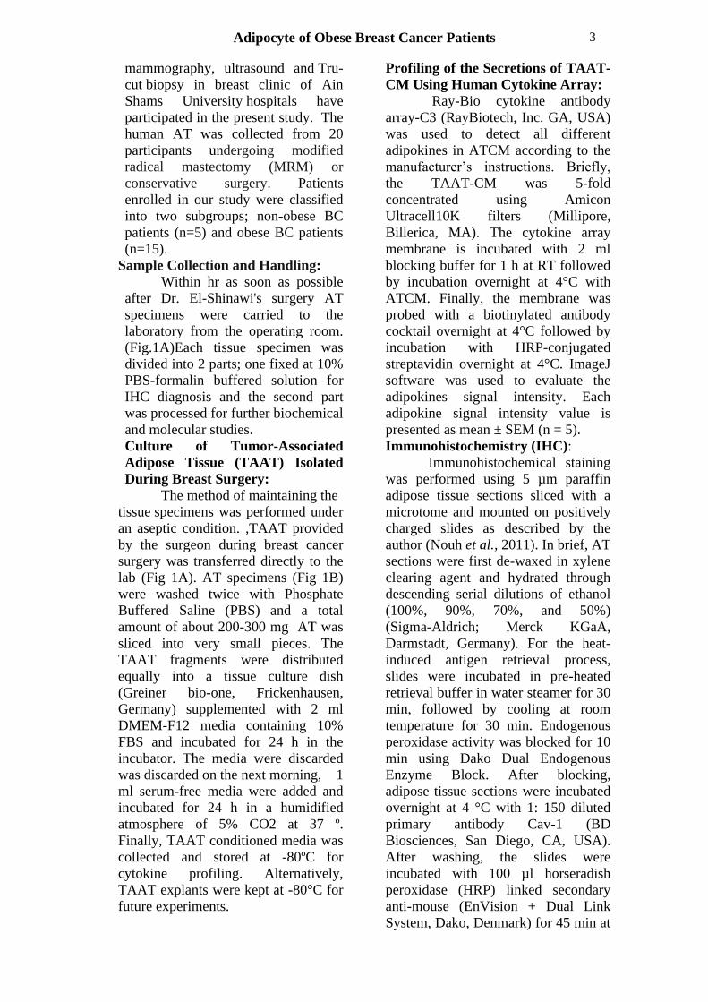
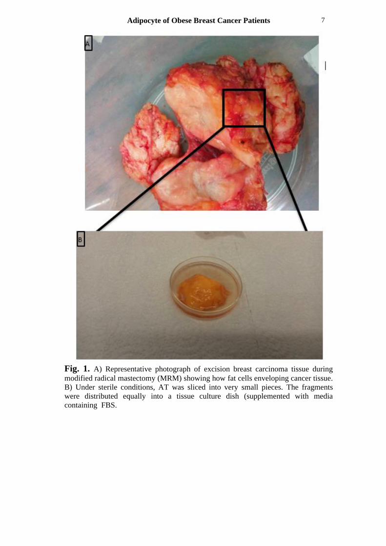
Profiling of Adipokines Secreted
From AT Explants BC Patients:
ATCM secreted of AT explants
from BC patients after 24 h was
subjected to RayBioTM cytokine
antibody array 3 cytokines (Fig. 2).
The secreted ATCM from BC patients
showed that the monocyte
chemoattractant protein (MCP-1),
interleukin (IL-6), and interleukin (IL-
8) cytokines are the highest adipokines
secreted by AT. The cytokines array
image was quantified using ImageJ
software.
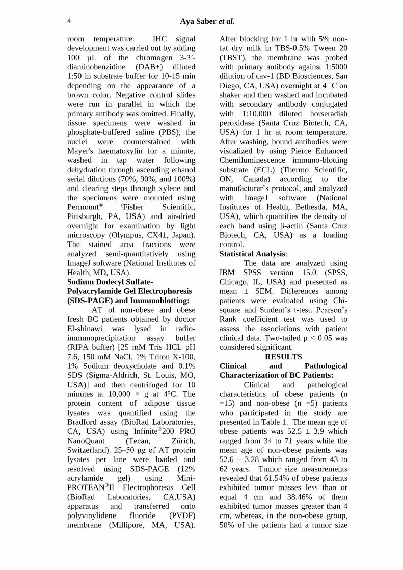
Overexpression of Caveolin-1
Protein in AT of Non-Obese Versus
Obese BC Patients:
Assessment of the expression of
Cav-1 in paraffin-embedded tissue
sections of non-obese versus obese AT
patients with BC using IHC to stain
Cav-1 (Figs. 3A, B& C). A statistically
significant difference between the
expressions of Cav-1 protein was
detected in obese compared to non-
obese tissues (P < 0.05).
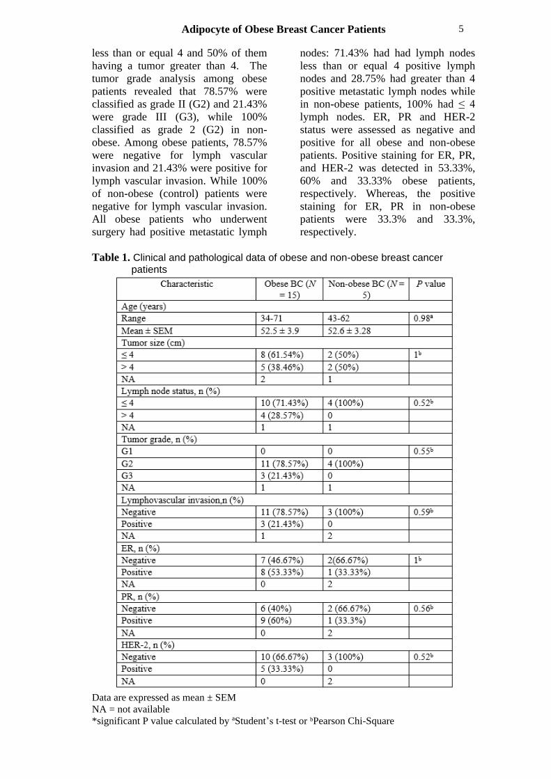
Caveolin-1 Protein Level Is Higher
in the AT of Obese Than Non-Obese
Breast Cancer Patients:
Immunoblotting analysis was
used for assessment of Cav-1
expression in tissue lysates of obese
vs. non-obese BC adipose tissue
patients to confirm IHC results.
Western Blot results (Figs. 4A, B& C)
showed the expression of Cav-1 (22
KDa) was significantly (p < 0.05)
overexpressed in obese (n = 10)
compared to non-obese patients (n = 5)
by ImageJ software and normalized
against the loading control B-actin.
Adipocyte of Obese Breast Cancer Patients 7
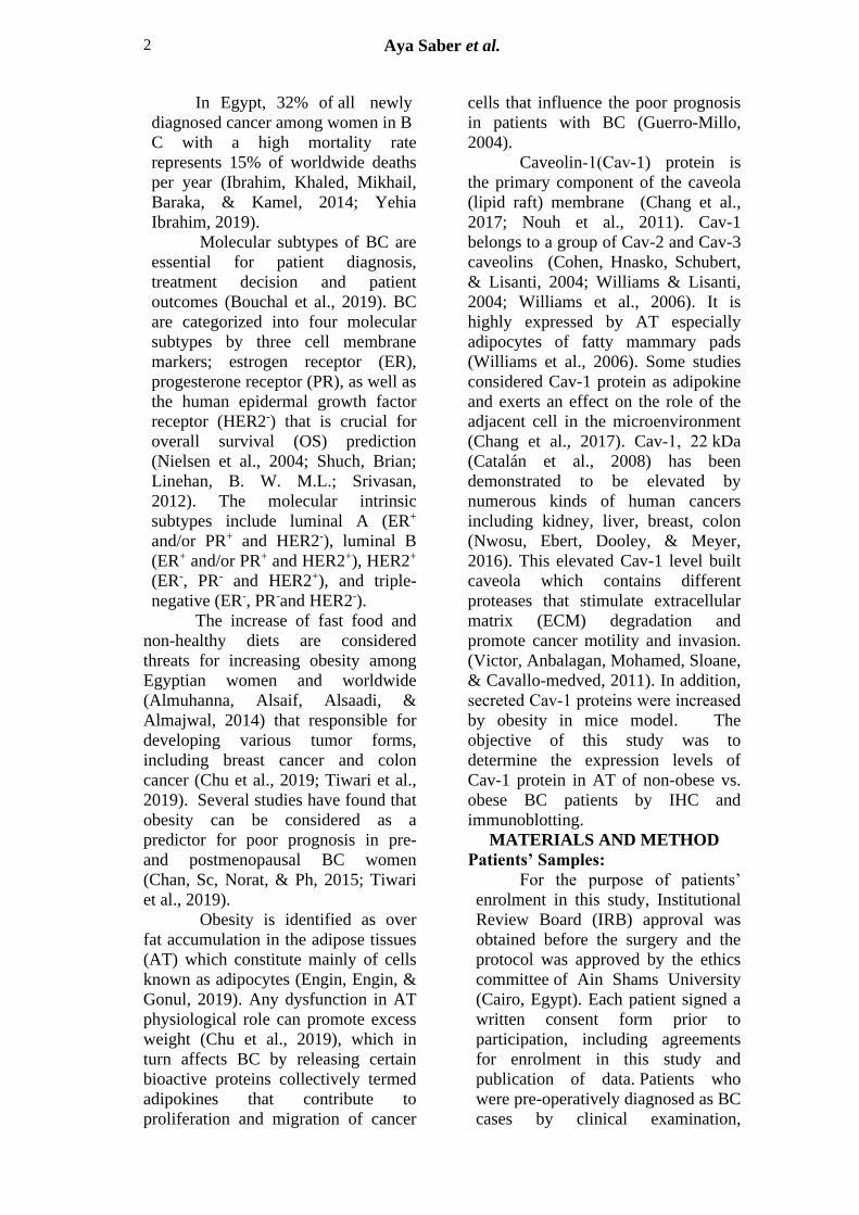
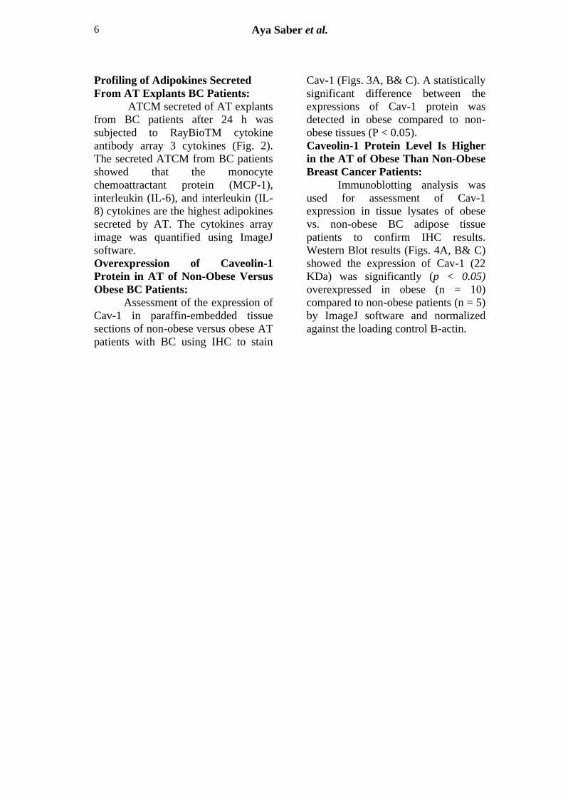
Fig. 1. A) Representative photograph of excision breast carcinoma tissue during
modified radical mastectomy (MRM) showing how fat cells enveloping cancer tissue.
B) Under sterile conditions, AT was sliced into very small pieces. The fragments
were distributed equally into a tissue culture dish (supplemented with media
containing FBS.
Aya Saber et al. 8
Fig. 2. Chart illustrates the secreted cytokines/chemokines from AT BC patients
after 24 hr. A) Representative RayBioTM cytokine antibody array 3 of secreted
ATCM from BC patients after 24 hr. B) Bars represent mean of signal intensity value
of each cytokine secreted by AT explants from BC patients (n = 5).The cytokine array
image was quantified using ImageJ software

Adipocyte of Obese Breast Cancer Patients 9
Fig. 3. Representative fields of immunostaining of Cav-1 (brown color) in paraffin
embedded adipose tissue sections showing high density of adipocyte cells positive for Cav-1
in (B) obese (n = 10) compared to (A) non-obese patients (n = 5). (F) Bars represent the area
fraction of obese relative to non-obese patients calculated by using ImageJ program. The data
represent the mean ± SEM. * P < 0.05 as determined by Student’s t test.
Fig. 4. Representative immunoblots membranes showing Cav-1 protein expression in (B)
adipose tissues of obese relative to (A) non-obese breast cancer patients. (A)1-5 Lanes
represent adipose tissue lysates of different non-obese patients showing weak expression of
Cav-1 (22 kDa). (B)1-5 Lanes represent adipose tissue lysates of different obese patients
showing increased expression of Cav-1 (22 kDa). (C) Bars represent the relative density
values of detected protein bands assessed by ImageJ software and normalized against the
loading control B-actin, showing statistically significant higher expression of Cav-1 in obese
(n = 10) than in non-obese (n = 5) adipose tissues. The data represent mean ± SEM. * P <
0.05 as determined by Student’s t test.
Aya Saber et al. 10
DISCUSSION
In the present study, our data
further demonstrated that the highest
secreted adipokines are IL-6, IL-8, and
MCP-1 and there is a high expression
of Cav-1 protein level in adipose
breast carcinoma tissues by adipocytes
of obese vs. non-obese.
As mentioned by the author
Catalan that there is a positive
correlation between the secretion of
MCP-1 and Cav-1(Catalán et al.,
2008). Our study also confirmed this
where we found more expression of
MCP-1 by obese breast cancer
patients. In other words, we found that
there is an increase also in secreted
Cav-1 in addition to MCP-1 but the
secretion increased significantly in
obese patients compared to non-obese
patients.
Furthermore, the expression of
IL-6 found to be correlated with the
expression of caveolin-1 and poor
prognosis in breast cancer patients
(Podar et al., 2003).
In the caveolae, the membrane
regions which is rich in cholesterols
and glycosphingolipids characterized
by obtaining the main protein Cav-1
(Chang et al., 2017). Besides previous
studies that Cav-1 implicated in
endocytosis and signaling, there is a
wide variety of studies showed that
Cav-1 is altered in several cancer types
such as liver, colon, breast, kidney,
prostate, and plays diverse functions in
cancer development by secretion of
MMPS which degrade ECM (Nwosu
et al., 2016). In addition, Cav1
promote breast cancer development
and metastasis by affecting various
aspects of epithelial–mesenchymal
transition (EMT) (Bailey & Liu, 2008;
Gai, Lu, Tu, Liang, & Zheng, 2014;
Goetz, Lajoie, Wiseman, & Nabi,
2008; Qian et al., 2019). As
mentioned in the study conducted by
Taher and colleagues that human and
mouse prostate cancer cell lines
promotes cancer cell survival and
growth in vitro via secreting Cav-1 and
thus considered as a biomarker and
therapeutic target for cancer treatment
(Tahir et al., 2001).
It should be mentioned that in
obesity, adipocytes are hypertrophied
with a change in the adipokine secretiv
e profile of obese adipose tissue.
Our findings agree with a study
conducted by Chang and co-workers
that obesity changed Cav-1 secretion
as reported that adipose tissues from
normal chow diet-fed mice far less
than that secreted high-fat diet-fed
mice and that lack of Cav-1 has been
linked to loss of fat tissue (Chang et
al., 2017). In addition, they found that
ERK1/2 might play a role in obese
subjects in comparison to non-obese
subjects. Interestingly, inhibition of
ERK1/2 activation in obese patients
with PD98059, a MEK1/2 inhibitor,
prevented the Cav-1 secretion (Chang
et al., 2017).
Our findings are in accordance
with a study that showed that
Cav‐1 expression may contribute to the
formation of more caveolae the
membrane lipid raft that encloses
different non-active proteases,
cytokines, inflammatory mediators
involved in cancer motility, invasion
and metastasis. In addition, we found
that Cav-1 secretion in CAAT is
markedly associated with obesity, so
the high expression of cav-1 may
emerge as a biomarker for obese breast
cancer patients and its targeting may
represent a therapeutic strategy.
However, our findings should be
verified in a large group of breast
cancer patients.
REFERENCES
Almuhanna, M. A., Alsaif, M.,
Alsaadi, M., & Almajwal, A.
(2014). Fast food intake and
prevalence of obesity in school
children in Riyadh City. Sudanese
Journal of Paediatrics, 14(1), 71–
80.
Bailey, K. M., & Liu, J. (2008).
Caveolin-1 up-regulation during
epithelial to mesenchymal
Adipocyte of Obese Breast Cancer Patients 11
transition is mediated by focal
adhesion kinase. Journal of
Biological Chemistry, 283(20),
13714–13724. https://doi.org/
10.1074/jbc.M709329200
Bouchal, P., Schubert, O. T., Faktor,
J., Capkova, L., Imrichova, H.,
Zoufalova, K., … Aebersold, R.
(2019). Breast Cancer
Classification Based on
Proteotypes Obtained by SWATH
Mass Spectrometry. Cell Reports,
28(3), 832–843.e7. https:// doi.
org/10.1016/j.celrep.2019.06.046
Catalán, V., Gómez-ambrosi, J.,
Rodríguez, A., Silva, C., Rotellar,
F., Gil, M. J., … Frühbeck, G.
(2008). Expression of caveolin-1
in human adipose tissue is
upregulated in obesity and
obesity-associated type 2 diabetes
mellitus and related to
inflammation, 948255400, 213–
219.https://doi.org/10.1111
/j.1365 -2265.2007.03021.x
Chan, D. S. M., Sc, M., Norat, T., &
Ph, D. (2015). Obesity and Breast
Cancer : Not Only a Risk Factor
of the Disease. https://doi.org/
10.1007/s11864-015-0341-9
Chang, C., Chen, C., Wen, H., Huang,
C., Hung, M., & Lu, H. (2017).
Caveolin-1 Secreted from
Adipose Tissues and Adipocytes
Functions as an Adipogenesis
Enhancer, 25(11), 1932–1940.
https://doi.org/10.1002/oby.21970
Chu, D., Nguyen, T., Phuong, T., Le,
N., Tien, B., & Tran, D. (2019).
cells The E ff ects of Adipocytes
on the Regulation of Breast
Cancer in the Tumor
Microenvironment : An Update,
1–19.
Cohen, A. W., Hnasko, R., Schubert,
W., & Lisanti, M. P. (2004). Role
of caveolae and caveolins in
health and disease. Physiological
Reviews, 84(4), 1341–1379.
https://doi.org/10.1152/physrev.0
0046.2003
Engin, A. B., Engin, A., & Gonul, I. I.
(2019). The effect of adipocyte –
macrophage crosstalk in obesity-
related breast cancer.
Gai, X., Lu, Z., Tu, K., Liang, Z., &
Zheng, X. (2014). Caveolin-1 is
up-regulated by GLI1 and
contributes to GLI1-driven EMT
in hepatocellular carcinoma.
PLoS ONE, 9(1). https://doi.org/
10.1371/journal.pone.0084551
Goetz, J. G., Lajoie, P., Wiseman, S.
M., & Nabi, I. R. (2008).
Caveolin-1 in tumor progression:
The good, the bad and the ugly.
Cancer and Metastasis Reviews,
27(4), 715–735. https://doi.org/
10.1007/s10555-008-9160-9
Guerro-Millo, M. (2004). Adipose
tissue and adipokines: For better
or worse. Diabetes and
Metabolism, 30(1), 13–19. https:
//doi.org/10.1016/s1262-3636
(07) 70084-8
Ibrahim, A. S., Khaled, H. M.,
Mikhail, N. N. H., Baraka, H., &
Kamel, H. (2014). Cancer
Incidence in Egypt : Results of
the National Population-Based
Cancer Registry Program, 2014.
MOKBEL, K., & MOKBEL, K.
(2019). Chemoprevention of
Breast Cancer With Vitamins and
Micronutrients: A Concise
Review. In Vivo, 33(4), 983–997.
https://doi.org/10.21873/invivo.11
568
Nielsen, T. O., Hsu, F. D., Jensen, K.,
Cheang, M., Karaca, G., Hu, Z.,
… Perou, C. M. (2004).
Immunohistochemical and
Clinical Characterization of the
Basal- Like Subtype of Invasive
Breast Carcinoma, 10(919),
5367–5374.
Nouh, M. A., Mohamed, M. M., El-
Shinawi, M., Shaalan, M. A.,
Cavallo-Medved, D., Khaled, H.
M., & Sloane, B. F. (2011).
Cathepsin b: A potential
prognostic marker for
inflammatory breast cancer.
Journal of Translational
Medicine, 9(1), 1. https://doi.org/
10.1186/1479-5876-9-1
Aya Saber et al. 12
Nwosu, Z. C., Ebert, M. P., Dooley, S.,
& Meyer, C. (2016). Caveolin-1
in the regulation of cell
metabolism : a cancer perspective.
Molecular Cancer, 1–12. https://
doi.org/10.1186/s12943-016-
0558-7
Podar, K., Tai, Y., Cole, C. E.,
Hideshima, T., Sattler, M.,
Hamblin, A., … Anderson, K. C.
(2003). Essential Role of
Caveolae in Interleukin-6- and
Insulin-like Growth Factor I-
triggered Akt-1-mediated
Survival of Multiple Myeloma
Cells *, 278(8), 5794–5801.
https://doi.org/10.1074/jbc.M208
636200
Qian, X. L., Pan, Y. H., Huang, Q. Y.,
Shi, Y. B., Huang, Q. Y., Hu, Z.
Z., & Xiong, L. X. (2019).
Caveolin-1: A multifaceted driver
of breast cancer progression and
its application in clinical
treatment. OncoTargets and
Therapy, 12, 1539–1552. https://
doi.org/10.2147/OTT.S191317
Shuch, Brian; Linehan, B. W. M.L.;
Srivasan, R. (2012). O ncologist
G, 1051–1062. https://doi.org/
10.1634/theoncologist.2011-0227
Tahir, S. A., Yang, G., Ebara, S.,
Timme, T. L., Satoh, T., Li, L., …
Thompson, T. C. (2001). Secreted
caveolin-1 stimulates cell
survival/clonal growth and
contributes to metastasis in
androgen-insensitive prostate
cancer. Cancer Research, 61(10),
3882–3885.
Tiwari, P., Blank, A., Cui, C.,
Schoenfelt, K. Q., Zhou, G., Xu,
Y.,Olopade, F. (2019).
Metabolically activated adipose
tissue macrophages link obesity
to triple-negative breast cancer,
216(6), 1345–1358.
Torre, L. A., Bray, F., Siegel, R. L., &
Ferlay, J. (2015). Global Cancer
Statistics , 2012, 65(2), 87–108.
https://doi.org/10.3322/caac.2126
2.
Victor, B. C., Anbalagan, A.,
Mohamed, M. M., Sloane, B. F.,
& Cavallo-medved, D. (2011).
Inhibition of cathepsin B activity
attenuates extracellular matrix
degradation and inflammatory
breast cancer invasion. Breast
Cancer Research, 13(6), R115.
https://doi.org/10.1186/bcr3058
Williams, T. M., & Lisanti, M. P.
(2004). The caveolin proteins.
Genome Biology, 5(3), 1–8.
https://doi.org/10.1186/gb-2004-
5-3-214
Williams, T. M., Sotgia, F., Lee, H.,
Hassan, G., Di Vizio, D.,
Bonuccelli, G., … Lisanti, M. P.
(2006). Stromal and epithelial
caveolin-1 both confer a
protective effect against
mammary hyperplasia and
tumorigenesis: Caveolin-1
antagonizes cyclin D1 function in
mammary epithelial cells.
American Journal of Pathology,
169(5), 1784–1801. https:
//doi.org/10.2353/ajpath.2006.060
590
Yedjou, C. G., Sims, J. N., Miele, L.,
Noubissi, F., Lowe, L., Fonseca,
D. D., … Tchounwou, P. B.
(2019). Health and Racial
Disparity in Breast Cancer.
Advances in Experimental
Medicine and Biology, 1152, 31–
49. https://doi.org/10.1007/978-3-
030-20301-6_3
Yehia Ibrahim, N. (2019). Clinico-
Epidemiological Study of Elderly
Breast Cancer in a Developing
Country: Egypt. Journal of
Cancer Treatment and Research,
7(1), 23. https://doi.org/
10.11648/ j.jctr.20190701.14.
Adipocyte of Obese Breast Cancer Patients 13
ARABIC SUMMARY
الخلايا الدهنية المكونة للنسيج الدهنى لمرضى سرطان الثدى ذ وات الوزن الزائد تتميز بزيادة انتاج بروتين الكافيولين-1 والذي يعتبر احدى المكونات الاساسية للانبعاج الكافبولى فى غشاء الخلية ويساهم فى تفاقم
سرطان الثدى
أية صابر1، محمد حسنى1، حسام طه محمد 1,2 ، محمد فا رس3، سلوى ثابت1، منى مصطفى محمد1، محمد الشناوي4
مصر ،12613قسم علم الحيوان، كلية العلوم، جامعة القاهرة، الجيزة، 1 ، الجيزة كلية بيوتكنولوجى، جامعة اكتوبر للعلوم الحديثة والفنون 2
قاهرةل، ازهرقسم علم الحيوان، كلية العلوم، جامعة الا 3
مصر ،11566الطب، جامعة عين شمس، القاهرة، قسم الجراحة العامة، كلية 4
زال سرطان الثديي لا يؤثر بعد سرطان الرئة أكثر أشكال السرطان انتشارا وثاني أشكال مميتة وقاتلة
يؤثر حيث انه ، في جميع أنحاء العالم والاسرة حياة المرأة على شرة أشخاص رأة واحدة في ععلى أكثر من ام
من كل %32في مصر، . وما أدى إلى ما يقرب من مليوني حالة من سرطان الثدي سنويام على مستوى العالم
من الوفيات على مستوى %15يمثل وحالات السرطان التي تم تشخيصها حديثا بين النساء هي سرطان الثدي،
المريض وتحسين لتشخيص مختلفة يئى لسرطان الثدي يمثل أنواع. أن التكوين البيولوجى والجزالعالم سنويا
اعتمادا على تعبير مستقبلات إلى أربعة أنواع فرعية جزيئية . وقد تم تصنيف المرضى نتائج المرضى
الهرمونات مثل هرمون الإستروجين وهرمون البروجستيرون بالإضافة إلى مستقبل عامل نمو البشرة وهذه
ومينال إيه الذي يمتاز بوجود مستقبل هرمون ة تنقسم إلى أربع أنواع هي النوع الأول هو ليالأنواع الجزيئي
الاستروجين و/أو مستقبل هرمون البروجستيرون، والنوع الثاني هو ليومينال بي الذي يمتاز بوجود مستقبل
هرمون الاستروجين و/أو مستقبل هرمون البروجستيرون بالإضافة إلى مستقبل عامل نمو البشرة أما النوع
و البشرة الموجب الذي يمتاز بوجود مستقبل عامل نمو البشرة فقط وأخيرا النوع الثالث فهو مستقبل عامل نم
من مستقبل هرمون الاستروجين أو مستقبل هرمون الرابع وهو السالب الثلاثي الذي يمتاز بعدم وجود أيا
ين سنة الماضية البروجستيرون أو مستقبل عامل نمو البشرة. وقد وجد ان التغير النمطى لحياة المراة فى العشر
ظهورالمسؤولة عن من العوامل ومن اهمها زيادة الوزن بسبب الاعتماد على الوجبات السريعة غير الصحية
مختلفة، بما في ذلك سرطان الثدي حميدة وسرطانية ورما عدة دراسات أن السمنة هي دالة التنبؤ اثبتت وقد .
لطمثالسيئ في حالات سرطان الثدي قبل وبعد انقطاع ا يتم تحديد السمنة على أنها زائدة عن الحد من تراكم .
بحيث يمكن لأي اختلال وظيفي في الأدوار تعزيز الوزن الزائد والسمنة، الذي الدهون في الأنسجة الدهنية
والتى يطلق بعض البروتينات الحيوية النشطة بشكل جماعي افرازسرطان الثدي عن طريق علىبدوره يؤثر
المرضى الذين يعانون تحفز نشاط هجرة الخلايا السرطانية التي و تسهم في انتشار اديبوكاينز وهىعليها مسمى
.من سرطان الثدي روتينبان ينتميو. الكافيوليين احد المكونات الرئيسية لغشاء الكافيولا المنبعج إلى عائلة من
لى مستوى عال بواسطة الأنسجة الدهنية، وهو ويتم التعبير عنه ع .3و كافيولا 2بروتينات اخرى كافيولا ثلاثة
، كاديبوكاينالدهون الثديية، لذا فهو يعمل الانسجةمتوفر بكثرة في التي ثبت أنها مرتفعة بأنواع عديدة من
او يتم قياس الهدف من هذه الدراسة أن يحدد لذلك فان بما في ذلك الكلى والكبد والثدي والقولون. اتالسرطان
فيولين فى المرضى ذوات الوزن الزائد والمعتدل باستخدام كيمياء الخلايا المناعية والامينوبلوتنج.مستوى الكا