Embed Size (px)
Citation preview

Proc. Natl. Acad. Sci. USAVol. 92, pp. 10099-10103, October 1995Cell Biology
Efficient gene transfer into human hepatocytes bybaculovirus vectorsCHRISTIAN HOFMANN*t, VOLKER SANDIG*t, GARY JENNINGS*, MICHAEL RUDOLPHt, PETER SCHLAG§,AND MICHAEL STRAUSS*11I*Max-Planck-Gesellschaft and Humboldt University, Max Delbruck Center for Molecular Medicine, and §Universitatsklinikum Rudolf Virchow,Robert-Rossle-Klinik, Robert-Rossle-Strasse 10, D-13122 Berlin, Germany; tUniversity of Potsdam, Faculty of Biology, Am Neuen Palais 10,D-14469 Potsdam, Germany; and IDanish Cancer Society, Division of Cancer Biology, Strandboulevarden 49, DK-2100 Copenhagen, Denmark
Communicated by Max L. Birnstiel, Research Institute of Molecular Pathology, Vienna, Austria, June 19, 1995
ABSTRACT Viral vectors are the most efficient tools forgene delivery, and the search for tissue-specific infectingviruses is important for the development of in vivo genetherapy strategies. The baculovirus Autographa californicanuclear polyhedrosis virus is widely used as a vector forexpression of foreign genes in insect cells, and its hostspecificity is supposed to be restricted to arthropods. Here wedemonstrate that recombinant A. californica nuclear polyhe-drosis virus is efficiently taken up by human hepatocytes viaan endosomal pathway. High-level reporter gene expressionfrom heterologous promoters was observed in human andrabbit hepatocytes in vitro. Mouse hepatocytes and some otherepithelial cell types are targeted at a considerably lower rate.The efficiency of gene transfer by baculovirus considerablyexceeds that obtained by calcium phosphate or lipid trans-fection. These properties of baculovirus suggest a use for it asa vector for liver-directed gene transfer but highlight apotential risk in handling certain recombinant baculoviruses.
Gene therapy is a powerful concept for the treatment of avariety of diseases (1-3). The liver is the organ where manygenetic diseases are manifested (4). The first clinical trial forthe treatment of familial hypercholesterolemia was based ontransfer of the low density lipoprotein receptor gene to hepa-tocytes in vitro and subsequent retransplantation (5). Since thisprotocol is very complicated and probably does not result insufficient therapeutic effects, it would be particularly impor-tant to have methods available that allow for gene transfer tothe liver in vivo (6, 7). Retroviruses integrate their genome intothat of the host cell but only in dividing cells (8); therefore,gene transfer to the liver requires stimulation of cell prolifer-ation- e.g., by partial hepatectomy (9-11). Adenoviral vectorsdeliver genes to the liver at very high efficiencies, approaching100% gene transduction to hepatocytes (12). However, themajor disadvantage of this type of vector is the instability of thetransferred genes in the target cells due to a lack of integrationand to immunological response (13). Recent modifications ofthe adenoviral viral vector system leading to a reduction of theimmune response resulted in a significant prolongation offunction of the transferred gene (14). The general disadvan-tage of viral vectors available so far for gene delivery to theliver is their lack of organ specificity.
Alternative means for gene targeting to the liver have beendeveloped, which are based on the concept of receptor tar-geting (15-21). Despite the fact that the generation of particlesfor receptor targeting has been optimized recently and aremarkable efficiency of in vivo gene transfer could beachieved (22), this is still not efficient enough for therapeuticapplication. Thus, it would be important to investigate the
abilities of other viruses with regard to hepatocyte-specificgene delivery.The baculovirusAutographa californica nuclear polyhedrosis
virus (AcNPV), which is used as a vector for protein overpro-duction in insect cells (23-25), was also studied in the pastregarding its ability to infect mammalian cells (26-29). Abor-tive infection of some cell types by AcNPV has been detected,but neither gene expression nor viral DNA replication could beobserved, most likely due to the restriction of viral promoterfunction to insect cells. These results suggested that the viruswould not be a risk factor for humans and even prompted thedevelopment of baculovirus vectors as a biological weaponagainst particular insects. Recently, a baculovirus expressingan insect-specific toxin has been used in a field trial (30).As infection of liver cells by AcNPV was not tested before,
we designed recombinant baculoviruses that should expressreporter genes provided the virus is taken up by hepatocytes.The results described in this report favor the use of baculovirusvectors for the development of strategies for liver-directedgene therapy but also point at a requirement for a carefulinvestigation of the potential risks connected with the unre-stricted use of this type of virus.
MATERIALS AND METHODSGeneration of Recombinant Baculoviruses. Recombinant
baculoviruses were constructed using the transfer vectorpVL1392 (31) (PharMingen). For AcNPV-PHTag the genecoding for a C-terminally truncated simian virus 40 (SV40)large tumor antigen (T antigen) (32) was cloned next to theviral polyhedrin (PH) promoter, whereas for AcNPV-CMVTagthis gene was fused to the cytomegalovirus (CMV) immediateearly promoter (33) and inserted into pVL1392 in the orien-tation opposite to the PH promoter. The Photinus pyralisluciferase reporter gene (34) was introduced into the baculo-virus vector either under control of the PH promoter (AcNPV-PHL) or under control of the CMV immediate early promoter(AcNPV-CMVL) fused to the luciferase gene before cloninginto the transfer vector. Viruses were generated according tostandard procedures (23, 35). Budded virus was concentratedfrom cell culture medium by sedimentation at 35,000 x g for30 min and purified by centrifugation in a 24-62% (wt/vol)linear sucrose gradient. The virus titer was estimated by plaqueassay on Sf9 cells.
Cell Culture. Primary human hepatocytes were obtained byperfusion with collagenase of small sections of normal tissuefrom resected livers of patients with liver metastases of coloncarcinoma as described (36). Primary mouse or rabbit hepa-tocytes were obtained by perfusion with collagenase of livers
Abbreviations: AcNPV, Autographa californica nuclear polyhedrosisvirus; SV40, simian virus 40; T antigen, tumor antigen; CMV, cyto-megalovirus; moi, multiplicity of infection; PH, polyhedrin.tThese authors contributed equally to this work.I1To whom reprint requests should be addressed at the * address.
10099
The publication costs of this article were defrayed in part by page chargepayment. This article must therefore be hereby marked "advertisement" inaccordance with 18 U.S.C. §1734 solely to indicate this fact.

10100 Cell Biology: Hofmann et al.
in situ. Primary hepatocytes were seeded onto a layer ofcollagen IV and grown in William's E medium without serum.Most experiments were carried out using an established hep-atocyte line (Huh7) derived from a human hepatocellularcarcinoma (37), which was grown in Dulbecco's modifiedEagle's medium containing 10% (vol/vol) fetal calf serum. Anumber of cell lines from different tissue origin were used(given in the legend to Fig. 3) and grown in the mediarecommended by the authors or by the American Type CultureCollection. Treatment with baculovirus was done 48 h afterseeding.
Incubation of Mammalian Cells with Recombinant Bacu-lovirus. Different cell types were treated with baculovirusunder similar conditions. Incubation of established cells wasgenerally done in complete medium containing 10% (vol/vol)heat-inactivated fetal calf serum for 1 h at 37C at the multi-plicity of infection (moi) indicated. Untreated serum caused aconsiderable decrease in gene transfer efficiencies. Primaryhepatocytes were treated with virus in the absence of serum.Treatment with a maximal virus concentration of 1500 moi percell did not cause any toxic side effect as indicated by trypanblue exclusion and an unchanged plating efficiency at 2 daysafter treatment.Assays for Reporter Enzyme Activities. Cells were lysed in
a buffer containing 100 mM potassium phosphate (pH 7.8),0.2% Triton X-100, and 1 mM dithiothreitol. For luciferaseassays, 50 p,l of cleared lysate was incubated with 180 ,ld ofreaction buffer containing 25 mM phosphate buffer (pH 7.8),4 mM EGTA, 15 mM MgSO4, 1 mM dithiothreitol, and 1 mMATP, and 50 Al of a 20 ,uM luciferin solution was injected.Relative light units were measured using a luminometer(Berthold, Wildbad, Germany). Values given in the figures arethose obtained from extracts of 104 cells. A background valueof 120 relative light units was substracted in all cases. Assaysfor f3-galactosidase activity were carried out using the Galacto-
Light kit (Tropix, Bedford, MA) essentially as described by themanufacturer.
RESULTSBaculovirus Infection of Hepatocytes. To explore the pos-
sibility that a recombinant baculovirus might be used to infecthuman hepatocytes, we constructed recombinant baculovi-ruses with the gene coding for a truncated large T antigen ofSV40 under the control of either the PH gene promoter ofbaculovirus or the immediate early promoter of CMV. Afterincubation of human hepatocellular carcinoma cells (Huh7)for 1 h with the virus at a moi of 1000, T antigen was visualizedby immunofluorescence. Whereas nuclei of hepatocytestreated with AcNPV-PHTag were negative, those treated withAcNPV-CMVTag were strongly positive (Fig. 1). The effi-ciency of obtaining T antigen-positive nuclei by treatment ofHuh7 cells with the latter virus was usually 80-100% at a moi1000 and still "50% at a moi of 100. The actual uptake of bothtypes of recombinant baculoviruses was confirmed by electronmicroscopy (Fig. 2).
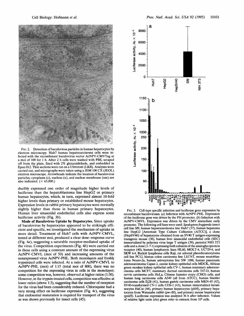
Specific Uptake by Hepatocytes. To investigate the speci-ficity of hepatocyte infection by baculovirus, we tested differ-ent hepatic and nonhepatic cell types for expression of theluciferase reporter gene. We could not detect luciferase ac-tivity in human or mouse hepatocytes or NIH 3T3 fibroblastcells on infection by AcNPV-PHL, whereas high levels ofactivity were found in Sf9 insect cells, which shows that the PHpromoter is inactive in the mammalian cells tested (Fig. 3a).However, if expression was driven by the CMV promoter, highlevels of luciferase activity were detected in various hepatic celltypes (Fig. 3b). In contrast, no or very low levels of expressionwere found in five lymphocytic cell lines, human and rodentcarcinoma lines of different tissue origin (lung, breast, kidney,and bladder), mouse fibroblasts, and various neuronal cells(Fig. 3b). The established hepatocarcinoma line Huh7 repro-
FIG. 1. Immunofluorescence detection of SV40 large T antigen expression in human hepatocytes after infection by recombinant baculoviruses.(a and b) AcNPV-CMVTag. (c and d) AcNPV-PHTag. In these recombinants, expression was driven by either the PH promoter or the CMVimmediate early promoter. Huh7 human hepatocarcinoma cells (37) grown on coverslips were incubated with virus at a moi of 1000 for 1 h. Thirty-sixhours later cells were fixed with methanol/acetone at -20°C for 5 min. For immunofluorescence detection, fixed cells were first incubated for 1h with the T antigen-specific monoclonal antibody 416, washed, incubated for 1 h with a biotinylated goat anti-mouse antibody, and finally incubatedwith streptavidin-Texas Red. (a and c) Immunofluorescence detection. (b and d) Phase-contrast micrograph. (Bars = 25 ,Lm.)
Proc. Natl. Acad. Sci. USA 92 (1995)
Proc. Natl. Acad. Sci. USA 92 (1995) 10101
FIG. 2. Detection of baculovirus particles in human hepatocytes byelectron microscopy. Huh7 human hepatocarcinoma cells were in-fected with the recombinant baculovirus vector AcNPV-CMVTag ata moi of 100 for 1 h. After 2 h cells were washed with PBS, scrapedoff from the plate, fixed with 2% glutaraldehyde, and embedded inEpon 812. Thin sections were cut on a Ultrotom (LKB). Analyses werecarried out, and micrographs were taken using a JEM 100 CX (JEOL)electron microscope. Arrowheads indicate the location of baculovirusparticles; cytoplasm (c), nucleus (n), and nuclear membrane (nm) arealso indicated. (x 63,000.)
ducibly expressed one order of magnitude higher levels ofluciferase than the hepatoblastoma line HepG2 or primaryhuman hepatocytes, which, in turn, expressed almost 10-foldhigher levels than primary or established mouse hepatocytes.Expression levels in rabbit primary hepatocytes were normallyslightly higher than those in human primary hepatocytes.Human liver sinusoidal endothelial cells also express someluciferase activity (Fig. 3b).Mode of Baculovirus Uptake by Hepatocytes. Since uptake
of baculovirus by hepatocytes appeared to be strikingly effi-cient and specific, we investigated the mechanism of uptake inmore detail. Treatment of Huh7 cells with AcNPV-CMVL,tested at different moi, produced a clear dose-response curve(Fig. 4a), suggesting a saturable receptor-mediated uptake ofthe virus. Competition experiments (Fig. 4b) were carried outin these cells using a constant amount of the expressing virusAcNPV-CMVL (moi of 50) and increasing amounts of thenonexpressed virus AcNPV-PHL. Both monolayers and freshlytrypsinized cells were infected. At a ratio of AcNPV-CMVL toAcNPV-PHL (moi) of 1:15 (total moi of 800), there was nocompetition for the expressing virus in cells in the monolayer;some competition was, however, observed at higher ratios (1:30).However, in the trypsin-treated cells, competition was effective atlower ratios (above 1:3), suggesting that the number of receptorsfor the virus had been considerably reduced. Chloroquine had avery strong effect on luciferase expression (Fig. 4c), suggestingthat endosomal maturation is required for transport of the virusas was shown previously for insect cells (45).
7 8000-°x' 6000-
.5-io 4000Dau0cu
!D 20000
-J
0
1150
1100
1050
0
X 1000x 250
.20 200co0co.A 1500-j
100
50
a
rl_. C\J 0
I a) cnI a
Iz
b
AO=C Ec-NVM.pei was d by th CMV 'im i earlyII Qi'I.C%1 -j-° _ C Z2z I <
FIG. 3. Cenl-type specific infection and luciferase gene expression byrecombinant baculoviruses. (a) Infection with AcNPV-PHL. Expressionof the luciferase gene was driven by the PH promoter. (b) Infection withAcNPV-CMVL. Expression was driven by the CMVaimmediate earlypromoter. The following cell lines were used: Spodoptera ugiperda insectcell line Sf9, human hepatocarcinoma line Huh7 (37), human hepatomaline HepG2 [American Type Culture Collection (ATCC), a clone(HepSV40) of hepatocytes obtained from an SV40 T antigen-expressingtransgenic mouse (38), human liver sinusoidal endothelial cells (SEC)immortalized by polyoma virus large T antigen (39), parental NIH3o3cells and a clone (1-7-1) expressing both subunits of the asialoglycoproteinreceptor (40), human lymphocyte lines HLst, MOLT-4, UC729-6, andSKW 6.4, Burkitt lymphoma cells Raji, rat adrenal pheochromocytomacehu ine PC12, human colon carcinoma line LS174T, mouse neuroblas-toma Neuro-2a, human astrocytoma line SW 1088, human pancreaticadenocarcinoma Capan 2, canine kidney epithelial cells MDCKN Africangreen monkey kidney epithelial cells CV-1, human mammary adenocar-cinoma cells MCF7, mammary ductual carcinoma cells T47-D, humancervix carcinoma cells HeLa, Chinese hamster ovary (CHO) cells, andhuman lung carcinoma cells A549 (all from ATCC), human bladdercarcinoma cells Ej28 (41), human gastric carcinoma cells MKN-45 (42),SV40-transformed CV-1 cells COS-1 (43), human immortalized kerati-nocytes HaCat (44), primary human hepatocytes (phH), primary hepa-tocytes from Watanabe rabbit (pw-rH), and primary mouse hepatocytes(pmH). Luciferase expression was analyzed 36 h after infection. Valuesof relative light units (rlu) given refer to extracts from 104 cells.
Cell Biology: Hofmann et al.

10102 Cell Biology: Hofmann et al.
El
= 106 -:
C
106,
1
4.
6)
It
C.)
4-
CI
c)
'.)u)
'e
moi
bHuh7trp
o Huh7
; 105~~
0.1 1 10 100
Ratio of virus titers
c
g 100.0%9
0
o 80.0%
v 60.0%
, 40.0%C)CZa 20.0%
o
C 0.5 0.1 0.01
Chloroquine, mM
FIG. 4. Effect of virus titer, virus competition, and inhibition ofendosomal maturation on baculovirus-mediated expression of theluciferase gene in Huh7 cells. (a) Effect of virus titer on luciferaseactivity. (b) Influence of competing nonspecific AcNPV-PHL virus onexpression levels obtained with AcNPV-CMVL. (c) Effect of chloro-quine treatment of cells. (a) Huh7 cells were infected with AcNPV-CMVL or AcNPV-PHL at increasing moi. (b) Huh7 cells were infectedwith AcNPV-CMVL at a fixed moi of 50 and increasing amounts ofAcNPV-PHL. (c) Huh7 cells were infected with AcNPV-CMVL at a
moi of 100. In the case of trypsin treatment, cells were incubated withtrypsin (500 jig/ml) for 3 min at 37°C, washed, and replated 1 h beforevirus application (Huh7trp cells). For treatment with chloroquine,cells were incubated with the agent for 4 h (3 h before infection andduring the infection time of 1 h). Control cells (C) were incubated withchloroquine 12 h after infection. Expression was analyzed 36 h afterinfection as described in Materials and Methods. rlu, Relative lightunits.
Efficiency of Gene Transfer into Hepatocytes. To gain someinsight into the efficiency of gene transfer by a baculovirus-derived vector, a comparative study was carried out in Huh7cells including calcium phosphate transfection, lipid transfec-tion (lipofection), and adenoviral vector infection. The ,B-ga-
Table 1. Comparison of various gene delivery methods regardingtransfer and expression efficiencies in human hepatocytes
,f-Galactosidaseactivity, rlu Positive
Method moi per 103 cells cells, %
Calcium phosphate 81,200 + 7,400 20Lipofectamine 72,300 + 6,800 15Baculovirus 10 94,600 + 8,400 12
100 512,300 62,600 50Adenovirus 10 53,100 + 4,600 30
100 151,800 + 17,200 95
Three-centimeter dishes were seeded with 1 x 10⁵ Huh7 cells theday before treatment. Four dishes were used for every method.Calcium phosphate transfection was according to standard protocols(41) and lipofection using either Lipofectin or Lipofectamine(GIBCO/BRL/Life Technologies) was done according to the proto-col provided by the manufacturer. Infection with either baculovirus oradenovirus vectors was done for 1 h at the moi given. The adenovirusharboring a Rous sarcoma virus promoter-driven gene coding fornuclear ,B-galactosidase was a kind gift of Andre Lieber and Mark Kay(University of Washington, Seattle). f3-Galactosidase assays were doneas described in Materials and Methods. Extract from every dish wastested for activity, and the mean values of three dishes are given.Histochemical staining with 5-bromo-4-chloro-3-indolyl 3-D-galactoside was carried out in parallel dishes. The number of blue cellswas counted under the microscope and is given as a percentage of thetotal number of cells. Three separate areas of a dish containing 1000cells were counted. rlu, Relative light units.
lactosidase reporter gene under control of the Rous sarcomavirus promoter was used to allow for quantification of both thetotal activity in the cells treated and the percentage of ex-pressing cells. The results are summarized in Table 1. The dataclearly show that both viral vector systems are more efficientthan calcium phosphate transfection or lipofection if used at amoi of 100. At the time of the assay (36 h after treatment),which is optimal for evaluating transient expression, totalreporter enzyme activity was higher with the baculovirusvector, whereas the actual number of expressing cells washigher with the adenoviral vector. Whereas adenoviral vectorsare toxic at moi >100, baculovirus can be used at a moi of 1000without problems, resulting in almost 100% gene transductionefficiency (see Fig. 1). Baculovirus-transduced genes remainhighly active over a period of at least 2 weeks in nondividingprimary hepatocytes but decline by more than one order ofmagnitude in growing established hepatocytes (V.S. and C.H.,unpublished observations).
DISCUSSIONOur results demonstrate that a recombinant baculovirus canefficiently infect human hepatocytes and can deliver functionalgenes to the nucleus. Using a promoter that is known to beactive in mammalian cells, we were able to show that humanhepatocytes infected by a recombinant virus express foreigngenes at high levels. These levels obtained with baculovirusescontaining test genes under control of the CMV promoter aremuch higher than those obtained by calcium phosphate co-precipitation or lipofection. The high efficiency and the degreeof specificity of gene transfer into hepatocytes due to thepresence of a particular receptor on hepatocyte cell mem-branes and inhibition of gene expression by inhibition ofendosomal maturation suggest involvement of the endocytoticpathway in the uptake route used by baculovirus in hepato-cytes. It was postulated at the beginning of this work that acandidate receptor could be the asialoglycoprotein receptor,since glycosylated baculovirus envelope proteins are missingterminal sialic acid residues and might, therefore, be recog-nized as degraded glycoproteins. However, a derivative ofNIH3T3 cells (1-7-1) expressing the cloned asialoglycoprotein
Proc. Natl. Acad. Sci. USA 92 (1995)
V.uv7

Proc. Nati. Acad. Sci. USA 92 (1995) 10103
receptor (40) did not show significant uptake of luciferase-expressing virus (see Fig. 3b), and asialoorosomucoid did notcompete with the virus for uptake by hepatocytes (data notshown). Thus, if there is a receptor for binding of baculovirusto, and uptake by, hepatocytes, it remains to be identified.From our experiments, it is obvious that human and mousehepatocytes differ considerably regarding the efficiency ofbaculovirus uptake, which may well be due to the structure orabundance of the potential receptor.Our findings may lead to the development of a new type of
vector for liver-directed gene therapy. If compared with theexisting tools for gene transfer to hepatocytes, recombinantbaculovirus is comparable to both artificial receptor-targetingparticles and retroviral vectors regarding the efficiency of genetransfer in vitro (unpublished results) and is even comparableto adenovirus vectors with regard to expression levels. How-ever, baculovirus is more specific for hepatocytes than ade-novirus and has the enormous advantage of most likely notexpressing its own viral genes in mammalian cells due to therestricted function of its promoters. Like adenovirus, baculo-virus normally does not integrate into the host genome (un-published data); hence, it can only be considered as a short-term expression vector so far. However, since stable expressionwas observed in resting primary hepatocytes over >2 weeks,longer lasting effects might be possible in liver tissue in vivo.
Furthermore, our results emphasize possible hazards forhumans in the unrestricted use of baculovirus as vectors forgene expression in insect cells in vitro or in insects in vivo. Ourdata have clearly shown that in hepatocytes there is nodetectable foreign gene expression from the PH promoter,which, in most cases, is used for foreign gene expression ininsect cells and is tightly regulated for late gene expression.Thus, properly designed vectors for insect cells should notharm investigators, even if their livers should somehow be-come infected. However, often little care is taken in construct-ing recombinants, and (for example, during cloning) prokary-otic DNA sequences remain in the final construct because theyare thought to pose no disadvantage in insect cells. Those oreven some baculoviral sequences may function as promoterelements under certain circumstances as was shown for the T7promoter (46). For both risk assessment and application togene therapy, further investigations on the efficiency of livertargeting and gene expression in vivo are required. If studies invarious animal models in vivo could confirm the striking abilityof baculovirus to selectively infect hepatocytes and immuno-logical problems could be ruled out, this would representimportant progress in the development of liver-directed genetherapy.
We would like to thank Dr. B. Fiebich (Freiburg) for helpfulsuggestions and Uta Fischer and Kordelia Hummel for expert tech-nical assistance. This work was partially supported by a grant fromFonds der Chemischen Industrie to M.S.
1. Anderson, W. F. (1992) Science 256, 808-813.2. Mulligan, R. C. (1993) Science 260, 926-932.3. Kay, M. & Woo, S. L. C. (1994) Trends Genet. 10, 253-257.4. Horwitz, A. L. (1991) Curr. Top. Microbiol. Immunol. 168, 185-
200.5. Grossman, M., Raper, S. E., Kozarsky, K, Stein, E. A., En-
gelhardt, J. F., Muller, D., Lupien, P. J. & Wilson, J. M. (1994)Nat. Genet. 6, 335-341.
6. Strauss, M. (1994) Gene Ther. 1, 156-164.7. Versland, M. R., Wu, C. H. & Wu, G. Y. (1992) Sem. Liver Dis.
12, 332-339.8. Miller, D. G., Adam, M. A. & Miller, A. D. (1990) MoI. Cell. Bio.
10, 4239-4242.9. Ferry, N., Duplessis, O., Houssin, D., Danos, 0. & Heard, J.-L.
(1991) Proc. Natl. Acad. Sci. USA 88, 8377-8381.10. Kay, M. A., Li, Q. T., Liu, T. J., Leland, F., Finegold, M. & Woo,
S. L. C. (1992) Hum. Gene Ther. 3, 641-647.
11. Kay, M. A., Rothenberg, S., Landen, C. N., Bellinger, D. A.,Leland, F., Toman, C., Finegold, M., Thompson, A. R., Read,M. S., Brinkhous, K. M. & Woo, S. L. C. (1993) Science 262,117-119.
12. Li, Q. T., Kay, M. A., Finegold, M., Stratford-Perricaudet, L. D.& Woo, S. L. C. (1993) Hum. Gene Ther. 4, 403-409.
13. Engelhardt, J. F., Ye, X., Doranz, B. & Wilson, J. M. (1994) Proc.Natl. Acad. Sci. USA 91, 6196-6200.
14. Yang, Y., Nunes, F. A., Berencsi, K., Gonczol, E., Engelhardt,J. F. & Wilson, J. M. (1994) Nat. Genet. 7, 362-369.
15. Wu, G. Y. & Wu, C. H. (1987) J. Bio. Chem. 262, 4429-4432.16. Wagner, E., Zenke, M., Cotten, M., Beug, H. & Birnstiel, M. L.
(1990) Proc. Natl. Acad. Sci. USA 87, 3410-3414.17. Wu, G. Y., Wilson, J. M., Shalaby, F., Grossman, M., Shafritz,
D. A. & Wu, C. H. (1991) J. Bio. Chem. 266, 14338-14342.18. Wagner, E., Cotten, M., Foisner, R. & Birnstiel, M. L. (1991)
Proc. Natl. Acad. Sci. USA 88, 4255-4259.19. Curiel, D. T., Wagner, E., Cotten, M., Birnstiel, M., Agarwal, S.,
Li, C.-M., Loechel, S. & Hu, P.-C. (1992) Hum. Gene Ther. 3,147-154.
20. Wagner, E., Zatloukal, K., Cotten, M., Kirlappos, H., Mechtler,K., Curiel, D. T. & Birnstiel, M. L. (1992) Proc. Natl. Acad. Sci.USA 89, 6099-6103.
21. Cristiano, R. J., Smith, L. C. & Woo, S. L. C. (1993) Proc. Natl.Acad. Sci. USA 90, 2122-2126.
22. Perales, J. C., Ferkol, T., Beegen, H., Ratnoff, 0. D. & Hanson,R. W. (1994) Proc. Natl. Acad. Sci. USA 91, 4086-4090.
23. Luckow, V. A. & Summers, M. D. (1988) BiolTechnology 6,47-55.
24. O'Reilly, D. R., Miller, L. K. & Luckow, V. A. (1992) BaculovirusExpression Vectors:A Laboratory Manual (Freeman, New York).
25. Vlak, J. M., Schlaeger, E.-J. & Bernard, A. R., eds. (1992)Proceedings of the Baculovirus and Recombinant Protein Produc-tion Workshop (Editiones Roche, Basel).
26. Tjia, S. T., Meyer zu Altenschildesche, G. & Doerfler, W. (1983)Virology 125, 107-117.
27. Groner, A., Granados, R. R. & Burand, J. P. (1984) Intervirology21, 203-209.
28. Carbonell, L. F., Klowden, M. J. & Miller, L. K. (1985) J. Virol.56, 153-160.
29. Carbonell, L. F. & Miller, L. K (1987) Appl. Environ. Microbiol.53, 1412-1417.
30. Cory, J. S., Hirst, M. L., Williams, T., Hails R. S., Goulson, D.,Green, B. M., Carty, T. M., Possee, R. D., Cayley, P. J. & Bishop,D. H. L. (1994) Nature (London) 370, 138-140.
31. PharMingen (1993) PharMingen Manual (PharMingen, SanDiego).
32. Strauss, M., Argani, P., Mohr, I. & Gluzman, Y. (1987) J. Virol.61, 3326-3330.
33. Hennighausen, L. & Fleckenstein, B. (1986) EMBO J. 5, 1367-1371.
34. deWet, J. R., Wood, K. V., DeLuca, M., Helinski, D. R. &Subramani, S. (1987) Mol. Cell. Biol. 7, 725-737.
35. Summers, M. D. & Smith, G. E. (1992) Tex. Agric. Exp. Stn. Bull.1555.
36. Hofmann, C., Sandig, V., Kirillowa, I., Jennings, G., Rudolph,M., Schlag, P. & Strauss, M. (1995) Biol. Chem. Hoppe-Seyler 376,173-178.
37. Nakabayashi, H., Taketa, K., Miyame, K., Yamane, T. & Sato, J.(1982) Cancer Res. 42, 3858-3863.
38. Paul, D., Brinster, R., Hohne, M., Ummelmann, E. & Pinkert, C.(1988) Exp. Cell Res. 175, 354-362.
39. Hering, S., Griffin, B. E. & Strauss, M. (1991) Exp. Cell Res. 195,1-7.
40. Shia, M. A. & Lodish, H. F. (1989) Proc. Natl. Acad. Sci. USA 86,1158-1162.
41. Hastings, R. J. & Franks, L. M. (1983) Br. J. Cancer 47, 233-244.42. Motoyama, T., Hojo, H., Suzuki, T. & Oboshi, S. (1979) Acta
Med. Biol. 27, 49-63.43. Gluzman, Y. (1981) Cell 23, 175-182.44. Boukamp, P., Petrussevska, R. T., Breitkreutz, D., Hornung, J.,
Markham, A. & Fusenig, N. E. (1988) J. Cell Biol. 106, 761-771.45. Charlton, C. A. & Volkman, L. E. (1993) Virology 197, 245-254.46. Sandig, V., Lieber, A., Biahring, S. & Strauss, M. (1993) Gene 131,
255-259.
Cell Biology: Hofmann et aL