Embed Size (px)
Citation preview
There are several minimally invasive endoscopic spine procedures to treat lumbar spinal stenosis (LSS).1,2) Due to technical barriers in handling the rigid endoscope and
limited availability of instruments, they have been only performed by experienced endoscopic surgeons.3) In the meantime, open laminectomy alone or with lumbar spinal fusion has been the standard surgical method for degen-erative spinal stenosis. However, it has been continuously associated with complications such as paraspinal muscle injury, excessive bleeding, and wound infection.4,5) Re-cently, favorable results of minimally invasive decompres-sion surgery designed to overcome these issues have been reported in comparison to the open decompression or fu-
Efficacy of Biportal Endoscopic Spine Surgery for Lumbar Spinal Stenosis
Dae-Jung Choi, DM, Ju-Eun Kim, MD*
Spine Center, Barun Hospital, Jinju,*Department of Orthopedic Surgery, Andong Hospital, Andong, Korea
Background: Biportal endoscopic spine surgery (BESS) is a recent addition to minimally invasive spine surgery treatments. It boasts excellent magnification and fine discrimination of neural structures. Selective decompression with preservation of facet joints for structural stability is also feasible owing to access to the spinal canal and foramen deeper inside. This study has a pur-pose to investigate clinical benefits of BESS for spinal stenosis in comparison to the other common surgical treatments such as microscopic decompression-only (DO) and fusion and instrumentation (FI).
Methods: From December 2013 to March 2015, 30 cases of DO, 48 cases of FI, and 66 consecutive cases of BESS for lumbar spi-nal stenosis (LSS) were enrolled to evaluate the relative clinical efficacy of BESS. Visual analog scale (VAS) for back pain and leg pain, postoperative hemoglobin, C-reactive protein (CRP) changes, transfusion, and postoperative complications were examined.
Results: All the patients were followed up until 6 months, and 98 patients (86.7%) for 2 years. At the 6-month follow-up, VAS for back pain improved from 6.8 to 2.8, 6.8 to 3.2, and 6.8 to 2.8 (p = 0.078) for BESS, DO, and FI, respectively; VAS for leg pain improved from 6.3 to 2.2, 7.0 to 2.5, and 7.2 to 2.5 (p = 0.291), respectively. Two cases in the BESS group underwent additional fo-raminal decompression, but no fusion surgery was performed. Postoperative hemoglobin changes for BESS, DO, and FI were −2.5, −2.4, and −1.3 mL, respectively. The BESS group had no transfusion cases, whereas 10 cases (33.3%) in DO and 41 cases (85.4%) in FI had transfusion (p = 0.000). CRP changes for BESS, DO, and FI were 0.32, 6.53, and 6.00, respectively, at day 2 postoperatively (p = 0.000); the complication rate for each group was 8.6% (two dural tears and one root injury), 6.7% (two dural tears), and 8.3% (two dural tears and two wound infections), respectively.
Conclusions: BESS for LSS showed clinical results not inferior to those of the other open surgery methods in the short-term. Stable hemodynamic changes with no need for blood transfusion and minimal changes in CRP were thought to cause less injury to the back muscles with minimal bleeding. Foraminal stenosis decompression should be simultaneously conducted with central decompression to avoid an additional surgery.
Keywords: Lumbar vertebrae, Spinal stenosis, Minimally invasive surgical procedures, Endoscopy
Original Article Clinics in Orthopedic Surgery 2019;11:82-88 • https://doi.org/10.4055/cios.2019.11.1.82
Copyright © 2019 by The Korean Orthopaedic AssociationThis is an Open Access article distributed under the terms of the Creative Commons Attribution Non-Commercial License (http://creativecommons.org/licenses/by-nc/4.0)
which permits unrestricted non-commercial use, distribution, and reproduction in any medium, provided the original work is properly cited.Clinics in Orthopedic Surgery • pISSN 2005-291X eISSN 2005-4408
Received August 10, 2018; Accepted October 21, 2018Correspondence to: Dae-Jung Choi, DMSpine Center, Barun Hospital, 7 Dongjin-ro, Jinju 52725, KoreaTel: +82-55-790-3000, Fax: +82-55-790-3030E-mail: [email protected]
83
Choi and Kim. Efficacy of Biportal Endoscopic Spine SurgeryClinics in Orthopedic Surgery • Vol. 11, No. 1, 2019 • www.ecios.org
sion surgery.6,7) These reports can be considered as strong evidence of the superiority of the recent techniques for LSS by eliminating causative lesions and saving innocent structures for spinal stability. Biportal endoscopic spine surgery (BESS), a recent addition to minimally invasive spine surgery procedures, provides a wider range of view and allows for free handling of various instruments under a much clearer and magnified view.8,9) The efficacy of BESS for LSS compared to other surgical treatments has not been reported because of it being relatively new and the short follow-up. In this study, we report the clinical results of BESS for LSS based on at least 2 years of follow-up.
METHODS
From December 2013 to March 2015, 84 cases of open decompression-only (DO) and fusion with instrumenta-tion (FI) after decompression and 66 cases of the BESS were performed for lumbar spinal diseases. The inclusion criterion was LSS with or without grade I degenerative spondylolisthesis. We excluded LSS with potential instabil-ity, such as LSS with disc herniation, spondylolytic spon-dylolisthesis, and grade II or more degenerative spondylo-listhesis. The surgical indication was the same for the three different methods regardless of severity and presence of combined foraminal stenosis. The patients with more than three-level LSS, however, were assigned to undergo DO rather than FI for preventing multilevel fusion and disabil-ity or BESS for shortening. DO or FI was performed from December 2013 to December 2014. BESS was performed from January 2015 to March 2015. Severity of spinal ste-nosis and presence of foraminal stenosis with or without spondylolisthesis did not affect the choice of surgical tech-nique.
To evaluate patient satisfaction, the visual analog scale (VAS) for back pain and leg pain was used. BESS re-quired the least amount of back muscle dissection so that the VAS score for back pain was evaluated in addition to VAS for leg pain in the assessment of successful decom-pression of causative lesions compressing the roots. To evaluate clinical efficacy, postoperative changes in hemo-globin and C-reactive protein (CRP) and cases of trans-fusion were compared among the three methods. These factors could serve as indirect indicators of the severity of back muscle injury. Postoperative complications such as dural tear, root injury, postoperative infection and epidural hematoma, were also investigated to overcome technical problems associated with the early learning period. Those factors could be comparable factors for technical risk or safety of performance. We conducted this study in com-
pliance with the principles of the Declaration of Helsinki. The protocol of this study was reviewed and approved by the Institutional Review Board of Andong Hospital (IRB No. 2018-005). All patients were informed and allowed that their clinical information could be used only for med-ical study.
Statistical AnalysisFrequency data such as the sex ratio, transfusion, dural tear, root injury, postoperative wound infection, and epi-dural hematoma were analyzed using chi-square test or Fisher exact test. Continuous data such as differences in pre-, post- and following-up results in terms of VAS of back pain, VAS of leg pain, and CRP were analyzed using Wilcoxon signed-ranks test as nonparametric test because the sample size of DO (n = 30) and BESS (n = 35) were relatively small. Statistical difference in the average of continuous data among the three methods were evaluated using one-way analysis of variance test. The acceptance of the null hypothesis was dependent on the p-value greater than the significance level 0.05. The statistical tool used was IBM SPSS ver. 21.0 (IBM Corp., Armonk, NY, USA).
Surgical Techniques Decompression-onlyUnilateral laminectomy bilateral decompression was per-formed under microscope. When approaching foraminal stenosis, contralateral decompression was performed using a curved foraminal Kerrison punch under a loupe because the deeper area of the contralateral foramen could be ac-cessed easier just by bending of the surgeon’s neck wearing a loupe and retracting back the muscle by a Taylor retrac-tor rather than titling the operating table under a steep angle of the microscopic view. Bony structures of facets, by minimal dissection of back muscles, were preserved as much as possible for stability using partial laminectomy. Contralateral approach for sufficient decompression of the opposite side and contralateral foramen, however, needed to sacrifice the interspinous ligament and some part of the proximal and distal spinous process in the case of severe narrowing of the interspinous space due to the collapsed disc space. Flavectomy was performed to the attachment sites below the proximal lamina, lateral recess and distal part with distal laminectomy. A drain tube was inserted in every case before closing muscles and the skin.
Fusion and instrumentationDecompression technique was the same as DO described above. After unilateral facetectomy, and the tip of the su-perior articular process was removed to expose the disc
84
Choi and Kim. Efficacy of Biportal Endoscopic Spine SurgeryClinics in Orthopedic Surgery • Vol. 11, No. 1, 2019 • www.ecios.org
space. Disc space preparation was performed for a cage with laminectomized chip bone with or without bone substitutes. Instrumentation was proceeded using percuta-neous screw fixation system under C-arm. Irrigation was performed about every 20 minutes during the procedure; in particular, disc space irrigation before inserting bone graft and a cage was mandatory to prevent deep wound in-fection. Drain insertion and the rest of the procedure were the same as those of DO.
Biportal endoscopic spine surgeryThe patient was placed in the prone position. Two stan-dard entry points were made about 2 cm apart at the margin of the interlaminar space confirmed on the antero-posterior view. Subcutaneous fascia and muscles were split using a blunt dilator. Using a 0° arthroscope, the targeted facet joint was identified with a freer elevator. Muscle de-tachment using a dilator in the interlaminar space before inserting of the arthroscope helped to secure sufficient visualization during the procedure. Fluent water flow over the inlets was checked to not increase the water pressure in the spinal canal and overcompress the dura which might increase the intracranial pressure. Natural pressure from a 3,000 mL saline bag at a level 50–80 cm higher than the level of the patient’s back could be sustained. The pressure was about 25–30 mmHg when using an infusion pump. After securing the water flow and catching the facet joint in front of the visual field, the rest of the procedure could proceed just as done in open surgery with the help of a high-speed burr, a Kerrison punch, and angled curettes.
To approach the contralateral side, sufficient work-ing space for the arthroscope and certain working instru-ments was secured by partial resection of the base (10 o’clock at left side approach) and distal end (12 o’clock) of
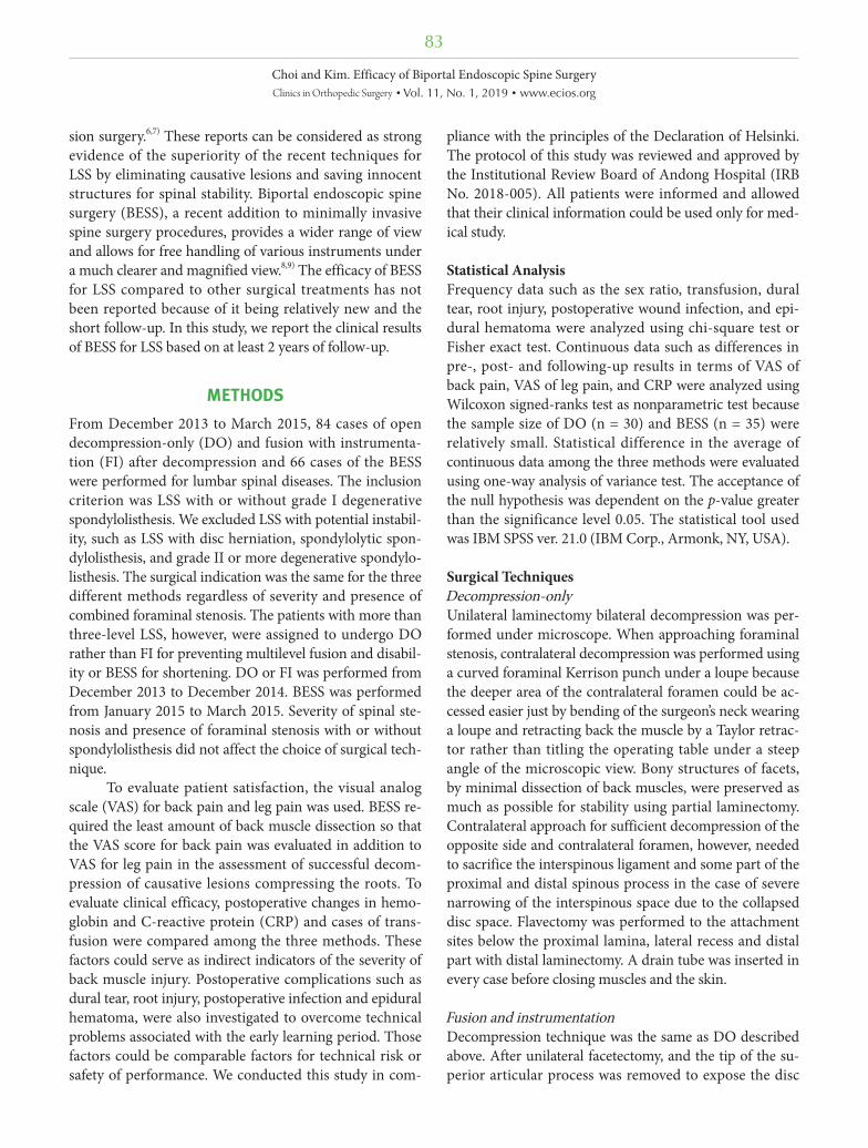
the spinous processes. During flavectomy of the contra-lateral side, the ligamentum flavum (LF) was detached or loosened from the margins of the medial and dorsal sides of the contralateral facet joint just as done on the ipsilat-eral side. Going forward, pushing an instrument under the tight LF could sever the dura or the nerve root; therefore, insertion of an instrument under the LF before securing the space under the LF was avoided. Considering dural central folding was hidden under epidural central fat tissue (Fig. 1), efforts were made to cross the midline between the LF and fat tissue rather than between the fat tissue and dura. Forceful detachment of LF at the midline from the dural central folding could make an incidental dural tear. Three of the four roots could be clearly observed in one segment during decompression. The exiting root of the ipsilateral side could not be easily observed. Muscle bleeding was controlled by a radiofrequency wand under low-voltage coagulation set. Intraspinal bleeding from the
*
Fig. 1. Intraoperative arthroscopic view of the central folding (asterisk). Midline folding is hidden under the epidural fat and the ligamentum flavum. Care should be taken not to injure the structure when crossing the midline to decompress the contralateral side.
Table 1. Demographics of the Study Groups
Variable BESS DO FI p-value
No. of cases 35 30 48 0.279
Age (yr), mean ± SD (range) 65.4 ± 11.8 (36–85) 65.2 ± 12.0 (33–84) 65.0 ± 8.8 (48–81) 0.118
Sex (male:female) 14:21 17:13 19:29 0.290
No. of levels 0.001
1 24 15 39
2 11 9 9
3 0 3 0
4 0 3 0
BESS: biportal endoscopic spine surgery, DO: decompression-only, FI: fusion and instrumentation, SD: standard deviation.
85
Choi and Kim. Efficacy of Biportal Endoscopic Spine SurgeryClinics in Orthopedic Surgery • Vol. 11, No. 1, 2019 • www.ecios.org
laminectomized bone edge was compressed by squashing a piece of bone wax on the bleeding sites. Bleeding from the epidural edge just after flavectomy from the small epi-dural vessels could be coagulated using a small sized (1.5-mm ball tip) radiofrequency wand which is commonly used in wrist arthroscopy.
RESULTS
BESS was performed on 35 patients (male, 14; female, 21; age, 65.4 ± 11.8 years), DO on 30 patients (male, 17; female, 13; age, 65.2 ± 12.0 years), and FI on 48 patients (male, 19; female, 29; age, 65.0 ± 8.8 years) (Table 1). All the patients were followed up for 6 months for VAS for
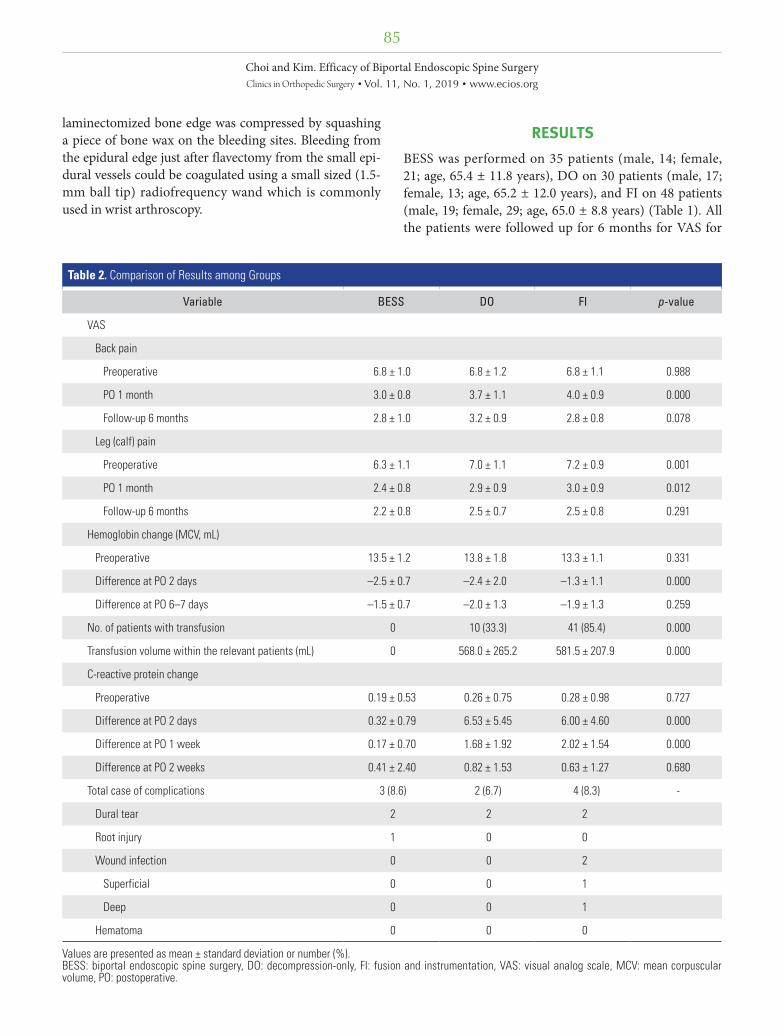
Table 2. Comparison of Results among Groups
Variable BESS DO FI p-value
VAS
Back pain
Preoperative 6.8 ± 1.0 6.8 ± 1.2 6.8 ± 1.1 0.988
PO 1 month 3.0 ± 0.8 3.7 ± 1.1 4.0 ± 0.9 0.000
Follow-up 6 months 2.8 ± 1.0 3.2 ± 0.9 2.8 ± 0.8 0.078
Leg (calf) pain
Preoperative 6.3 ± 1.1 7.0 ± 1.1 7.2 ± 0.9 0.001
PO 1 month 2.4 ± 0.8 2.9 ± 0.9 3.0 ± 0.9 0.012
Follow-up 6 months 2.2 ± 0.8 2.5 ± 0.7 2.5 ± 0.8 0.291
Hemoglobin change (MCV, mL)
Preoperative 13.5 ± 1.2 13.8 ± 1.8 13.3 ± 1.1 0.331
Difference at PO 2 days –2.5 ± 0.7 –2.4 ± 2.0 –1.3 ± 1.1 0.000
Difference at PO 6–7 days –1.5 ± 0.7 –2.0 ± 1.3 –1.9 ± 1.3 0.259
No. of patients with transfusion 0 10 (33.3) 41 (85.4) 0.000
Transfusion volume within the relevant patients (mL) 0 568.0 ± 265.2 581.5 ± 207.9 0.000
C-reactive protein change
Preoperative 0.19 ± 0.53 0.26 ± 0.75 0.28 ± 0.98 0.727
Difference at PO 2 days 0.32 ± 0.79 6.53 ± 5.45 6.00 ± 4.60 0.000
Difference at PO 1 week 0.17 ± 0.70 1.68 ± 1.92 2.02 ± 1.54 0.000
Difference at PO 2 weeks 0.41 ± 2.40 0.82 ± 1.53 0.63 ± 1.27 0.680
Total case of complications 3 (8.6) 2 (6.7) 4 (8.3) -
Dural tear 2 2 2
Root injury 1 0 0
Wound infection 0 0 2
Superficial 0 0 1
Deep 0 0 1
Hematoma 0 0 0
Values are presented as mean ± standard deviation or number (%).BESS: biportal endoscopic spine surgery, DO: decompression-only, FI: fusion and instrumentation, VAS: visual analog scale, MCV: mean corpuscular volume, PO: postoperative.
86
Choi and Kim. Efficacy of Biportal Endoscopic Spine SurgeryClinics in Orthopedic Surgery • Vol. 11, No. 1, 2019 • www.ecios.org
back pain and leg pain. Ninety-eight patients (86.7%) were followed up for at least 2 years; i.e., 33 patients (94.3%), 24.5 ± 3.7 months after BESS; 25 patients (83.3%), 30.5 ± 2.4 months after DO; and 40 patients (83.3%), 34.9 ± 3.6 months after FI.
At the preoperative assessment and 1-month and 6-month follow-up, VAS for back pain improved from 6.8 ± 1.0 via 3.0 ± 0.8 to 2.8 ± 1.0 with BESS, 6.8 ± 1.2 via 3.7 ± 1.1 to 3.2 ± 0.9 with DO, and 6.8 ± 1.1 via 4.0 ± 0.9 to 2.8 ± 0.8 with FI (1-month follow-up, p = 0.000; 6-months follow-up, p = 0.078). VAS for leg pain improved from 6.3 ± 1.1 via 2.4 ± 0.8 to 2.2 ± 0.8, 7.0 ± 1.1 via 2.9 ± 0.9 to 2.5 ± 0.7, and 7.2 ± 0.9 via 3.0 ± 0.9 to 2.5 ± 0.8 (p = 0.012, p = 0.291) with BESS, DO, and FI, respectively.
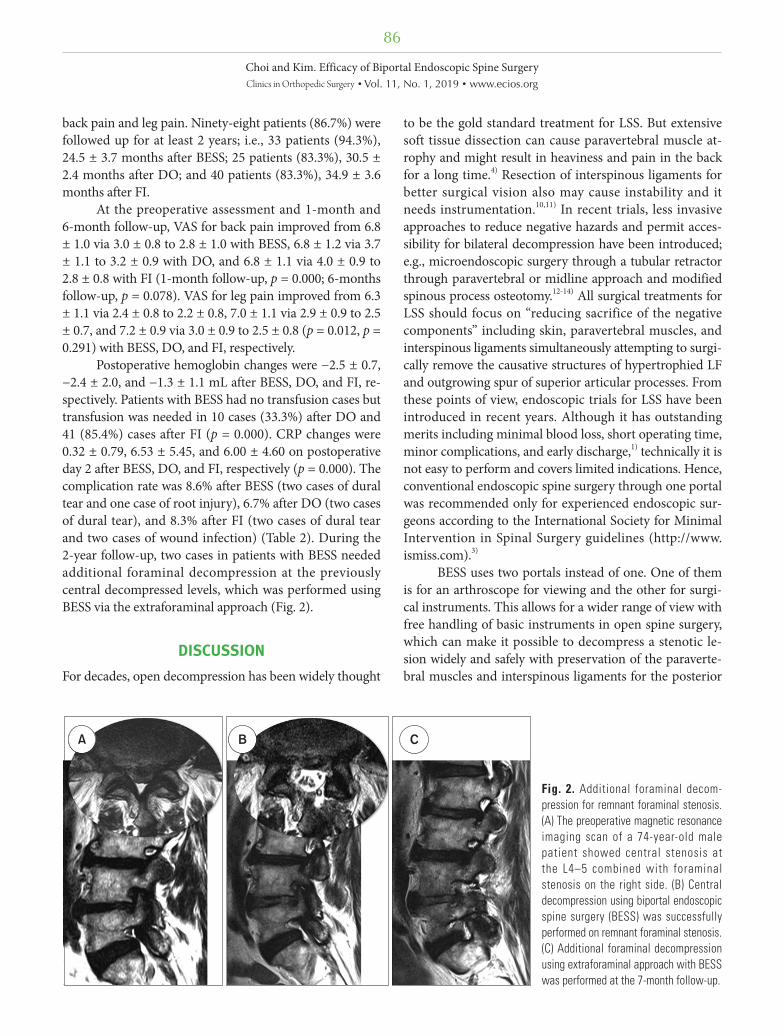
Postoperative hemoglobin changes were −2.5 ± 0.7, −2.4 ± 2.0, and −1.3 ± 1.1 mL after BESS, DO, and FI, re-spectively. Patients with BESS had no transfusion cases but transfusion was needed in 10 cases (33.3%) after DO and 41 (85.4%) cases after FI (p = 0.000). CRP changes were 0.32 ± 0.79, 6.53 ± 5.45, and 6.00 ± 4.60 on postoperative day 2 after BESS, DO, and FI, respectively (p = 0.000). The complication rate was 8.6% after BESS (two cases of dural tear and one case of root injury), 6.7% after DO (two cases of dural tear), and 8.3% after FI (two cases of dural tear and two cases of wound infection) (Table 2). During the 2-year follow-up, two cases in patients with BESS needed additional foraminal decompression at the previously central decompressed levels, which was performed using BESS via the extraforaminal approach (Fig. 2).
DISCUSSION
For decades, open decompression has been widely thought
to be the gold standard treatment for LSS. But extensive soft tissue dissection can cause paravertebral muscle at-rophy and might result in heaviness and pain in the back for a long time.4) Resection of interspinous ligaments for better surgical vision also may cause instability and it needs instrumentation.10,11) In recent trials, less invasive approaches to reduce negative hazards and permit acces-sibility for bilateral decompression have been introduced; e.g., microendoscopic surgery through a tubular retractor through paravertebral or midline approach and modified spinous process osteotomy.12-14) All surgical treatments for LSS should focus on “reducing sacrifice of the negative components” including skin, paravertebral muscles, and interspinous ligaments simultaneously attempting to surgi-cally remove the causative structures of hypertrophied LF and outgrowing spur of superior articular processes. From these points of view, endoscopic trials for LSS have been introduced in recent years. Although it has outstanding merits including minimal blood loss, short operating time, minor complications, and early discharge,1) technically it is not easy to perform and covers limited indications. Hence, conventional endoscopic spine surgery through one portal was recommended only for experienced endoscopic sur-geons according to the International Society for Minimal Intervention in Spinal Surgery guidelines (http://www. ismiss.com).3)
BESS uses two portals instead of one. One of them is for an arthroscope for viewing and the other for surgi-cal instruments. This allows for a wider range of view with free handling of basic instruments in open spine surgery, which can make it possible to decompress a stenotic le-sion widely and safely with preservation of the paraverte-bral muscles and interspinous ligaments for the posterior
A B C
Fig. 2. Additional foraminal decom-pression for remnant foraminal stenosis. (A) The preoperative magnetic resonance imaging scan of a 74-year-old male patient showed central stenosis at the L4–5 combined with foraminal stenosis on the right side. (B) Central decompression using biportal endoscopic spine surgery (BESS) was successfully performed on remnant foraminal stenosis. (C) Additional foraminal decompression using extraforaminal approach with BESS was performed at the 7-month follow-up.
87
Choi and Kim. Efficacy of Biportal Endoscopic Spine SurgeryClinics in Orthopedic Surgery • Vol. 11, No. 1, 2019 • www.ecios.org
integrity of the spinal column. The results of this study showed that patients who underwent BESS had lower back heaviness and back pain especially in the early healing period 1 month after the surgery. It might be because of preservation of the paravertebral muscles, which was quite different from the DO or FI procedures. BESS also showed comparable results to those of the other procedures in terms of leg and back pain improvement, which means that it could be a possible alternative technique for suffi-cient decompression of the dura and roots in LSS.
CRP is an acute inflammatory protein that increases at sites of infection or inflammation. There is now growing evidence that CRP plays important roles in inflammatory processes.15,16) On postoperative one or two, the CRP level was significantly lower in the patients with BESS, which was normalized within 7 days. This means that tissue de-struction was much less in the BESS procedure and pos-sible inflammatory debris could be irrigated better during the procedure compared with the other procedures. CRP changes after all three procedures should not be consid-ered as the occurrence of infection in the early postopera-tive period. However, early normalization in the BESS patients strongly supports that intravenous antibiotics could be necessary only within 1 or 2 days for prophylaxis because the endoscopic surgery involves lots of saline ir-rigation and small skin incisions for inlets. Postoperative hemoglobin level was two points lower than the preop-erative level because of hydration by saline during sur-gery. Conversely, the FI procedure caused a less decrease possibly because of the higher frequency of transfusion. In the BESS patients, there was no transfusion required compared to 33% and 85% for DO and FI, respectively. Decompression for symptomatic relief in the patients with LSS without transfusion was supposed to be recommend-able during BESS in the presence of multi-level stenosis or senile patients with hemodynamic instability.
The neurological complication rate for BESS was 8.6% with two cases of dura tear and one case of contralat-eral root injury associated with technical problems. It was related to the learning curve period and the preventive strategies in the procedure were described in the surgical technique.17,18) To reduce the occurrence of this techni-cal issue, the most important factor is to keep the surgical field clear by blocking epidural bleeding. In the surgical technique described above, fluent water flow and bleeding control from edged bone or epidural small vessels were ensured before proceeding with flavectomy or laminec-tomy especially on the contralateral side. If the bleeding cannot be controlled with every effort, lowering the dia-stolic blood pressure to around 100 mmHg can be helpful
in some cases. However, increasing the saline pressure by raising the height of the saline bag or squeezing the bag is not recommended. This is because if there is no good out-flow of saline, the surgical field becomes turbid with small bleeds even with a high input pressure of saline.
During the learning curve, there were similar dis-turbances such as prolonged operating time for one level decompression due to the turbid surgical field by small bleeds and frequent dura tears due to insufficient infor-mation on the endoscopic view and surgical anatomy. There was also incomplete decompression especially in the patients with central stenosis and foraminal stenosis due to unskilled decompression.7) Radicular pain from foraminal stenosis could be easily masked by claudication with shorter distance walking. However, when the patients tried to walk longer distances after central decompression, the fatigue and buttock pain because of foraminal stenosis were attenuated and became intolerable.
This study has some limitations. First, there were too few cases for each procedure to evaluate other techni-cal complications. Second, because of the technical issues with BESS performed in the early learning period, there were higher neurological complication rates, and some patients were dissatisfied with decompression. Third, fu-sions involving more than three levels were not performed to avoid the risk of early revision surgery due to nonunion, wound infection, longer rehabilitation, and screw loosen-ing in senile patients. These patients were intentionally as-signed to the DO procedure. Therefore, the clinical scores of DO patients were not supposed to be lower than those of BESS patients with shorter level decompression. The differences in the number of level of surgery among proce-dures might also have biased the clinical results. Therefore, prospective, head-to-head studies with matched levels and mid- and long-term follow-ups are needed to clarify the efficacy of BESS.
BESS as an alternative treatment for LSS showed clinical efficacy comparable to that of the other open surgeries in the short-term follow-up. Stable hemody-namic results without transfusion that led to less amount of bleeding because of minimal back muscle dissection through two inlets could be another merit of this tech-nique. The presence of combined foraminal stenosis should be carefully evaluated before surgery, and forami-nal decompression should be included during surgical planning to avoid additional surgery in the future.
CONFLICT OF INTEREST
No potential conflict of interest relevant to this article was
88
Choi and Kim. Efficacy of Biportal Endoscopic Spine SurgeryClinics in Orthopedic Surgery • Vol. 11, No. 1, 2019 • www.ecios.org
reported.
REFERENCES
1. Polikandriotis JA, Hudak EM, Perry MW. Minimally in-vasive surgery through endoscopic laminotomy and fo-raminotomy for the treatment of lumbar spinal stenosis. J Orthop. 2013;10(1):13-6.
2. Wong AP, Smith ZA, Lall RR, Bresnahan LE, Fessler RG. The microendoscopic decompression of lumbar stenosis: a review of the current literature and clinical results. Minim Invasive Surg. 2012;2012:325095.
3. Birkenmaier C, Komp M, Leu HF, Wegener B, Ruetten S. The current state of endoscopic disc surgery: review of controlled studies comparing full-endoscopic procedures for disc herniations to standard procedures. Pain Physician. 2013;16(4):335-44.
4. Hu ZJ, Fang XQ, Zhou ZJ, Wang JY, Zhao FD, Fan SW. Effect and possible mechanism of muscle-splitting approach on multifidus muscle injury and atrophy after posterior lumbar spine surgery. J Bone Joint Surg Am. 2013;95(24):e192(1-9).
5. Huang YH, Ou CY. Significant blood loss in lumbar fu-sion surgery for degenerative spine. World Neurosurg. 2015;84(3):780-5.
6. Choi DJ, Jung JT, Lee SJ, Kim YS, Jang HJ, Yoo B. Biportal endoscopic spinal surgery for recurrent lumbar disc hernia-tions. Clin Orthop Surg. 2016;8(3):325-9.
7. Choi DJ, Choi CM, Jung JT, Lee SJ, Kim YS. Learning curve associated with complications in biportal endoscopic spinal surgery: challenges and strategies. Asian Spine J. 2016;10(4):624-9.
8. Scholler K, Alimi M, Cong GT, Christos P, Hartl R. Lum-bar spinal stenosis associated with degenerative lumbar spondylolisthesis: a systematic review and meta-analysis of secondary fusion rates following open vs minimally invasive decompression. Neurosurgery. 2017;80(3):355-67.
9. Forsth P, Olafsson G, Carlsson T, et al. A randomized, con-
trolled trial of fusion surgery for lumbar spinal stenosis. N Engl J Med. 2016;374(15):1413-23.
10. Tai CL, Hsieh PH, Chen WP, Chen LH, Chen WJ, Lai PL. Biomechanical comparison of lumbar spine instability between laminectomy and bilateral laminotomy for spinal stenosis syndrome: an experimental study in porcine model. BMC Musculoskelet Disord. 2008;9:84.
11. Hartmann F, Janssen C, Bohm S, Hely H, Rommens PM, Gercek E. Biomechanical effect of graded minimal-invasive decompression procedures on lumbar spinal stability. Arch Orthop Trauma Surg. 2012;132(9):1233-9.
12. Pao JL, Chen WC, Chen PQ. Clinical outcomes of microen-doscopic decompressive laminotomy for degenerative lum-bar spinal stenosis. Eur Spine J. 2009;18(5):672-8.
13. Takaso M, Nakazawa T, Imura T, et al. Less invasive and less technically demanding decompressive procedure for lumbar spinal stenosis: appropriate for general orthopaedic surgeons? Int Orthop. 2011;35(1):67-73.
14. Mikami Y, Nagae M, Ikeda T, Tonomura H, Fujiwara H, Kubo T. Tubular surgery with the assistance of endoscopic surgery via midline approach for lumbar spinal canal steno-sis: a technical note. Eur Spine J. 2013;22(9):2105-12.
15. Gewurz H, Mold C, Siegel J, Fiedel B. C-reactive protein and the acute phase response. Adv Intern Med. 1982;27:345-72.
16. Sproston NR, Ashworth JJ. Role of C-reactive protein at sites of inflammation and infection. Front Immunol. 2018;9:754.
17. Turner JA, Ersek M, Herron L, Deyo R. Surgery for lumbar spinal stenosis: attempted meta-analysis of the literature. Spine (Phila Pa 1976). 1992;17(1):1-8.
18. Nowitzke AM. Assessment of the learning curve for lumbar microendoscopic discectomy. Neurosurgery. 2005;56(4):755-62.


![Percutaneous Endoscopic Lumbar Spine Surgery for …1.1. Lumbar Disc Herniation (LDH) Lumbar disc herniation [1][2] [3] (Figure 1(a)) is a medical condition affecting the spine in](https://img.dokumen.tips/doc/110x75/5f01e5a17e708231d4019244/percutaneous-endoscopic-lumbar-spine-surgery-for-11-lumbar-disc-herniation-ldh.jpg)