Embed Size (px)
Citation preview
SECTION 10
_____________________________________________
Effects of Electromagnetic Fields From
Wireless Communication
upon the Blood-Brain Barrier
Leif G. Salford, MD, PhD
Senior Professor Department of Neurosurgery
Lund University
Henrietta Nittby, MD, PhD
Department of Neurosurgery
Lund University Hospital
Bertil R. R. Persson PhD, MD h.c.
Prof. em. Department of Medical Radiation Physics
Lund University
Prepared for the BioInitiative Working Group
September 2012
2
I. INTRODUCTION
The Blood-Brain Barrier
Some organs of crucial importance for the function of our bodies are protected from exposure
to potentially harmful compounds in the blood. Thus the brain, the eyes (which are
protrusions of the brain), the testes and the follicles of the ovaries have special barriers
between the capillaries and the tissue. In the normal brain, the passage of compounds over
this barrier, the Blood-Brain Barrier (BBB), is highly restricted.
The BBB is a hydrophobic barrier formed by the vascular endothelial cells of the capillaries
in the brain with tight junctions between them leaving no openings between the vessel lumen
and the surrounding brain. The existence of the mammalian BBB was discovered in the late
19th
century by the German bacteriologist Paul Ehrlich and his student, Edwin Goldman. Paul
Ehrlich found, that when he injected dyes into the systemic blood circulation, the brain tissue
did not take up any of the stain. A barrier surrounding the brain tissue at the site of the brain
micro vessels seemed to be a logic explanation to these findings.
There is scientific evidence that the BBB exists not only in vertebrates, but also in insects (1),
crustaceans and cephalopod molluscs (such as the cuttlefish) (2) and in elasmobranchs
(cartilaginous fishes such as sharks) (3) and helices (landsnails) (4), maintaining ionic
integrity of the neuronal bathing fluid.
The BBB seems to be present very early in the foetal development. Also, at an early stage,
there seems to be a cerebrospinal fluid barrier, which excludes cerebrospinal fluid (CST)
protein from the brain extracellular space (5).
BBB Anatomy and Physiology
The tight junctions of the BBB are composed of tight junction proteins (occludin, claudin and
zonula occludens, where the zonula occludens is the intracellular peripheral membrane
protein that anchors claudin and occludin to the actin cytoskeleton (6). An important part is
3
the binding of claudin proteins on opposing membranes, where claudin-5 in particular is
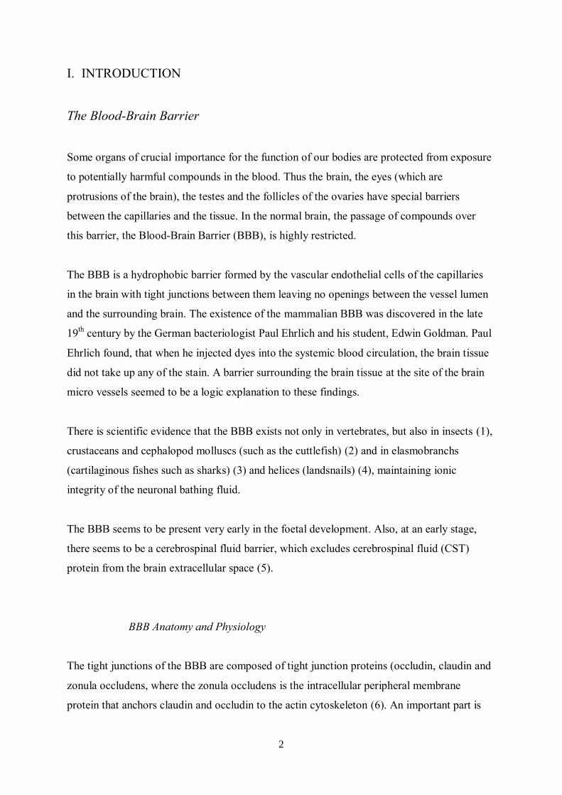
crucial in the BBB (7). Astrocytes are surrounding the outer surface of the endothelial cells
with protrusions, called end feet, and are implicated in the maintenance, functional regulation
and repair of the BBB. The astrocytes form a connection between the endothelium and the
neurons and constitute a second barrier to hydrophilic molecules (see Figure 1).
Fig. 1. The mammalian BBB
Other periendothelial accessory structures of the BBB include pericytes and a bilayer basal
membrane which surrounds the endothelial cells and pericytes. The basement membrane
(basal lamina) supports the ablumenal surface of the endothelium and may act as a barrier to
passage of macromolecules. The pericytes are a type of macrophages, expressing macrophage
markers with capacity for phagocytosis but also for antigen presentation. In fact, the pericytes,
which cover about 25% o the capillary surface (8), seem to be in a position to significantly
contribute to central nervous system (CNS) immune mechanisms (9). The pericytes also have
other functional roles: with their capability for contractility they seem to serve as a smooth
muscle equivalent, and through regulation of endothelial cells they maintain the stability of
blood vessels (9). Additionally, the pericytes seem to be highly involved in many diseases,
both infectious and autoimmune, and also in other diseases such as Alzheimer’s by production

4
of amyloid. Also, by regulating their vascular permeability, the pericytes are supposed to play
an important role in inflammatory diseases (9).
Physiologically, the microvasculature of the central nervous system (CNS) differs from that
of peripheral organs. It is characterized not only by its tight junctions, which seal cell-to-cell
contacts between adjacent endothelial cells, but also by the low number of pinocytotic
vesicles for nutrient transport through the endothelial cytoplasm and its lack of fenestrations,
and the five-fold higher number of mitochondria in BBB endothelial cells compared to
muscular endothelia in rat (10). All this speaks in favour of an energy-dependent
transcapillary transport. These above-described membrane properties of the BBB control the
bidirectional exchange of molecules between the general circulation and the central nervous
system. By at least four mechanisms, the endothelial cells directly control the flux of solutes
into the brain parenchyma. Firstly, the tight junctions and low number of pinocytotic vesicles
guarantee that proteins cannot pass freely into the brain parenchyma.
Secondly, solutes which are not highly lipid soluble, or which do not bind to selective
transporters with high affinity, are excluded from free exchange. By means of this lipid
solubility, carbon dioxide and oxygen, among many others, are able to enter the brain
interstitial fluid passively, whereas the passage of, for example sugars and many amino acids,
depends on other, active mechanisms. Thirdly, the BBB has a capacity to metabolize certain
solutes, such as drugs and nutrients (11). Fourthly, active transporters maintain the levels of
certain solutes at specific values within the brain interstitial fluid, made possible by active
transport against the concentration gradients. These enzyme systems are differently
distributed between the luminal and the ablumenal membranes of the endothelial cells, thus
gaining the BBB polarity properties. For example, Na+-K
+-ATPase is located on the
antilumenal membrane (12).
It has been proposed that the active transport across the brain capillaries might be the most
important mechanism for the regulation of the internal milieu within the brain parenchyma.
Also, it has been proposed that this mechanism, requiring energy to function properly, might
be the one most sensitive to disease and that interference with this active transport could
play an important part in the neurological dysfunction seen in many metabolic disorders (12).
It is important to have information on possible differences between homo and other mammals.
The mammalian brain at large seems to have a uniform anatomy of its BBB constituents
5
preserved through the evolution, and very little information about differences between
mammalian species has been available. However, recently very interesting observations have
been published. Humans have evolved protoplasmic astrocytes that are both larger (27-fold
greater volume) and far more elaborate than their rodent counterparts. These astrocytes reside
near blood vessels, and their processes contribute to the BBB (13). When the end feet of
human and rodent protoplasmic astrocytes are compared, it is shown that nearly all astrocytes
in both species contact the vasculature, but in the human brain, the end feet completely
encompass the vessels while the rodent astrocytes form rosettes of end feet around the
vasculature. The number of mithochondria is however equally abundant in human and rodent
end feet (14).
Comparisons between mammalian species concerning enzymatic functions in the BBB are
few in number. Similarities are described: mouse vs human (15) and rat vs human (16), while
differences are demonstrated between rodent and dog BBB leading to the conclusion that the
canine BBB may be preferable to that of the rat as a model for studies of glucose transport
relevant to human brain (17).
In summary, the BBB serves as a regulatory system that stabilizes and optimizes the fluid
environment of the brain’s intracellular compartment (18-20). The intact BBB protects the
brain from damage, whereas the dysfunctioning BBB allows influx of normally excluded
hydrophilic molecules into the brain tissue. This might lead to cerebral oedema, increased
intracranial pressure, and in the worst case, irreversible brain damage.
II. DISRUPTION OF THE BLOOD-BRAIN BARRIER
The normal selective permeability of the BBB can be altered in several pathological
conditions such as epileptic seizures (21) or extreme hypertension (22)and also transient
openings of the BBB might lead to permanent tissue damage (22). Considering the ensuing
leakage of substances from the blood circulation into the brain tissue, harmful substances
might disrupt the cellular balance in the brain tissue and in the worst case, even carcinogenic
substances might pass into the brain tissue. It has also been shown that an increased
permeability of the BBB is seen in cases of oxidative stress (23), where BBB dysfunction and

6
neurodegeneration were shown to be mediated through an excitotoxicity mechanism by the
serine protease tissue plasminogen activator, with NO and ONOO- as downstream mediators
(23).
Opening of the BBB thus can have detrimental effects and since it has been shown for a few
decades that EMFs have the potency to increase the permeability of this barrier, a major
debate is going on in society with increasing intensity. In the following, we try to clarify the
actual status of the available evidence in the field.
Early Studies
In early studies on the effects of low-intensity EMFs on the BBB, various compounds were
injected intravenously, followed by EMF exposure and comparisons of the penetration into
the brain tissue between sham and exposed animals.
Frey et al. (25) found increases in the BBB permeability of rats to fluorescein after 30 min of
exposure to both pulsed and continuous waves (CWs) at 1.2GHz with average power densities
of 0.2mW/cm2. Similar observations were made in a study with 180 animals by Oscar and
Hawkins (26). Exposure of anaesthetized rats for 20 min to 1.3GHz of pulsed EMFs with
average power densities of 0.3mW/cm2 resulted in leakage of 14C-mannitol, dextran, and
inulin into the cerebellar brain tissue, as well as inulin and dextran leakage from capillaries
into hypothalamic and medullar tissue. Also, BBB permeability to mannitol was investigated
in un-anaesthetised rats, which were exposed to pulsed radiation or sham exposed for 20 min.
The animals were sacrificed at different time intervals after the exposure. BBB permeability
was seen in the groups sacrificed 8 min and 4 h after exposure, but to a much lesser extent in
those sacrificed after 8 h. Finally, the permeation of mannitol through the BBB was found to
be a very definite function of exposure parameters such as power density, pulse width, and the
number of pulses per second. However, in later studies, Oscar et al. (27) emphasised that
changes of BBB permeability after microwave exposure partly could be explained by an
increase of local cerebral blood flow. In accordance with this, they concluded that their initial
findings (26) might be of less magnitude than originally thought (Table 1).
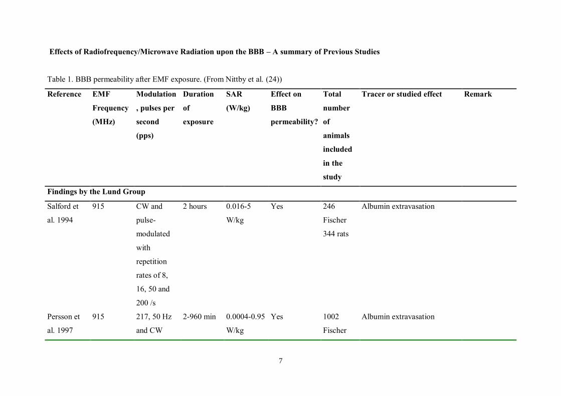
7
Effects of Radiofrequency/Microwave Radiation upon the BBB – A summary of Previous Studies
Table 1. BBB permeability after EMF exposure. (From Nittby et al. (24))
Reference EMF
Frequency
(MHz)
Modulation
, pulses per
second
(pps)
Duration
of
exposure
SAR
(W/kg)
Effect on
BBB
permeability?
Total
number
of
animals
included
in the
study
Tracer or studied effect Remark
Findings by the Lund Group
Salford et
al. 1994
915 CW and
pulse-
modulated
with
repetition
rates of 8,
16, 50 and
200 /s
2 hours 0.016-5
W/kg
Yes 246
Fischer
344 rats
Albumin extravasation
Persson et
al. 1997
915 217, 50 Hz
and CW
2-960 min 0.0004-0.95
W/kg
Yes 1002
Fischer
Albumin extravasation
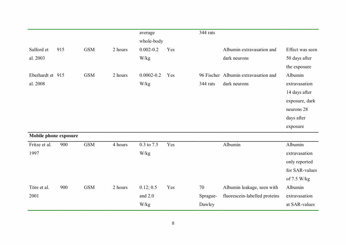
8
average
whole-body
344 rats
Salford et
al. 2003
915 GSM 2 hours 0.002-0.2
W/kg
Yes Albumin extravasation and
dark neurons
Effect was seen
50 days after
the exposure
Eberhardt et
al. 2008
915 GSM 2 hours 0.0002-0.2
W/kg
Yes 96 Fischer
344 rats
Albumin extravasation and
dark neurons
Albumin
extravasation
14 days after
exposure, dark
neurons 28
days after
exposure
Mobile phone exposure
Fritze et al.
1997
900 GSM 4 hours 0.3 to 7.5
W/kg
Yes Albumin Albumin
extravasation
only reported
for SAR-values
of 7.5 W/kg
Töre et al.
2001
900 GSM 2 hours 0.12; 0.5
and 2.0
W/kg
Yes 70
Sprague-
Dawley
Albumin leakage, seen with
fluorescein-labelled proteins
Albumin
extravasation
at SAR-values
9
of 0.5 and 2.0
W/kg
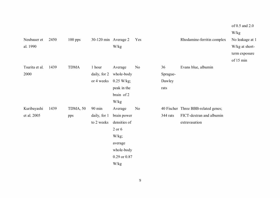
Neubauer et
al. 1990
2450 100 pps 30-120 min Average 2
W/kg
Yes Rhodamine-ferritin complex No leakage at 1
W/kg at short-
term exposure
of 15 min
Tsurita et al.
2000
1439 TDMA 1 hour
daily, for 2
or 4 weeks
Average
whole-body
0.25 W/kg;
peak in the
brain of 2
W/kg
No 36
Sprague-
Dawley
rats
Evans blue, albumin
Kuribayashi
et al. 2005
1439 TDMA, 50
pps
90 min
daily, for 1
to 2 weeks
Average
brain power
densities of
2 or 6
W/kg;
average
whole-body
0.29 or 0.87
W/kg
No 40 Fischer
344 rats
Three BBB-related genes;
FICT-dextran and albumin
extravasation

10
Finnie et al.
2001
898.4 GSM 1 hour Whole-
body of 4
W/kg
No 60 mice Albumin extravasation
Finnie et al.
2002
900 GSM 1 hour
daily, 5
days a
week for
104 weeks
Average
whole-body
0.25; 1.0;
2.0 and 4.0
W/kg
No 207 mice Albumin extravasation
Franke et al.
2005b
1800 GSM 1 to 5 days Average 0.3
W/kg
No Sucrose permeation In vitro model
of BBB
Schirmacher
et al. 2000
1800 GSM 4 days Average 0.3
W/kg
No Sucrose permeation In vitro model
of BBB
Franke et al.
2005a
1966 UMTS 1 to 3 days Average 1.8
W/kg
No Sucrose and albumin
permeation
In vitro model
of BBB
Cosquer et al.
2005
2450 500 pps 45 min Average
whole-body
2 W/kg
No Rats Scopolamine methylbromide
extravasation
Indirect
investigation of
BBB opening
by
performance in
radial arm
maze
11
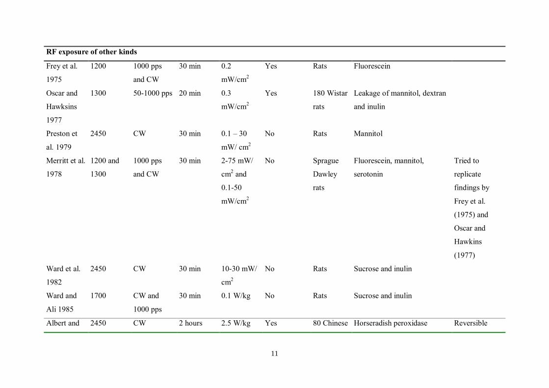
RF exposure of other kinds
Frey et al.
1975
1200 1000 pps
and CW
30 min 0.2
mW/cm2
Yes Rats Fluorescein
Oscar and
Hawksins
1977
1300 50-1000 pps 20 min 0.3
mW/cm2
Yes 180 Wistar
rats
Leakage of mannitol, dextran
and inulin
Preston et
al. 1979
2450 CW 30 min 0.1 – 30
mW/ cm2
No Rats Mannitol
Merritt et al.
1978
1200 and
1300
1000 pps
and CW
30 min 2-75 mW/
cm2 and
0.1-50
mW/cm2
No Sprague
Dawley
rats
Fluorescein, mannitol,
serotonin
Tried to
replicate
findings by
Frey et al.
(1975) and
Oscar and
Hawkins
(1977)
Ward et al.
1982
2450 CW 30 min 10-30 mW/
cm2
No Rats Sucrose and inulin
Ward and
Ali 1985
1700 CW and
1000 pps
30 min 0.1 W/kg No Rats Sucrose and inulin
Albert and 2450 CW 2 hours 2.5 W/kg Yes 80 Chinese Horseradish peroxidase Reversible
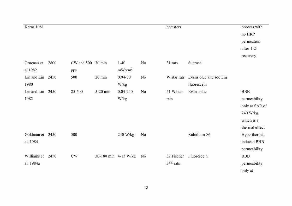
12
Kerns 1981 hamsters process with
no HRP
permeation
after 1-2
recovery
Gruenau et
al 1982
2800 CW and 500
pps
30 min 1-40
mW/cm2
No 31 rats Sucrose
Lin and Lin
1980
2450 500 20 min 0.04-80
W/kg
No Wistar rats Evans blue and sodium
fluorescein
Lin and Lin
1982
2450 25-500 5-20 min 0.04-240
W/kg
No 51 Wistar
rats
Evans blue BBB
permeability
only at SAR of
240 W/kg,
which is a
thermal effect
Goldman et
al. 1984
2450 500 240 W/kg No Rubidium-86 Hyperthermia
induced BBB
permeability
Williams et
al. 1984a
2450 CW 30-180 min 4-13 W/kg No 32 Fischer
344 rats
Fluorescein BBB
permeability
only at
13
hyperthermic
levels > 41C
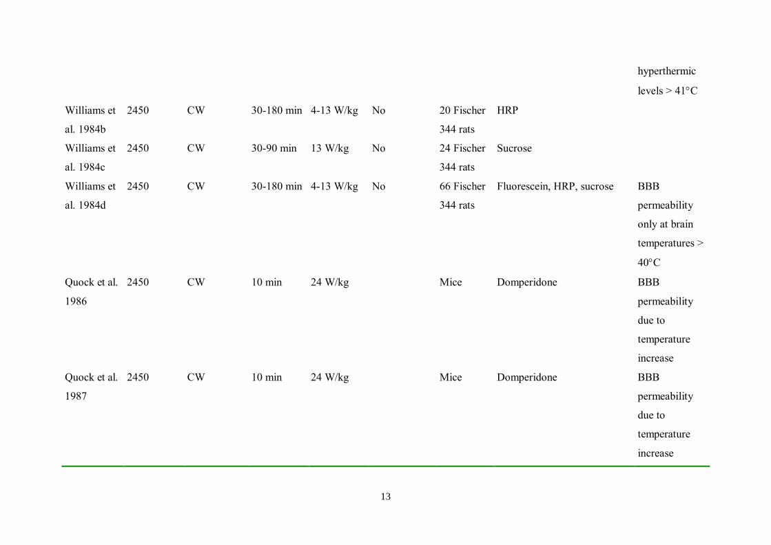
Williams et
al. 1984b
2450 CW 30-180 min 4-13 W/kg No 20 Fischer
344 rats
HRP
Williams et
al. 1984c
2450 CW 30-90 min 13 W/kg No 24 Fischer
344 rats
Sucrose
Williams et
al. 1984d
2450 CW 30-180 min 4-13 W/kg No 66 Fischer
344 rats
Fluorescein, HRP, sucrose BBB
permeability
only at brain
temperatures >
40C
Quock et al.
1986
2450 CW 10 min 24 W/kg Mice Domperidone BBB
permeability
due to
temperature
increase
Quock et al.
1987
2450 CW 10 min 24 W/kg Mice Domperidone BBB
permeability
due to
temperature
increase

14
Moriyama
et al. 1991
2450 CW 21 Sprague
Dawley
rats
HRP BBB
permeability
due to
temperature
increase
Nakagawa
et al. 1994
2450 CW Japanese
monkeys
BBB
permeability
due to
temperature
increase
MRI exposure Magnetic
field
Shivers et
al. 1987
23 min 0.15 T static
magnetic
field
Yes HRP Standard MRI
procedure
Preston et
al. 1989
23 min 4.7 T static
magnetic
field
No Rats Sucrose Standard MRI
procedure
Prato et al. 65 23 min x 2 0.15 T static Yes 43 Diethylenetriaminepentaacetic Standard MRI
15
1990 magnetic
field
Sprague
Dawley
rats
acid (DTPA) procedure
Prato et al.
1994
23 min x 2 1.5 T static
magnetic
field
Yes 50 rats Standard MRI
procedure
Garber et al.
1989
0.3-0.5 T
static
magnetic
field
Yes Rats Mannitol Standard MRI
procedure
Adzamli et
al. 1989
No Standard MRI
procedure
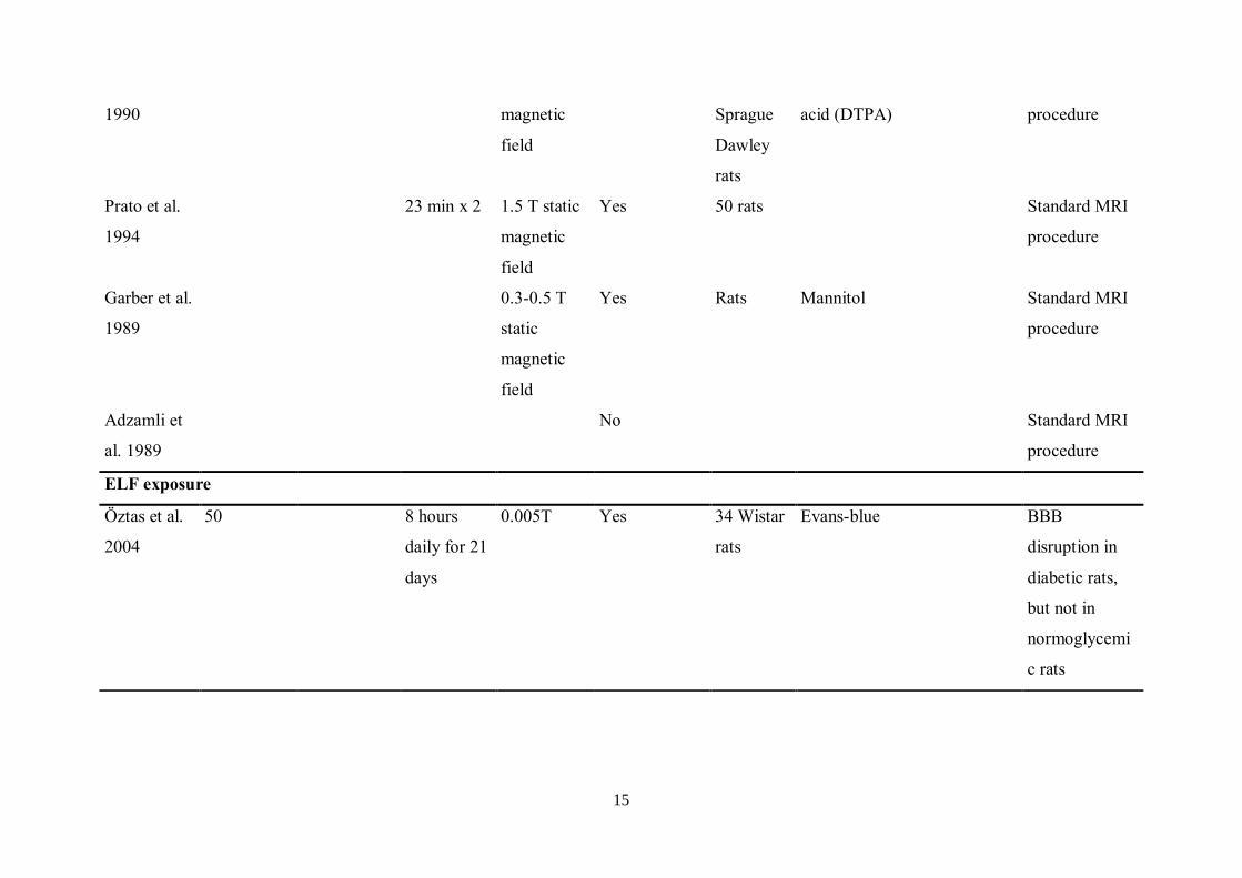
ELF exposure
Öztas et al.
2004
50 8 hours
daily for 21
days
0.005T Yes 34 Wistar
rats
Evans-blue BBB
disruption in
diabetic rats,
but not in
normoglycemi
c rats
16
In an attempt to repeat the findings of Oscar and Hawkins (26), Preston et al. (28) found no
increase in the uptake of 14C-mannitol in anaesthetised rats after 2450MHz CW exposure for
30 min at power densities of 0.1 to 30mW/cm2. Preston et al. further concluded that the
increased BBB permeability, which had been observed by Oscar and Hawkins (26) in
cerebellum and medulla, possibly had been misinterpreted and was not due to the EMF
exposure. Rather, changes in blood flow and water influx or egress were supposed to be
responsible for the BBB permeability in these caudal parts of the brain. Also, further attempts,
made by Merritt et al. (1978) (29), to replicate the findings of Oscar and Hawkins from 1977,
resulted in the conclusion that no repetition of the initial findings could be made. Merritt et al.
(29) tried to replicate also the findings of Frey et al. (25), but reported that no changes were
seen.
However, Frey commented upon this in an article in 1998, where he pointed out that, in fact,
statistical analysis by the editor and reviewer of the data from the study by Merritt et al.
provided a confirmation of the findings of Frey et al. (25) (30).
No alteration of BBB permeation of 14C-sucrose and 3H-inulin was found by Ward et al.
(31)after exposure of anaesthetised rats to CW at 2450MHz for 30 min at power densities of 0,
10, 20, or 30 mW/cm2 after correction for thermal effects. Similarly, Ward and Ali (32)
observed no permeation after 1.7GHz exposure at SAR of 0.1 W/kg, using the same exposure
duration and injected tracers as Ward et al. (31). Absence of EMF induced BBB permeability
was also reported by Gruenau et al. (33), after injection of 14C-sucrose in conscious rats and
exposure 30 min pulsed energy (2.8GHz at 0, 1, 5, 10, or 15mW/cm2) or continuous wave
(2.8 GHz, 0, 10, or 40 mW/cm2).
Proof of EMF-induced BBB permeability was put forward by Albert and Kerns (34), who
exposed un-anaesthetised Chinese hamsters to 2,450MHz CWs for 2 h at SARs of 2.5 W/kg.
In one-third of the exposed animals there was an increased permeability of the BBB to
horseradish peroxidase (HRP) and the endothelial cells of these irradiated animals had a 2–3-
fold higher number of pinocytotic vesicles with HRP than the sham animals. The mechanism
of BBB permeability seemed to be reversible, since animals allowed to recover for 1 or 2 h
after the EMF exposure had almost no HRP permeation. A total number of 80 animals were
included in this study.

17
Temperature Dependence
In further studies, more attention was directed towards the effects of hyperthermia, resulting
from exposure at high SAR-levels, on BBB permeability.
A study correlating changes of BBB permeability with the quantity of absorbed microwave
energy by Lin and Lin (35), using Evans blue and sodium fluorescein as indicators of BBB
permeation, showed that 20 min of 2,450MHz exposure of anaesthetised Wistar rats caused
no alteration of BBB permeability even at SAR values of 80 W/kg. Notably, the same lack of
alteration was observed also at lower SAR-values, down to 0.04 W/kg. In further studies by
the same group (36), no permeation of Evans blue could be observed after exposure to
2,450MHzB RFs for 5–20 min when the SAR-values ranged from 0.04–200 W/kg. Not until a
SAR-value of 240 W/kg, with ensuing rise in brain temperature to 43ºC, was applied, the
BBB permeability increased. These observations of demonstrable increases of BBB
permeability associated with intense, microwave-induced hyperthermia were supported by
another study by the same group (37).
In a series of EMF exposures at 2,450MHz CW, Williams et al. (38-40) concluded that
increase of BBB permeability might not be explained by microwave exposure, but rather
temperature increases and technically derived artefacts such as increase of the cerebral blood
volume and a reduction in renal excretion of the tracer. Significantly elevated levels of
sodium fluorescein (38) were found only in the brains of conscious rats made considerably
hyperthermic by exposure to ambient heat for 90 min or 2,450MHz CW microwave energy
for 30 or 90 min, but this was at high SAR values, 13 W/kg—far beyond the ICNIRP limit of
2 W/kg (41) —and not comparable to the experiments performed by, among others, our group,
as described below.
With more research into the area of EMF induced BBB permeability, it became evident that
with high-intensity EMF exposure resulting in tissue heating, the BBB permeability is
temperature dependent (42). Thus, the importance of differentiating between thermal and non-
thermal effects on the integrity of the BBB was realized. This is the reason why studies with
increases of BBB permeability due to exposure to SAR-values well above recommended
18
exposure levels (43-46) need to be considered from another point of view, as compared to
those focusing on the non-thermal effects of EMFs.
Continued Studies—MRI and BBB Permeability
Following the increasing use of magnetic resonance imaging (MRI), the effects of MRI
radiation upon BBB permeability were investigated more thoroughly. MRI entails the
concurrent exposure of subjects to a high-intensity static field, a radiofrequency field, and
time-varying magnetic field. Shivers et al. (47) observed that exposure to a short (23 min)
standard (of those days) clinical MRI procedure at 0.15 Tesla (T) temporarily increased the
permeability of the BBB to horseradish peroxidase (HRP) in anaesthetised rats. This was
revealed by electron microscopy (EM), to be due to an amplified vesicle-mediated transport
of HRP across the microvessel endothelium, to the ablumenal basal lamina and extracellular
compartment of the brain parenchyma. This vesicle-mediated transport also included
transendothelial channels. However, no passage of the tracer through disrupted
interendothelial tight junctions was present.
During the next few years, more groups studied the effects of MRI exposure on the BBB
permeability by injection of radioactive tracers into rats. One supported (48)while others
contradicted (49, 50) the initial findings made by Shivers et al. (47). Garber et al. exposed rats
to MRI procedures at 1.5, 0.5, and 0.3 T with RFs of 13, 21, and 64 MHz, respectively (48).
Brain mannitol concentration was significantly increased at 0.3 T and 0.5 T but not at 1.5 T.
No decrease in plasma mannitol concentration of MRI exposed animals was found and thus
the authors concluded that effects of MRI associated energies on mannitol transport do not
occur measurably in the body, and might be more specific to brain vasculature. Preston et al.
(50) found no significant permeation of blood-borne 14C-sucrose into brain parenchyma in
anesthetized rats subjected to 23 min of MRI at 4.7 T and RFs at 12.5 kHz. However, the
authors pointed out that if the MRI effect was focal and excess tracer counts were found only
in restricted sites, there could have been MRI induced extravasation of sucrose that was not
detected, due to the preponderance of normal tissue counts. When Preston et al. (50)
compared the lack of BBB leakage in their study to the MRI induced leakage which had been
observed by Shivers et al. (47), they also concluded that certain characteristics of electric and

19
magnetic fields, which were present in the study by Shivers et al. but not in their own work,
could have been critical to the observed effects.
In 1990, further studies by the Shivers-Prato group were presented (51) and the group could
now quantitatively support its initial findings, in a series of 43 Sprague-Dawley rats. The
BBB permeability to diethylenetriaminepentaacetic acid (DTPA) increased in rats after two
sequential 23 min MRI exposures at 0.15 T. It was suggested that the increased BBB
permeability could result from a time-varying magnetic field mediated stimulation of
endocytosis. Also, the increased BBB permeability could be explained by exposure-induced
increases of intracellular Ca2+
in the vascular endothelial cells. Since the Ca2+
is an
intracellular mediator, increases of BBB permeability could possibly be initiated in this way.
A few years later, in a series of 50 rats, the Shivers - Prato group also found that the BBB
permeability in rats is also altered by exposure to MRI at 1.5T for 23 min in 2 subsequent
exposure sessions (52).
Studies by the Lund Group
Two of us found these observations highly interesting:
- the neurosurgeon (LGS) in the hope to utilize possible applications of EMF to
make the blood-brain barrier (BBB) more penetrable to chemotherapy, in order
to treat brain cancers more effectively. An intact BBB keeps out chemotherapy
agents, allowing cancer cells to hide behind the BBB.
- the radiophysicist (BRRP) interested in possible adverse effects of the MRI
technique.
After a visit to Shivers’ group in London Ontario in 1988, we started work in Lund in 1988,
studying the effects of MRI on rat brain and we found, by the use of Evans Blue, the same
increased permeability over BBB for albumin (53).
This work was continued by separating the constituents of the MRI field: RF, undulant
magnetic field, and static magnetic field. Since RF turned out to be the most efficient
component of the MRI, the following studies focused mainly on the RF effects. Striving for
20
investigating the actual real-life situation, endogenous substances, which naturally circulate in
the vessels of the animals, were used. In line with this, albumin and also fibrinogen leakage
over the BBB were followed after identification of albumin with rabbit antibodies (see Figure
2 and 3) and rabbit anti-human fibrinogen.
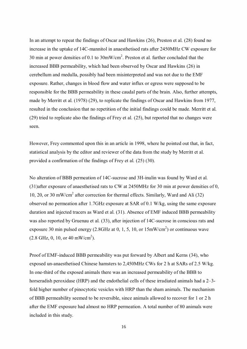
Figure 2. Albumin extravasation in rat brain (material from Persson et al. 1997)(54).
Left: control brain with albumin staining in hypothalamus, which serves as an inbuilt-control
of the staining method, since the hypothalamus lacks BBB, and one occasional staining.
Right: Brain of EMF exposed rat, with multiple albumin positive foci.
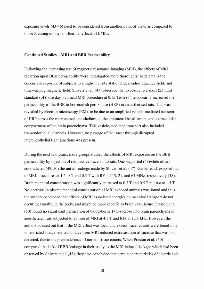
Figure 3. Albumin extravasation around vessels in the brain of an EMF exposed rat.
The work by Blackman et al. (55, 56) made the ground laid the groundwork for studies on the
frequency modulation 16 Hz and its harmonies harmonics 4 and 8 Hz. A carrier wave of 915
MHz was used. At the suggestion of Östen Mäkitalo (Telia), a pioneer in mobile phone

21
development, who introduced 50 Hz (DUX) and 217 Hz (GSM) modulation in new digital
wireless communication systems, we also included theses frequencies. This paralleled the
first BBB study results that were published in 1992-1994 (57-59).
The result of our continued work, comprising more than 1000 animals, with exposure to both
CWs and pulsed modulated waves, in the most cases lasting for 2 h, showed that there was a
significant difference between the amount of albumin extravasation in the exposed animals as
compared to the controls. In the exposed group 35–50% of the animals had a disrupted BBB
as seen by the amount of albumin leakage, while the corresponding leakage in the sham
exposed animals was only 17% (for results see Figure 4) (54).
0 (CW) 4 8,3 16 50 217 All All-pulsed0,0
0,1
0,2
0,3
0,4
0,5
0,6
0,7
0,8
0,9
1,0
p=0.4
Con
trols
0.17
58
p < 0.00005
p = 0.5p = 0.6
p = 0.6
p < 0.00005
Exposed: SAR = 1.7 - 8.3 W/kg
Number of rats: 132 20 32 6 73
Fra
cti
on
of
pa
tho
log
ica
l ra
ts
Modulation Frequency / Hz
0 (CW) 4 8,3 16 50 217 All All-pulsed0,0
0,1
0,2
0,3
0,4
0,5
0,6
0,7
0,8
0,9
1,0
Con
trols
0.17
177
p = 0.40p < 0.00005
p < 0.003p = < 0.002
p = 0.3
p = 0.01
Exposed: SAR = (0,11 - 0.95) W/kg
Number
of rats: 209 56 91 18 12 32
Fra
cti
on
of
pa
tho
log
ica
l ra
ts
Modulation Frequency / Hz
0 (CW) 4 8,3 16 50 217 All All-pulsed0,0
0,1
0,2
0,3
0,4
0,5
0,6
0,7
0,8
0,9
1,0
Con
trols
0,17
135
p < 0.00005p = 0.9
p<0.00005
p = 0.5p = 0.5
p < 0.01
Exposed: SAR = (2 - 8) 10-2 W/kg
No. of rats: 178 41 48 20 26 43
Fra
cti
on
of
pa
tho
log
ica
l ra
ts
Modulation Frequency / Hz
0 (CW) 4 8,3 16 50 217 All All-pulsed0,0
0,1
0,2
0,3
0,4
0,5
0,6
0,7
0,8
0,9
1,0
Con
trols
0.17
111
p < 0.00005p < 0.001
p < 0.0002p = 0.02p < 0.0001
p = 0.6
12
Exposed: SAR = 4.10-4 - 8.10-3 W/kg
Number
of rats: 111 52 23 6 18
Fra
ctio
n o
f p
ath
olo
gic
al ra
ts
Modulation Frequency / Hz
1 – 8 W/kg 100 – 950 mW/kg
20-80 mW/kg 0.4-8 mW/kg
Figure 4. Albumin extravasation score as a result of EMF exposure (results from the study by
Persson et al. (54)).
22
The fact that sham-exposed control animals also show some amount of albumin extravasation
(see Figure 4), is most likely due to our very sensitive methods for immune histological
examination. However, it is hard to explain the fact that although all animals in the 1997
series were inbred Fischer 344 rats, only every second animal, at the most, showed albumin
leakage after EMF exposure. The question, what might protect the remaining 50% of the
exposed animals from BBB disruption, is highly intriguing. It should be noted that in our
large series, only in one single animal fibrinogen leakage has been observed (54).
Another conclusion from the 1997 study is that the number of pathological leakages in
exposed animals is more frequent, and also more severe, per animal compared to the controls.
This is an interesting observation as the prevailing opinion is that pulse modulated
electromagnetic fields are more potent in causing biological effects.
In a statistical re-evaluation of our material published in 1997, where only exposed rats with a
matched unexposed control rat are included, we found for the most interesting modulation
frequency 217 Hz, i.e. that of GSM, that at SAR-values of 0.2 to 4 mW/kg 48 exposed rats
had a significantly increased albumin leakage (p < 0.001) as compared their 48 matched
controls. On the other hand, SAR-values of 25-50 mW/kg, gave no significant difference
between 22 exposed rats vs their matched controls (Wilcoxon´s Rank Test, 2-sided p-value)
(60).
In all our earlier studies we showed albumin extravasation immediately after exposure as
described above. In later years we have performed a series of experiments where the animals
were allowed to survive for 7 days (61), 14 days, 28 days (62) or 50 days (63) after one single
2-hour exposure to the radiation from a GSM mobile phone. All were exposed in TEM-cells
to a 915 MHz carrier wave as described below. The peak power output from the GSM mobile
phone fed into the TEM-cells was 1 mW, 10 mW, 100 mW and 1000 mW per cell
respectively for the 7-14-28-days survival animals, resulting in average whole-body SAR of
0.12 mW/kg, 1.2 mW/kg, 12 mW/kg and 120 mW/kg for four different exposure groups
SAR-values of 2, 20 and 200 mW/kg mW/kg for 2 hours for the 50-days survival animals.
Albumin extravasation over the BBB after GSM exposure seemed to be time-dependent, with
significantly increased albumin in the brain parenchyma of the rats, which had survived for 7
and 14 days, but not for those surviving 28 days. After 50 days, albumin extravasation was
23
significantly increased again, with albumin-positive foci around the finer blood vessels in
white and gray matter of the exposed animals.
In connection to the albumin passage over the BBB, albumin also spread in the surrounding
brain tissue. A significantly increased uptake of albumin in the cytoplasm of neurons could be
seen in the GSM exposed animals surviving 7 and 14 days after exposure, but not in those
surviving 28 or 50 days.
Neuronal uptake
Extravasated albumin rapidly diffused down to, and beyond, concentrations possible to
demonstrate accurately immunohistologically. However, the initial albumin leakage into the
brain tissue (seen within hours in ~40% of exposed animals in our previous studies) most
likely started a vicious circle of further BBB opening.
It has been postulated that albumin is the most likely neurotoxin in serum (64). Hassel et al.
(65) have demonstrated that injection of albumin into the brain parenchyma of rats gives rise
to neuronal damage. When 25 μl of rat albumin is infused into rat neostriatum, 10 and 30, but
not 3 mg/ml albumin causes neuronal cell death and axonal severe damage. It also causes
leakage of endogenous albumin in and around the area of neuronal damage. Albumin in the
dose 10 mg/ml is approximately equivalent to 25% of the serum concentration.
It is less likely that the albumin leakage demonstrated in our experiments locally reaches such
concentrations. However, we have seen that in the animals surviving 28 and 50 days after 2
hours of GSM exposure, there was a significantly increased incidence of neuronal damage as
compared to the sham controls. In the 7-days and 14-days survival animals, on the other hand,
no such increase of neuronal damage was seen.
In the 50-days post-exposure survival study, a 2 h exposure to GSM at SAR values 200, 20,
and 2 mW/kg resulted in a significant (p = 0.002) neuronal damage in rat brains of the
exposed animals as compared to the controls 50 days after the exposure occasion (Salford et
al., 2003)(63). We have followed up this observation, as mentioned above, in a study where
96 animals were sacrificed 14 and 28 days respectively after an exposure for 2 h to GSM
mobile phone electromagnetic fields at SAR values 0 (controls), 0.12, 1.2, 12 and 120
mW/kg. Significant neuronal damage is seen after 28 days and albumin leakage after 14. Our

24
findings may support the hypothesis that albumin leakage into the brain is the cause for the
neuronal damage observed after 28 and 50 days (62).
The damaged neurons in the above mentioned studies took the shape of so-called dark
neurons. Three main characteristics of the damaged dark neurons have been proposed (66): (i)
irregular cellular outlines, (ii) increased chromatin density in the nucleus and cytoplasm and
(iii) intensely and homogenously stained nucleus. The damaged dark neurons found in the 50
days-survival animals were investigated regarding signs of apoptotic markers, but we found
no positive staining for Caspase-3, a marker for apoptosis (Bexell et al. unpublished results).
However, the albumin leakage out in the neuropil in connection to EMF exposure might start
other deleterious processes, leading to the formation of the dark neurons.
A group in Turkey performed similar experiments. However, also the presumed protective
effects of the antioxidant Ginko biloba (Gb) were examined by Ilhan et al. (67). About 22
female Wistar rats were exposed to a 900 MHz electromagnetic GSM near-field signal for 1 h
a day for 7 days. In the GSM only group, the pathological examination revealed scattered and
grouped dark neurons in all locations, but especially in the cortex, hippocampus and basal
ganglia, mixed in among normal neurons. A combined non-parametric test for the four groups
revealed that the distributions of scores differed significantly between the control and the
GSM only exposure group (p < 0.01).
Long-term study, including studies of memory and behaviour
In a recent long-term study from our laboratory, rats were exposed to GSM radiation 2 hours
weekly during 55 weeks (two different exposure groups with 0.6 mW/kg and 60 mW/kg at the
initiation of the exposure period). After this protracted exposure, behaviour and memory of
the exposed animals were tested. Whereas the behaviour of the animals was not affected, the
GSM exposed rats had significantly impaired episodic memory as compared to the sham
controls (68). After the finalization of these tests, that is 5-7 weeks after the last exposure, the
animals were sacrificed by perfusion fixation. Albumin extravasation, an indicator of BBB
leakage, was increased in about 1 animal in each group of low GSM exposed, high GSM
exposed, sham exposed and cage control rats. About 40 % of the animals had neuronal
damage. GFAP staining, as an indicator of glial reaction, revealed positive results in 31-69 %
of the animals for different groups and the aggregation product lipofuscin was increased in

25
44-71 % of the animals for different groups. With the Gallyas staining (aiming at cytoskeletal
structures), no changes were seen. When comparing the results between the different groups,
it turned out that there was no statistically significant difference for any of these parameters
due to GSM exposure (69). When comparing these findings to those from animals which had
been exposed only once for 2 hours, it seems likely that during the 55 weeks of repeated
exposure, albumin leakage at an initial stage of the experimental period might have been
absorbed after some time, and that at a certain, but unknown, time point during this
protracted, more than 1 year long-exposure period, some adaptation process might have been
activated. However, this could not compensate for cognitive alterations, demonstrated by the
episodic memory tests.
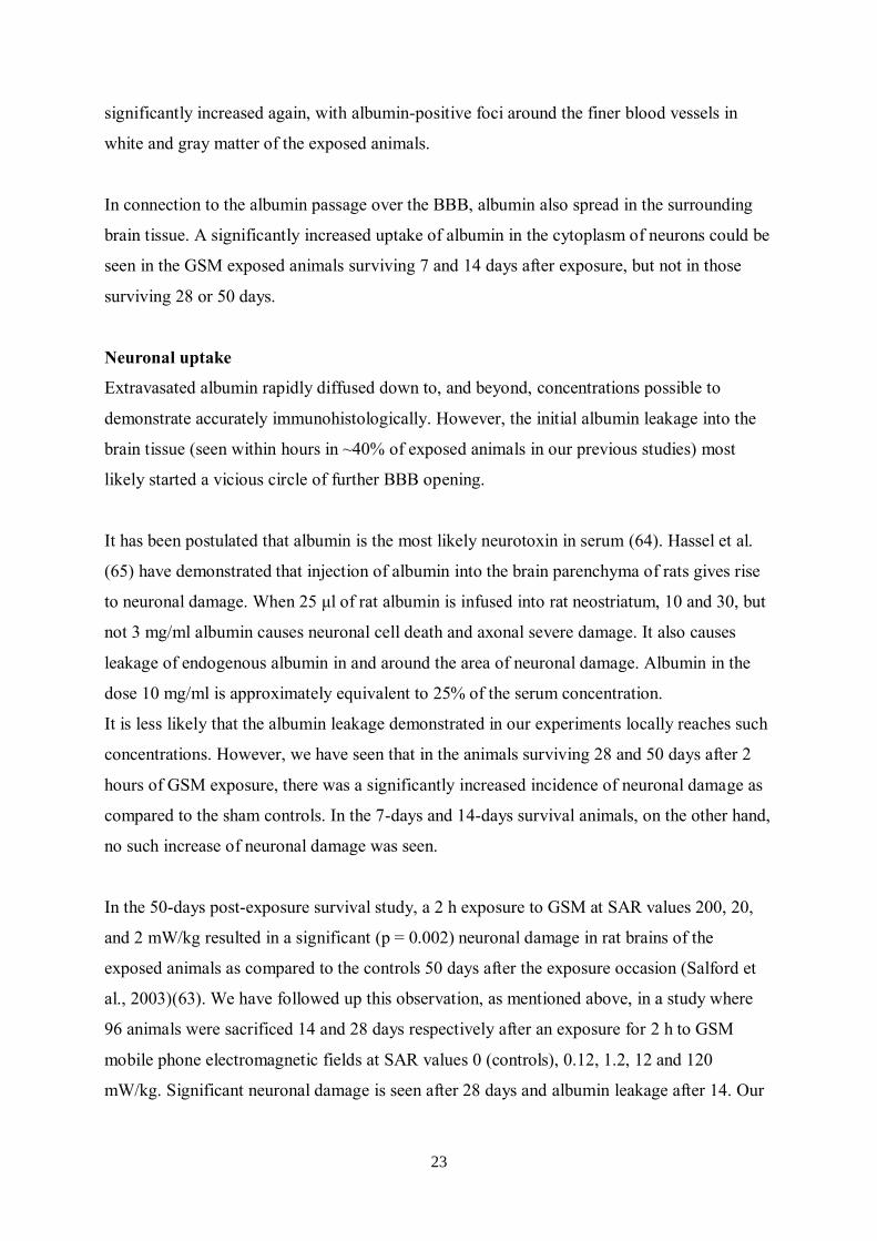
TEM-cells
In the majority of our studies, EMF exposure of the animals has been performed in transverse
electromagnetic transmission line chambers (TEM-cells, see Figure 5) (53, 54, 59, 61-63, 68-
71). These TEM-cells are known to generate uniform electromagnetic fields for standard
measurements. Each TEM-cell has two compartments, one above and one below the center
septum. Thus, two animals can be exposed at a time. The animals are un-anaesthetized during
the whole exposure. Since they can move and turn in the TEM-cells as they like, the
component of stress-induced immobilization (described by Stagg et al. (72)) is effectively
minimized. Through our studies, we have concluded that the amount of albumin leakage is
neither affected by the sex of the animals, nor their placement in the upper or lower
compartments of the TEM-cells.
26
The TEM cell
Figure 5. TEM-cells for EMF exposure.
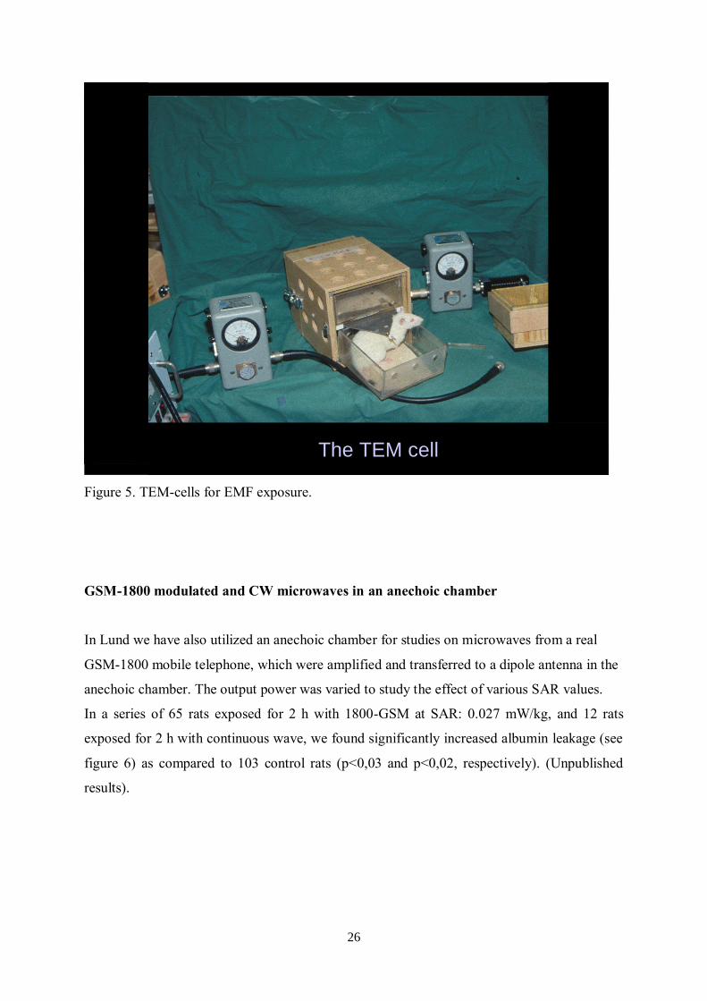
GSM-1800 modulated and CW microwaves in an anechoic chamber
In Lund we have also utilized an anechoic chamber for studies on microwaves from a real
GSM-1800 mobile telephone, which were amplified and transferred to a dipole antenna in the
anechoic chamber. The output power was varied to study the effect of various SAR values.
In a series of 65 rats exposed for 2 h with 1800-GSM at SAR: 0.027 mW/kg, and 12 rats
exposed for 2 h with continuous wave, we found significantly increased albumin leakage (see
figure 6) as compared to 103 control rats (p<0,03 and p<0,02, respectively). (Unpublished
results).

27
Figure 6.
Pathological leakage around vessels demonstrated by immunostaining against albumin.
Fischer 344 rat exposed for 2 h with 1800-GSM at SAR: 0.027 mW/kg
Other Studies on BBB Permeability, Focusing on the Effects of RF EMFs of the
Type Emitted by Mobile Phones
With the increasing use of mobile phones, much attention has been directed towards the
possible effects on BBB permeability, after exposure to the type of RF EMFs emitted by the
different sorts of mobile phones.
Repetitions of our initial findings of albumin leakage have been made by Fritze et al. (73),
with 900 MHz exposure of rats for 4 h at brain power densities ranging from 0.3–7.5 W/kg.
Albumin extravasation into the brain tissue was seen, with significant difference between
controls and rats exposed reported for 7.5 W/kg, which is a thermal level. However, Fisher
exact probability test (two-tailed) performed on the reported results, reveals significant ( p <
0.01, Fisher exact probability test) difference for the subthermal level group (SAR 0.3 W/kg
plus 1.3 W/kg, compared to sham exposed and cage control animals) where in total 10 out of
20 animals showed one or more extravasations direct after exposure (Salford et al. (20)).
28
Another group, working in Bordeaux, and led by Prof Pierre Aubineau, has also demonstrated
evidence of albumin leakage in rats exposed for 2 h to 900 MHz at non thermal SAR-values,
using fluorescein-labeled proteins. The results were presented at two meetings by Töre et al.
(74, 75). The findings are very similar to those of our group, described above.
At the BEMS meeting in 2002 in Quebec City in Canada, the Aubineau-Töre group presented
results from exposure GSM-900 EMFs at SAR values of 0.12, 0.5, and 2.0 W/kg. Seventy
Sprague-Dawley rats were included in the study. In addition to normal sham and normal GSM
exposed rats, also rats subjected to chronic dura mater neurogenic inflammation, induced by
bilateral sympathetic superior cervical ganglionectomy, were included. Arterial blood
pressure was measured during the exposure, and Töre et al. (74, 75) concluded that the
pressure variations (100–130mm Hg) were well below those limits, which are considered to
be compatible with an opening of the BBB of rats. In order to induce opening of the BBB in
rats, arterial blood pressure needs to reach values of 170 mmHg, according to Töre et al. (74,
75). At SAR of 2 W/kg a marked BBB permeabilization was observed, but also at the lower
SAR-value of 0.5 W/kg, permeabilization, although somewhat more discrete, was present
around intracranial blood vessels, both those of the meninges and of the brain parenchyma.
Comparing the animals, which had been subjected to ganglionectomy, to the other animals,
Töre et al. made an interesting observation: as expected, albumin extravasation was more
prominent in the sympathectomised sham-exposed rats as compared to normal exposed rats.
This was due to the fact that the sympathectomised rats were in a chronic inflammation-prone
state with hyper-development of pro-inflammatory structures, such as the parasympathetic
and sensory inputs as well as mast cells, and changes in the structure of the blood vessels.
Such an inflammation-prone state has a well-known effect on the BBB leakage. However,
when comparing sham-exposed sympathectomised rats to GSM-exposed sympathectomised
rats, a remarkable increase in albumin leakage was present in the GSM exposed
sympathectomised rats compared to the sham rats. In the GSM-exposed sympathectomised
rats, both brain areas and the dura mater showed levels of albumin leakage resembling those
observed in positive controls after osmotic shock. Indeed, more attention should be paid to
this finding, since it implicates that the sensitivity to EMF-induced BBB permeability
depends not only on power densities and exposure modulations, but also on the initial state of
health of the exposed subject.
In rats, uptake of a systemically administered rhodamine-ferritin complex through the BBB
also has been observed, after exposure to pulsed 2.45GHz EMFs at average power densities of

29
2 W/kg by Neubauer et al. (76). The authors observed that the magnitude of BBB
permeability depended on power density and duration of exposure. Exposure to a lower power
density (1 W/kg) and shorter duration of the exposure (15 min) did not alter the BBB
permeability, as compared to higher power densities (SAR 2 W/kg) and longer duration of
exposure (30–120 min). The microtubules seemed to play a vital role in the observed BBB
permeability, since treatment with colchicine, which inhibits microtubular function, resulted
in near-complete blockade of rhodamine-ferritin uptake. The mechanism underlying the
observed leakage was presumed to be correlated to pinocytotic-like transport.
In other studies, no effect of EMF exposure has been observed on the BBB integrity. With
exposure to 1,439MHz EMFs, 1 h daily during 2 or 4 weeks (average whole-body energy
doses of 0.25 W/kg) no extravazation of serum albumin trough the BBB was observed in a
series of 36 animals by Tsurita et al.(77). However, in this small material only 12 animals in
total were EMF exposed (6 rats exposed for 2 weeks and 6 rats exposed for 4 weeks). Also,
lack of interference with the BBB function of rats was found after 1,439MHz exposure for 90
min/d for 1–2 weeks at average brain power densities of either 2 or 6W/kg by Kuribayashi et
al.(78). A total number of 40 animals were included in the study.
Finnie et al. (79) came to the conclusion that no increase in albumin leakage over the BBB
resulted from EMF exposure in a series of 60 mice. With whole body exposure of mice to
GSM-900 EMFs for 1 h at a SAR of 4 W/kg or sham exposure, no difference in albumin
extravazation was observed between the different groups. Also, free-moving cage controls
were included in the study, and interestingly, there was no significant difference between
these non-restrained mice as compared to the sham and EMF-exposed animals. Thus, the
authors concluded that there were no stress-related exposure module confinement effects on
the BBB permeability.
Finnie et al. (80) continued to investigate more long-lasting exposure effects. In a series of
experiments, a total of 207 mice were exposed 60 min daily, 5 days per week for 104 weeks at
average whole body SARs of 0.25, 1.0, 2.0, and 4.0W/kg. This led to a minor disruption of
the BBB, as seen by the use of endogenous albumin as a vascular tracer. However, it should
be added that the authors performed no statistical analyses to evaluate the albumin leakage
through the small vessels in the brain. In an answer to correspondence in the same journal
(81), the authors presented the original data from the long-term study in one table, from which
30
one can conclude that non-leptomeningeal albumin leaking vessels were seen in few sham-
exposed animals, and in one-third of the animals in the 0.25 W/kg group and to a lesser extent
in the higher SAR groups.
The fact that some research groups observe albumin leakage/transport over the BBB after
EMF exposure and others do not, has led to a rather intense debate between the researchers
but also in society, which is puzzled by the divergent findings. A major concentration of the
involved research groups took place at Schloss Reisensburg in Germany in 2003, where the
technical approaches in the studies of BBB effects were discussed. Two world-renowned
researchers in the BBB field, Dr. David Begley of Kings College, London, and Prof. Olaf
Poulsen of Copenhagen, Denmark, chaired the FGF/COST 281 Reisensburg, November 2–6
meeting. They made the final statement as a summary of the meeting: ‘‘It seems clear that RF
fields can have some effects on tissues’’. The statement was made to a large extent on the
basis of the concordant findings of the Bordeaux group, represented by Prof. Aubineau, and
the Lund group, represented by Prof. Salford and Prof. Persson.
The histopathological examinations of the brains are not uncomplicated. Some laboratories
that have tried to replicate our studies have not been able to demonstrate the albumin leakage.
We have recently had problems with the albumin staining due to change of suppliers of
avidin, biotin, serum and antibodies. The lateral hypothalamic nuclei in the immediate vicinity
of the third ventricle are well known for their normally insufficient BBB. This has served as
an inbuilt control of adequate albumin staining in all our experiments since 1990. In our study
on combined effects of RF- and ELF-EMF, for the first time, we could not demonstrate
albumin extravasation in basal hypothalamus. Not until our third attempt with new staining
material, we got our positive control and could also demonstrate albumin leakage in the
exposed brains (61).
The biological effects of RF exposure depend on many parameters, such as mean power level
and the time variations of the power (82) and whether in vivo or in vitro experiments are
performed. In the in vivo situation, different kinds of animals, and also the same kind of
animals but of different breeds, might react differently. It might not necessarily be the
strongest RF fields that give rise to the most obvious biological effects (54, 63). In many
cases, the weak and precisely tuned EMFs have the most important biological function; two
examples of this are cellular communication and protein folding. It seems quite likely that in

31
different experimental set-ups, and in different living organisms, the signal has to be tuned to
different properties in order to cause any effect. This could perhaps in some part explain why,
in some cases, there are quite obvious effects of RF exposure, whereas in others, no such
effects can be seen.
Other Studies on BBB permeability and neuronal damage
As has been mentioned above (p. 26) Ilhan et al. (67), in 2004 reported neuronal damage in
female Wistar rats, which had been exposed to a 900 MHz electromagnetic GSM near-field
signal for 1 h. a day for 7 days. They found scattered and grouped dark neurons in the cortex,
hippocampus and basal ganglia, mixed in among normal neurons. A combined non-parametric
test for the four groups revealed that the distributions of scores differed significantly between
the control and the GSM only exposure group (p < 0.01).
Later, Masuda et al. (83) tried to replicate the findings by our group of albumin extravasation
and dark neurons. F344 rats (n=64) were exposed to 915 MHz signals for 2 hours (SAR of 0,
0.02, 0.2 and 2 W/kg), and albumin extravasation and dark neurons were investigated 14 and
50 days after the exposure. No albumin extravasation was seen, neither in control or exposed
rats, and no difference in the occurrence of dark neurons could be found due to EMF
exposure. An interesting difference as compared to the studies by Salford et al. mentioned
above, was that animals, after perfusion fixation, were left in a 4ºC storage for 18 hours
before the brains were removed. The question is whether this might have led to dilution of the
very sensitive albumin extravasation, which is often more pronounced in the circumventrical
organs as compared to the brain extravasates (personal communications with our
neuropathologist Arne Brun). This might explain the fact, that no albumin extravasation could
be seen in neither the cage control animals, the shams or the GSM exposed animals.
Another study by Mason and his group at Brooks Airforce Resarch Laboratory, San Antonio,
also tried to confirm our findings of albumin extravastion by using the same type of TEM-
cells for EMF Exposure (84), although the exposure parameters where somewhat different
with only 30-min exposure, including only male rats of the Fischer 344 CD-VAF strain and
utilizing only the upper compartment of the TEM cells. Exposure was at whole-body SAR
values of 0.002 to 20 W/kg. Regarding extracellular albumin accumulation, the results were
32
not formally analyzed, as motivated by too low scores of albumin. Regarding intracellular
albumin uptake, no significant difference between the different groups was reported. However,
as presented in the paper by McQuade et al.(84), at the lowest SAR of 1.8 mW/kg at 16 Hz, of
33 exposed rats, 11 had 2 or 3 positivities (33% of the animals) and 22 had none or 1
positivity. In the sham animals, 18% were positive and among the cage controls only 12%.
These results are reminiscent of prior work by the Lund group reporting that 17% of the sham
animals had some albumin leakage, while only at the most 50% of the identical and equally
handled, but RF exposed animals displayed albumin extravasation (60).
In a third study aiming to replicate the Lund findings of dark neurons, a group in Bordeaux
(85) exposed 14 weeks old Fischer 344 rats (which, however, were restrained in a rocket-type
exposure setup), to the GSM-900 signal for 2 h at various brain-averaged SARs (0, 0.14 and
2.0 W/kg). Eight rats were included in each of these groups.
Albumin leakage and neuronal degeneration was evaluated 14 and 50 days after exposure.
It was reported that no statistically significant albumin leakage was observed and that
neuronal degeneration assessed using cresyl-violet or the more specific marker Fluoro-Jade B,
was not significantly different among the tested groups. Here we want to point out that the
Bordeaux group makes a major deviation from the way we have evaluated the occurrence of
dark neurons in the tissue slices. While we counted the overall number of dark neurons, de
Gannes et al. (85) chose to subdivide the slices into 12 different small regions, which were
compared individually to each other (fig 3 in the publication). This gave the effect that a clear
overall difference in number of observed dark neurons between animals 50 days after
exposure to 2 W/kg for two hours versus sham exposed, disappeared in the statistics. On the
contrary, if all the numerical values for the bars representing the scored dark neurons
observed in each brain zone and region 50 days after exposure to 2 W/kg are compared to all
those of the sham animals, a highly significant difference (Kruskall-Wallis) between animals
exposed to 2 W/kg and sham is demonstrated (Mann-Whitney) p = 0.003! This is in
concordance with the Lund experience!
Indirect studies and studies on the blood cerebrospinal fluid barrier
The integrity of the BBB has also been investigated indirectly. Cosquer et al. (86) treated rats
with the muscarinic antagonist scopolamine methylbromide, which is known to induce
33
memory impairments, followed by EMF exposure at 2.45GHz for 45 min at average whole
body SARs of 2W/kg. Opening of the BBB after EMF exposure was hypothesised to affect
the performance in a radial arm maze. However, no such alterations were observed and the
authors concluded that no BBB opening seemed to have occurred. In agreement with this, no
albumin extravasation was noticed.
Ushiyama et al. (87) investigated the effects on the blood cerebrospinal fluid barrier after RF-
EMF exposure. With a microperfusion method, cerebrospinal fluid from rat brain was
collected in vivo. Fluorescent intensity of FITC-albumin in perfusate was measured. Rats
exposed to 1.5GHz RFs during 30 min at SAR-values of 0.5, 2.0, 9.5W/kg for adult rats and
0.6, 2.2, 10.4W/kg for juvenile rats, respectively, were compared to sham-exposed controls.
Under these conditions, no increase in FITC-albumin was seen in the cerebrospinal fluid of
exposed rats as compared to sham exposed controls. It was concluded that no effect on the
function of the blood cerebrospinal fluid barrier was seen.
In a recent study, the permeability of the human BBB after mobile phone exposure was
assessed measuring blood levels of S100B and transthyretin in human volunteers by
Söderqvist et al. (88). S100B is a calcium-binding protein, and it has been shown to be
increased in serum after damage to the BBB. Transthyretin, also known as pre-albumin, is
synthesised both in the liver and the choroid plexus. 30 min of GSM-900-like exposure at
SAR-values of 1 W/kg was used. No difference was seen regarding S100, but transthyretin
was increased 60 min after the termination of exposure as compared to the control situation.
The concentrations of S100B and transthyretin were also analysed 30 min prior to provocation
and after 30 min rest, showing a decrease after 30 min rest, which was suggested, might be
due to less stress after the 30 min rest. Thus, it is interesting that despite this decline, which
might be due to relaxation, still an increase in thransthyretin could be measured 30 min after
exposure. It was also put forward, that it could not be excluded that the thransthyretin rise
might be a compensation to the previous decrease, and that new studies including more
participants and also a sham group would be needed.
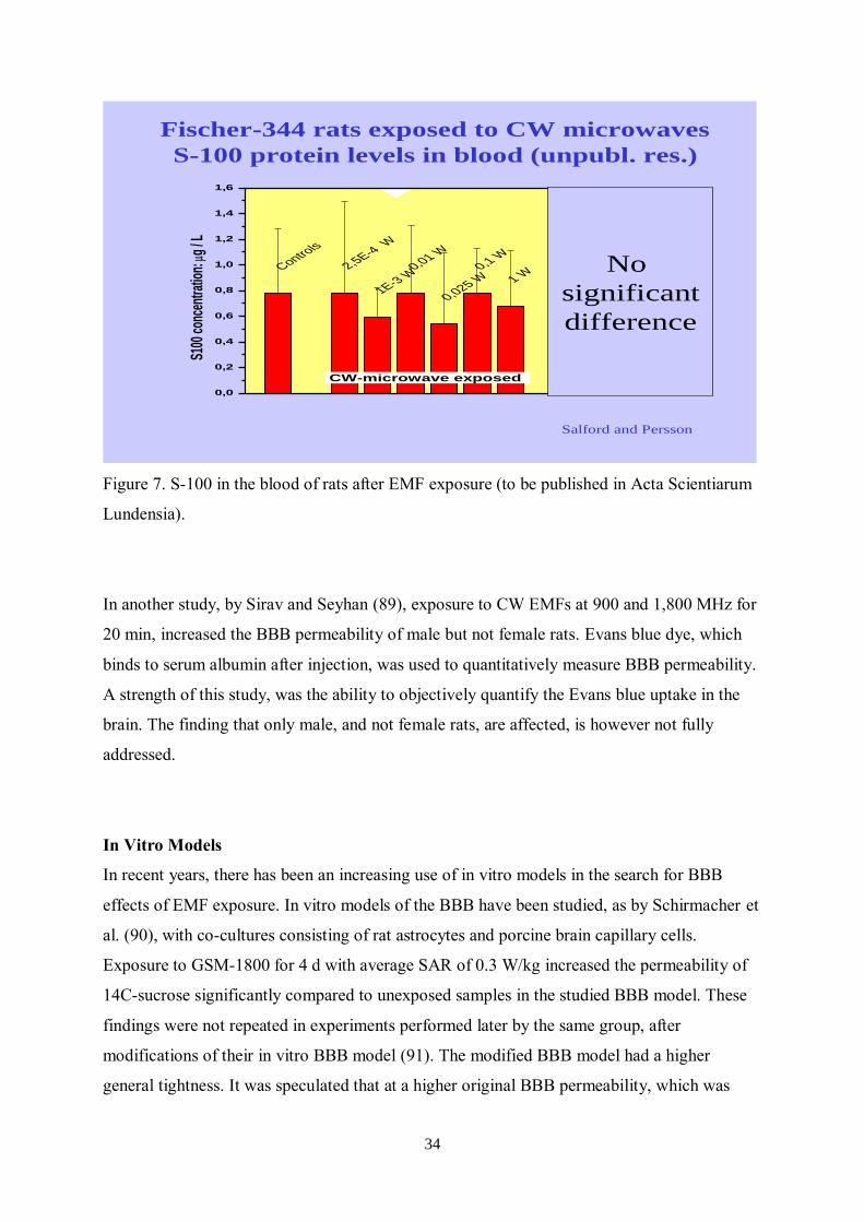
We have in the past investigated whether MW exposure, CW and at different SAR levels
might enhance S-100 protein levels in the blood of a large proportion of our rats. We could
conclude that no significant differences were seen (see Figure 7 below) (to be published).
34
Controls
2,5E-4 W
1E-3 W0,01 W
0,025 W0,1 W
1 W
EMX
CW+EMX
0,0
0,2
0,4
0,6
0,8
1,0
1,2
1,4
1,6
CW-microwave exposed
S100
con
cent
ratio
n:
g / L
Fischer-344 rats exposed to CW microwaves
S-100 protein levels in blood (unpubl. res.)
Salford and Persson
No
significant
difference
Figure 7. S-100 in the blood of rats after EMF exposure (to be published in Acta Scientiarum
Lundensia).
In another study, by Sirav and Seyhan (89), exposure to CW EMFs at 900 and 1,800 MHz for
20 min, increased the BBB permeability of male but not female rats. Evans blue dye, which
binds to serum albumin after injection, was used to quantitatively measure BBB permeability.
A strength of this study, was the ability to objectively quantify the Evans blue uptake in the
brain. The finding that only male, and not female rats, are affected, is however not fully
addressed.
In Vitro Models
In recent years, there has been an increasing use of in vitro models in the search for BBB
effects of EMF exposure. In vitro models of the BBB have been studied, as by Schirmacher et
al. (90), with co-cultures consisting of rat astrocytes and porcine brain capillary cells.
Exposure to GSM-1800 for 4 d with average SAR of 0.3 W/kg increased the permeability of
14C-sucrose significantly compared to unexposed samples in the studied BBB model. These
findings were not repeated in experiments performed later by the same group, after
modifications of their in vitro BBB model (91). The modified BBB model had a higher
general tightness. It was speculated that at a higher original BBB permeability, which was

35
present in the first study by Schirmacher et al. (90), the cultures were more susceptible to the
RF EMFs. Using porcine brain microvascular endothelial cell cultures as an in vitro model of
the BBB, no effects on barrier tightness, transport behavior, and integrity of tight junction
proteins were observed after exposure to UMTS EMFs at 1.966 GHz for 1–3 d at different
field strengths at 3.4–34 V/m, generating a maximum SAR of 1.8 W/kg (92).
In the search after the mechanism underlying non thermal EMF effects, Leszczynski et al.
(93) observed human endothelial cells, with the interesting finding that GSM-900 exposure
for 1 h with SAR-values of 2 W/kg resulted in changes in the phosphorylation status of many
proteins. Among the affected pathways, the hsp27/p38MAPK stress response pathway was
found, with a transient phosphorylation of hsp27 as a result of the mobile phone exposure.
This generated the hypothesis that the mobile-phone induced hsp27-activation might stabilize
stress fibers and in this way cause an increase in the BBB permeability. Furthermore, it was
also suggested that several brain-damaging factors might all contribute to the mobile phone-
induced effects observed in the brain and other structures as well.
Further perspectives of the importance of the BBB including the human situation
BBB in the Context of Alzheimer’s Disease and the findings by the Zlokovic Group
The BBB, as mentioned previously, is of essential role for maintaining an accurate brain
function. As described by Zlokovic (94), in a review regarding BBB in correlation to
neurodegenerative disorders, BBB breakdown can be due to tight junction disruption,
alterations of angiogenesis or vessel regression, hypoperfusion, inflammatory response and
alterations of the transport of molecules across the BBB (94). Further, as Zlokovic
hypothesises, this might contribute to neurodegenerative disorders, such as Alzheimer’s
disease (AD), Parkinson’s disease, multiple sclerosis and amyotrophic lateral sclerosis.
In the review by Zlokovic (94), a neurovascular disease pathway is presented, regarding
possible genesis of AD, where it is suggested that changes in vascular genes and receptors in
brain capillaries and small arteries might disrupt BBB functions, leading to an accumulation
36
of amyloid beta (Aβ), a neuroinflammatory response and BBB breakdown and further on
accumulation of Aβ, loss of the BBB to clear Aβ (due to affected synaptic transmission,
neuronal injury and recruitment of microglia) and secretion of proinflammatory cytokines.
Ultimately, this is suggested to lead to disappearance of the capillary unit, increasing Aβ
deposits and synaptic and neuronal loss (94).
This observation might explain how vascular disease contributes to Alzheimer's disease (AD)
risk; the heterogeneity of AD; and supports the idea that exclusively focusing on amyloid is
likely to be disappointing.
Neuronal injury resulting from vascular defects that are not related to amyloid-beta but is
related to damage results from a breakdown of the blood-brain barrier and a reduction in
blood flow (94). Although Amyloid beta definitely has an important role in Alzheimer's
disease it's very important to investigate other leads, perhaps where amyloid-beta isn't as
centrally involved.
Human apolipoprotein E has three isoforms: APOE2, APOE3 and APOE4. APOE4 is a major
genetic risk factor for Alzheimer's disease and is associated with Down's syndrome dementia
and poor neurological outcome after traumatic brain injury and haemorrhage. Neurovascular
dysfunction is present in normal APOE4 carriers and individuals with APOE4-associated
disorders. In mice, lack of APOE leads to blood-brain barrier (BBB) breakdown, whereas
APOE4 increases BBB susceptibility to injury. How APOE genotype affects brain
microcirculation remains elusive. Using different APOE transgenic mice, including mice with
ablation and/or inhibition of cyclophilin A (CypA), it has been shown show that expression of
APOE4 and lack of murine APOE, but not APOE2 and APOE3, leads to BBB breakdown by
activating a proinflammatory CypA-nuclear factor-kappa B-matrix-metalloproteinase-9
pathway in pericytes. These findings suggest that CypA is a key target for treating APOE4-
mediated neurovascular injury and the resulting neuronal dysfunction and degeneration. The
data reviewed above support an essential role of neurovascular and BBB mechanisms in
contributing to both, onset and progression of AD (95, 96).
37
BBB in the context of Alzheimer’s Disease – Importance of EMF Exposure
In this context, the findings of Arendash et al., that long-term EMF reduced brain Aβ
deposition through Aβ anti-aggregation actions in AD mice, are highly interesting (97). It was
also found, by Mori and Arendash et al., that long-term exposure to high frequency EMF
treatment prevented cognitive impairment in AD transgenic (Tg) mice and improved memory
in normal mice and that an increase in neuronal activity could be observed in the EMF
exposed groups (98). Furthermore, it was found by the group that EMF treatment enhances
brain mitochondrial functions in AD Tg as well as normal mice and that no increase in brain
temperature could be found in connection to the EMF exposure (99). An interesting aspect in
this context, is the role of mitochondria for many cellular functions, including reactive oxygen
species generation, apoptosis, and Ca2+ homeostasis as was mentioned by Dragicevic et al.
and reviewed by Nicholls (99, 100).
In the first mentioned study by Arendash et al. (97), mice were EMF exposed with start at
young age or at adult age. In the young-age group, 24 mice were divided into 4 subgroups:
n=6 were Tg controls, n=6 were Tg animals treated with EMF, n=6 were non-transgenic (NT)
controls and n=6 were NT animals treated with EMF. 2.5, 4-5 and 6-7 months after daily
GSM-900 EMF exposure (two 1-hour sessions daily, at SAR 0.25 W/kg), the animals were
evaluated by cognitive tests. At the end of the study, Aβ in the brains was evaluated by
immunohistochemistry. No effect on cognitive functions was observed after 2 months of
exposure. However, for the Tg+EMF mice with start of EMF exposure at young age, the
cognitive function was maintained after 6-7 months of exposure, while it deteriorated in the
Tg group. In a final task for NT mice after 7 months of EMF, the EMF actually improved the
mnemonic function. In the adult-age group, Tg animals had impaired cognitive functions at
the age of 4 months. 28 Tg and NT mice were included. After long-term EMF exposure (2, 5
and 8 months) the memory was tested. While 2 months of EMF exposure had no effect, 5
months of exposure had positive effects only on NT mice, and 8 months of exposure had
beneficial effects for the Tg mice, with better results in the Tg+EMF group as compared to
the Tg controls. Also the NT+EMF mice had an improved function as compared to NT
controls after 8 months. Staining for Aβ revealed lower values on both hippocampus and the
entorhinal cortex in the Tg+EMF group as compared to the Tg control group. Hippocampal

38
tissue from Tg mice were then exposed to EMF for 4 days, after which it was shown that the
Aβ amount had decreased as compared to non-exposed control tissue. It was also reported
that a t1° temperature increase was observed in EMF exposed animals during exposure, but
not in between exposure sessions (97).
In the study by Mori and Arendash (98), n=6 mice were Tg controls, carrying the mutant
APPK670N , n=10 mice were Tg treated with EMF, n=4 mice were NT controls and n=5
mice were NT treated with EMF. EMF exposed animals were placed in a Faraday cage,
receiving two 2-hour periods of EMF treatment at GSM-900 frequencies, pulse modulated at
SAR 0.25-1.05 W/kg. The neuronal expression of c-Fos was taken as an indicator of neuronal
activity. With immunohistochemistry, it was found that c-Fos was increased in both the
NT+EMF group, as well as in the Tg+EMF group in the entorhinal cortex. However, only this
one brain region was analyzed, since c-Fos expression was too low in other regions, which the
authors hypothesised might be due to that c-Fos in an early response gene, and that at a
certain time after stimulation, when the animals were sacrificed, the expression had already
declined in other regions, such as hippocampus. In a cognitive test (Y-maze), it was found
that EMF improved the performance in both NT and Tg group as compared to untreated
controls. It should also be noted, that despite the very interesting findings, the number of
included animals is quite small (98).
EMF and 18
FDG Uptake – Recent Studies
The question whether EMF exposure from mobile phones has neuronal effects in the human
situation was recently addressed by an American research group led by Volkow et al.,
conducting a PET study on 18
F-fluorodeoxyglucose (18
FDG) uptake (101). Though PET-
studies on humans in correlation to EMF exposure have also been previously made, the
purpose of this study was to extend the study material and use the more direct measure of
brain glucose metabolism by the uptake of 18
FDG instead of the previously used CBF
(cerebral blood flow) measure, which might be a more indirect sign of neuronal activity and
also reflect short-term alterations (60s) as compared to the more long-lasting ones observed
with 18
FDG (suggested to be in the range of 30 min). 18
FDG is actively transported across the
BBB into the cells, where it is phosphorylated, and is, among others, used as a prognostic
value for following low-grade brain tumours, where an increased uptake in previously low-
39
grade tumours is an indicator of anaplastic transformation (for review into the topic of 18
FDG
and brain tumours (102).
(space)
In the study by Volkow et al. (101), in total, 47 persons were involved, and effects upon brain
glucose metabolism of EMF exposure were evaluated using PET with injection of 18
FDG.
PET scans were performed both with and without EMF exposure (50 min of GSM-900 with
maximum SAR of 0.901 W/kg), and the participants were blinded to the exposure situation.
Whereas whole-brain metabolism was not affected, there were regional differences, in the
right orbitofrontal cortex and the lower part of the right superior temporal gyrus (that is, the
same side as the mobile phone was placed at) with increased metabolism in the exposure
situation of about 7% as compared to control. There was a positive correlation between the
strength of the E-field from the phones and the brain activation. Interestingly, it was
hypothesized that RF-EMF exposure might increase the excitability of brain neurons.
Following the study by Volkow et al. (101), Kwon et al. (103) also investigated effects of
GSM-900 exposure upon brain 18
FDG uptake. Thirteen persons were exposed to GSM-900
for 30 minutes to the right side of the head, and all subjects were also sham-exposed, and
blinded to the exposure situation (SAR-values of maximum 0.74 W/kg in the head and 0.23
W/kg in the brain tissue). Contrary to the findings of Volkow et al. (101), the study by Kwon
et al. (103) demonstrated a decrease in brain 18
FDG uptake after GSM-900 exposure, with
decreased uptake values in the temporoparietal junction. A volume-of-interest analysis
focused upon the right temporal lobe, showed a decreased 18
FDG uptake in the anterior
inferior temporal cortex. No effects on task performance were found, and no correlation
between temperature or 18
FDG uptake (a temperature increase of <0.21°C was found on the
skin on the exposed side of the head) (103).
In the animal situation, Frilot et al. investigated the effect of ELF magnetic field exposure (2.5
G at 60 Hz) upon 18
FDG uptake in rats, comparing uptake with and without EMF exposure.
An increased glucose uptake was found in the hindbrain when the field was orthogonally to
the sagittal plane, but not when the angle varied randomly between the field and sagittal
plane. These effects were hypothesized to be coupled to induction of electric field on the gate
of ion channels (104).

40
Possible connection between BBB leakage and nerve cell injury
It has been suggested that BBB leakage is the major reason for nerve cell injury,
such as that seen in dark neurons in stroke-prone spontaneously hypertensive rats (105). Much
speaks in favour of this possibility. The parallel findings in the Lund material of neuronal
uptake of albumin and dark neurons may support the hypothesis that albumin leakage into the
brain is the cause for the neuronal damage observed after 28 and 50 d. It should, however, be
pointed out that the connection is not yet proven (Figure 8).
7d 14 d 28 d 50 d
Albumin 0.04 0.02 ns 0.04
foci
Neuronal 0.02 0.005 ns ns
albumin
Dark ns ns 0.01 0.001
neurons
Exposed vs sham
© Salford et al
Figure 8. Results from the Lund group (61-63)
Also, other unwanted and toxic molecules in the blood may leak into the brain tissue in
parallel with the albumin, and concentrate in and damage the neurons and glial cells of the
brain. In favour of a causal connection between albumin and neuronal damage is a series of
experiments performed in rats by another group at Lund University; albumin leaks into the
brain and neuronal degeneration is seen in areas with BBB disruption in several
circumstances: after intracarotid infusion of hyperosmolar solutions in rats (106) in the stroke

41
prone hypertensive rat (105); and in acute hypertension by aortic compression in rats (22).
Furthermore, it has been shown in other laboratories that epileptic seizures cause
extravasation of plasma into brain parenchyma (21), and in the clinical situation the cerebellar
Purkinje cells are heavily exposed to plasma constituents and degenerate in epileptic patients .
There are indications that an already disrupted BBB is more sensitive to the RF fields than an
intact BBB (74, 91). It has been stated by other researchers that albumin is the most likely
neurotoxin in serum (64). It has been demonstrated that injection of albumin into the brain
parenchyma of rats gives rise to neuronal damage. When 25 micro-litres of rat albumin is
infused into rat neostriatum, 10 and 30, but not 3 mg/ ml albumin causes neuronal cell death
and axonal severe damage (65). It also causes leakage of endogenous albumin in and around
the area of neuronal damage. However, it is still unclear whether the albumin leakage
demonstrated in our experiments locally reaches such concentrations.
Possible mechanisms
Microarray analysis of the expression of all the rats’ genes in cortex and hippocampus, after
exposure to GSM RFs or sham exposure for 6 h, has shown interesting differences between
exposed animals and controls as described by Nittby et al. (107). Genes of interest for
membrane transport show highly significant differences. This may be of importance in
conjunction with our earlier findings of albumin leakage into neurons around capillaries in
exposed animals. It can be noted here that among the significantly altered genes from these
evaluations, two variants of the gene RGS4 are up-regulated in hippocampal tissue from
exposed rats as compared to the sham-exposed rats (unpublished results). RGS is a regulator
of G protein signalling, and it has been proposed that RGS4 might regulate BBB permeability
in mammals, in a way corresponding to the role of its Loco homolog G protein coupled
receptor (GPCR) in developing and maintaining the BBB permeability of Drosophila (7).
It has also been suggested in other connections that manifestations of BBB disruption might
also be mediated by the formation of free radicals, such as O2-, H2O2, and hydroxyl radical,
which are supposed to oxidize cell membrane lipids by virtue of the high concentration of
polyunsaturated fatty acids in these membrane constituents (108). As an example of this, it
was reported by Chan et al.(109), that treatment of the brain of rats with a free-radical

42
generating system resulted in lipid-peroxidation, and an increased permeation of Evans blue
due to barrier breakdown.
Recently, a detailed molecular mechanism, by means of which mobile phone radiation might
exert its effects, has been proposed (110). By using Rat1 and HeLa cells, it was shown that
EMF exposure resulted in rapid activation of ERK/ MAPKs (mitogen-activated protein
kinase). The activation of these ERKs was mediated by reactive oxygen species (ROS),
resulting in a signalling cascade ultimately affecting transcription, by the central key role of
ERKs in signalling pathways.
In the continued search for the mechanisms behind EMF mediated effects, their interaction
with calcium-45 transport in bio-membranes has been studied (111) and Ca2+-efflux over
plasma membranes has been observed in plasma vesicles from spinach exposed to ELF
magnetic fields (112). With this model, quantum mechanical theoretical models for the
interaction between magnetic fields and biological systems are tested. The model proposed by
Blanchard and Blackman (113), in which it is assumed that biologically active ions can be
bound to a channel protein and in this way alter the opening state of that channel, could in this
way be quantitatively confirmed. Thus, the membrane is one site of interaction between the
magnetic fields and the cell, and more specifically, the Ca2+-channels, are one of the targets.
More recently, new models for the interaction between magnetic fields and hydrogen nuclei
also have been proposed.
EMF-induced Ca2+-efflux over plasma membranes, understandably, can have many different
effects on the target cells. Some agents that increase the BBB permeability act through a
contractile mechanism that widens the intercellular junctions of the capillary endothelium. An
increase of free Ca2+ should mediate these changes, thereby resulting in measurable
alterations of intracellular Ca2+-levels in brain capillary cells after exposure to BBB-
disrupting agents (108).
Another hypothesis is that EMF-induced intracellular Ca2+-alterations might affect Ets genes,
which are transcription factors expressed in different tissues (114). In this context, we could
add that in our gene expression material from GSM-exposed rats vs., sham-exposed rats, one
Ets variant gene is actually significantly up-regulated in hippocampus and one Ets1 gene is
significantly up-regulated in cortex of the exposed animals.
43
EMF induced BBB permeability – with the aim of medical use
In the attempt to further try to understand the underlying mechanisms of the RF effects, we
recently undertook a study upon snail nociception, with 1-hour GSM-1800 exposure of the
land snail H. pomatia. This revealed, that the exposure induced analgesia in the snail model,
with a significantly increased latency of reaction when placed on a hot plate, as compared to
when only sham exposed. The vast knowledge about the physiology of the snail, its
neurotransmission systems and it simplicity as compared the mammals may provide a tool for
successful continued search for the mechanisms behind the effects of the GSM EMF upon
biology (115).
In a recent study by Kuo et al (116), it was described how EMFs might be utilized to facilitate
transport across the BBB. In an in vitro model, human micro-vascular endothelial cells were
co-cultured with human astrocytes. Effects of EMF upon P-glycoprotein (P-gp) and multi-
drug resistance -associated proteins (MRP) were tested in connection to treatment with anti-
retroviral drugs, where the MRPs and P-gp are known to play an important role in multidrug
resistance, which is encountered in carcinomas and therapies for acquired immune-deficiency
(Kuo et al. 2012). With increasing EMF frequencies up to 900 MHz (both 715MHz and 900
MHz), the endocytotic uptake of calcein was increased (5mW, square wave with amplitude
modulation at 20 MHz for 4 hours). Treatment with EMF could also inhibit expression of
MRP and P-gp after treatment with anti-retroviral drugs, indicating that it might be useful in
order to deliver antiretroviral proteins into the brain, by decreasing the efflux of the drugs due
to the MRPs and P-gl.
Kuo et al. (117) also showed that EMF exposure (915 MHz EMFs at 5 mW with 20 MHz
amplitude modulation for 4 hours) in combination with cationic solid lipid nanoparticles
(CSLNs) could increase the transport of the antiretroviral drug Saquinavir 22-fold across
human brain-microvascular endothelial cells (as compared to a 17-fold increase when only
CSLNs were used).
44
Conclusions
In this review, we have reported the results of our group’s research during the last 24 years,
and the results of similar, but seldom identical, experiments of several other groups around
the world. When summing up what we have described here, we are convinced that RF
electromagnetic fields have effects upon biology, and we believe that it is more probable than
unlikely, that non-thermal electromagnetic fields from mobile phones and base stations do
have effects also upon the human brain. However, in this context, it is also important to point
out, that the studies from our laboratory, as well as most studies presented above and available
in literature, have been performed using animals and not humans. Thus no definitive
conclusions can be drawn regarding effects of mobile phone use upon the human BBB.
However, studies in humans utilizing radiopharmaceuticals have been performed by Volkow
et al. (101) upon brain glucose metabolism, and as was described by Saha et al. (118) already
in 1994, studies with PET or SPECT and radiopharmaceuticals are used in brain imaging.
Further, a tool to directly study the human BBB has recently been described (119). It is based
upon a non-radioactive methodology for in vivo non-invasive, real-time imaging of BBB
permeability for conventional drugs, using nitroxyl radicals as spin-labels and MRI. In this
connection, it should be mentioned though, that MRI has the drawback of possibly itself
influence upon the results.
Based upon what has been presented here, we feel that the WHO IARC classification of RFR
at the level 2B is adequate at present.
The question whether existing FCC/IEE and/or ICNIRP public safety limits and reference
levels are adequate to protect the public is not easily answered. The reported studies on EMF
induced BBB disruption have shown partially contradictory results from different laboratories.
However, the fact that an abundance of studies do show effects is an important warning. This
is true even if it can be summarized that the effects most often are weak and are seen in about
40% of the exposed animals.
45
However, we have stressed the following opinion in several publications during the past
years: - “The intense use of mobile phones, not least by youngsters, is a serious memento. A
neuronal damage may not have immediately demonstrable consequences, even if repeated. It
may, however, in the long run, result in reduced brain reserve capacity that might be unveiled
by other later neuronal disease or even the wear and tear of ageing. We can not exclude that
after some decades of (often), daily use, a whole generation of users, may suffer negative
effects such as autoimmune and neuro-degenerative diseases maybe already in their middle
age”.
One remarkable observation, which we have made in our studies throughout the years, is that
exposure with whole-body average power densities below 10 mW/kg gives rise to a more
pronounced albumin leakage than higher power densities, all at non-thermal levels. These
very low SAR-values, such as 1 mW/kg, exist at a distance of more than one meter away from
the mobile phone antenna and at a distance of about 150–200 m from a base station.
Further, when a mobile phone operating at 915 MHz (and its antenna) is held 1.4 cm from the
human head, the very low SAR levels of 10 mW/kg exist in deep-lying parts of the human
brain such as the basal ganglia, and the power density of 1 mW/kg and less is absorbed in
thalamus bilaterally.
With this information as a background, it is difficult to recommend safety limits as the
function of existing mobile systems might not allow for limits that produce SAR levels below
1 or 0,1 mW/kg in the human brain, which are reported to cause a pathological leakage of the
BBB and to neuronal damage.
Demonstrated effects on the BBB, as well as a series of other effects upon biology (120) have
given rise to scientific concern and to public anxiety. It is up to the society and our politicians
and also the providers of the radiofrequency-emitting technologies to support continued
research in order to understand the nature of the effects, thereby neutralizing or at least
reducing them. Also, it should be kept in mind that proven effects on biology also means that
positive potentials might be revealed. This might be useful in medical applications, for
example a controlled opening of the BBB would enable previously excluded pharmaceuticals
to reach their targets within the brain tissue.

46
Acknowledgements
A large proportion of this communication is based upon our article in Electromagnetic
Biology and Medicine: Nittby H, Grafström G, Eberhardt JL, Malmgren L, Brun A, Persson
BRR, Salford LG. (2008) Radiofrequency and Extremely Low-Frequency Electromagnetic
Field Effects on the Blood-Brain Barrier Electromagnetic Biology and Medicine, 27: 103–126
(24) and we thank the Editor for the permission to utilize the material.
A large proportion of the Lund experiments have been performed together with Arne Brun,
Jacob L. Eberhardt, Gustav Grafström and Lars Malmgren, to whom we address warm thanks
for stimulating collaboration during 24 years.
References
1. J.J. Carlsson SD, Hilgers LS. Blood barriers of the insect. Annu Rev Entomol 45:(2000).
2. N.J. Abbottand Y. Pichon. The glial blood-brain barrier of crustacea and cephalopods: a
review. Journal de physiologie. 82:304-313 (1987). 3. T. Gotowand P.H. Hashimoto. Plasma membrane organization of astrocytes in elasmobranchs
with special reference to the brain barrier system. Journal of neurocytology. 13:727-742
(1984).
4. M. Reinecke. The glial cells of the cerebral ganglia of Helix pomatia L. (Gastropoda, Pulmonata). II. Uptake of ferritin and 3H-glutamate. Cell and tissue research. 169:361-382
(1976).
5. H.M. Dziegielewska KM, Jones SE, Reader M, Saunders NR. . Proteins in cerebrospinal fluid and plasma of postnatal Monodelphis domestica (grey shorttailed
opossum). Comp Biochem Physiol. 92B:569-576 (1989).
6. J.A. Alberts B. Cell junctions, cell adhesion and the extracellular matrix. In G. S (ed.),
Molecular Biology of the Cell, Garland Publishing, New york, 2002. 7. R. Danemanand B.A. Barres. The blood-brain barrier--lessons from moody flies. Cell. 123:9-
12 (2005).
8. R.N. Frank, S. Dutta, and M.A. Mancini. Pericyte coverage is greater in the retinal than in the cerebral capillaries of the rat. Investigative ophthalmology & visual science. 28:1086-1091
(1987).
9. W.E. Thomas. Brain macrophages: on the role of pericytes and perivascular cells. Brain research Brain research reviews. 31:42-57 (1999).
10. W.H. Oldendorf, M.E. Cornford, and W.J. Brown. The large apparent work capability of the
blood-brain barrier: a study of the mitochondrial content of capillary endothelial cells in brain
and other tissues of the rat. Annals of neurology. 1:409-417 (1977). 11. J.F. Ghersi-Egea, A. Minn, and G. Siest. A new aspect of the protective functions of the
blood-brain barrier: activities of four drug-metabolizing enzymes in isolated rat brain
microvessels. Life sciences. 42:2515-2523 (1988). 12. A.L. Betz, J.A. Firth, and G.W. Goldstein. Polarity of the blood-brain barrier: distribution of
enzymes between the luminal and antiluminal membranes of brain capillary endothelial cells.
Brain research. 192:17-28 (1980). 13. N.A. Oberheim, X. Wang, S. Goldman, and M. Nedergaard. Astrocytic complexity
distinguishes the human brain. Trends in neurosciences. 29:547-553 (2006).
14. N.A. Oberheim, T. Takano, X. Han, W. He, J.H. Lin, F. Wang, Q. Xu, J.D. Wyatt, W. Pilcher,
J.G. Ojemann, B.R. Ransom, S.A. Goldman, and M. Nedergaard. Uniquely hominid features
47
of adult human astrocytes. The Journal of neuroscience : the official journal of the Society for
Neuroscience. 29:3276-3287 (2009).
15. W.A. Banks, V.B. Kumar, M.W. Franko, J.W. Bess, Jr., and L.O. Arthur. Evidence that the species barrier of human immunodeficiency virus-1 does not extend to uptake by the blood--
brain barrier: comparison of mouse and human brain microvessels. Life sciences. 77:2361-
2368 (2005). 16. P. Hsiao, L. Sasongko, J.M. Link, D.A. Mankoff, M. Muzi, A.C. Collier, and J.D. Unadkat.
Verapamil P-glycoprotein transport across the rat blood-brain barrier: cyclosporine, a
concentration inhibition analysis, and comparison with human data. The Journal of
pharmacology and experimental therapeutics. 317:704-710 (2006). 17. D.Z. Gerhart, R.L. Leino, N.D. Borson, W.E. Taylor, K.M. Gronlund, A.L. McCall, and L.R.
Drewes. Localization of glucose transporter GLUT 3 in brain: comparison of rodent and dog
using species-specific carboxyl-terminal antisera. Neuroscience. 66:237-246 (1995). 18. W. Oldendorf. Permeability of the BBB. In T. D (ed.), The Nervous System, Raven Press,
New York, 1975, pp. 229–289.
19. S. Rapoport. BBB in Physiology and Medicine, Raven Press, New York, 176. 20. L.G. Salford, Persson, B., Malmgren, L., & Brun. Téléphonie Mobile et Barrière Sang-
Cerveau. In P. Marco (ed.), Téléphonie mobile—effects potentiels sur la santé des ondes
électromagnétiques de haute fréquence, Emburg, Belgium, 2001, pp. 141–152.
21. A. Mihalyand B. Bozoky. Immunohistochemical localization of extravasated serum albumin in the hippocampus of human subjects with partial and generalized epilepsies and epileptiform
convulsions. Acta neuropathologica. 65:25-34 (1984).
22. T.E. Sokrab, B.B. Johansson, H. Kalimo, and Y. Olsson. A transient hypertensive opening of the blood-brain barrier can lead to brain damage. Extravasation of serum proteins and cellular
changes in rats subjected to aortic compression. Acta neuropathologica. 75:557-565 (1988).
23. S.R. Parathath, S. Parathath, and S.E. Tsirka. Nitric oxide mediates neurodegeneration and
breakdown of the blood-brain barrier in tPA-dependent excitotoxic injury in mice. Journal of cell science. 119:339-349 (2006).
24. H. Nittby, G. Grafstrom, J.L. Eberhardt, L. Malmgren, A. Brun, B.R. Persson, and L.G.
Salford. Radiofrequency and extremely low-frequency electromagnetic field effects on the blood-brain barrier. Electromagnetic biology and medicine. 27:103-126 (2008).
25. A.H. Frey, S.R. Feld, and B. Frey. Neural function and behavior: defining the relationship.
Annals of the New York Academy of Sciences. 247:433-439 (1975). 26. K.J. Oscarand T.D. Hawkins. Microwave alteration of the blood-brain barrier system of rats.
Brain research. 126:281-293 (1977).
27. K.J. Oscar, S.P. Gruenau, M.T. Folker, and S.I. Rapoport. Local cerebral blood flow after
microwave exposure. Brain research. 204:220-225 (1981). 28. E. Preston, E.J. Vavasour, and H.M. Assenheim. Permeability of the blood-brain barrier to
mannitol in the rat following 2450 MHz microwave irradiation. Brain research. 174:109-117
(1979). 29. J.H. Merritt, A.F. Chamness, and S.J. Allen. Studies on blood-brain barrier permeability after
microwave-radiation. Radiation and environmental biophysics. 15:367-377 (1978).
30. A.H. Frey. Headaches from cellular telephones: are they real and what are the implications? Environmental health perspectives. 106:101-103 (1998).
31. T.R. Ward, J.A. Elder, M.D. Long, and D. Svendsgaard. Measurement of blood-brain barrier
permeation in rats during exposure to 2450-MHz microwaves. Bioelectromagnetics. 3:371-
383 (1982). 32. T.R. Wardand J.S. Ali. Blood-brain barrier permeation in the rat during exposure to low-
power 1.7-GHz microwave radiation. Bioelectromagnetics. 6:131-143 (1985).
33. S.P. Gruenau, K.J. Oscar, M.T. Folker, and S.I. Rapoport. Absence of microwave effect on blood-brain barrier permeability to [14C]sucrose in the conscious rat. Experimental neurology.
75:299-307 (1982).
34. E.N. Albertand J.M. Kerns. Reversible microwave effects on the blood-brain barrier. Brain
research. 230:153-164 (1981).

48
35. J.C. Linand M.F. Lin. Studies on microwave and blood-brain barrier interaction.
Bioelectromagnetics. 1:313-323 (1980).
36. J.C. Linand M.F. Lin. Microwave hyperthermia-induced blood-brain barrier alterations. Radiation research. 89:77-87 (1982).
37. H. Goldman, J.C. Lin, S. Murphy, and M.F. Lin. Cerebrovascular permeability to 86Rb in the
rat after exposure to pulsed microwaves. Bioelectromagnetics. 5:323-330 (1984). 38. W.M. Williams, W. Hoss, M. Formaniak, and S.M. Michaelson. Effect of 2450 MHz
microwave energy on the blood-brain barrier to hydrophilic molecules. A. Effect on the
permeability to sodium fluorescein. Brain research. 319:165-170 (1984).
39. W.M. Williams, M. Del Cerro, and S.M. Michaelson. Effect of 2450 MHz microwave energy on the blood-brain barrier to hydrophilic molecules. B. Effect on the permeability to HRP.
Brain research. 319:171-181 (1984).
40. W.M. Williams, J. Platner, and S.M. Michaelson. Effect of 2450 MHz microwave energy on the blood-brain barrier to hydrophilic molecules. C. Effect on the permeability to
[14C]sucrose. Brain research. 319:183-190 (1984).
41. ICNIRP. Guidelines for limiting exposure to time-varying electric, magnetic and electromagnetic fields Health Physiology:494–522 (1998).
42. W.M. Williams, S.T. Lu, M. Del Cerro, and S.M. Michaelson. Effect of 2450 MHz
microwave energy on the blood-brain barrier to hydrophilic molecules. D. Brain temperature
and blood-brain barrier permeability to hydrophilic tracers. Brain research. 319:191-212 (1984).
43. R.M. Quock, J.M. Fujimoto, T.K. Ishii, and D.G. Lange. Microwave facilitation of
methylatropine antagonism of central cholinomimetic drug effects. Radiation research. 105:328-340 (1986).
44. R.M. Quock, F.J. Kouchich, T.K. Ishii, and D.G. Lange. Microwave facilitation of
domperidone antagonism of apomorphine-induced stereotypic climbing in mice.
Bioelectromagnetics. 8:45-55 (1987). 45. E. Moriyama, M. Salcman, and R.D. Broadwell. Blood-brain barrier alteration after
microwave-induced hyperthermia is purely a thermal effect: I. Temperature and power
measurements. Surgical neurology. 35:177-182 (1991). 46. M. Nakagawa, K. Matsumoto, H. Higashi, T. Furuta, and T. Ohmoto. Acute effects of
interstitial hyperthermia on normal monkey brain--magnetic resonance imaging appearance
and effects on blood-brain barrier. Neurologia medico-chirurgica. 34:668-675 (1994). 47. R.R. Shivers, M. Kavaliers, G.C. Teskey, F.S. Prato, and R.M. Pelletier. Magnetic resonance
imaging temporarily alters blood-brain barrier permeability in the rat. Neuroscience letters.
76:25-31 (1987).
48. H.J. Garber, W.H. Oldendorf, L.D. Braun, and R.B. Lufkin. MRI gradient fields increase brain mannitol space. Magnetic resonance imaging. 7:605-610 (1989).
49. I.K. Adzamli, Jolesz, E. A., Blau, M. An assessment of BBB integrity under MRI
conditions: brain uptake of radiolabelled Gd-DTPA and In-DTPA-IgG. J Nucl Med:839-840 (1989).
50. E. Preston, Buffler, K., Haas, N. Does magnetic resonance imaging compromise integrity of
the BBB? Neuroscience letters:46–50 (1989). 51. F.S. Prato, J.R. Frappier, R.R. Shivers, M. Kavaliers, P. Zabel, D. Drost, and T.Y. Lee.
Magnetic resonance imaging increases the blood-brain barrier permeability to 153-gadolinium
diethylenetriaminepentaacetic acid in rats. Brain research. 523:301-304 (1990).
52. F.S. Prato, J.M. Wills, J. Roger, H. Frappier, D.J. Drost, T.Y. Lee, R.R. Shivers, and P. Zabel. Blood-brain barrier permeability in rats is altered by exposure to magnetic fields associated
with magnetic resonance imaging at 1.5 T. Microscopy research and technique. 27:528-534
(1994). 53. B.A. Salford LG, Eberhardt J, Malmgren L, Persson BRR. Electromagnetic field-induced
permeability of the blood-brain barrier shown by immunohistochemical methods. Interaction
mechanism of low-level electromagnetic fields, living systems, Oxford University Press,
Oxford, 1992, pp. 251–258.

49
54. B.R.R. Persson, Salford, L. G., Brun, A. BBB permeability in rats exposed to electromagnetic
fields used in wireless communication. Wireless Networks:455–461 (1997).
55. C.F. Blackman, S.G. Benane, J.R. Rabinowitz, D.E. House, and W.T. Joines. A role for the magnetic field in the radiation-induced efflux of calcium ions from brain tissue in vitro.
Bioelectromagnetics. 6:327-337 (1985).
56. C.F. Blackman, L.S. Kinney, D.E. House, and W.T. Joines. Multiple power-density windows and their possible origin. Bioelectromagnetics. 10:115-128 (1989).
57. L.G. Salford, A. Brun, J. Eberhardt, L. Malmgren, and B. Persson. Electromagnetic field-
induced permeability of the blood-brain barrier shown by immunohistochemical methods. In
B. Nordénand C. Ramel (eds.), Interaction Mechanism of Low-Level Electromagnetic Fields in Living Systems, Oxford University Press, Oxford, 1992, pp. 251-258.
58. L.G. Salford, A. Brun, J.L. Eberhardt, and B.R.R. Persson. Permeability of the blood-brain-
barrier induced by 915 MHz electromagnetic-radiation, continuous wave and modulated at 8, 16, 50 and 200 Hz. Bioelectrochemistry and Bioenergetics. 30:293-301 (1993).
59. L.G. Salford, A. Brun, K. Sturesson, J.L. Eberhardt, and B.R. Persson. Permeability of the
blood-brain barrier induced by 915 MHz electromagnetic radiation, continuous wave and modulated at 8, 16, 50, and 200 Hz. Microscopy research and technique. 27:535-542 (1994).
60. N.H. Salford LG, Brun A, Grafström G, Eberhardt J, Malmgren L, Persson BRR. Non-thermal
effects of EMF upon the mammalian brain: the Lund experience. Environmentalist:493–500
(2007). 61. H. Nittby, A. Brun, J. Eberhardt, L. Malmgren, B.R. Persson, and L.G. Salford. Increased
blood-brain barrier permeability in mammalian brain 7 days after exposure to the radiation
from a GSM-900 mobile phone. Pathophysiology. 16:103-112 (2009). 62. J.L. Eberhardt, B.R. Persson, A.E. Brun, L.G. Salford, and L.O. Malmgren. Blood-brain
barrier permeability and nerve cell damage in rat brain 14 and 28 days after exposure to
microwaves from GSM mobile phones. Electromagnetic biology and medicine. 27:215-229
(2008). 63. L.G. Salford, A.E. Brun, J.L. Eberhardt, L. Malmgren, and B.R. Persson. Nerve cell damage
in mammalian brain after exposure to microwaves from GSM mobile phones. Environmental
health perspectives. 111:881-883; discussion A408 (2003). 64. S. Eimerland M. Schramm. Acute glutamate toxicity and its potentiation by serum albumin are
determined by the Ca2+ concentration. Neuroscience letters. 130:125-127 (1991).
65. B. Hassel, E.G. Iversen, and F. Fonnum. Neurotoxicity of albumin in vivo. Neuroscience letters. 167:29-32 (1994).
66. T. Sugimoto, G.J. Bennett, and K.C. Kajander. Transsynaptic degeneration in the superficial
dorsal horn after sciatic nerve injury: effects of a chronic constriction injury, transection, and
strychnine. Pain. 42:205-213 (1990). 67. A. Ilhan, A. Gurel, F. Armutcu, S. Kamisli, M. Iraz, O. Akyol, and S. Ozen. Ginkgo biloba
prevents mobile phone-induced oxidative stress in rat brain. Clinica chimica acta; international
journal of clinical chemistry. 340:153-162 (2004). 68. H. Nittby, G. Grafstrom, D.P. Tian, L. Malmgren, A. Brun, B.R. Persson, L.G. Salford, and J.
Eberhardt. Cognitive impairment in rats after long-term exposure to GSM-900 mobile phone
radiation. Bioelectromagnetics. 29:219-232 (2008). 69. G. Grafstrom, H. Nittby, A. Brun, L. Malmgren, B.R.R. Persson, L.G. Salford, and J.
Eberhardt. Histopathological examinations of rat brains after long-term exposure to GSM-900
mobile phone radiation. Brain Research Bulletin. 77:257-263 (2008).
70. J. Van Hese, Martens, L., De Zutter, D., De Wagter, C., Malmgren, L., Persson, B. R. R, & Salford, L. G. Simulations of the effect of inhomogenities in TEM transmission cells using the
FDTD-method. IEEE Transactions on Electromagnetic Compatibility:292–298 (1991).
71. L. Martens, van Hese, J. Electromagnetic field calculations used for exposure experiments on small animals in TEM-cells. Bioelectrochem Bioenerget:73–81 (1993).
72. R.B. Stagg, Havel, L. H. III, Pastorian, K., Cain, C., Adey, W. R., & Byus, C. V. Effect of
immobilization and concurrent exposure to a pulse-modulated microwave field on core body
temperature, plasma ACTH and corticosteroid, and brain ornithine decarboxylase, Fos and Jun mRNA. Radiation research:584–592 (2001).
50
73. K. Fritze, C. Sommer, B. Schmitz, G. Mies, K.A. Hossmann, M. Kiessling, and C. Wiessner.
Effect of global system for mobile communication (GSM) microwave exposure on blood-
brain barrier permeability in rat. Acta neuropathologica. 94:465-470 (1997). 74. F. Töre, Dulou, P. E., Haro, E., Veyret, B., & Aubineau, P. Two-hour exposure to 2-W/kg,
900-MHZ GSM microwaves induces plasma protein extravasation in rat brain and dura mater.,
Proceedings of the 5th International congress of the EBEA, Helsinki, Finland, 2001, pp. 43-45. 75. F. Töre, Dulou, P. E., Haro, E., Veyret, B., & Aubineau, P. Effect of 2 h GSM-900 microwave
exposures at 2.0, 0.5 and 0.12 W/kg on plasma protein extravasation in rat brain and dura
mater., Proceedings of the 24th annual meeting of the BEMS2002, pp. 61-62.
76. C. Neubauer, A.M. Phelan, H. Kues, and D.G. Lange. Microwave irradiation of rats at 2.45 GHz activates pinocytotic-like uptake of tracer by capillary endothelial cells of cerebral cortex.
Bioelectromagnetics. 11:261-268 (1990).
77. G. Tsurita, H. Nagawa, S. Ueno, S. Watanabe, and M. Taki. Biological and morphological effects on the brain after exposure of rats to a 1439 MHz TDMA field. Bioelectromagnetics.
21:364-371 (2000).
78. M. Kuribayashi, J. Wang, O. Fujiwara, Y. Doi, K. Nabae, S. Tamano, T. Ogiso, M. Asamoto, and T. Shirai. Lack of effects of 1439 MHz electromagnetic near field exposure on the blood-
brain barrier in immature and young rats. Bioelectromagnetics. 26:578-588 (2005).
79. J.W. Finnie, P.C. Blumbergs, J. Manavis, T.D. Utteridge, V. Gebski, J.G. Swift, B. Vernon-
Roberts, and T.R. Kuchel. Effect of global system for mobile communication (gsm)-like radiofrequency fields on vascular permeability in mouse brain. Pathology. 33:338-340 (2001).
80. J.W. Finnie, P.C. Blumbergs, J. Manavis, T.D. Utteridge, V. Gebski, R.A. Davies, B. Vernon-
Roberts, and T.R. Kuchel. Effect of long-term mobile communication microwave exposure on vascular permeability in mouse brain. Pathology. 34:344-347 (2002).
81. J.W. Finnieand P.C. Blumbergs. Mobile telephones and brain vascular leakage. Pathology.
36:96-97 (2004).
82. J. Bach Andersen, Mogensen, P., Pedersen, G. F. . Possible exposures from future mobile communication systems. Review of Radio Science 1999–2002, Wiley- Interscience, New
York, 2002, pp. 935–941.
83. H. Masuda, A. Ushiyama, M. Takahashi, J. Wang, O. Fujiwara, T. Hikage, T. Nojima, K. Fujita, M. Kudo, and C. Ohkubo. Effects of 915 MHz electromagnetic-field radiation in TEM
cell on the blood-brain barrier and neurons in the rat brain. Radiation research. 172:66-73
(2009). 84. J.M. McQuade, J.H. Merritt, S.A. Miller, T. Scholin, M.C. Cook, A. Salazar, O.B. Rahimi,
M.R. Murphy, and P.A. Mason. Radiofrequency-radiation exposure does not induce detectable
leakage of albumin across the blood-brain barrier. Radiation research. 171:615-621 (2009).
85. F.P. de Gannes, B. Billaudel, M. Taxile, E. Haro, G. Ruffie, P. Leveque, B. Veyret, and I. Lagroye. Effects of head-only exposure of rats to GSM-900 on blood-brain barrier
permeability and neuronal degeneration. Radiation research. 172:359-367 (2009).
86. B. Cosquer, A.P. Vasconcelos, J. Frohlich, and J.C. Cassel. Blood-brain barrier and electromagnetic fields: effects of scopolamine methylbromide on working memory after
whole-body exposure to 2.45 GHz microwaves in rats. Behavioural brain research. 161:229-
237 (2005). 87. A. Ushiyama, Masuda, H, Hirota S, Wake K, Kawai H, Watanabe S, Taki M, Ohkubo C.
Biological effect on blood cerebrospinal fluid barrier due to radio frequency electromagnetic
fileds exposure of the rat brain in vivo. Environmentalist 489–492 (2007).
88. F. Soderqvist, M. Carlberg, K. Hansson Mild, and L. Hardell. Exposure to an 890-MHz mobile phone-like signal and serum levels of S100B and transthyretin in volunteers.
Toxicology letters. 189:63-66 (2009).
89. S.N. Sirav B. Blood–brain barrier disruption by continuous-wave radio frequency radiation. Electromagnetic biology and medicine:215–222 (2009).
90. A. Schirmacher, S. Winters, S. Fischer, J. Goeke, H.J. Galla, U. Kullnick, E.B. Ringelstein,
and F. Stogbauer. Electromagnetic fields (1.8 GHz) increase the permeability to sucrose of the
blood-brain barrier in vitro. Bioelectromagnetics. 21:338-345 (2000).

51
91. H. Franke, E.B. Ringelstein, and F. Stogbauer. Electromagnetic fields (GSM 1800) do not
alter blood-brain barrier permeability to sucrose in models in vitro with high barrier tightness.
Bioelectromagnetics. 26:529-535 (2005). 92. H. Franke, J. Streckert, A. Bitz, J. Goeke, V. Hansen, E.B. Ringelstein, H. Nattkamper, H.J.
Galla, and F. Stogbauer. Effects of Universal Mobile Telecommunications System (UMTS)
electromagnetic fields on the blood-brain barrier in vitro. Radiation research. 164:258-269 (2005).
93. D. Leszczynski, S. Joenvaara, J. Reivinen, and R. Kuokka. Non-thermal activation of the
hsp27/p38MAPK stress pathway by mobile phone radiation in human endothelial cells:
molecular mechanism for cancer- and blood-brain barrier-related effects. Differentiation; research in biological diversity. 70:120-129 (2002).
94. B.V. Zlokovic. The blood-brain barrier in health and chronic neurodegenerative disorders.
Neuron. 57:178-201 (2008). 95. R.D. Bell, E.A. Winkler, I. Singh, A.P. Sagare, R. Deane, Z. Wu, D.M. Holtzman, C.
Betsholtz, A. Armulik, J. Sallstrom, B.C. Berk, and B.V. Zlokovic. Apolipoprotein E controls
cerebrovascular integrity via cyclophilin A. Nature. 485:512-516 (2012). 96. R.D. Belland B.V. Zlokovic. Neurovascular mechanisms and blood-brain barrier disorder in
Alzheimer's disease. Acta Neuropathol. 118:103-113 (2009).
97. G.W. Arendash, J. Sanchez-Ramos, T. Mori, M. Mamcarz, X. Lin, M. Runfeldt, L. Wang, G.
Zhang, V. Sava, J. Tan, and C. Cao. Electromagnetic Field Treatment Protects Against and Reverses Cognitive Impairment in Alzheimer's Disease Mice. J Alzheimers Dis. 19:191-210
(2010).
98. T. Moriand G.W. Arendash. Long-Term Electromagnetic FieldTreatment Increases Brain Neuronal Activity: Linkage to Cognitive Benefit andTherapeutic Implications for Alzheimer's
Disease. J Alzheimers Dis. 1:102 (2011).
99. N. Dragicevic, P.C. Bradshaw, M. Mamcarz, X. Lin, L. Wang, C. Cao, and G.W. Arendash.
Long-term electromagnetic field treatment enhances brain mitochondrial function of both Alzheimer’s transgenic mice and normal mice: a mechanism for electromagnetic field-induced
cognitive benefit? . Neuroscience. 185:135-149 (2011).
100. D. Nicholls. Mitochondrial Bioenergetics, Aging, and Aging-Related Disease. Sci Aging Knowl Environ. 2002:12 (2002).
101. N.D. Volkow, D. Tomasi, G.-J. Wang, P. Vaska, J.S. Fowler, F. Telang, D. Alexoff, J. Logan,
and C. Wong. Effects of Cell Phone Radiofrequency Signal Exposure on Brain Glucose Metabolism. Jama-Journal of the American Medical Association. 305:808-813 (2011).
102. W. Chen. Clinical applications of PET in brain tumors. Journal of Nuclear Medicine. 48:1468-
1481 (2007).
103. M.S. Kwon, V. Vorobyev, S. Kannala, M. Laine, J.O. Rinne, T. Toivonen, J. Johansson, M. Teras, H. Lindholm, T. Alanko, and H. Hamalainen. GSM mobile phone radiation suppresses
brain glucose metabolism. Journal of Cerebral Blood Flow and Metabolism. 31:2293-2301
(2011). 104. C. Frilot, II, S. Carrubba, and A.A. Marino. Transient and Steady-State Magnetic Fields
Induce Increased Fluorodeoxyglucose Uptake in the Rat Hindbrain. Synapse. 65:617-623
(2011). 105. K. Fredriksson, H. Kalimo, C. Nordborg, B.B. Johansson, and Y. Olsson. Nerve cell injury in
the brain of stroke-prone spontaneously hypertensive rats. Acta neuropathologica. 76:227-237
(1988).
106. T.S. Salahuddin, H. Kalimo, B.B. Johansson, and Y. Olsson. Observations on exsudation of fibronectin, fibrinogen and albumin in the brain after carotid infusion of hyperosmolar
solutions. An immunohistochemical study in the rat indicating longlasting changes in the brain
microenvironment and multifocal nerve cell injuries. Acta neuropathologica. 76:1-10 (1988). 107. W.B. Nittby H, Krogh M, Grafström G, Berlin H, Rehn G, Eberhardt JL, Malmgren L,
Persson BRR, Salford LG. Exposure to radiation from global system for mobile
communications at 1, 800 MHz significantly changes gene expression in rat hippocampus and
cortex. Environmentalist:458–465 (2008).

52
108. H. Davson, Segal, M. B. Breakdown of the barriers and cerebral edema. In D. H (ed.),
Physiology if the CSF and the BBBs, CRC Press, Inc1996, pp. 538–539.
109. P.H. Chan, R.A. Fishman, J. Caronna, J.W. Schmidley, G. Prioleau, and J. Lee. Induction of brain edema following intracerebral injection of arachidonic acid. Annals of neurology.
13:625-632 (1983).
110. J. Friedman, S. Kraus, Y. Hauptman, Y. Schiff, and R. Seger. Mechanism of short-term ERK activation by electromagnetic fields at mobile phone frequencies. The Biochemical journal.
405:559-568 (2007).
111. B.R.R. Persson, Salford, L. G. EMF interaction with calcium-45 transport in biomembranes.
Nanobiology. 3:483–490 (1992). 112. C.L. Baureus Koch, M. Sommarin, B.R. Persson, L.G. Salford, and J.L. Eberhardt. Interaction
between weak low frequency magnetic fields and cell membranes. Bioelectromagnetics.
24:395-402 (2003). 113. J.P. Blanchardand C.F. Blackman. Clarification and application of an ion parametric
resonance model for magnetic field interactions with biological systems. Bioelectromagnetics.
15:217-238 (1994). 114. V. Romano-Spicaand N. Mucci. Biological effects of EMF exposure on Ets genes.
Radiatsionnaia biologiia, radioecologiia / Rossiiskaia akademiia nauk. 43:528-530 (2003).
115. H. Nittby, M.K. Moghadam, W. Sun, L. Malmgren, J. Eberhardt, B.R. Persson, and L.G.
Salford. Analgetic effects of non-thermal GSM-1900 radiofrequency electromagnetic fields in the land snail Helix pomatia. International Journal of Radiation Biology. 88:245-252 (2012).
116. L.C.-H. Kuo Y-C. Expression of P-glycoprotein and multidrug resistance-associated protein
on human brain-microvascular endothelial cells with electromagnetic stimulation. Colloids and Surfaces B: Biointerfaces 57– 62 (2012).
117. Y.C. Kuoand H.H. Chen. Effect of electromagnetic field on endocytosis of cationic solid lipid
nanoparticles by human brain-microvascular endothelial cells. Journal of drug targeting.
18:447-456 (2010).
118. Saha, G. B., W. J. Macintyre, et al. (1994). "RADIOPHARMACEUTICALS FOR
BRAIN IMAGING." Seminars in Nuclear Medicine 24(4): 324-349.
119. Zhelev Z, Bakalova R, Aoki I, et al. Nitroxyl radicals as low toxic spin-labels for non-
invasive magnetic resonance imaging of blood–brain barrier permeability for
conventional therapeutics. Chem Commun 2009; 1: 53-5
120. G.J. Hyland. Physics and biology of mobile telephony. Lancet. 356:1833-1836 (2000).


![moDel legislation For electromagnetic FielDs protection1].pdf · moDel legislation For electromagnetic FielDs protection. ... Model legislation for electromagnetic fields ... If a](https://img.dokumen.tips/doc/110x75/5b166fc57f8b9a4a6d8befdf/model-legislation-for-electromagnetic-fields-1pdf-model-legislation-for-electromagnetic.jpg)