Embed Size (px)
Citation preview

ORIGINAL ARTICLE
Effects of Early IL-17A Neutralization on DiseaseInduction in a Primate Model of ExperimentalAutoimmune Encephalomyelitis
Yolanda S. Kap & S. Anwar Jagessar & Nikki van Driel &Erwin Blezer & Jan Bauer & Marjan van Meurs &
Paul Smith & Jon D. Laman & Bert A. ‘t Hart
Received: 30 June 2010 /Accepted: 28 July 2010 /Published online: 12 August 2010# The Author(s) 2010. This article is published with open access at Springerlink.com
Abstract We report on the effect of antibody-mediatedneutralization of interleukin (IL)-17A in a non-humanprimate experimental autoimmune encephalomyelitis(EAE) model induced with recombinant human myelinoligodendrocyte glycoprotein (rhMOG). We tested ahuman-anti-human IL-17A-antibody in two doses (3 and30 mg/kg) against placebo (PBS). The treatment was started1 day before EAE induction and continued throughout theexperiment. Although all monkeys developed clinicallyevident EAE, the onset of neurological signs was delayed insome monkeys from both treatment groups. Total CNSlesion volumes, demyelination, or inflammation did not
differ between the different groups. Immune profilingrevealed an altered distribution of IL-17A producing cells inthe lymphoid organs of antibody-treated monkeys. Compara-ble numbers of IL-17A producing cells were observed in thebrain. RhMOG-induced T cell proliferation in the lymphnodes was slightly reduced after anti-IL-17A antibodytreatment. To summarize, we found that anti-IL-17A antibodyas a single treatment from disease induction effects a trendtowards delayed neurological disease progression in themarmoset EAE model, although the effect did not reachstatistical significance. This suggests a role of IL-17A in latestage disease in the marmoset EAE model, but IL-17A maynot be the dominant pathogenic cytokine.
Keywords Th17 . IL-23 . Antibody therapy .
Non-human primate . Neuroimmunology
Introduction
Multiple sclerosis (MS) is a progressive neurologicaldisease of unknown origin characterized by the progressiveaccumulation of inflammation and degeneration in the greyand white matter of the central nervous system (CNS). Theaccumulation of injury involves a combined cellular andhumoral autoimmune attack on the protective myelinsheaths around axons leading to disturbed saltatory pulseconduction (Compston and Coles 2008; Sospedra andMartin 2005). This immunopathogenic process is modeledin the experimental autoimmune encephalomyelitis (EAE)model, an experimental disease induced in geneticallysusceptible laboratory animals including mice, rats, guineapigs, and monkeys. The EAE model in common marmo-sets, a small-sized neotropical primate, combines a remarkable
Guarantor of the work: Dr. Bert A. 't Hart (Chairman of theDepartment of Immunobiology, BPRC)
Y. S. Kap : S. A. Jagessar :N. van Driel : P. Smith :B. A. ‘t Hart (*)Department of Immunobiology,Biomedical Primate Research Centre,P.O. Box 3306, 2280 GH Rijswijk, The Netherlandse-mail: [email protected]
Y. S. Kap :M. van Meurs : J. D. Laman : B. A. ‘t HartDepartment of Immunology, Erasmus Medical Centre,Rotterdam, The Netherlands
Y. S. Kap : S. A. Jagessar :M. van Meurs : J. D. Laman :B. A. ‘t HartMS Centre ErasMS,Rotterdam, The Netherlands
E. BlezerImage Sciences Institute, University Medical Center Utrecht,Utrecht, The Netherlands
J. BauerBrain Research Institute, University of Vienna,Vienna, Austria
J Neuroimmune Pharmacol (2011) 6:341–353DOI 10.1007/s11481-010-9238-3
clinical and neuropathological similarity with MS and a highgenetic and immunological proximity to humans ('t Hart andMassacesi 2009). The model is therefore exquisitely suitablefor translational research into immunopathogenic mecha-nisms and the efficacy evaluation of therapeutic antibodies,which because of their high target specificity do not cross-react with lower species. The marmoset EAE model thusforms a bridge narrowing the gap between EAE models andMS ('t Hart et al. 2004; Kap et al. 2010).
Research conducted in the past years has shown that the100% disease incidence and variable progression rate ofneurological deficit is associated with the mobilization ofhighly reactive T cells present in the normal repertoire withvariable specificities for myelin oligodendrocyte glycopro-tein (MOG) (Jagessar et al. 2010; Kap et al. 2008). Whereasdisease induction could be mapped to the Caja-DRB1*W1201 restricted activation of T helper (Th)1 cellsspecific for MOG24–36 (Brok et al. 2000), the rate of diseaseprogression appeared to be regulated by natural killer-like Tcells specific for MOG34–56 (Jagessar et al. 2010; Kap et al.2008). Further research confirmed that this T cell subset iscapable to induce clinical signs and pathology in the CNSwhite and grey matter without the support of innate antigenreceptor ligands in the inoculum or autoantibodies againstrhMOG or the inducing peptides (Jagessar et al. 2010).These data led to the hypothesis that MOG34–56-specific Tcells have a central pathogenic role in the regulation ofdisease progression in the marmoset EAE model.
The most obvious immunological characteristics ofMOG34-56-specific T cell lines generated from EAE-affected monkeys were specific cytolytic activity towardsMOG34–56-pulsed target cells and production of IL-17A(Jagessar et al. 2010; Kap et al. 2008). However, typicalTh1 cell-derived cytokines, such as interferon (IFN)-γ ortumor necrosis factor alpha (TNF-α), that are abundantlyproduced by the MOG24–36-specific Th1 cells involved inEAE initiation (Brok et al. 2000) were not produced byMOG34–56-specific T cells (Jagessar et al. 2010). This maywell be due to the fact that for the activation ofencephalitogenic Th1 cells in mice as well as in marmosetsstrong bacterial adjuvants are needed, i.e. completeFreund's adjuvant (CFA) (Billiau and Matthys 2001). Bycontrast, our data in the marmoset EAE model demonstratesthat encephalitogenic T cells specific for MOG34–56 canalso be activated by immunization with MOG34–56 inincomplete Freund's adjuvant (IFA) (Jagessar et al. 2010).
These observations prompted us to test whetherantibody-mediated neutralization of IL-17A modulatesdisease progression in the EAE model in marmosets,elicited by a single immunization with recombinant proteinof the extracellular domain of human MOG (MOG1–125) inCFA. The most prevalent disease course in this model isclinically characterized by the sudden appearance of
progressively accumulating motor defects after an episodeof variable length, during which MRI-detectable lesions donot lead to evident neurological symptoms (Brok et al.2000; Kap et al. 2008). A typical relapsing-remittingdisease course is only occasionally observed. Our previousstudies have revealed that intervention with an antibodyagainst the shared p40 subunit of IL-12 and IL-23 has aclear beneficial effect in the rhMOG-induced EAE model ('tHart et al. 2005b; Brok et al. 2002). This supports animportant pathogenic role of IL-12 and IL-23, which maybe exerted via the inhibition of IL-17A production, as IL-23promotes IL-17A production (Langrish et al. 2005).
Here, we report that prophylactic treatment with anti-IL-17Amonoclonal antibody (mAb) at doses achieving plasma throughlevels above 1 μg/ml induced a moderate delay of EAE onset.These data indicate that IL-17A is only one of several cytokinesdriving progression of the marmoset EAE model to clinicallyevident neuroinflammatory disease; absence of IL-17A beingpossibly compensated by Th1 cytokines that are abundantlyproduced in the rhMOG/CFA model
Material and methods
Animals
The common marmoset monkeys used in this study wererandomly selected from the outbred colony kept atthe Biomedical Primate Research Centre (Rijswijk, TheNetherlands) under conventional and not SPF conditions.Animals were only included after a complete physical,hematological, and biochemical check-up had been per-formed. During the study, monkeys were pair-housed inspacious cages and remained under intensive veterinary care.The daily diet consisted of commercial food pellets for NewWorld monkeys (Special Diet Services, Witham, Essex,UK), supplemented with rice, raisins, peanuts, marshmal-lows, biscuits, fresh fruit, grasshoppers, and maggots.Drinking water was provided ad libitum. In accordance withthe Dutch law on animal experimentation, all study protocolsand experimental procedures have been reviewed andapproved by the Institute's Ethics Committee.
The study comprised 22 male and two female monkeys,M05011 and M04049. The average age of the monkeys was33±16 months, which is adult age for marmosets. Theaverage body weight was 377±35 g.
RhMOG-induced EAE
EAE was induced by immunization with a recombinantprotein encompassing the extracellular domain of humanMOG residues 1–125, which was produced in Escherichiacoli and purified as previously described (Kerlero de Rosbo
342 J Neuroimmune Pharmacol (2011) 6:341–353
et al. 1997). The inoculum, containing 100 μg rhMOG in300 μl phosphate buffered saline (PBS) emulsified with300 μl CFA containing mycobacterium butyricum (DifcoLaboratories, Detroit, MI), was injected at four locationsinto the dorsal skin under ketamin anesthesia (40 mg/kg;AST Pharma, Oudewater, The Netherlands).
Clinical signs were scored daily by two independentobservers using a previously documented semi-quantitative scale ('t Hart et al. 2008): 0=no evidentclinical signs; 0.5=apathy, loss of appetite, alteredwalking pattern without ataxia; 1=lethargy, anorexia, tailparalysis, tremor; 2=ataxia, optic disease; 2.25=monopa-resis; 2.5=paraparesis, sensory loss; 3=para- or hemi-plegia. For ethical reasons monkeys were sacrificedbefore or once complete paralysis of hind limbs (score≥3.0) was observed, or at the pre-determined endpoint ofthe study (post-sensitization day (psd) 113). Body weightmeasurements of conscious monkeys, which is used asurrogate disease marker, were performed three timesper week.
Monkeys selected for necropsy were first deeply sedatedby intramuscular injection of ketamin (50 mg/kg) andsubsequently euthanised by infusion of pento-barbitalsodium (Euthesate®; Apharmo, Duiven, The Netherlands).
Reactivity and dosing regimen of anti-IL-17A mAb
The test substance was produced by UCB Celltech (UK)as a humanized IgG4κ mAb specific for human IL-17A,coded as 497.g2. The antibody has been extensivelycharacterized in vitro in terms of bioassay, affinity forIL-17A, and cross-reactivity against marmoset IL-17A.The affinity of the antibody with marmoset IL-17A istwofold lower than with human IL-17A when assessedby Biacore and fourfold less potent in a bioassaycompared with humans.
The animals were subcutaneously injected once aweek starting 1 day before immunization until the pre-determined end of the study at day 113. Animals wererandomly assigned to three experimental groups. Eightanimals received 3 mg/ml/kg anti-IL-17A mAb diluted inPBS, eight animals were injected with 30 mg/ml/kg anti-IL-17A mAb diluted in PBS, and eight control animalsreceived sterile PBS (1 ml/kg) as placebo treatment. Allanimals received the same volume per kg body weight.One monkey (M04063) in the 30 mg/kg antibody dosegroup succumbed at psd 69 unexpectedly without priorsigns of EAE and was therefore excluded from furtheranalyses. Autopsy revealed that the cause of death wasnot related to the test substance or EAE, but toperforation of the gastro-intestinal tract by plant material,possibly originating from the branches used for cageenrichment.
Blood sampling and plasma levels of anti-IL-17A mAb
Venous blood was collected into heparinized vacutainers(Greiner, Sölingen, Germany) under ketamin anesthesia(40 mg/kg; AST Pharma, Oudewater, The Netherlands) atpsd 0, 6, 34, 62, and at necropsy.
After centrifugation plasma was collected and storedfrozen at −20°C until analysis of test substance levels wasperformed. Test substance plasma levels were determinedby ELISA. Microtitre plates were coated with human IL-17A (R&D Systems, Minneapolis, MN) at 0.5 μg/mL inPBS overnight, blocked with PBS/1% BSA, glazed withPBS/5% lactose/0.1% BSA, dried, sealed in foil pouches,and stored at 2–8°C. The standard curve was prepared bymaking serial doubling dilutions of the 497.g2 top standard(starting at 200 ng/mL) in PBS/1% BSA/1% BGG/1%human plasma. 50 μL of each standard, interassay control(IAC), and sample (diluted at least 1/100) were added to theappropriate wells containing 50 μL PBS/1% BSA/1%BGG. The IAC concentrations were nominally 80, 20, and8 ng/mL. Standards, IAC, and samples were tested induplicate. The plate was covered and incubated with agitationat RT for 2 h. The plate was washedwith PBS/0.1%Tween-20four times and incubated with goat anti-human Kappa-HRPconjugate (1/10,000) in PBS/1% BSA/1% BGG at RT for30 min. The plate was washed again with PBS/0.1% Tween-20 four times and incubated with 100 μL Tetramethylbenzidine substrate for 10 min. The reaction was stoppedwith 50 μL/well of 2.5 M H2SO4 and measured at 450 nm(and 630 nm as a reference).
Magnetic resonance images
Post-mortem magnetic resonance images (MRI) of onebrain hemisphere were recorded to assess differences inthe CNS lesion load between treated and controlmonkeys (Blezer et al. 2007). Half of the brain collectedat necropsy was fixed in 4% buffered formalin andtransferred into buffered saline containing sodium azideafter two weeks.
MRI experiments were performed on a 9.4 T horizontalbore MRI scanner (Varian, Palo Alto, CA). The formalin-fixed brains were submerged in a non-magnetic oil(Fomblin; perfluorinated polyether, Solvay Solexis, Weesp,The Netherlands) to prevent unwanted susceptibilityartifacts.
The following quantitative images were obtained (fieldof view=2.5×2.5 cm2, matrix=256×256, slice thickness=0.75 mm, number of slices=41, number of experiments=2);
– T2 maps, which were calculated from the imagesobtained of a multi echo sequence using the followingparameters: repetition time=4,000 ms, echo spacing=
J Neuroimmune Pharmacol (2011) 6:341–353 343

14.75 ms, echo train length=4. T2 maps were the resultof a mono-exponential fit of the MRI signal intensitiesas a function of TE. The T2-weighted images with TEof 14.75 ms were used for region of interest (ROI)analyses.
– Magnetization transfer ratio (MTR) maps were calcu-lated from two T1-weighted spin echo images withand without a magnetization transfer-saturation pulse,with MTR=100*[(Munsaturated-Msaturated)/Munsa-turated]. Repetition time=1675 ms; echo time=23 ms; MT-pulse: 8.19 ms Gaussian-shaped pulse,nominal flip angle 1,000, offset −9.4 kHz.
ROI were defined using the free available medicalimage processing, analysis and visualization (MIPAVversion 4.3.0, National Institutes of Health, Bethesda,MD) package. ROI of lesions, defined as areas withabnormal increased signal intensities, were automaticallyoutlined in all T2-weighted images containing whitematter structures using the level-set method of MIPAV.Volumes, T2, and MTR values were calculated fromthese ROI.
Histology and immunohistochemistry
Frozen and fixed tissues were processed for histologicaland immunohistochemical techniques as previouslydescribed ('t Hart et al. 1998; Laman et al. 1998).
Demyelination was visualized by staining myelin andmyelin degradation products with Klüver Barrera method(Luxol Fast Blue with Periodic Acid-Schiff). Inflamma-tion was visualized by staining infiltrating cells withhematoxylin and eosin. Of each animal eight sections,approximately 6 cm2 in total, were analyzed.
Snap-frozen sections of the brain, spleen, and axillary(ALN), inguinal (ILN), and cervical lymph nodes (CLN)were used to determine the number of IL-17A-producingcells. Sections of 6 μm were thaw mounted on gelatin/chrome alum coated glass slides and stored overnight inhumidified atmosphere. Next day, sections were air-driedfor 1 h at room temperature. Within 2 weeks sectionswere fixed at room temperature in fresh acetone contain-ing 0.02% H2O2. After air-drying, sections were washedwith PBS containing 0.05% Tween-20 (Sigma-AldrichChemie, Zwijndrecht, The Netherlands) and incubatedwith 1% blocking reagent (TSA HRP kit, Invitrogen,Molecular Probes, Carlsbad, CA) at room temperature for1 h. After blocking, sections were incubated for 1 hwith a primary antibody against IL-17A (eBio64Cap17,eBiosciences, San Diego, CA). All incubations wereperformed at room temperature and between incubationssections were washed with 1% blocking reagent. Primaryantibody was detected by incubation for 1 h with
biotinylated rabbit anti-mouse IgG (DAKO, Glostrup,Denmark) and streptavidin-HRP (TSA HRP kit, Invitro-gen) for 30 min, both diluted in 1% blocking reagent.Sections were incubated for 10 min with tyramide-biotindissolved in amplification buffer (TSA HRP kit, Invi-trogen) containing 0.15% H2O2. Next, sections wereagain incubated for 30 min with streptavidin-HRP. Toreveal peroxidase activity, sections were incubated withAEC (3-amino-9-ethylcarbazole, Sigma) substrate for10 min resulting in a bright red precipitate. Sections werecounterstained with hematoxylin and embedded withglycerol-gelatin. In control stainings in which the firstantibody was omitted or using an isotype control, noimmunopositive cells could be detected. For the lymphnodes, the number of IL-17A producing cells is the meanof the number of IL-17A-positive cells of three non-adjacent sections.
Analysis of T cell reactivities
Ex vivo T cell responses against different antigens wereanalyzed as described previously (Brok et al. 2000).Briefly, PBMC were isolated by density gradient centri-fugation over lymphocyte separation medium (Axis-Shield PoC AS, Oslo, Norway). Mononuclear cell(MNC) suspensions were prepared from asepticallyremoved spleen and ALN. Cells were cultured intriplicate for detection of proliferative responses towardsrhMOG and a panel of MOG peptides. All antigens weretested at 5 μg/ml. After 48 h of culture, 0.5 μCi/well oftritiated thymidine (3H-Thy) was added and incorporationof radiolabel was determined after 18 h using a matrix9600 β-counter (Packard 9600; Packard InstrumentCompany, Meriden, CT). Results are expressed as themean stimulation index (SI), which is defined asthe counts per minute (cpm) of stimulated cells dividedby the cpm of unstimulated cells. SI values above 2.0were considered to be relevant.
The phenotype of proliferating cells was determined asdescribed previously (Kap et al. 2008). Briefly, 4×10⁶
viable MNC from ALN or spleen were suspended in 1 mlPBS and incubated for 7 min at room temperature withCFSE (final concentration 1.5 μM; Fluka, Deisenhofen,Germany). Labeled MNC were cultured for 7 days withrhMOG or MOG peptides. For flow cytometric analysiswe used the following commercially available labeledmonoclonal antibodies directed against human CDmarkers: Alexa Fluor 700-labeled anti-CD3 (BDBiosciences), APC-labeled anti-CD4 (DAKO, Glostrup,Denmark), biotinylated anti-CD8 (Serotec, Düsseldorf,Germany), and streptavidin PerCP (BD Biosciences).Flow cytometric analysis was performed on a FACSortusing FACSDiva software (BD Biosciences).
344 J Neuroimmune Pharmacol (2011) 6:341–353
Statistics
Log-rank test, one-way ANOVA, and student's t test wereperformed to statistically analyze the results. Results wereconsidered statistically different at p<0.05.
Results
Plasma levels of anti-IL-17A mAbin rhMOG-immunized monkeys
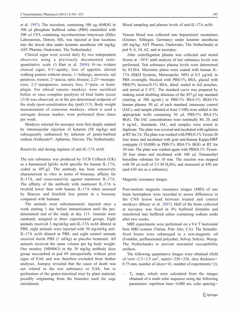
The marmosets included in the study were randomlyassigned to three treatment groups. The control group(n=8) received 1 ml/kg PBS and the two treatment groupsreceived anti-IL-17A mAb at 3 mg/ml/kg (n=8) or 30 mg/ml/kg (n=7). The test substances were subcutaneouslyadministered once per week starting 1 day before immuni-zation. Plasma levels of the anti-IL-17A mAb weredetermined 7 days after each administration (Fig. 1). Asexpected, plasma concentrations of the antibody in the3 mg/kg antibody dose group were about tenfold lowercompared with the 30 mg/kg antibody dose group. In twoanimals (M04099 and M03144) of the 3 mg/kg antibodydose group plasma antibody trough levels, measured 1 weekafter administration, were remarkably low, i.e., below 1 μg/mlat psd 34, while these were above 1 μg/ml in all the othermonkeys of this group. In the 30 mg/kg antibody doseanimals, the plasma levels of the test substance were above10 μg/ml from psd 6 throughout the experiment.
Clinical signs after anti-IL-17A mAb treatment
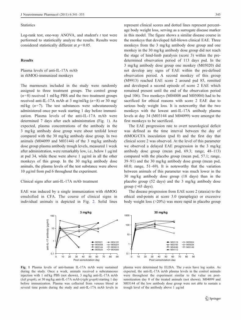
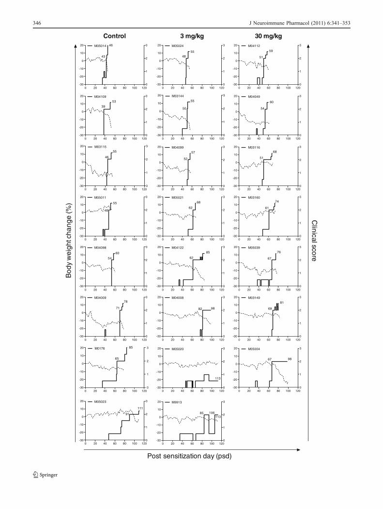
EAE was induced by a single immunization with rhMOGemulsified in CFA. The course of clinical signs inindividual animals is depicted in Fig. 2. Solid lines
represent clinical scores and dotted lines represent percent-age body weight loss, serving as a surrogate disease markerin this model. The figure shows a similar disease course inthe monkeys that developed full-blown clinical EAE. Threemonkeys from the 3 mg/kg antibody dose group and onemonkey in the 30 mg/kg antibody dose group did not reachthe stage of hind-limb paralysis (score 3) within the pre-determined observation period of 113 days psd. In the3 mg/kg antibody dose group one monkey (M05020) didnot develop any signs of EAE within the pre-definedobservation period. A second monkey of this group(M9913) reached EAE score 2 around psd 85, remittedand developed a second episode of score 2 EAE whichremained present until the end of the observation period(psd 106). Two monkeys (M04008 and M05004) had to besacrificed for ethical reasons with score 2 EAE due toserious body weight loss. It is noteworthy that the twomonkeys with the lowest anti-IL-17A antibody plasmalevels at day 34 (M03144 and M04099) were amongst thefirst monkeys to be sacrificed.
The EAE progression rate to overt neurological deficitwas defined as the time interval between the day ofrhMOG/CFA inoculation (psd 0) and the first day thatclinical score 2 was observed. At the level of this parameterwe observed a delayed EAE progression in the 3 mg/kgantibody dose group (mean psd, 69.3; range, 48–113)compared with the placebo group (mean psd, 57.1; range,39–91) and the 30 mg/kg antibody dose group (mean psd,60.0; range, 51–69). It is noteworthy that the variationbetween animals of this parameter was much lower in the30 mg/kg antibody dose group (18 days) than in theplacebo group (52 days) and the 3 mg/kg antibody dosegroup (>65 days).
The disease progression from EAE score 2 (ataxia) to theethical end-points at score 3.0 (paraplegia) or excessivebody weight loss (>20%) was more rapid in placebo group
Fig. 1 Plasma levels of anti-human IL-17A mAb were sustainedduring the study. Once a week, animals received a subcutaneousinjection with 1 ml/kg PBS (not shown), 3 mg/kg anti-IL-17A mAb(left graph), or 30 mg/kg anti-IL-17A mAb (right graph) starting 1 daybefore immunization. Plasma was collected from venous blood atseveral time points during the study and anti-IL-17A mAb levels in
plasma were determined by ELISA. The y-axis have log scales. Asexpected, the anti-IL-17A mAb plasma levels in the control animalswere throughout the experiment similar to the value on post-sensitization day 0 of the treated animals (not shown). M04099 andM03144 of the low antibody dose group were not able to sustain atrough level of the antibody above 1 μg/ml
J Neuroimmune Pharmacol (2011) 6:341–353 345
Post sensitization day (psd)
-30
-20
-10
0
10
20
0
1
2
3M05020
113
-30
-20
-10
0
10
20
0
1
2
3M05004
67 98
-30
-20
-10
0
10
20
0
1
2
3M0176
65
85
0 20 40 60 80 100 120-30
-20
-10
0
10
20
0
1
2
3M9913
85 106113
0 20 40 60 80 100 120
0 20 40 60 80 100 1200 20 40 60 80 100 1200 20 40 60 80 100 120
0 20 40 60 80 100 1200 20 40 60 80 100 1200 20 40 60 80 100 120
0 20 40 60 80 100 1200 20 40 60 80 100 1200 20 40 60 80 100 120
0 20 40 60 80 100 1200 20 40 60 80 100 1200 20 40 60 80 100 120
0 20 40 60 80 100 1200 20 40 60 80 100 1200 20 40 60 80 100 120
0 20 40 60 80 100 1200 20 40 60 80 100 1200 20 40 60 80 100 120
0 20 40 60 80 100 1200 20 40 60 80 100 1200 20 40 60 80 100 120
-30
-20
-10
0
10
20
0
1
2
3M05023
91
111
Control 30 mg/kg
-30
-20
-10
0
10
20
0
1
2
3M05014
43
46
-30
-20
-10
0
10
20
0
1
2
3M04112
51
59
-30
-20
-10
0
10
20
0
1
2
3M05024
4855
-30
-20
-10
0
10
20
0
1
2
3M0410953
39
-30
-20
-10
0
10
20
0
1
2
3M04049
54
60
-30
-20
-10
0
10
20
0
1
2
3M03144
50
55
-30
-20
-10
0
10
20
0
1
2
3M03115
46
55
-30
-20
-10
0
10
20
0
1
2
3M03116
51
68
-30
-20
-10
0
10
20
0
1
2
3M04099
52
57
-30
-20
-10
0
10
20
0
1
2
3M05011
48
55
-30
-20
-10
0
10
20
0
1
2
3M03160
61
74
-30
-20
-10
0
10
20
0
1
2
3M05021
62
68
-30
-20
-10
0
10
20
0
1
2
3M04098
54
60
-30
-20
-10
0
10
20
0
1
2
3M05039
67
76
-30
-20
-10
0
10
20
0
1
2
3M04122
62
85
-30
-20
-10
0
10
20
0
1
2
3M04009
71
78
-30
-20
-10
0
10
20
0
1
2
3M03149
69
81
-30
-20
-10
0
10
20
0
1
2
3M04008
82 98
3 mg/kg
Clinical score
Bod
y w
eigh
t cha
nge
(%)
346 J Neuroimmune Pharmacol (2011) 6:341–353

monkeys (mean, 10.8 days; range, 3–20) than in monkeysfrom the 3 mg/kg antibody (mean, 12.9; range, 5–28) and30 mg/kg antibody (mean, 13.7; range, 6–31) dose group.Statistical evaluation of both disease phases (score, 0–2 andscore, 2 to >2) was done by survival analysis, but nosignificant differences were found (Fig. 3).
In summary, these data show a moderate inhibitory effectof the antibody treatment on the EAE course, althoughdifferences between the groups did not reach statisticalsignificance.
Effect of anti-IL-17A mAb on white matter lesion load
Prior to sectioning for histological examination, post-mortem T2-weighted MR images were made of theformalin-fixed cerebral hemispheres to visualize andquantitate the total lesion load. The analyzed parameterswere lesion volume, T2, and MTR values of white matterlesions. In fixed brains, an increased T2 signal reflectsdemyelination, while a reduction of MTR values correlateswell with the intensity of macrophage infiltration (Blezeret al. 2007).
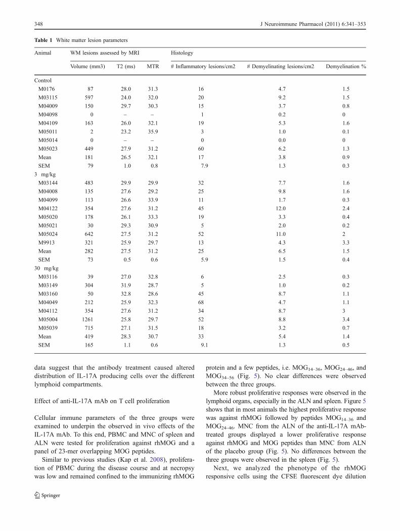
Significant differences were not observed between thethree groups (Table 1). In the analyzed hemispheres of twoanimals of the control group cerebral white matter lesionswere absent. White matter lesion volumes were slightlyincreased in the 30 mg/kg antibody dose group comparedwith the placebo group. Especially monkey M05004 hadclinical score 2 for a long time, which might explain thehigh lesion load. The lesions in the 3 and 30 mg/kg
antibody dose group had a slightly higher T2 and lowerMTR value compared with placebo (Table 1).
These results were confirmed by the quantitativeimmunohistochemistry data (Table 1). Both inflammationand demyelination were moderately increased, although notsignificant, in the two antibody-treated groups comparedwith the placebo group.
For the correct interpretation of these data it is pertinent toemphasize that lesion formation in this model starts wellbefore neurological deficits can be observed. Moreover, mostanimals were killed at the stage of clinically evident EAE,which occurred at different time intervals after immunization.It may therefore not be surprising that major effects of theantibody treatment on the CNS pathology were not observed,neither in positive nor in negative direction.
IL-17A expression in the brain and lymphoid organs
To assess the effect of anti-IL-17A mAb treatment on insitu expression of IL-17A, cryosectioned brains andlymphoid organs were examined with immunohistochemistry.In the brains of control and anti-IL-17A mAb-treated animalsIL-17A-positive cells were found in the perivascular space orin the vicinity of blood vessels (Fig. 4a). No differences wereobserved between placebo and anti-IL-17A mAb-treatedanimals.
IL-17A-producing cells in secondary lymphoid organswere mainly found in the red pulp of the spleen and in themedulla of the lymph nodes. We observed a trend towardshigher numbers of IL-17A producing cells in the spleen ofanimals dosed with 3 mg/kg anti-IL-17AmAb compared withanimals that had received placebo or 30 mg/kg antibody, butdifferences were not significant (Fig. 4a–b). By contrast, wedetected lower numbers of IL-17A producing cells in theALN and ILN, which drain the immunization sites, and thebrain-draining CLN, in both treatment groups comparedwith the placebo group (Fig. 4a–b). Taken together, these
Fig. 2 Clinical score and body weight loss of rhMOG-immunizedmarmosets. Shown are the clinical score (solid line, right y-axis) andthe body weight change in percentages compared with day 0 (dottedline, left y-axis) of the placebo group (left graphs), the low dose group(middle graphs), and the high-dose group (right graphs). Numbers inthe figure represent the time points (psd) when neurological signs(score ≥2) were first observed and the day of sacrifice
�
Fig. 3 Survival curves. Survival time to score 2 (a) and survival timeto day of sacrifice (b) are shown. p Values indicated in the graph arethe results of comparing three groups. Comparing the survival to theday of sacrifice of the control group versus only the 3 mg/kg groupresulted in a p value of 0.1610. When two animals of the 3 mg/kg
group with low anti-IL-17A mAb plasma levels (M04099 andM03144) are included in the control group instead of the 3 mg/kggroup, the p value is 0.0428. According to the Bonferroni correctionthis is not significant, but it is highly suggestive for a delay in diseaseprogression
J Neuroimmune Pharmacol (2011) 6:341–353 347
data suggest that the antibody treatment caused altereddistribution of IL-17A producing cells over the differentlymphoid compartments.
Effect of anti-IL-17A mAb on T cell proliferation
Cellular immune parameters of the three groups wereexamined to underpin the observed in vivo effects of theIL-17A mAb. To this end, PBMC and MNC of spleen andALN were tested for proliferation against rhMOG and apanel of 23-mer overlapping MOG peptides.
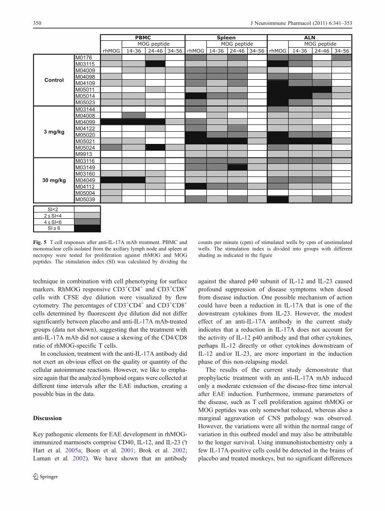
Similar to previous studies (Kap et al. 2008), prolifera-tion of PBMC during the disease course and at necropsywas low and remained confined to the immunizing rhMOG
protein and a few peptides, i.e. MOG14–36, MOG24–46, andMOG34–56 (Fig. 5). No clear differences were observedbetween the three groups.
More robust proliferative responses were observed in thelymphoid organs, especially in the ALN and spleen. Figure 5shows that in most animals the highest proliferative responsewas against rhMOG followed by peptides MOG14–36 andMOG24–46. MNC from the ALN of the anti-IL-17A mAb-treated groups displayed a lower proliferative responseagainst rhMOG and MOG peptides than MNC from ALNof the placebo group (Fig. 5). No differences between thethree groups were observed in the spleen (Fig. 5).
Next, we analyzed the phenotype of the rhMOGresponsive cells using the CFSE fluorescent dye dilution
Table 1 White matter lesion parameters
Animal WM lesions assessed by MRI Histology
Volume (mm3) T2 (ms) MTR # Inflammatory lesions/cm2 # Demyelinating lesions/cm2 Demyelination %
Control
M0176 87 28.0 31.3 16 4.7 1.5
M03115 597 24.0 32.0 20 9.2 1.5
M04009 150 29.7 30.3 15 3.7 0.8
M04098 0 – – 1 0.2 0
M04109 163 26.0 32.1 19 5.3 1.6
M05011 2 23.2 35.9 3 1.0 0.1
M05014 0 – – 0 0.0 0
M05023 449 27.9 31.2 60 6.2 1.3
Mean 181 26.5 32.1 17 3.8 0.9
SEM 79 1.0 0.8 7.9 1.3 0.3
3 mg/kg
M03144 483 29.9 29.9 32 7.7 1.6
M04008 135 27.6 29.2 25 9.8 1.6
M04099 113 26.6 33.9 11 1.7 0.3
M04122 354 27.6 31.2 45 12.0 2.4
M05020 178 26.1 33.3 19 3.3 0.4
M05021 30 29.3 30.9 5 2.0 0.2
M05024 642 27.5 31.2 52 11.0 2
M9913 321 25.9 29.7 13 4.3 3.3
Mean 282 27.5 31.2 25 6.5 1.5
SEM 73 0.5 0.6 5.9 1.5 0.4
30 mg/kg
M03116 39 27.0 32.8 6 2.5 0.3
M03149 304 31.9 28.7 5 1.0 0.2
M03160 50 32.8 28.6 45 8.7 1.1
M04049 212 25.9 32.3 68 4.7 1.1
M04112 354 27.6 31.2 34 8.7 3
M05004 1261 25.8 29.7 52 8.8 3.4
M05039 715 27.1 31.5 18 3.2 0.7
Mean 419 28.3 30.7 33 5.4 1.4
SEM 165 1.1 0.6 9.1 1.3 0.5
348 J Neuroimmune Pharmacol (2011) 6:341–353

Fig. 4 Differential expressionof IL-17A in the brain andlymphoid organs. IL-17Aexpression was detected byimmunohistochemistry. a (200×)Representative examples of eachgroup for brain, spleen, andaxillary (ALN), inguinal (ILN),and cervical (CLN) lymphnodes. b IL-17A positive cellsper mm2 (mean ± SEM). Onlyone CLN of the control groupwas analyzed. No IL-17Aproducing cells were detected inthe ILN of the 30 mg/kg treatedgroup. Statistical differencesbetween the numbers of IL-17Apositive cells were analyzed byOne-way ANOVA. Results wereconsidered statistically differentat p<0.05. No statisticaldifferences were found betweenthe three groups
J Neuroimmune Pharmacol (2011) 6:341–353 349
technique in combination with cell phenotyping for surfacemarkers. RhMOG responsive CD3+CD4+ and CD3+CD8+
cells with CFSE dye dilution were visualized by flowcytometry. The percentages of CD3+CD4+ and CD3+CD8+
cells determined by fluorescent dye dilution did not differsignificantly between placebo and anti-IL-17A mAb-treatedgroups (data not shown), suggesting that the treatment withanti-IL-17A mAb did not cause a skewing of the CD4/CD8ratio of rhMOG-specific T cells.
In conclusion, treatment with the anti-IL-17A antibody didnot exert an obvious effect on the quality or quantity of thecellular autoimmune reactions. However, we like to empha-size again that the analyzed lymphoid organs were collected atdifferent time intervals after the EAE induction, creating apossible bias in the data.
Discussion
Key pathogenic elements for EAE development in rhMOG-immunized marmosets comprise CD40, IL-12, and IL-23 ('tHart et al. 2005a; Boon et al. 2001; Brok et al. 2002;Laman et al. 2002). We have shown that an antibody
against the shared p40 subunit of IL-12 and IL-23 causedprofound suppression of disease symptoms when dosedfrom disease induction. One possible mechanism of actioncould have been a reduction in IL-17A that is one of thedownstream cytokines from IL-23. However, the modesteffect of an anti-IL-17A antibody in the current studyindicates that a reduction in IL-17A does not account forthe activity of IL-12 p40 antibody and that other cytokines,perhaps IL-12 directly or other cytokines downstream ofIL-12 and/or IL-23, are more important in the inductionphase of this non-relapsing model.
The results of the current study demonstrate thatprophylactic treatment with an anti-IL-17A mAb inducedonly a moderate extension of the disease-free time intervalafter EAE induction. Furthermore, immune parameters ofthe disease, such as T cell proliferation against rhMOG orMOG peptides was only somewhat reduced, whereas also amarginal aggravation of CNS pathology was observed.However, the variations were all within the normal range ofvariation in this outbred model and may also be attributableto the longer survival. Using immunohistochemistry only afew IL-17A-positive cells could be detected in the brains ofplacebo and treated monkeys, but no significant differences
Fig. 5 T cell responses after anti-IL-17A mAb treatment. PBMC andmononuclear cells isolated from the axillary lymph node and spleen atnecropsy were tested for proliferation against rhMOG and MOGpeptides. The stimulation index (SI) was calculated by dividing the
counts per minute (cpm) of stimulated wells by cpm of unstimulatedwells. The stimulation index is divided into groups with differentshading as indicated in the figure
350 J Neuroimmune Pharmacol (2011) 6:341–353

between groups were observed. The data show a trendtowards increased numbers of IL-17A-positive cells in thespleen in the 3 mg/kg group and a decreased number in theaxillary and inguinal lymph nodes of both groups comparedwith placebo animals (not significant). The higher numberof Th17 cells in the spleen of monkeys that have beentreated with anti-IL-17A antibody compared with placebomonkeys may be explained by feedback regulation. Theobservation that IL-17A receptor deficient mice producemore IL-17A (Smith et al. 2008) indicates that IL-17Aproduction may be regulated by a negative feedback loop.In conclusion, the current results fail to show a robust effectof the anti-IL-17A mAb treatment on the EAE course inrhMOG/CFA-immunized marmosets.
The moderate protection against EAE induction is inagreement with data reported by some groups in mouseEAE models. Also in IL-17A−/− mice (Komiyama et al.2006), in mice treated with anti-IL-17A mAb (Chen et al.2006; Hofstetter et al. 2005; Langrish et al. 2005), and inmice vaccinated against IL-17A (Rohn et al. 2006) onlymoderately delayed onset of clinical signs has beenobserved. However, other groups have demonstrated morerobust effects of IL-17A on the EAE course in mice.Examples are by vaccination with IL-17A-OVA complexesthat induce IL-17A blocking antibodies (Uyttenhove andVan Snick 2006) or adoptive transfer of IL-17+ and IL-17−
T cells (Jager et al. 2009; Komiyama et al. 2006; Langrishet al. 2005). Furthermore, in the chronic-relapsing EAEmodel in Biozzi ABH mice, prophylactic dosing with ananti-IL-17A antibody produced a small delay in onset of theacute phase of disease, whereas treatment with the antibodystarted after the acute phase or during the secondary-progressive phase was very efficacious, and could evenreverse the chronic motor disability (Smith, unpublishedobservations). Other groups suggested that Th17 are notpathogenic at all (Haak et al. 2009) or that besides Th17cells, Th1 cells are also essential in EAE (Kroenke et al.2008; Lees et al. 2008; O'Connor et al. 2008).
How can it be explained that IL-17A neutralization doesnot fully protect against EAE in the rhMOG-inducedmarmoset model? One possible explanation is the highcomplexity of the model. Unraveling of the pathogenicmechanism showed that the disease is initiated by MOG24–
36-specific Th1 cells, which induce small inflammatorylesions (Brok et al. 2000). The moderate inflammatoryinjury caused by Th1 cells can be dramatically amplified byantibodies against conformational epitopes of MOG (Gen-ain et al. 1995), which are clearly produced in the rhMOGmodel. Another subset of highly reactive T cells withnatural killer-cytotoxic T-lymphocyte characteristics andspecific for MOG34–56 becomes involved at a later stage inthe disease (Kap et al. 2008). Our previously reportedstudies in marmoset EAE models induced with MOG34–56
in CFA (Kap et al. 2008) or IFA (Jagessar et al. 2010)demonstrated that the same clinical and pathological end-points can be reached via different immunopathogenicroutes. It can thus be envisaged that after neutralization ofIL-17A in the complex rhMOG/CFA-induced model otherpathogenic mechanisms, such as Th1 cells, may come intoplay. It could also be possible that Th1 and Th17 cells act indifferent phases of the disease. In the previously reportedstudy in the rhMOG marmoset model it was observed thatTh1 cells specific for MOG24–36 are engaged in EAEinduction, whereas MOG34–56-specific T cells, which pro-duce mainly IL-17A, mediate disease progression (Jagessaret al. 2010; Kap et al. 2008). This suggests that, consistentwith the results in the Biozzi ABH mouse model, neutral-izing IL-17A in a later phase of the disease is more effective.
Yet, another possible explanation is that IL-17A,although being the signature cytokine of Th17 cells, is notthe dominant pathogenic factor produced by Th17 cells.This explanation is supported by data in mice. IL-12/IL-23p40−/− mice are completely resistant to EAE, whereasonly clinical scores and not the disease incidence is reducedin IL-17−/− mice (Becher et al. 2002; Cua et al. 2003; Granet al. 2002; Komiyama et al. 2006). Moreover, treatment ofmice with anti-IL-23 antibody prevented either EAEinduction or relapse, in contrast to the anti-IL-17A antibodytreatment that blocked relapse, but had a smaller effect onthe induction phase (Chen et al. 2006). This may suggestthat other Th17 cytokines induced by IL-23 may bepathogenetically more relevant than IL-17A in the induc-tion phase, such as IL-21 or IL-22 (reviewed in (Fouser etal. 2008)). In addition, Chen et al. suggested that IL-23 mayalso directly activate macrophages to produce inflammatorycytokines, such as IL-6, IL-1, and TNF-α, which may causeinflammation and demyelination even when IL-17A isneutralized (Chen et al. 2006).
Is there a place for an IL-17A neutralizing antibody inthe treatment of MS? Axtell et al. reported that on thebasis of clinical response to IFN-β two groups may bedistinguished in the MS patient population. Responsive-ness to IFN-β treatment requires a Th1 cytokine profile,whereas cases with a Th17 dominated cytokine profileare non-responders to IFN-β treatment (Axtell et al.2010). It is tempting to speculate on the basis of these andour data that non-responder cases to IFN-β treatment inthe MS population may benefit from treatment with IL-17A antibody. The ideal study design to obtain proof-of-principle may be a parallel effectivity analysis in themarmoset EAE model, where in one sibling of a chimerictwin EAE is induced with MOG34–56 in CFA, which is aTh1 prone model (Jagessar et al. 2010; Kap et al. 2008)and in the other twin sibling with MOG34–56 in IFA, whichis a more Th17 prone disease (Jagessar et al. 2010; Kapet al. 2008).
J Neuroimmune Pharmacol (2011) 6:341–353 351
In summary, we found that treatment with anti-IL-17Aantibody induces a moderate delay of clinical EAE inmarmosets, but that EAE is not completely abrogated. Thissuggests a pathogenic role for IL-17A in the marmoset EAEmodel and maybe in MS, but IL-17A may not be the onlykey pathogenic cytokine.
Acknowledgements The authors like to thank Fred Batenburg forexcellent biotechnical assistance and daily care of the monkeys, JacoBakker DVM, Gerco Braskamp DVM and Merei Keehnen DVM forexpert veterinary care, Tom Haaksma and Dr. Ivanela Kondovafor autopsy of the monkeys. The authors thank Henk van Westbroekfor the artwork. The anti-IL-17A antibody was provided by UCBCelltech (UK). The study was financially supported by UCB Celltech.
Conflicts of Interest The authors of this manuscript do not reportconflict of interest.
Open Access This article is distributed under the terms of theCreative Commons Attribution Noncommercial License which per-mits any noncommercial use, distribution, and reproduction in anymedium, provided the original author(s) and source are credited.
References
Axtell RC, de Jong BA, Boniface K, van der Voort LF, Bhat R, DeSarno P, Naves R, Han M, Zhong F, Castellanos JG, Mair R,Christakos A, Kolkowitz I, Katz L, Killestein J, Polman CH, deWaal MR, Steinman L, Raman C (2010) T helper type 1 and 17cells determine efficacy of interferon-beta in multiple sclerosisand experimental encephalomyelitis. Nat Med 16:406–412
Becher B, Durell BG, Noelle RJ (2002) Experimental autoimmuneencephalitis and inflammation in the absence of interleukin-12. JClin Invest 110:493–497
Billiau A, Matthys P (2001) Modes of action of Freund's adjuvants inexperimental models of autoimmune diseases. J Leukoc Biol70:849–860
Blezer EL, Bauer J, Brok HP, Nicolay K, 't Hart BA (2007)Quantitative MRI-pathology correlations of brain white matterlesions developing in a non-human primate model of multiplesclerosis. NMR Biomed 20:90–103
Boon L, Brok HP, Bauer J, Ortiz-Buijsse A, Schellekens MM, Ramdien-Murli S, Blezer E, van Meurs M, Ceuppens J, de Boer M, 't HartBA, Laman JD (2001) Prevention of experimental autoimmuneencephalomyelitis in the common marmoset (Callithrix jacchus)using a chimeric antagonist monoclonal antibody against humanCD40 is associated with altered B cell responses. J Immunol167:2942–2949
Brok HP, Uccelli A, Kerlero De Rosbo N, Bontrop RE, RoccatagliataL, de Groot NG, Capello E, Laman JD, Nicolay K, Mancardi GL,Ben-Nun A, 't Hart BA (2000) Myelin/oligodendrocyteglycoprotein-induced autoimmune encephalomyelitis in commonmarmosets: the encephalitogenic T cell epitope pMOG24–36 ispresented by a monomorphic MHC class II molecule. J Immunol165:1093–1101
Brok HP, van Meurs M, Blezer E, Schantz A, Peritt D, Treacy G,Laman JD, Bauer J, 't Hart BA (2002) Prevention of experimen-tal autoimmune encephalomyelitis in common marmosets usingan anti-IL-12p40 monoclonal antibody. J Immunol 169:6554–6563
Chen Y, Langrish CL, McKenzie B, Joyce-Shaikh B, Stumhofer JS,McClanahan T, Blumenschein W, Churakovsa T, Low J, PrestaL, Hunter CA, Kastelein RA, Cua DJ (2006) Anti-IL-23 therapyinhibits multiple inflammatory pathways and ameliorates auto-immune encephalomyelitis. J Clin Invest 116:1317–1326
Compston A, Coles A (2008) Multiple sclerosis. Lancet 372:1502–1517
Cua DJ, Sherlock J, Chen Y, Murphy CA, Joyce B, Seymour B,Lucian L, To W, Kwan S, Churakova T, Zurawski S,Wiekowski M, Lira SA, Gorman D, Kastelein RA, SedgwickJD (2003) Interleukin-23 rather than interleukin-12 is thecritical cytokine for autoimmune inflammation of the brain.Nature 421:744–748
Fouser LA, Wright JF, Dunussi-Joannopoulos K, Collins M (2008)Th17 cytokines and their emerging roles in inflammation andautoimmunity. Immunol Rev 226:87–102
Genain CP, Nguyen MH, Letvin NL, Pearl R, Davis RL, Adelman M,Lees MB, Linington C, Hauser SL (1995) Antibody facilitationof multiple sclerosis-like lesions in a nonhuman primate. J ClinInvest 96:2966–2974
Gran B, Zhang GX, Yu S, Li J, Chen XH, Ventura ES, Kamoun M,Rostami A (2002) IL-12p35-deficient mice are susceptible toexperimental autoimmune encephalomyelitis: evidence for redun-dancy in the IL-12 system in the induction of central nervous systemautoimmune demyelination. J Immunol 169:7104–7110
Haak S, Croxford AL, Kreymborg K, Heppner FL, Pouly S, Becher B,Waisman A (2009) IL-17A and IL-17F do not contribute vitally toautoimmune neuro-inflammation in mice. J Clin Invest 119:61–69
Hofstetter HH, Ibrahim SM, Koczan D, Kruse N, Weishaupt A, ToykaKV, Gold R (2005) Therapeutic efficacy of IL-17 neutralizationin murine experimental autoimmune encephalomyelitis. CellImmunol 237:123–130
Jager A, Dardalhon V, Sobel RA, Bettelli E, Kuchroo VK (2009) Th1,Th17, and Th9 effector cells induce experimental autoimmuneencephalomyelitis with different pathological phenotypes. JImmunol 183:7169–7177
Jagessar SA, Kap YS, Heijmans N, van Driel N, van Straalen L,Bajramovic JJ, Brok HPM, Blezer ELA, Bauer J, Laman JD, HartBA (2010) Induction of progressive demyelinating autoimmuneencephalomyelitis in common marmoset monkeys using MOG34-56 peptide in incomplete Freund adjuvant. J Neuropathol ExpNeurol 69:372–385
Kap YS, Smith P, Jagessar SA, Remarque E, Blezer E, Strijkers GJ,Laman JD, Hintzen RQ, Bauer J, Brok HP, 't Hart BA (2008) Fastprogression of recombinant human myelin/oligodendrocyte glyco-protein (MOG)-induced experimental autoimmune encephalomy-elitis in marmosets is associated with the activation of MOG34–56-specific cytotoxic T cells. J Immunol 180:1326–1337
Kap YS, Laman JD, 't Hart BA (2010) Experimental AutoimmuneEncephalomyelitis in the Common Marmoset, a Bridge BetweenRodent EAE and Multiple Sclerosis for Immunotherapy Develop-ment. J Neuroimmune Pharmacol 5:220–230
Kerlero de Rosbo N, Hoffman M, Mendel I, Yust I, Kaye J,Bakimer R, Flechter S, Abramsky O, Milo R, Karni A, Ben-Nun A(1997) Predominance of the autoimmune response to myelinoligodendrocyte glycoprotein (MOG) in multiple sclerosis: reactivityto the extracellular domain of MOG is directed against three mainregions. Eur J Immunol 27:3059–3069
Komiyama Y, Nakae S, Matsuki T, Nambu A, Ishigame H, Kakuta S,Sudo K, Iwakura Y (2006) IL-17 plays an important role in thedevelopment of experimental autoimmune encephalomyelitis. JImmunol 177:566–573
Kroenke MA, Carlson TJ, Andjelkovic AV, Segal BM (2008) IL-12- and IL-23-modulated T cells induce distinct types of EAEbased on histology, CNS chemokine profile, and response tocytokine inhibition. J Exp Med 205:1535–1541
352 J Neuroimmune Pharmacol (2011) 6:341–353
Laman JD, van Meurs M, Schellekens MM, de Boer M, Melchers B,Massacesi L, Lassmann H, Claassen E, 't Hart BA (1998) Expressionof accessory molecules and cytokines in acute EAE in marmosetmonkeys (Callithrix jacchus). J Neuroimmunol 86:30–45
Laman JD, 't Hart BA, Brok H, Meurs M, Schellekens MM, Kasran A,Boon L, Bauer J, Boer M, Ceuppens J (2002) Protection ofmarmoset monkeys against EAE by treatment with a murineantibody blocking CD40 (mu5D12). Eur J Immunol 32:2218–2228
Langrish CL, Chen Y, Blumenschein WM, Mattson J, Basham B,Sedgwick JD, McClanahan T, Kastelein RA, Cua DJ (2005) IL-23 drives a pathogenic T cell population that induces autoim-mune inflammation. J Exp Med 201:233–240
Lees JR, Iwakura Y, Russell JH (2008) Host T cells are the mainproducers of IL-17 within the central nervous system duringinitiation of experimental autoimmune encephalomyelitis inducedby adoptive transfer of Th1 cell lines. J Immunol 180:8066–8072
O'Connor RA, Prendergast CT, Sabatos CA, Lau CW, Leech MD,Wraith DC, Anderton SM (2008) Cutting edge: Th1 cells facilitate theentry of Th17 cells to the central nervous system during experimentalautoimmune encephalomyelitis. J Immunol 181:3750–3754
Rohn TA, Jennings GT, Hernandez M, Grest P, Beck M, Zou Y, Kopf M,Bachmann MF (2006) Vaccination against IL-17 suppresses autoim-mune arthritis and encephalomyelitis. Eur J Immunol 36:2857–2867
Smith E, Stark MA, Zarbock A, Burcin TL, Bruce AC, Vaswani D,Foley P, Ley K (2008) IL-17A inhibits the expansion of IL-17A-producing T cells in mice through “short-loop” inhibition via IL-17 receptor. J Immunol 181:1357–1364
Sospedra M, Martin R (2005) Immunology of multiple sclerosis.Annu Rev Immunol 23:683–747
't Hart BA, Massacesi L (2009) Clinical, pathological, and immunologicaspects of the multiple sclerosis model in common marmosets(Callithrix jacchus). J Neuropathol Exp Neurol 68:341–355
't Hart BA, Bauer J, Muller HJ, Melchers B, Nicolay K, Brok H,Bontrop RE, Lassmann H, Massacesi L (1998) Histopathologicalcharacterization of magnetic resonance imaging-detectable brainwhite matter lesions in a primate model of multiple sclerosis: acorrelative study in the experimental autoimmune encephalomy-elitis model in common marmosets (Callithrix jacchus). Am JPathol 153:649–663
't Hart BA, Laman JD, Bauer J, Blezer E, van Kooyk Y, Hintzen RQ(2004) Modelling of multiple sclerosis: lessons learned in a non-human primate. Lancet Neurol 3:588–597
't Hart BA, Blezer EL, Brok HP, Boon L, de Boer M, Bauer J, LamanJD (2005a) Treatment with chimeric anti-human CD40 antibodysuppresses MRI-detectable inflammation and enlargement of pre-existing brain lesions in common marmosets affected by MOG-induced EAE. J Neuroimmunol 163:31–39
't Hart BA, Brok HP, Remarque E, Benson J, Treacy G, Amor S, HintzenRQ, Laman JD, Bauer J, Blezer EL (2005b) Suppression of ongoingdisease in a nonhuman primate model of multiple sclerosis by ahuman-anti-human IL-12p40 antibody. J Immunol 175:4761–4768
't Hart BA, Hintzen RQ, Laman JD (2008) Preclinical assessment oftherapeutic antibodies against human CD40 and humaninterleukin-12/23p40 in a nonhuman primate model of multiplesclerosis. Neurodegener Dis 5:38–52
Uyttenhove C, Van Snick J (2006) Development of an anti-IL-17Aauto-vaccine that prevents experimental auto-immune encephalo-myelitis. Eur J Immunol 36:2868–2874
J Neuroimmune Pharmacol (2011) 6:341–353 353