Embed Size (px)
Citation preview
www.elsevier.com/locate/pharmbiochembeh
Pharmacology, Biochemistry and
Effects of chlordiazepoxide on single-unit activity in the septal region of
the freely moving rat: aversive vs. non-aversive contexts
Earl Thomas*, Craig E. Strickland, Elna Yadin, Debra A. Burock
Department of Psychology, Bryn Mawr College, 101 N. Merion Avenue, Bryn Mawr, PA 19010, United States
Received 10 March 2004; received in revised form 22 October 2004; accepted 29 October 2004
Available online 25 November 2004
Abstract
Evidence suggests that stimuli that have the property of inhibiting fear in a Pavlovian fear conditioning paradigm increase cellular activity
in the lateral septum, a result consistent with the idea that the lateral septum is actively involved in the inhibition of fear. The experiments
reported here were designed to determine if an anxiolytic drug with fear-inhibiting properties would also increase neuronal activity in the
lateral septum in a manner that might relate to its mechanism of action as an anxiolytic. An experiment was performed to compare the effects
of the benzodiazepine anxiolytic chlordiazepoxide (CDP) upon single-unit activity in the septal region of the rat brain during Pavlovian
aversive conditioning with the effects of CDP in a non-aversive context. During Pavlovian conditioning there was a decrease in unit activity
in the more lateral regions of the septum, the dorsolateral and ventrolateral nuclei, when a stimulus signaling footshock (CS+) was presented.
This conditioned suppression of unit activity was blocked by an intraperitoneal injection of CDP. Additionally, CDP increased baseline unit
activity in these regions in the absence of conditioned stimuli. In the more medial regions of the septum, the intermediate lateral septum, we
observed few consistent changes either to the conditioned stimuli or to the drug. In a non-aversive context CDP had either no effect at low to
moderate doses, or a suppressant effect at a higher dose. The results support a fear-relief hypothesis of lateral septal functioning and suggest
the lateral septum as a possible site for the anxiolytic action of benzodiazepines.
D 2004 Elsevier Inc. All rights reserved.
Keywords: Single unit; Benzodiazepine; Chlordiazepoxide; Pavlovian conditioning; Fear; Septum
1. Introduction
There is accumulating evidence that the lateral septum
plays an important role in the inhibition of fear and anxiety.
For example, in common laboratory tests of anxiety such as
the Vogel (Vogel et al., 1971) water conflict test, electrical
stimulation of the lateral septum has the same anti-conflict
action as the anxiolytic benzodiazepines (BZDs) (Yadin et
al., 1993), suggesting an anxiolytic action of septal
stimulation. Electrical stimulation of the lateral septum also
has a suppressing effect on species-specific defense
responses, indicative of fear, induced in rats by lesions of
the ventromedial nucleus (Brayley and Albert, 1977).
Lesions of the lateral septum, on the other hand, appear to
be pro-conflict in the conflict test, suggesting an anxiogenic
0091-3057/$ - see front matter D 2004 Elsevier Inc. All rights reserved.
doi:10.1016/j.pbb.2004.10.018
* Corresponding author. Tel.: +1 610 526 5013; fax: +1 610 526 7476.
E-mail address: [email protected] (E. Thomas).
effect of the lesion (Yadin et al., 1993). Consistent with an
anxiogenic effect of lateral septal lesions is the finding that
such lesions facilitate contextual fear conditioning in a
Pavlovian model of fear conditioning (Sparks and LeDoux,
1995).
Single- and multiple-unit recording experiments in the
septum also support a role for the septum in the modulation
of fear conditioning (Thomas and Yadin, 1980; Thomas et
al., 1991; Yadin and Thomas, 1981). The recording data are
consistent with a fear-relief role for the lateral septum and a
possible fear excitatory function for the medial septal
regions. When animals were tested in a Pavlovian differ-
ential conditioning paradigm with an aversive US, cells in
the lateral septum increased their rates of firing in the
presence of stimuli that signaled relief or safety and
inhibited their rates of firing in the presence of stimuli that
signaled fear. Most medial cells in this study were not
sensitive to the conditioning contingencies. This accords
Behavior 80 (2005) 151–159
E. Thomas et al. / Pharmacology, Biochemistry and Behavior 80 (2005) 151–159152
with data from other experiments which found virtually no
associative conditioning in medial septal cells in appetitive
conditioning in rats reinforced with a food US (Segal, 1973)
or conditioning of the nictitating membrane response in the
rabbit with an air-puff US (Berger and Thompson, 1978).
However, in contrast to the data from other laboratories
we did find that there are at least a proportion of medial
septal cells that do show evidence of associative condition-
ing. In such cells, the preponderant response appeared to be
the reverse of what we observed in the lateral septum. These
medial septal cells were activated by a conditioned exciter
of fear. It is possible therefore that there is some contribution
by the medial septum to fear conditioning and the
expression of fear.
Since cells in the septum respond in a consistent manner
to stimuli associated with fear and fear-relief, it would be of
interest to determine if the same cells respond appropriately
to pharmacological agents which affect fear. Specifically,
since lateral septal cells are activated by stimuli which have
fear- relief properties, it seems reasonable that anxiolytic
agents such as the BZDs might have a similar effect on cells
in the lateral septum. The lateral septum contains moder-
ately high densities of GABA-linked benzodiazepine
receptors (Speth et al., 1980). These receptors may be of
importance in mediating the behavioral effects of BZDs
since both the GABA agonist muscimol and the BZD
anxiolytics midazolam and chlordiazepoxide have anxio-
lytic effects when administered directly into the septum
(Drugan et al., 1986; Grishkat, 1991).
The behavioral effects of BZDs are substantially different
in the aversive context compared to the non-aversive
context. So, for instance, moderate doses of BZDs increase
behavior suppressed by conditioned fear but tend not to
affect behavior that hasn’t been suppressed (Cook and
Davidson, 1978). Accordingly, one of the key goals of the
present study was determine if the effects of BZDs on septal
unit activity in the two contexts parallel their effects on
behavior in a manner that might account for the behavioral
effects of the drug. For the aversive context we chose a
Pavlovian fear conditioning paradigm where it is possible to
assess the effect of BZDs both upon baseline spontaneous
unit activity as well as the effect on conditioned changes in
unit activity. For the non-aversive context animals were
placed in the conditioning chamber but no conditioned or
unconditioned stimuli were presented.
2. Methods
2.1. Subjects
Subjects were male Sprague–Dawley rats. Animals were
between 90 and 110 days old and weighed approximately
350–400 g at the time of surgery. Animals were individually
housed in a light and temperature controlled animal colony.
The animals were maintained on a 12-h dark and 12-h light
cycle and were provided with ad lib food and water
throughout the experiment. Care and use of animals were
approved by the Institutional Animal Care and Use
Committee and experiments were carried out in accordance
with the National Institutes of Health Guide for the Care
and Use of Laboratory Animals.
2.2. Apparatus
The recording chamber was constructed of clear Plex-
iglas with a metal grid floor. The chamber measured
25�22�34 cm and was located inside an electrically
shielded, wooden, sound-attenuating cubicle. The cubicle
contained a 7.5-W houselight, a 15-W light used as a visual
conditioned stimulus (CS), and a loudspeaker, all of which
were located on the cubicle wall opposite the uncovered
Plexiglas wall. The house light remained on at all times.
White noise (70 db) was presented continuously except
during the auditory CS which was an 800 Hz, 70 db tone.
The unconditioned stimulus (US) was a 1-s footshock
delivered through the grid floor. It consisted of a 100 pulse/s
square wave of 1 ms duration. The US was delivered by
Grass stimulator (model S44) connected in series with a
constant current unit (Grass model CCU1).
Recording electrodes consisted of a bundle of eight
nichrome wires each 50 microns in diameter and Teflon
insulated to the tip. These wires were soldered to female
Amphenol pins which were inserted into a plug on the
animal’s head. The wires were twisted together and cut on a
458 angle before being implanted. This configuration yields
a distance of approximately 50 Am between the shortest and
longest electrode tips and simulates a microdrive allowing
units to be sampled at slightly different dorso-ventral
coordinates.
2.3. Surgery
The animals were anesthetized with sodium pentobarbital
(42 mg/kg, i.p.). All electrodes were chronically implanted
using standard stereotaxic procedures. Coordinates for the
lateral septum were taken from Paxinos and Watson (1998)
and were as follows: 0.6 mm anterior to bregma, 0.4 mm
lateral to the midline, and 6.0 mm ventral to the skull
surface. The plug included a ground wire which was
wrapped around one of four stainless steel screws inserted
in the skull. The screws and the plug were secured to the
skull surface using dental cement.
2.4. Recording procedure
Single-unit activity was recorded using a high input
impedance amplification system. Field-effect transistors
were cemented to the plug on the animal’s head. The
headstage amplifier was connected in a voltage follower
configuration and recording was done differentially through
electrode pairs in the site. This configuration minimizes
E. Thomas et al. / Pharmacology, Biochemistry and Behavior 80 (2005) 151–159 153
undifferentiated multiple unit activity virtually eliminates
movement artifact as well as EMG artifact. The signal was
passed through a bandpass filter (500–3000 Hz) and a high
gain amplifier (A–M instruments model 1800). The final
gain on the amplifiers was 10,000. The signal was
monitored on a digital oscilloscope and computer screen.
Recording and control of the conditioning experiment was
accomplished by two computers in communication with
each other. One computer served to discriminate and isolate
units according to criteria described below. The signal from
the amplifier was digitized and analyzed by a commercial
software package which isolated the units being studied
(Datawave Technologies). When a unit which satisfied the
criteria was detected by the first computer a TTL pulse was
delivered to a second computer which stored the data,
controlled all the environmental events, and displayed an
on-line, real-time histogram of unit activity during each
conditioning trial. The TTL pulse also triggered the
oscilloscope so that the unit waveform could be monitored,
thus allowing for further monitoring of unit specificity.
2.5. Data analysis
Unit isolation was performed using cluster analysis
provided by the software mentioned above. Single units
were determined by applying a number of acceptance
criteria which correspond to various parameters of the
waveform. These criteria include spike width and height,
peak magnitude, peak and valley time. In addition the unit
with the identified rate and form had to be observable in a
single electrode in the bundle. All acceptable units had to
have a signal to noise ratio of at least 3:1. The application of
these criteria as well as constant on line visual monitoring of
the waveform gave reasonable assurance that single units
were indeed isolated. Each of the eight electrodes in the
bundle was sampled to determine if an acceptable unit could
be obtained from it. Since conditioning was conducted over
several sessions the acceptance parameters for unit identi-
fication were saved from session to session and applied on
successive sessions. Waveforms on each day were compared
in order to determine with reasonable certainty that the same
unit was being sampled. If the unit was lost then the
electrodes were sampled again, a search was made for new
units and new acceptance parameters were defined. Units
were often kept over several sessions.
2.6. Procedure
Separate groups of animal were tested in a Pavlovian
aversive conditioning paradigm and in a non-aversive
context.
2.6.1. Pavlovian conditioning
Twenty animals were run in this condition. Those
animals with acceptable unit activity were given the
following conditioning sequence. On the first day they
were given a habituation session. In this session animals
were presented with 40 trials in which the 10-s CSs were
presented without US presentations. CS+ and CS� trials
were randomly ordered according to a modified Gellermann
(Gellermann, 1933) sequence in which no CS of a given
type was presented on more than three consecutive trials.
Intertrial intervals were randomly determined with a mean
of 70 s. Subsequently, the animals received 12 sessions of
Pavlovian differential conditioning. On these sessions one
CS (CS+) was paired with US. For these trials the CS
duration was 11 s with a 10-s CS–US interval. The CS and
US overlapped for 1 s and coterminated. The other CS
(CS�) was presented in the absence of the US. Animals
received one session per day and each session consisted of
40 trials, 20 CS+ and 20 CS� trials. Prior to the first
conditioning session the shock intensity was adjusted for
each animal so that it produced a flinch with minimal
vocalization. This shock intensity was used throughout the
remainder of the conditioning sessions. The magnitude of
the footshock for all animals ranged from 0.6 to 1.0 ma. For
half of the animals the tone served as CS+ and the light as
CS�. For the other half the light served as CS+ and the tone
as CS�. On each trial unit activity was recorded during a 10
s pre-CS period, during the 10 s CS–US interval and for 10 s
following the termination of the US. Unit activity was
evaluated separately for CS+ and CS� trials.
Following the last day of conditioning the animals were
tested for the effects of (CDP) on baseline and conditioned
unit responding. Testing was conducted in two 40-trial
conditioning sessions carried out in the same day. In the first
session the animal was injected with the vehicle (saline, 1
ml/kg) 15 min prior to the start of the session. Immediately
after the end of the vehicle session animals were injected
with CDP and 15 min later were given a second 40 trial
conditioning session. A baseline session in which vehicle
was injected prior to testing was run 48 h after the drug
session to ensure that unit activity had returned to baseline.
This sequence was run three times for three different doses
of drug. The order of the doses of CDP administered was
10, 5, and 20 mg/kg i.p. At least 1 week was allowed to
elapse between each dose in order to ensure clearance of the
drug and its active metabolites.
2.6.2. Non-aversive context
The subjects for this condition were 14 male Sprague–
Dawley rats. Animals were tested on three doses of CDP.
Based upon a relatively flat dose–response curve for the
doses selected for the aversive context, it was decided, for
this group, to move the assessment of the dose–response
function toward a lower dose. Therefore the three doses
selected for this condition were 2, 5 and 10 mg/kg. For each
dose animals were habituated in the recording chamber for
30 min. Baseline unit activity was then recorded for 15 min.
Subsequently the animal was removed for the chamber and
administered saline (1 ml/kg i.p.) and placed back in the
chamber. Fifteen minutes after the saline injection, unit
E. Thomas et al. / Pharmacology, Biochemistry and Behavior 80 (2005) 151–159154
activity was recorded for another 15 min. Finally, the animal
was again removed from the chamber and injected with
(CDP) in concentrations of either 2, 5 or 10 mg/kg. The
animal was then replaced in the chamber and 15 min
following CDP administration an additional 15 min of unit
activity was recorded. Each animal received each dose of
CDP, one per session. At least 1 week was allowed to elapse
between doses. The order of doses for each animal was
randomized.
As in experiment 1 units had a variable life span and did
not necessarily last for the entire dosage regimen. If a unit
was lost a search was made for another unit. If none was
found the animal was terminated. For several of the animals
the same unit was maintained for the entire dosage regimen.
2.7. Histology
Following the last recording session the animals were
overdosed with pentobarbital and the brains were perfused
with formalin. Frozen sections were taken at 40 Am.
Electrode placements were verified by a photographic
procedure adapted from Guzman-Flores, Alcarez, and
Fernandez-Guardiola (Guzman-Flores et al., 1958).
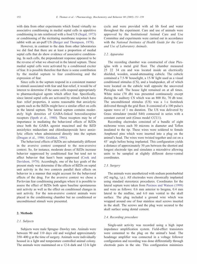
Fig. 2. Photomicrographs depicting representative placements of electrodes.
(A) Dorsolateral septum. (B) Intermediolateral septum. Arrows point
toward the electrode tips.
3. Results
3.1. Conditioning data
Histological verification of the recording electrodes for
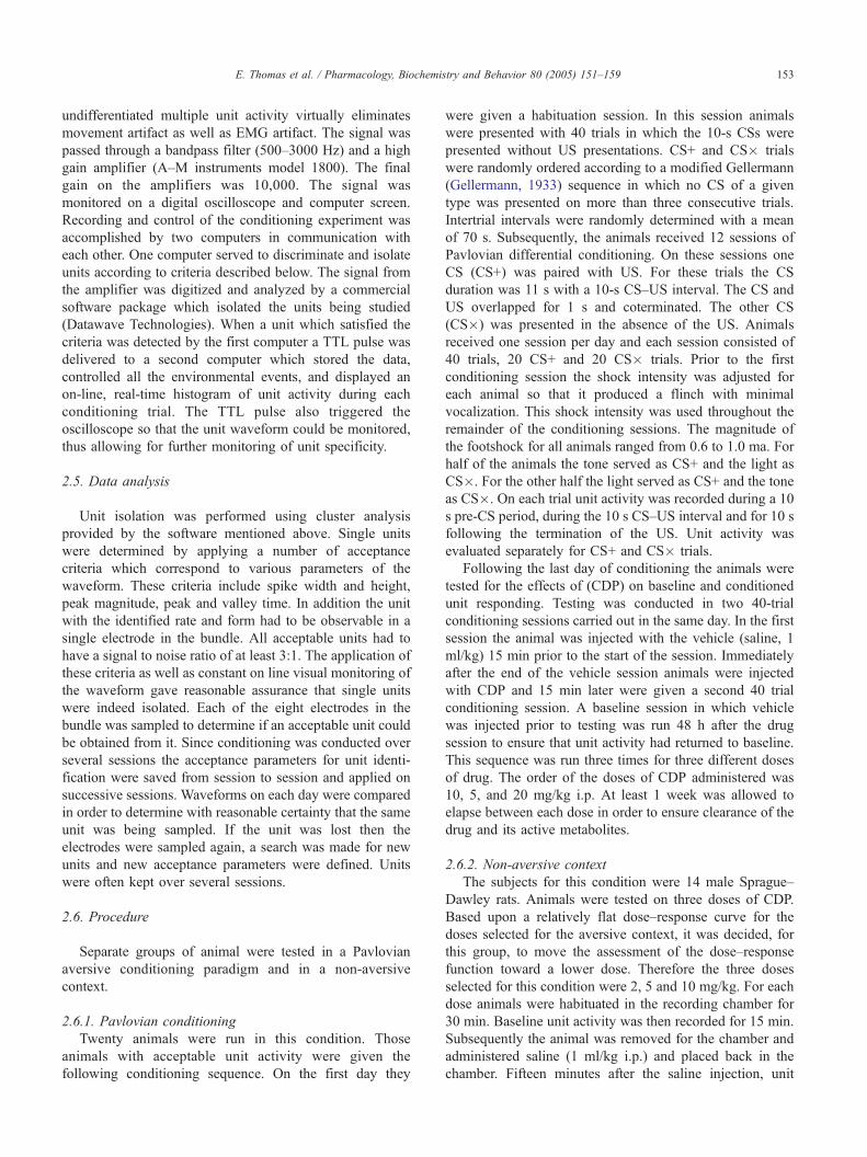
animals in the conditioning experiment is shown in Fig. 1.
Twenty animals which completed the sequence had electro-
Fig. 1. Histological representation of electrode placements. .=Dorsolateral/ventral group; o=intermediate septal group. Plates adapted from Paxinos
and Watson (1998).
des in the lateral septal area. Two distinct groups of sites may
be discerned, a lateral group with electrodes tips located in the
dorsolateral/ventrolateral region of the septum (LSD/V) and a
more medial group with tips in the intermediolateral septum
(LSI). Eleven sites were in the LSD/V and nine in the LSI.
Fig. 2 presents photographs of representative animals with
electrodes in LSD (A) and LSI (B).
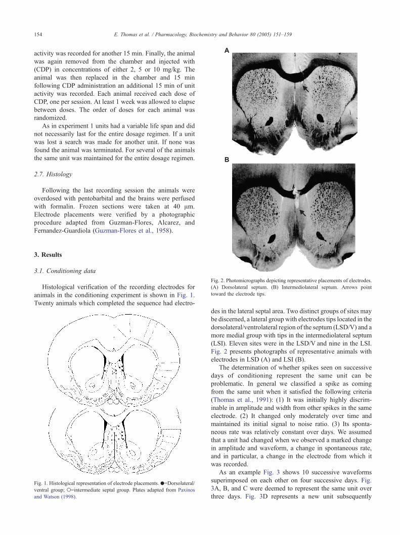
The determination of whether spikes seen on successive
days of conditioning represent the same unit can be
problematic. In general we classified a spike as coming
from the same unit when it satisfied the following criteria
(Thomas et al., 1991): (1) It was initially highly discrim-
inable in amplitude and width from other spikes in the same
electrode. (2) It changed only moderately over time and
maintained its initial signal to noise ratio. (3) Its sponta-
neous rate was relatively constant over days. We assumed
that a unit had changed when we observed a marked change
in amplitude and waveform, a change in spontaneous rate,
and in particular, a change in the electrode from which it
was recorded.
As an example Fig. 3 shows 10 successive waveforms
superimposed on each other on four successive days. Fig.
3A, B, and C were deemed to represent the same unit over
three days. Fig. 3D represents a new unit subsequently

Fig. 3. Superimposed waveforms of spikes meeting the multiple criteria of a
single unit over successive days. Each figure consists of 10 successive
overlapping spikes. (A)–(C) depict a single unit maintained over three
successive days. (D) depicts a new unit subsequently found in the same
animal. The time bar represents 1 ms duration. The vertical bar represents
100 AV.
E. Thomas et al. / Pharmacology, Biochemistry and Behavior 80 (2005) 151–159 155
discovered in the same animal. Thus, although the same
units were not necessarily maintained throughout the
conditioning session it was still possible to derive a
pseudo-learning curve for the groups of animals designated
as LSD/V and LSI. For the derivation of this curve if a unit
was lost during training another was substituted for it.
Cellular activity is expressed as a unit activity ratio which
compares the difference in the number of unit spikes
between the CS and the Pre-CS period divided by the sum
of the two (CS�PRE)/(CS+PRE). For each animal the unit
activity ratio was determined for each trial and daily means
calculated across the 20 CS+ and CS� trials of the session.
Fig. 4. Mean unit activity during Pavlovian conditioning for the two histological g
LSI=intermediate septal group. (A) represents the course of conditioning over four
session for which the statistical analysis was performed. The median baseline rate
baseline rate for group LSI was 2.62 spikes/s (range, 0.79–4.44 spikes/s). The
significant.
This method allows for symmetrical depiction of excitation
and inhibition around a zero point, with negative values
reflecting suppression of activity during the CS compared
with the pre-CS period and positive values reflecting
facilitation during the CS. The scores vary from �1.00
(total suppression) to +1.00 (maximum increase in firing)
with zero indicating no change. This derivation compensates
to a large extent for differences in baseline rates of units and
minimizes the effect of substitution of one unit for another.
The learning curve actually contains data from twenty-six
different units. The baseline rate for the cells ranged from
0.19 to 26.0 spikes/s with a median rate of 3.10 spikes/s.
Using this procedure it may be seen in Fig. 4A that there
is a progressive decrease in the unit activity ratio for CS+ in
the LSD/V group over sessions, and no change in unit
activity to CS�. Cells in this septal region discriminate CS+
from CS� and in particular suppress activity to the
conditioned fear stimulus. There were no consistent changes
in unit activity to either CS in the LSI group.
These overall observations were supported by repeated
measures analysis of variance (ANOVA) performed on the
last conditioning session prior to the administration of drug,
depicted in Fig. 4B. The ANOVA revealed an effect of
histologic group [F(1,14)=12.48, pb0.01] and a CS effect
[F(1,14)=4.64, pb0.05]. Fig. 5 depicts the results of a
typical conditioning session for an animal in the LSD group,
represented as peristimulus time histogram summed over the
20 trials. As can be seen unit activity is suppressed during
CS+ compared to the Pre-CS period whereas no suppression
is seen during CS�.
roups. H=habituation, one session only. LSD/V=dorsolateral/ventral group,
blocks of three sessions. (B) represents the state of conditioning on the final
for group LSD/V was 2.91 spikes/s (range, 0.19–6.90 spikes/s). The median
difference in baseline rates between the two groups was not statistically
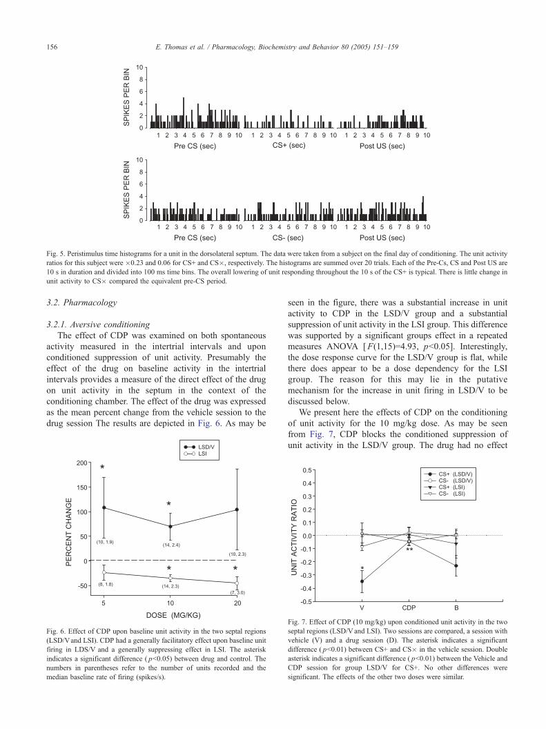
Fig. 5. Peristimulus time histograms for a unit in the dorsolateral septum. The data were taken from a subject on the final day of conditioning. The unit activity
ratios for this subject were �0.23 and 0.06 for CS+ and CS�, respectively. The histograms are summed over 20 trials. Each of the Pre-Cs, CS and Post US are
10 s in duration and divided into 100 ms time bins. The overall lowering of unit responding throughout the 10 s of the CS+ is typical. There is little change in
unit activity to CS� compared the equivalent pre-CS period.
E. Thomas et al. / Pharmacology, Biochemistry and Behavior 80 (2005) 151–159156
3.2. Pharmacology
3.2.1. Aversive conditioning
The effect of CDP was examined on both spontaneous
activity measured in the intertrial intervals and upon
conditioned suppression of unit activity. Presumably the
effect of the drug on baseline activity in the intertrial
intervals provides a measure of the direct effect of the drug
on unit activity in the septum in the context of the
conditioning chamber. The effect of the drug was expressed
as the mean percent change from the vehicle session to the
drug session The results are depicted in Fig. 6. As may be
Fig. 6. Effect of CDP upon baseline unit activity in the two septal regions
(LSD/Vand LSI). CDP had a generally facilitatory effect upon baseline unit
firing in LDS/V and a generally suppressing effect in LSI. The asterisk
indicates a significant difference ( pb0.05) between drug and control. The
numbers in parentheses refer to the number of units recorded and the
median baseline rate of firing (spikes/s).
seen in the figure, there was a substantial increase in unit
activity to CDP in the LSD/V group and a substantial
suppression of unit activity in the LSI group. This difference
was supported by a significant groups effect in a repeated
measures ANOVA [F(1,15)=4.93, pb0.05]. Interestingly,
the dose response curve for the LSD/V group is flat, while
there does appear to be a dose dependency for the LSI
group. The reason for this may lie in the putative
mechanism for the increase in unit firing in LSD/V to be
discussed below.
We present here the effects of CDP on the conditioning
of unit activity for the 10 mg/kg dose. As may be seen
from Fig. 7, CDP blocks the conditioned suppression of
unit activity in the LSD/V group. The drug had no effect
Fig. 7. Effect of CDP (10 mg/kg) upon conditioned unit activity in the two
septal regions (LSD/Vand LSI). Two sessions are compared, a session with
vehicle (V) and a drug session (D). The asterisk indicates a significant
difference ( pb0.01) between CS+ and CS� in the vehicle session. Double
asterisk indicates a significant difference ( pb0.01) between the Vehicle and
CDP session for group LSD/V for CS+. No other differences were
significant. The effects of the other two doses were similar.

E. Thomas et al. / Pharmacology, Biochemistry and Behavior 80 (2005) 151–159 157
on CS� in this group nor on either CS for the LSI group.
ANOVA shows a Groups effect [F(1,14)=5.00, pb0.05],
Groups by CS interaction [F(1,14)=5.99, pb0.05], Drug
effect [F(1,14)=9.93, pb0.01], and Groups by Drug
interaction [F(1,14)=18.38, pb0.001]. The two other doses
had similar effects upon the conditioned suppression of
unit activity. The unit activity ratio for CS+ in the LSD/V
group was �0.28 for the control and �0.03 for the 20 mg/
kg group. A t-test revealed this difference to be significant
t(9)=2.81, p=0.025. For the 5 mg/kg dose the unit activity
ratio for CS+ was =0.14 and �0.08 for vehicle and drug,
respectively. This difference did not reach statistical
significance.
Fig. 8. Histological representation of electrode placements for animals in
the non-aversive context. .=Dorsolateral/ventral group; o=intermediate
septal group. Plates adapted from Paxinos and Watson (1998).
Fig. 9. Effect of CDP upon spontaneous unit activity in the two septal
regions (LSD/V and LSI). In the non-aversive context the effect of
CDP upon unit activity was minimal except at the highest dose. The
asterisk indicates a significant difference ( pb0.05) between drug and
control.
3.2.2. Non-aversive context
Fig. 8 shows the placement of the electrodes in the
animals that completed the experiment. The electrode tips
were distributed in the regions LSD/V and LSI. Fig. 9
presents a plot of the percent change in unit activity in the
CDP session compared to the immediately preceding saline
session for each dose of CDP. As may be seen in the figure
CDP had no facilitatory effect upon unit activity in either
region of the septum. A one-sample t-test comparing the
percent change to zero yielded significance only at the 10
mg/kg dose t(10)=2.57, p=0.03. As may be seen the effect
of CDP at that dose was to suppress unit activity in LSD/V.
A similar trend is apparent for the effect of CDP in LSI.
Again the only significant effect was at the 10 mg/kg dose
t(3)=11.14, p=0.002.
4. Discussion
The principal finding of the present study was that cells
in the lateral septum, which are inhibited by aversive
conditioned stimuli in the awake freely moving animal,
increase their rate of firing in response to the benzodiaze-
pine anxiolytic chlordiazepoxide. In general the results of
this study confirm the results of our previous research
(Thomas et al., 1991) that cells in the more lateral regions of
the septum respond to the hedonic properties of conditioned
stimuli. The effect of Pavlovian aversive conditioning was
to decrease unit activity to CS+ in the dorsolateral and
ventrolateral septal nuclei but not in the more medial
intermediate nucleus.

E. Thomas et al. / Pharmacology, Biochemistry and Behavior 80 (2005) 151–159158
The recording of the response of single neurons to
pharmacological agents is one of a variety of methods for
determining the mechanism of therapeutic action of drugs.
Such recording in the awake freely moving animal has the
significant advantage of being able to relate such responses
directly to behavior. In this case the ability to record unit
activity in the context of aversive conditioning can supply
important information relating BZDs, unit activity, and fear.
Thus, the benzodiazepine CDP reduced the conditioned
suppression of unit activity in the lateral cell groups. Such a
reduction is highly consistent with its anxiolytic properties
and parallels its effect on behavior suppressed by aversive
conditioning (Cook and Davidson, 1978).
Equally germane to the putative mechanism of action of
CDP is the finding that in the aversive context CDP
increased spontaneous unit activity of these cells in the
baseline period in the absence of the CS. In contrast CDP
did not increase spontaneous unit activity in a non-aversive
context. The literature indicates that in aversive condition-
ing not only does the conditioned cue elicit fear, but the
context does as well (Fanselow, 2000; Phillips and
LeDoux, 1992; Sparks and LeDoux, 1995). It seems likely
then, that in the aversive paradigm some degree of
contextual conditioning took place, exerting a tonic
inhibitory effect on unit firing in the experimental environ-
ment. This is in agreement with research that shows that
the septum plays a substantial role in contextual con-
ditioning (Sparks and LeDoux, 1995).
The absence of an increase in unit activity in response to
the administration of CDP in the non-aversive context is in
striking contrast to the effect of CDP in the aversive context.
It appears that in the absence of a fear stimulus CDP has
little effect at doses which in the aversive context increase
unit activity in LSD/V. These data are consistent with those
of Givens and Breese (Givens and Breese, 1990a,b) who
found that ethanol, which binds to the BZD-GABA-Cl�
complex, has little effect on neurons in LS in urethane
anesthetized as well as unanesthetized rats. On the basis of
our results it might be expected that ethanol, which has
anxiolytic properties akin to CDP, would elevate septal
firing in an aversive context.
If, as we have proposed, the lateral septum is involved in
the relief of fear, then the increased unit activity in the
lateral septum to CDP in the aversive context would suggest
that benzodiazepines might exert their anxiolytic effect by
activating neurons in the lateral septum and related regions
that have been inhibited by fear. The mechanism whereby
BZDs might increase cellular activity in the lateral septum
remains at this time a matter of speculation. The BZDs work
primarily via the BZD-GABA-Cl� complex and therefore
would be expected to have a generally inhibitory action on
cellular activity. This has typically been the case in most
structures tested including the hippocampus (Chou and
Wang, 1977; Steffensen and Henriksen, 1992), amygdala
(Chou and Wang, 1977), dentate gyrus (Steffensen and
Henriksen, 1992), substantia nigra (Ross et al., 1982), locus
coeruleus (Grant et al., 1980), and in the case of the present
experiment, in intermediolateral septum.
It should be noted, however, that electron micrographs
reveal GABA–GABA synapses in interneurons in the lateral
septum (Onteniente et al., 1987). The synapsing of
GABAergic boutons onto other GABAergic neurons is
important because this connection allows for a possible
disinhibitory mechanism within the septum following
increased GABAergic activity. Thus, BZDs may have a
facilitatory effect on lateral septal neurons by means of a
disinhibition mechanism. It may be that the complexity of
GABAergic connections within the lateral septum accounts
for the lack of a simple dose response effect of CDP in our
experiment. It is of interest that the anxiolytic effect of direct
application of BZDs or GABA agonists into the lateral
septum also does not show a simple dose response effect
(Drugan et al., 1986; Grishkat, 1991).
BZDs, in addition to their anxiolytic effect, have been
shown to have substantial effects upon several forms
learning and memory. These include spatial tasks such as
the Morris water maze (McNaughton and Morris, 1987)
and non-spatial working memory tasks (Olaman and
McNaughton, 2001). These tasks are particularly sensitive
to hippocampal damage and very likely reflect the effect of
BZDs on hippocampal functioning. Pavlovian fear con-
ditioning also appears to be impaired in a variety of
situations. However, the effects of BZDs on fear con-
ditioning have been variable and context dependent (Harris
and Westbrook, 2001). To the extent that there is an
impairment of fear conditioning this likely represents a
diminution of amygdala function (Davis et al., 1994). A
possibility that bears further research is that the lateral
septum may have an effect on the expression and learning
of fear by modulating activity in the amygdala.
The lateral septum is strategically situated to serve as a
link between the hippocampus and the brainstem emotional
circuits as well between the hippocampus and the amygdala
(Sheehan et al., 2004; Thomas, 1988). It receives a
substantial input from the hippocampus (Risold and
Swanson, 1997). The lateral septum projects to the central
nucleus of the amygdala either directly (Volz et al., 1990) or
via the bed nucleus of the stria terminalis (Jakab and
Leranth, 1995). There is evidence that the behavioral effects
of the septum are mediated by the amygdala, especially the
central nucleus (Grishkat, 1991; Melia et al., 1992). Recent
research in our laboratory (Thomas and Sancar, 2001) has
shown that activation of the lateral septum by electrical
stimulation has a profound inhibitory effect upon cells in the
central nucleus of the amygdala likely mediated by GABAA
receptors.
Acknowledgments
This study was supported in part by National Institutes of
Health grant MH 54674.

E. Thomas et al. / Pharmacology, Biochemistry and Behavior 80 (2005) 151–159 159
References
Berger TW, Thompson RF. Neuronal plasticity in the limbic system during
classical conditioning of rabbit nictitating membrane response: II.
Septum and mammillary bodies. Brain Res 1978;156:293–314.
Brayley KM, Albert DJ. Suppression of VMH-lesion induced reactivity and
aggressiveness in the rat by stimulation of the lateral septum, but not by
medial septum or cingulate cortex. J Comp Physiol Psychol 1977;91:
290–9.
Chou DT, Wang SC. Unit activity of amygdala and hippocampal
neurons: effects of morphine and benzodiazepines. Brain Res 1977;
126:427–40.
Cook L, Davidson AB. Behavioral pharmacology: animal models involving
aversive control of behavior. In: Lipton MA, DiMascio A, Kullam KF,
editors. Psychopharmacology: a generation of progress. New York7
Raven Press; 1978. p. 563–7.
Davis M, Rainnie D, Cassell M. Neurotransmission in the rat amygdala
related to fear and anxiety. Trends Neurosci 1994;17:208–14.
Drugan RC, Skolnik P, Paul SM, Crawley JM. Low doses of muscimol
produce anticonflict actions in the lateral septum of the rat. Neuro-
pharmacology 1986;25:203–5.
Fanselow MS. Contextual fear, gestalt memories, and the hippocampus.
Behav Brain Res 2000;110:73–81.
Gellermann LW. Chance orders of alternating stimuli in visual discrim-
ination experiments. J Genet Psychol 1933;42:207–8.
Givens BS, Breese GR. Electrophysiological evidence that ethanol alters
function of medial septal area without affecting lateral septal function.
J Pharmacol Exp Ther 1990;253:95–103.
Givens BS, Breese GR. Site-specific enhancement of gamma-aminobutyric
acid-mediated inhibition of neural activity by ethanol in the rat medial
septal area. J Pharmacol Exp Ther 1990;254:528–38.
Grant SJ, Huang YH, Redmond Jr DE. Benzodiazepines attenuate single
unit activity in the locus coeruleus. Life Sci 1980;27:2231–6.
Grishkat HL. Modulation of anxiety: a role for the septum and amygdala in
the effects of benzodiazepine anxiolytics. Psychology. Bryn Mawr
College.
Guzman-Flores CM, Alcarez M, Fernandez-Guardiola A. Rapid procedure
to localize electrodes in experimental neurophysiology. Boletin del
Instituto de Estudios Medicos y Biologicos, Universidad Nacional de
Mexico 1958;16:26–31.
Harris JA, Westbrook RF. Contextual control over the expression of fear in
rats conditioned under a benzodiazepine. Psychopharmacology (Berl)
2001;156:92–7.
Jakab RL, Leranth C. Septum. In: Paxinos G, editor. The rat nervous
system. New York7 Academic Press; 1995. p. 593–6.
McNaughton N, Morris RG. Chlordiazepoxide, an anxiolytic benzodia-
zepine, impairs place navigation in rats. Behav Brain Res 1987;24:
39–46.
Melia KR, Sananes CB, Davis M. Lesions of the central nucleus of the
amygdala block the excitatory effects of septal ablation on the acoustic
startle reflex. Physiol Behav 1992;51:175–80.
Olaman SJ, McNaughton N. Chlordiazepoxide specifically impairs non-
spatial reference memory in the cued radial arm maze in rats. Pharmacol
Biochem Behav 2001;70:133–9.
Onteniente B, Geffard M, Campistron G, Calas A. An ultra-structural study
of GABA-immunoreactive neurons and terminals in the septum of the
rat. J Neurosci 1987;7:48–54.
Paxinos G, Watson C. The rat brain in stereotaxic coordinates. New York7
Academic Press; 1998.
Phillips RG, LeDoux JE. Differential contribution of the amygdala and
hippocampus to cued and contextual fear conditioning. Behav Neurosci
1992;106:274–85.
Risold PY, Swanson LW. Connections of the rat lateral septal complex.
Brain Res Brain Res Rev 1997;24:115–95.
Ross RJ, Waszczak BL, Lee EK, Walters JR. Effects of benzodiazepines on
single unit activity in the substantia nigra pars reticulata. Life Sci
1982;31:1025–35.
Segal M. Flow of conditioned responses in the limbic telencephalic system
of the rat. J Neurophysiol 1973;36:840–54.
Sheehan TP, Chambers RA, Russell DS. Regulation of affect by the
lateral septum: implications for neuropsychiatry. Brain Res Rev 2004;
46:71–117.
Sparks PD, LeDoux JE. Septal lesions potentiate freezing behavior to
contextual but not to phasic conditioned stimuli in rats. Behav Neurosci
1995;109:184–8.
Speth RC, Johnson RW, Regan J, Reisine T, Kobayashi RM, Bresolin N,
et al. The benzodiazepine receptor of the mammalian brain. Fed Proc
1980;39:3032–8.
Steffensen SC, Henriksen SJ. Comparison of the effects of ethanol and
chlordiazepoxide on electrophysiological activity in the fascia dentata
and hippocampus regio superior. Hippocampus 1992;2:201–11.
Thomas E. Forebrain mechanisms in the relief of fear: the role of the lateral
septum. Psychobiology 1988;16:36–44.
Thomas E., Sancar F.. Electrical stimulation of the lateral septum and
intraseptal benzodiazepines modulate activity of neurons in the central
nucleus of the amygdala of the rat. In: Abstract Viewer/Itinerary
Planner. Washington, D.C7 Society for Neuroscience; 2001 Prog.
No. 177.12.
Thomas E, Yadin E. Multiple unit activity in the septum during Pavlovian
aversive conditioning: evidence for an inhibitory role for the septum.
Exp Neurol 1980;69:50–60.
Thomas E, Yadin E, Strickland CE. Septal unit activity during classical
conditioning: a regional comparison. Brain Res 1991;547:303–8.
Vogel JR, Beer B, Clody DE. A simple and reliable conflict procedure for
testing anti-anxiety agents. Psychopharmacologia 1971;21:1–7.
Volz H-P, Rehbein G, Triepel J, Knuepfer MM, Stumpf H, Stock G.
Afferent connections of the nucleus centralis amygdalae. Anat Embryol
1990;181:177–94.
Yadin E, Thomas E. Septal correlates of conditioned inhibition and
excitation. J Comp Physiol Psychol 1981;95:331–40.
Yadin E, Thomas E, Grishkat HL, Strickland CE. The role of the lateral
septum in anxiolysis. Physiol Behav 1993;53:1077–83.