Embed Size (px)
Citation preview

© 2017 Dental Press Journal of Orthodontics Dental Press J Orthod. 2017 Sept-Oct;22(5):67-7467
original article
Effect of TiO2 nanoparticles incorporation on
antibacterial properties and shear bond strength of
dental composite used in Orthodontics
Ahmad Sodagar1, Mohamad Sadegh Ahmad Akhoundi1, Abbas Bahador2,Yasamin Farajzadeh Jalali3, Zahra Behzadi4, Farideh Elhaminejad4, Amir Hossein Mirhashemi1
Introduction: Plaque accumulation and bond failure are drawbacks of orthodontic treatment, which requires composite for bonding of brack-ets. As the antimicrobial properties of TiO2 nanoparticles (NPs) have been proven, the aim of this study was to evaluate the antimicrobial and mechanical properties of composite resins modified by the addition of TiO2 NPs. Methods: Orthodontics composite containing 0%, 1%, 5% and 10% NPs were prepared. 180 composite disks were prepared for elution test, disk agar diffusion test and biofilm inhibition test to collect the counts of microorganisms on three days, measure the inhibition diameter and quantify the viable counts of colonies consequently. For shear bond strength (SBS) test, 48 intact bovine incisors were divided into four groups. Composites containing 0%, 1%, 5% and 10% NPs were used for bonding of bracket. The bracket/tooth SBS was measured by using an universal testing machine. Results: All concentration of TiO2 NPs had a significant effect on creation and extension of inhibition zone. For S. mutans and S. sanguinis, all concentration of TiO2 NPs caused reduction of the colony counts. Composite containing 10% TiO2 NPs had significant effect on reduction of colony counts for S. mutans and S. sanguinis in all three days. The highest mean shear bond strength belonged to the control group, while the lowest value was seen in 10% NPs composite. Conclusions: Incorporating TiO2 nanoparticles into composite resins confer antibacterial properties to adhesives, while the mean shear bond of composite containing 1% and 5% NPs still in an acceptable range. Keywords: Titanium dioxide, nanoparticles. Dental bonding. Antimicrobial orthodontic adhesive. Shear strength.
1 Tehran University of Medical Sciences, Dental Research Center, Dentistry Research Institute, Faculty of Dentistry, Department of Orthodontics (Tehran, Iran).
2 Tehran University of Medical Sciences, Faculty of Medicine, Department of Microbiology (Tehran, Iran).
3 Ilam University of Medical Sciences, Faculty of Dentistry, Department of Orthodontics (Ilam, Iran).
4 Tehran University of Medical Sciences, Faculty of Dentistry (Tehran, Iran).
» The authors report no commercial, proprietary or financial interest in the products or companies described in this article.
DOI: https://doi.org/10.1590/2177-6709.22.5.067-074.oar
How to cite: Sodagar A, Akhoundi MSA, Bahador A, Jalali YF, Behzadi Z, Elhaminejad F, Mirhashemi AH. Effect of TiO2 nanoparticles incorpora-tion on antibacterial properties and shear bond strength of dental composite used in Orthodontics. Dental Press J Orthod. 2017 Sept-Oct;22(5):67-74. DOI: https://doi.org/10.1590/2177-6709.22.5.067-074.oar
Submitted: December 13, 2015 - Revised and accepted: February 04, 2017
Contact address: Amir Hossein Mirhashemi – Dental Research Center, Den-tistry Research Institute, Department of Orthodontics, Tehran University of Medical Sciences, Tehran, Iran – E-mail: [email protected]
Introdução: o acúmulo de placa e as descolagens de braquetes são algumas desvantagens presentes no tratamento ortodôntico, no qual se requer o uso de materiais compósitos para a colagem dos braquetes. Objetivo: tendo em vista que as propriedades antimicrobianas das nanopartículas (NPs) de TiO2 já foram confirmadas, o objetivo do presente estudo foi avaliar as propriedades antimicrobianas e mecânicas de resinas compostas modificadas pela adição de NPs de TiO2. Métodos: compósitos ortodônticos contendo 0%, 1%, 5% e 10% de NPs foram preparados. Cento e oitenta discos de compósito foram preparados para o teste de eluição, o ensaio de difusão em ágar por disco, e o ensaio de inibição da formação de biofilme, para se calcular as contagens de microrganismos ao longo de três dias, medir o diâmetro da inibição e, consequentemente, quantificar as contagens de colônias viáveis. Para o teste de resistência da colagem ao cisalhamento (SBS), 48 incisivos bovinos intactos foram divididos em quatro grupos, nos quais os compósitos contendo 0%, 1%, 5% e 10% de NPs foram utilizados para colagem dos braquetes. A SBS da interface braquete/dente foi medida em uma máquina universal de ensaios. Resultados: todas as concentrações de NPs de TiO2 apresentaram efeito significativo na formação e na extensão da zona de inibição. Para o S. mutans e o S. sanguinis, todas as concentrações de NPs de TiO2 causaram redução na contagem das colônias. O compósito contendo 10% de NPs de TiO2 apresentou uma diminuição significativa na contagem de colônias de S. mutans e S. sanguinis durante os três dias. A média mais alta da SBS foi observada no grupo controle, enquanto o valor mais baixo foi observado para o compósito com 10% de NPs. Conclusões: a incorporação de nanopartículas de TiO2 nas resinas compostas lhes conferiu propriedades antibacterianas, e o valor médio da SBS das resinas contendo 1% e 5% de NPs apresentou-se dentro de uma faixa aceitável. Palavras-chave: Nanopartículas de dióxido de titânio. Colagem ortodôntica. Adesivo ortodôntico antibacteriano. Resistência ao cisalhamento.

© 2017 Dental Press Journal of Orthodontics Dental Press J Orthod. 2017 Sept-Oct;22(5):67-7468
Effect of TiO2 nanoparticles incorporation on antibacterial properties and shear bond strength of dental composite used in Orthodonticsoriginal article
INTRODUCTIONBonding technique with resin-based composite as an
adhesive agent has been primarily used in Orthodontics for securing orthodontic brackets to the surface of the teeth. Unfortunately, in spite of the fact that bonding technique have many advantages — such as high esthet-ic and simple procedure —, still have some drawbacks such as plaque accumulation, development of white spot lesions and bond failure, which cause prolonging the treatment course, imposing high cost, consuming more chair time and a less than optimal esthetic result occurs after treatment, due to demineralization of enamel adja-cent to the brackets, specially around the bracket mar-gin, due to the surface exposed to composite.1-6
Several methods have been used to inhibit biofilm growth, which contributes to dental caries. For instance, one group of such efforts has been evaluation of effec-tiveness of incorporating different antimicrobial agents in the adhesives; two of the most common examples are fluoride and chlorhexidine.7-12
Today one of the most important advances in den-tal material field is the application of nanotechnology to resin composites. Many studies investigated the ef-fect of antimicrobial nanoparticles incorporated into composite resins to prevent plaque accumulation and bacterial adhesion. Nanoparticles are believed to pen-etrate into the cell wall of bacteria efficiently due to their smaller size, effectively exerting their antibacte-rial properties.13,14 A study evaluated the antibacterial effect of composite with different concentration of in-corporated nanosilver, which has been used in Medi-cine as an antimicrobial agent. The result of this study showed that nanosilver containing composite could confer surface antibacterial activity without significant difference on shear bond strength.15 Another study demonstrated that addition of nanosilver significant-ly decreased the shear bond strength of orthodontic brackets; however, the obtained mean value was still higher than the estimated force required for orthodon-tic treatment.16 Silver/hydroxyapatite nanoparticles were shown to decrease the shear bond strength as their concentration increased.17 Evidence shows that addi-tion of ZnO nanoparticles to composite resin increases the mechanical properties such as compressive strength and flexural modulus.18 ZnO nanoparticles also possess antimicrobial properties and inhibit the formation of biofilm.19 Chitosan is also used in many industries and
is available in nano and microforms.20,21 Recently, it has been shown that chitosan has antibacterial activity against a wide range of bacteria. Chitosan nanoparti-cles also enhance the antibacterial properties of com-posite resins without compromising their shear bond strength.22 It has also been demonstrated that chitosan nanoparticles in conjunction with ZnO nanoparticles could improve the antimicrobial effect of composite resins, while the obtained shear bond strength values were approximately similar to conventional orthodon-tic composites.23
Antimicrobial properties of TiO2 nanoparticles and their application in Medicine, Dentistry and other sci-ence have been widely known. Haghi et al24 evaluated the antimicrobial effect of TiO2 NPs on pathogenic strain of E. coli and they showed that the TiO2 NPs cause little pores in bacterial cell walls, leading to in-creased permeability and cell death. Based on this study, nano-TiO2 has efficient antibacterial effect and can be used as an antibacterial agent for different purposes.
We decided to assess the effect of incorporating dif-ferent percentages (wt %) of TiO2 nanoparticles into composite resin to obtain a composite with excellent antimicrobial properties and sufficient bond strength to enamel surface for use in Orthodontics.
MATERIAL AND METHODSNanocomposite preparation
For the fabrication of composite containing dif-ferent concentration of TiO2 NPs, 400 mg of TiO2 NP powder was manually mixed with 3600 mg of Transbond XT composite (3M Unitek, Monrovia, CA, USA), in order to obtain 4000 mg of compos-ite containing TiO2 NPs with 10% concentration. To fabricate composite containing 5% TiO2 NPs, 1200 mg of composite containing 10% TiO2 NPs was mixed with 1200 mg of plain composite. To fab-ricate composite containing 1% TiO2 NPs, 240 mg of 10% TiO2 NPs was mixed with 2160 mg of plain composite. Thus, 2560 mg of composite containing 10% TiO2 NPs, 2400 mg of composite containing 5% TiO2 NPs and 2400 mg of composite containing 1% TiO2 NPs were thus obtained.
Shear bond strength testForty-eight bovine central incisors with no visible
cracks or caries were disinfected in 0.5% chloramine-T
© 2017 Dental Press Journal of Orthodontics Dental Press J Orthod. 2017 Sept-Oct;22(5):67-7469
original articleSodagar A, Akhoundi MSA, Bahador A, Jalali YF, Behzadi Z, Elhaminejad F, Mirhashemi AH
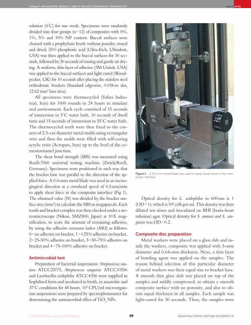
Figure 1 - A 0.6-mm metal blade was used to apply shear force to the com-posite interface.
solution (4°C) for one week. Specimens were randomly divided into four groups (n = 12) of composites with 0%, 1%, 5% and 10% NP content. Buccal surfaces were cleaned with a prophylaxis brush without powder, rinsed and dried; 35% phosphoric acid (Ultra-Etch, Ultradent, USA) was then applied to the buccal surfaces for 30 sec-onds, followed by 30 seconds of rinsing and gentle air dry-ing. A uniform, thin layer of adhesive (3M Unitek, USA) was applied to the buccal surfaces and light cured (Wood-pecker, UK) for 10 seconds after placing the stainless steel orthodontic brackets (Standard edgewise, 0.018-in slot, 12.62 mm2 base area).
All specimens were thermocycled (Vafaei Indus-trial, Iran) for 1000 rounds in 24 hours to simulate oral environment. Each cycle consisted of 15 seconds of immersion in 5°C water bath, 10 seconds of dwell time and 15 seconds of immersion in 55°C water bath. The thermocycled teeth were then fixed to the cor-ners of 2.5-cm diameter metal molds using rectangular wire and then the molds were filled with self-curing acrylic resin (Acropars, Iran) up to the level of the ce-mentoenamel junction.
The shear bond strength (SBS) was measured using Roell-7060 universal testing machine (Zwick/Roell, Germany). Specimens were positioned in such way that the bracket base was parallel to the direction of the ap-plied force. A 0.6-mm metal blade was used in an inciso-gingival direction at a crosshead speed of 0.5 mm/min to apply shear force to the composite interface (Fig 1). The obtained value (N) was divided by the bracket sur-face area (mm2) to calculate the SBS in megapascals. Each tooth and bracket complex was then checked under a ste-reomicroscope (Nikon, SMZ800, Japan) at 10 X mag-nification, to score the amount of remaining adhesive, by using the adhesive remnant index (ARI) as follows: 0 = no adhesive on bracket, 1 = < 25% adhesive on bracket, 2 = 25-50% adhesive on bracket, 3 = 50-75% adhesive on bracket and 4 = 75-100% adhesive on bracket.
Antimicrobial testPreparation of bacterial suspensions: Streptococcus mu-
tans ATCC25175, Streptococcus sanguinis ATCC10556 and Lactobacillus acidophilus ATCC4356 were supplied in liophilised form and incubated in broth, in anaerobic and 37°C conditions for 48 hours. 108 CFU/ml microorgan-ism suspensions were prepared by spectrophotometer for determining the antimicrobial effect of TiO2 NPs.
Optical density for L. acidophilus in 600 nm is 1 (OD = 1); which is 108 cells per ml. This density was then diluted ten times and inoculated on BHI (brain-heart infusion) agar. Optical density for S. mutans and S. san-guinis was OD = 0.2.
Composite disc preparationMetal washers were placed on a glass slab and in-
side the washers, composite was applied with 5-mm diameter and 0.64-mm thickness. Next, a thin layer of bonding agent was applied on the samples. The reason behind selection of this particular diameter of metal washers was their equal size to bracket base. A smooth thin glass slob was placed on top of the samples and mildly compressed, to obtain a smooth composite surface with no porosity, and also to ob-tain equal thickness in all samples. Each sample was light-cured for 30 seconds. Then, the samples were

© 2017 Dental Press Journal of Orthodontics Dental Press J Orthod. 2017 Sept-Oct;22(5):67-7470
Effect of TiO2 nanoparticles incorporation on antibacterial properties and shear bond strength of dental composite used in Orthodonticsoriginal article
separated from the washers and sterilized in Iran’s Nuclear Science and Technology gamma radiation center with 25 KGy dosages (Fig 2).
Biofilm inhibitionThree-day biofilms were generated on composite
discs (n = 36) using 24-well plates. Each well was inocu-lated with adjusted bacterial inoculum. Biofilms were grown at 37°C. At the end of the third day, each disc was rinsed with sterile saline solution to remove loosely adsorbed proteins and biofilm matrix residues. To count the colony forming units (CFUs) responsible for biofilm formation, specimens were sonicated in sterile saline so-lution and then vortexed. CFU/ml of the microorgan-ism present in the suspension was counted with drop-plate method using rapid dilution in microtiter plates.
Disc Agar Diffusion test (DAD)Antibacterial activity of discs via solubility and dif-
fusion of TiO2 NPs was examined by DAD. Com-posite discs (n = 36) were placed, 2 cm apart, on BHI agar plates, which were inoculated with a 200 μL bac-terial solution (≈108 CFU/mL) by a sterile swab. After 48-hour incubation, the bacterial growth inhibition di-ameter was optically measured.
Antibacterial properties of eluted componentsThis test was used to assess the antimicrobial activity of
TiO2 NPs released from composite discs. Composite discs were placed in tubes containing 0.5 ml of BHI broth. After 3, 15 and 30 days, the discs were transferred to 15-ml plas-tic tubes and 50 μL of the bacterial culture was added to each tube containing 5 ml of TSA (final concentration of 105 CFUs/ml in 1 ml of medium). The tubes were shaken at 300 rpm at 37oC for 24 hours. The obtained suspension was serially diluted in microtiter plates and spread cultured in TSA. The bacterial colonies (CFUs/ml) were counted using drop-plate method (Fig 3).
Statistical analysisShear bond strength test results were analyzed using
one-way ANOVA followed by post-hoc Tukey’s HSD test. The Kruskal-Wallis test was also applied to analyze the ARI results. Antimicrobial test results were ana-lyzed with multiple statistical tests. One-way ANOVA was first used for biofilm inhibition test, followed by Tukey HSD test. Kruskal-Wallis test was used to ana-lyze data attained from DAD test. Two-way ANOVA was first used to analyze day × concentration relation in eluted components test. For those groups with signifi-cant difference, one-way ANOVA was used.
Figure 2 - Composite disc preparation.
Figure 3 - Antibacterial properties of eluted com-ponents test.
© 2017 Dental Press Journal of Orthodontics Dental Press J Orthod. 2017 Sept-Oct;22(5):67-7471
original articleSodagar A, Akhoundi MSA, Bahador A, Jalali YF, Behzadi Z, Elhaminejad F, Mirhashemi AH
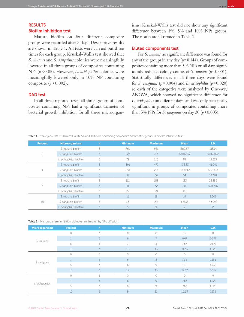
RESULTSBiofilm inhibition test
Mature biofilm on four different composite groups were recorded after 3 days. Descriptive results are shown in Table 1. All tests were carried out three times for each group. Kruskal-Wallis test showed that S. mutans and S. sanguinis colonies were meaningfully lowered in all three groups of composites containing NPs (p < 0.05). However, L. acidophilus colonies were meaningfully lowered only in 10% NP containing composite (p < 0.002).
DAD testIn all three repeated tests, all three groups of com-
posites containing NPs had a significant diameter of bacterial growth inhibition for all three microorgan-
isms. Kruskal-Wallis test did not show any significant difference between 1%, 5% and 10% NPs groups. The results are illustrated in Table 2.
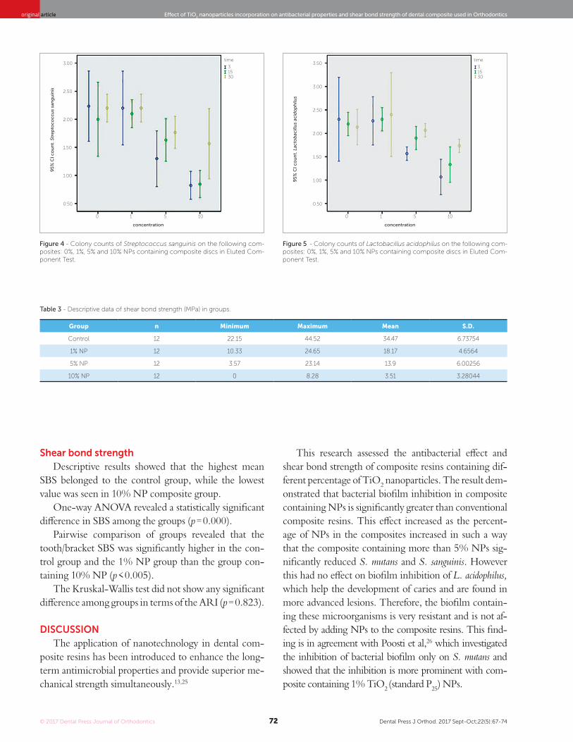
Eluted components testFor S. mutans no significant difference was found for
any of the groups in any day (p = 0.144). Groups of com-posites containing more than 5% NPs on all days signif-icantly reduced colony counts of S. mutans (p < 0.001). Statistically differences in all three days were found for S. sanguinis (p = 0.004) and L. acidophilus (p = 0.020) so each of the categories were analyzed by One-way ANOVA, which showed no significant difference for L. acidophilus on different days, and was only statistically significant in groups of composites containing more than 5% NPs for S. sanguinis on day 30 (p < 0.005).
Percent Microorganisms n Minimum Maximum Mean S.D.
0
S. mutans biofilm 3 761 981 889.67 115.14
S. sanguinis biofilm 3 523 701 630.6667 94.69072
L. acidophilus biofilm 3 72 110 89 19.313
1
S. mutans biofilm 3 391 472 435.33 41.041
S. sanguinis biofilm 3 168 201 181.6667 17.21434
L. acidophilus biofilm 3 39 66 54 13.748
5
S. mutans biofilm 3 112 158 133 23.259
S. sanguinis biofilm 3 41 52 47 5.56776
L. acidophilus biofilm 3 27 29 28 1
10
S. mutans biofilm 3 11 18 14 3.606
S. sanguinis biofilm 3 1.3 2.2 1.7333 4.5092
L. acidophilus biofilm 3 5 9 7 2
Table 1 - Colony counts (CFU/mm2) in 1%, 5% and 10% NPs containing composite and control group, in biofilm inhibition test.
Table 2 - Microorganism inhibition diameter (millimeter) by NPs diffusion.
Microorganisms Percent n Minimum Maximum Mean S.D.
S. mutans
0 3 0 0 0 0
1 3 6 7 6.67 0.577
5 3 7 8 7.67 0.577
10 3 10 13 11.33 1.528
S. sanguinis
0 3 0 0 0 0
1 3 6 8 7.33 1.155
5 3 6 9 8 1.732
10 3 12 13 12.67 0.577
L. acidophilus
0 3 0 0 0 0
1 3 6 9 7.67 1.528
5 3 6 9 7.67 1.528
10 3 9 11 10.33 1.155
© 2017 Dental Press Journal of Orthodontics Dental Press J Orthod. 2017 Sept-Oct;22(5):67-7472
Effect of TiO2 nanoparticles incorporation on antibacterial properties and shear bond strength of dental composite used in Orthodonticsoriginal article
Shear bond strengthDescriptive results showed that the highest mean
SBS belonged to the control group, while the lowest value was seen in 10% NP composite group.
One-way ANOVA revealed a statistically significant difference in SBS among the groups (p = 0.000).
Pairwise comparison of groups revealed that the tooth/bracket SBS was significantly higher in the con-trol group and the 1% NP group than the group con-taining 10% NP (p < 0.005).
The Kruskal-Wallis test did not show any significant difference among groups in terms of the ARI (p = 0.823).
DISCUSSIONThe application of nanotechnology in dental com-
posite resins has been introduced to enhance the long-term antimicrobial properties and provide superior me-chanical strength simultaneously.13,25
This research assessed the antibacterial effect and shear bond strength of composite resins containing dif-ferent percentage of TiO2 nanoparticles. The result dem-onstrated that bacterial biofilm inhibition in composite containing NPs is significantly greater than conventional composite resins. This effect increased as the percent-age of NPs in the composites increased in such a way that the composite containing more than 5% NPs sig-nificantly reduced S. mutans and S. sanguinis. However this had no effect on biofilm inhibition of L. acidophilus, which help the development of caries and are found in more advanced lesions. Therefore, the biofilm contain-ing these microorganisms is very resistant and is not af-fected by adding NPs to the composite resins. This find-ing is in agreement with Poosti et al,26 which investigated the inhibition of bacterial biofilm only on S. mutans and showed that the inhibition is more prominent with com-posite containing 1% TiO2 (standard P25) NPs.
Table 3 - Descriptive data of shear bond strength (MPa) in groups.
Group n Minimum Maximum Mean S.D.
Control 12 22.15 44.52 34.47 6.73754
1% NP 12 10.33 24.65 18.17 4.6564
5% NP 12 3.57 23.14 13.9 6.00256
10% NP 12 0 8.28 3.51 3.28044
Figure 4 - Colony counts of Streptococcus sanguinis on the following com-posites: 0%, 1%, 5% and 10% NPs containing composite discs in Eluted Com-ponent Test.
Figure 5 - Colony counts of Lactobacillus acidophilus on the following com-posites: 0%, 1%, 5% and 10% NPs containing composite discs in Eluted Com-ponent Test.
time time
concentration concentration
95
% C
I co
un
t. S
trep
toco
ccu
s sa
ng
uin
is
95
% C
I co
un
t. L
acto
bac
illu
s ac
ido
ph
ilus
30 30
3.00
3.00
3.50
2.00
2.00
1.001.00
2.50
2.50
1.50
1.50
0.50 0.50
0 05 51 110 10
15 153 3
© 2017 Dental Press Journal of Orthodontics Dental Press J Orthod. 2017 Sept-Oct;22(5):67-7473
original articleSodagar A, Akhoundi MSA, Bahador A, Jalali YF, Behzadi Z, Elhaminejad F, Mirhashemi AH
For other nanoparticles such as Ag, higher antibacte-rial effect than conventional composite has been shown only in direct contact with S. mutans. However, bacte-rial growth was not significantly different in compos-ites with or without NPs when BHI containing S. mu-tans was used15. On the other hand, some other studies proved that nanosilver and nanosilica filler-containing composites prevent enamel demineralization around orthodontic bracket, and showed silver NPs have an-timicrobial effect against refractory bacteria.27,28,29 Sil-ver NPs create a dark grey color change in composites, which defies the esthetic purposes.
Other nanoparticles — such as chitosan, ZnO and combination of chitosan and ZnO — in dental compos-ites cause inhibition of bacterial biofilm, and this effect is increased as the percentage of NPs increases in composite. Although the greatest effect was seen in composite con-taining 10% NPs on the first day of testing,19,23 the shear bond strength of composite was reduced as the percent-age of NPs increased. The obtained shear bond strength in <5% NPs groups were in an acceptable range.22
The results of eluted component test showing continuity of antibacterial effect indicated signifi-cant reduction of the colony count of S. mutans and S. sanguinis, only for 10%NPs group. Similar results were showed in other NPs such as ZnO, chitosan and the combination of ZnO and chitosan, compared to TiO2. They showed that NPs maintain their prop-erties for a long time, up to three weeks, since just a slight increase in the number of colonies was ob-served, unlike L. acidophilus that by increasing time the number of colonies remains constant.19,22,23
Composites containing TiO2 nanoparticles have a significantly different shear bond strength from that of the original Transbond XT composites — used as gold standard. The bond strength decreased as the percentage of nanoparticles increased in the composite. The high-est mean SBS belonged to the control group, while the lowest value was seen in 10% NP composite group. However, there was no significant difference between the obtained shear bond strength values in composite containing less than 5% nanoparticles and they were within the acceptable range of 6-8Mpa. However this may not be true for 10% NP group.
These results are in contrast to the results of other26 study that showed equal bond strengths in the composites containing TiO2 (standard P25) and the control group.
They just evaluated 1% nanoparticles incorporated into the composite. They concluded that adding TiO2 nanoparticles to orthodontic composite couldn’t com-promise the shear bond strength and debonding pattern of composite resins.26 Similar results were obtained by Elsaka et al.30 after incorporating TiO2 nanoparticles to glass-ionomer powder. They showed 3% concen-tration of TiO2 improved mechanical properties of composite resins.
CONCLUSIONIncorporating TiO2 nanoparticles into composite
resins confer antibacterial properties to adhesives, while the mean shear bond of composite containing 1% and 5% nanoparticles still in an acceptable range.
Author contributionsConception or design of the study: AS, AHM. Data
acquisition, analysis or interpretation: AS, ZB, FE, AHM. Writing the article: YFJ, AHM. Critical revi-sion of the article: MSAA, AHM. Final approval of the article: YFJ. Obtained funding: AS, MSAA, AHM. Overall responsibility: AS, YFJ, AHM.
© 2017 Dental Press Journal of Orthodontics Dental Press J Orthod. 2017 Sept-Oct;22(5):67-7474
Effect of TiO2 nanoparticles incorporation on antibacterial properties and shear bond strength of dental composite used in Orthodonticsoriginal article
1. William A Brantley; Theodore Eliades. Orthodontic Material: Sienttific and clinical
aspect. New York: Thieme Medical Publishers. 2002
2. Allaker RP. The use of nanoparticles to control oral biofilm formation. J Dent Res.
2010 Nov;89(11):1175-86.
3. Bernd W. Zimmer YR. Assessing patient-specific decalcification risk in fixed
orthodontic treatment and its impact on prophylactic procedures. Am J Orthod
Dentofacial Orthop. 2004 Sep;126(3):318-24.
4. Demling A, Heuer W, Elter C, Heidenblut T, Bach FW, Schwestka-Polly R, et al.
Analysis of supra- and subgingival long-term biofilm formation on orthodontic
bands. Eur J Orthod. 2009 Apr;31(2):202-6.
5. Schmit JL, Staley RN, Wefel JS, Kanellis M, Jakobsen JR, Keenan PJ. Effect of
fluoride varnish on demineralization adjacent to brackets bonded with RMGI
cement. Am J Orthod Dentofacial Orthop 2002 Aug;122:125-34.
6. Arhun N, Arman A, Cehreli SB, Arikan S, Karabulut E, Gulsahi K. Microleakage
beneath ceramic and metal brackets bonded with a conventional and an
antibacterial adhesive system. Angle Orthod 2006 Nov;76:1028-34.
7. Imazato S, Ebi N, Takahashi Y, et al. Antibacterial activity of bactericide-
immobilized filler for resin-based restoratives. Biomaterials 2003
Seo;24(20):3605-9.
8. Yamamoto K, Ohashi S, Aono M, et al. Antibacterial activity of silver ions
implanted in SiO2 filler on oral streptococci. Dent Mater 1996 Jul;12(4):227-9.
9. Syafiuddin T, Hisamitsu H, Toko T, et al. In vitro inhibition of caries around a
resin composite restoration containing antibacterial filler. Biomaterials 1997
Aug;18(15):1051-7.
10. Sodagar A, Bahador A, Khalil S, et al. The effect of TiO2 and SiO2 nanoparticles
on flexural strength of poly (methyl methacrylate) acrylic resins. J prosthodont
Res 2013 Jan;57(1): 15-9.
11. Leung D, Spratt DA, Pratten J, et al. Chlorhexidine-releasing methacrylate dental
composite materials. Biomaterials 2005 Dec;26(34): 7145-53.
12. Jedrychowski JR, Caputo AA, Kerper S. Antibacterial and mechanical properties
of restorative materials combined with chlorhexidines. J Oral Rehabil 1983
Sep;10(5):373-81.
13. Uysal T, Yagci A, Uysal B, Akdogan G. Are nano-composites and nano-ionomers
suitable for orthodontic bracket bonding? Eur J Orthod. 2010 Feb;32(1):78-82.
14. Yacoby I, Benhar I. Antibacterial nanomedicine. Nanomedicine (Lond). 2008;
3(3): 329-41.
15. AMiresmaeili, M. Atai, K.Mansouri , N.Farhadian, Effect of nanosilver incorporation
on antibacterial properties and bracket bond strength of composite resin, Iranian
Journal of Orthodontics. 2012;7:14-19
16. Rodriguez-Arguelles MC, Sieiro C, Cao R, Nasi L. Chitosan and silver
nanoparticles as pudding with raisins with antimicrobial properties. J Colloid
Interface Sci. 2011 Dec 1;364(1):80-4.
17. Akhavan A, Sodagar A, Mojtahedzadeh F, Sodagar K. Investigating the effect
of incorporating nanosilver/nanohydroxyapatite particles on the shear bond
strength of orthodontic adhesives. Acta Odontol Scand. 2013 Sep;71(5):1038-42.
REFERENCES
18. Tavassoli Hojati S, Alaghemand H, Hamze F, Ahmadian Babaki F, Rajab-Nia R,
Rezvani MB, et al. Antibacterial, physical and mechanical properties of flowable
resin composites containing zinc oxide nanoparticles. Dent Mater. 2013
May;29(5):495-505.
19. Aydin Sevinc B, Hanley L. Antibacterial activity of dental composites containing
zinc oxide nanoparticles. J Biomed Mater Res B Appl Biomater. 2010
Jul;94(1):22-31.
20. Pinto RJ, Fernandes SC, Freire CS, Sadocco P, Causio J, Neto CP, et al.
Antibacterial activity of optically transparent nanocomposite films based on
chitosan or its derivatives and silver nanoparticles. Carbohydr Res. 2012 Feb
1;348:77-83.
21. Fujimoto T, Tsuchiya Y, Terao M, Nakamura K, Yamamoto M. Antibacterial effects
of chitosan solution against Legionella pneumophila, Escherichia coli, and
Staphylococcus aureus. Int J Food Microbiol. 2006 Nov 1;112(2):96-101.
22. Sodagar A, Bahador A. Evaluating the effect of Nano-Chitosan particle
incorporation with dental Composite used in Orthodontics on shear bond
strength and antimicrobial properties 1392.
23. Mirhashemin AH, Bahador A, Kassaee MZ, Daryakenari G, Antimicrobial Effect
of Nano-Zinc Oxide and Nano-Chitosan Particles in Dental Composite Used in
Orthodontics, J Med Bacteriol 2013;2:1-10
24. Haghi M, Hekmatafshar M, Janipour MB, Gholizadeh SS, Faraz MK, Sayyadifar
F, Ghaedi M. Antibacterial effect of TiO2 nanoparticles on pathogenic strain
of E. coli. International Journal of Advanced Biotechnology and Research.
2012;3(3):621-4.
25. Initiative Nn. nanotechnology, big things from a tiny world. 2012.
26. poosti M. shear bond strenght and antibacterial effect of orthodontic composite
containing TiO2 nanoparticles. Eur J Orthod. 2013 Oct;35(5)676-9.
27. Alt V, Bechert T,Steinrucke P, An in vitro assessment of the antibacterial properties
and cytotoxicity of nanoparticulate silver bone cement. Biomaterials 2004 Aug
25(18):4383-91
28. Ahn SJ, Lee SJ, Kook JK, Lim BS, Experimental antimicrobial orthodontic
adhesives using nanofillers and silver nanoparticles. Dent Mater. 2009
Feb;25(2):206-13
29. Sodagar A, Bahador A, Azizi B. Evaluation and comparison of antibacterial
effect of nano Ag in Polymethyl methacrylate Acrylic in Orthodontics., Tehran
University of Medical Sciences, School of Dentistry. 2009 ;Num 4643.
30. Elasak SE, Hamaouda IM,Swain MV, Taitanium dioxide nanoparticles addition to
a conventional glass-inomer restorative: influence on physical and antibacterial
properties. J Dent. 2011 Sep;39(9): 589-89


![DENTA Flexible Engineering 2016 [compleet]](https://img.dokumen.tips/doc/110x75/589f1be91a28ab7b208b5403/denta-flexible-engineering-2016-compleet.jpg)

