Embed Size (px)
Citation preview
J. mar. biol. Ass. India, 47 (1) : 31 - 35, Jan. - June 2005
Effect of salinity on the hemocyte profile and phagocytosis in the Indian edible oyster, Crassostrea madrasensis (Preston)
Gijo Ittoop*, K.C. George, Rani Mary George, K.S. Sobhana, N.K. Sanil and P. C. Nisha
Central Marine Fisheries Research Institute, Cochin-682018, Kerala, India *E mail: [email protected]
Abstract
Groups of Indian edible oysters (Crassostrea madrasensis) of average size 6.6 + 1.5 cm x 4.2 + 0.2 cm, were maintained at different salinities of 6, 12,25 and 35 ppt for a period of 1 month. On
termination of the experiment, hemolymph samples were withdrawn from adductor muscle sinuses and analyzed for total hemocyte counts, differential hemocyte counts and phagocytic
function of the hemocytes. The results revealed that the total hemocyte counts were signifi- cantly high at 35 ppt and it was mainly contributed by semigranulocytes. In animals main- tained at all other salinity levels, hemocyte counts did not show significant variations. The phagocytic index was significantly low (p < 0.05) in oysters maintained at 6 and 35 ppt salinities compared to other treatment groups.
Key words: Crassostrea madrasensis, salinity, hemocyte count, phagocytosis
Introduction Rasmussen et al., 1985). The hyalinocytes
Molluscs are osmoconformers and are the immature cells which mature into
therefore environmental factors such as the other types of cells (Balquet and Poder,
salinity and temperature definitely influ- 1985). Phagocytosis is the most important
ence their defense mechanisms (Chu, cellular defense mechanism and granulo-
2000). The defense system of molluscs cytes are more actively phagocytic com-
includes cellular and humoral factors. Cel- pared to other cell types (Anderson and
lular factors comprise of hemocytes and Anderson, 2000).
these are of prime importance in the defenses. The hemocytes of the mollusc are generally classified as granulocytes and hyalinocytes based on the presence or absence of granules in the cytoplasm (Cheng and Foley, 1975; Rodrich and Ulrich, 1984). The granulocytes are fur- ther classified into semigranulocytes and granulocytes (Moore and Lowe, 1977;
Since the hemolymph of the marine molluscs follows the osmotic strength and ionic composition of the ambient water, the hemocytes are directly exposed to sali- nity variations (Gilles, 1979). The present study was intended to understand the influence of different levels of salinities on the hemocyte pattern as well as on the function of hemocytes i.e. phagocytosis.

32 Gijo lttoop et al.
The authors wish to express their deep sense of gratitude to the Director and staff of the Central Marine Fisheries Research Institute for the facilities provided to carry out the study. The financial support ex- tended from the AP cess hmd of the Indian Council of Agricultural Research during the period of the study is gratefully ac- knowledged.
Material and methods
Maintenance of C. madrasensis
The edible oysters, C. madrasensis (mean size 6.6 k 1.5 cm x 4.2 -e 0.2 cm) were collected from the backwaters of Cochin around Vypeen Islands, Kerala. The sa- linity recorded from the sample collection sites ranged from 10-15ppt. The experi- ments were carried out in fiberglass tanks of 50 liters capacity holding 30 liters of filtered and aerated seawater. Through out the course of experiment, the animals were fed on Chaetoceros sp. ad libitum. Four groups of animals were exposed to different salinities viz. 6, 12, 24 and 35 ppt. Each group had three replicates with 15 animals each. Every day the tanks were cleaned after siphoning out the dirt and feacal matter and 50% of water was ex- changed. Water quality parameters were checked frequently and were maintained at optimum levels. The animals were exposed to respective salinities for a pe- riod of 1 month, before hemolymph Sam- pling was done.
Hemolymph collection
The hemolymph samples were collected from the adductor muscle sinuses follow-
ing the method of Chen (1996). A notch was filed on the dorsal aspect of the shell valve, adjacent to adductor muscle and about 0.5ml - 2ml of hemolymph was collected from each animal using a 27- gauge needle attached to a 5-ml sterile syringe. The hemolymph samples were immediately stored at 4°C until analysis.
Total and differential hemocyte count
The total and differential hemocyte counts were estimated using hemocy- tometer according to the method of Nakayama et al. (1997) after staining with 0.3% of May-Gruenwald's eosin-methyl- ene blue solution. The total count was expressed as mean number of cells in each of the four squares x lo4 cells/ml of hemolymph.
Based on the staining characteristics using May-Gruenwald's stain, the hemocytes were classified as granulocytes, semigranulocytes and hyalinocytes (Gijo, 2004). For differential count, a total of 200 cells were counted and the percent- age of each type of cells such as granulocy- tes, semigranulocytes, and hyalinocytes was calculated.
Phagocytosis
Powdered yeast was used for phago- cytic studies as per the method of Bayne et al. (1979) with slight modification. Formalin killed yeast cells were washed 3 times with sterile filtered 2% seawater and was dispersed in appropriate quantities of 2% sterile seawater to have a density of 4 x lo6 cells per ml.
Hemocyte monolayers were prepared
Efect of snlinity on hemocyte profile and phagocytosis in edible oyster 33
on glass slides, rinsed with 2% seawater and incubated with yeast suspension in 2% seawater for 60 minutes at 25" C. Subsequently, the slides were rinsed three times with 2% seawater, fixed in 10% methanol for 15 minutes, air dried and stained with dilute Giemsa (diluted 10 times with distilled water) for 20 minutes, differentiated in acetone and mounted in DPX. The cells were observed under light microscope and about 200 cells were counted on each slide. The phagocytic index was calculated as per the following formula.
Phagocytic index =
Number of hemocytes with phagocytosed
yeast cells x 100
Total number of hemocytes counted
Results
Total and diflevential hemocyte coirnt
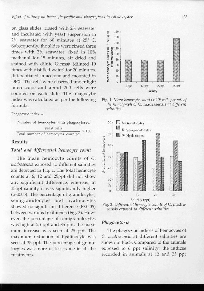
The mean hemocyte counts of C. nzndrnsensis exposed to different salinities are depicted in Fig. 1. The total hemocyte counts at 6, 12 and 25ppt did not show any significant difference, whereas, at 35ppt salinity it was significantly higher (p<0.05). The percentage of granulocytes, semigranulocytes and hyalinocytes showed no sigruficant difference (P<0.05) between various treatments (Fig. 2). How- ever, the percentage of semigranulocytes was high at 25 ppt and 35 ppt, the maxi- mum increase was seen at 25 ppt. The maximum reduction of hyalinocyte was seen at 35 ppt. The percentage of granu- locytes was more or less same in all the treatments.
6 ppt 12 ppt 25 ppt 35 ppt
Salinity
Fig. 1. Mean hemocyte count ( x lo4 cells per ml) of fhe hemolymph of C . madrasensis at diflerent salinities
Semigranulocytes
6 12 2 5 3 5
Salinity (ppt) Fig. 2. Diferential hemocyte counts of C . madra-
sensis exposed to diflerent salinities
Phagocytosis
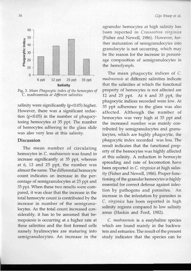
The phagocytic indices of hemocytes of C. mndrnsrnsis at different salinities are shown in Fig.3. Compared to the animals exposed to 6 ppt salinity, the indices recorded in animals at 12 and 25 ppt
34 Gijo Ittoop et al.
6 ppt 12 ppt 25 ppt 35 ppt
Salinity
Fig. 3. Mean Phagocytic index of the hemocytes of C. madrasensis at diferent salinities
salinity were significantly (p<0.05) higher. However, there was a significant reduc- tion (p<0.05) in the n ~ m b e r of phagocy- tosing hemocytes at 35 ppt. The number of hemocytes adhering to the glass slide was also very less at this salinity.
Discussion
agranular hemocytes at high salinity has been reported in Crassostrea virginica (Fisher and Newell, 1986). However, fur- ther maturation of semigranulocytes into granulocyte is not occurring, which may be the reason for the increase in percent- age composition of semigranulocytes in the hemolymph.
The mean phagocytic indices of C. madrasensis at different salinities indicate that the salinities at which the functional property of hemocytes is not affected are 12 and 25 ppt. At 6 and 35 ppt, the phagocytic indices recorded were low. At 35 ppt adherence to the glass was also affected. Although the number of hemocytes was very high at 35 ppt and the increased number was mainly con- tributed by semigranulocytes and granu- locytes, which are highly phagocytic, the phagocytic index recorded was less. The result indicates that the f~mctional prop- The mean number of circulating erty of the hemocytes was highly affected
hemocytes in C. madrasensis was fo~md to at this salinity. A reduction in hemocyte increase significantly at 35 ppt, whereas spreading and rate of locomotion have at 6, 12 and 25 ppt, the number was
almost the same. The differential hemocyte been reported in C , virginica at high salin-
count indicates an increase in the per- ity (Fisher and Newell, 1986). Proper fumc-
centage of semigranulocytes at 25 ppt and tioning of the granular hemocytes is highly
35 ppt. When these two results were com- essential for correct defense against infec-
pared, it was clear that the increase in the tion by pathogens and parasites. An
total hemocyte count is contributed by the increase in the infestation by parasites in C. virginica has been reported in high increase in number of the semigranu-
locytes. As the total count increased con- salinity regions compared to low salinity
siderably, it has to be assumed that he- areas (Haskin and Ford, 1982).
mopoiesis is occurring at a higher rate at C. madrasensis is a euryhaline species these salinities and the first formed cells which are fomd mainly in the backwa- namely hyalinocytes are maturing into ters and estuaries. The result of the present semigranulocytes. An increase in the study indicates that the species can be
E f i c t of salinity o n hemocyte profile and phagocytosis in edible oyster 35
cultured at salinities ranging from 12 to 25 ppt, without compromising the defense system. At 6 ppt, even though the total hemocyte count was normal, the phago- cytic index was significantly low com- pared to 12 and 25 ppt. At higher salini- ties hemocyte profile and function are affected, indicating a stressf~~l condition for the species as the immune system is compromised.
References
Anderson, C.B. and R.S. Anderson. 2000. Imm~~notoxicity of environmental pollutants in marine invertebrates. In: Fingerman, M. and Nagabhushanam R. (Eds.), Marine biotechnol- ogj. Published by Science Publishers Inc., NH, USA. 225pp.
Balquet, G. and M. Poder. 1985. A consideration of the cellular reactions in bivalve molluscs, with emphasis on haemocytic diseases. In: Ellis E.A. (Ed.), Fish and shellfislt Pathologj. Academic press, London. p. 381-385.
Bayne C.J., M.N. Moore, T.H. Carefoot and R.J. Thompson. 1979. Haemolymph functions in M!/tilus californianus: The cytochemistry of haemocytes and their response to foreign im- plants and haemolymph factors in phagocyto- sis. ].lnvertebr. Patlzol., 34: 1-20.
Chen, J.H. 1996. Haemolymph collection in the Abalone (Haliotis diversicolor). Actn Zoologica Taizonnica, 7 : 61-71.
Cheng, T.C. and D.A. Foley. 1975. Haemolymph cells of the bivalve mollusc Mrrcennria mercennria: An electron microscopical study. J. Invertebr. Pnthol., 26: 341-351.
Chu, F.L. E. 2000. Defense mechanisms of marine bivalves. In: Fingerman, M. and Nagabhushanam R. (Eds.), Mnrine biotechnol- ogy. Published by Science Publishers Inc., NH, USA. 225pp.
Fisher, W.S. and R.I.E. Newell. 1986. Salinity effects on the activity of granular hemocytes of Ameri- can oyster, Crassostrea virginica. Biol. B I ~ . , 170: 122-134.
Gilles, R 1979. Mechanisnzs of osmoreg~ilntion in nni- mals. Wiley Interscience. New York. 667 pp.
Haskin, H.H. and S.E. Ford. 1982. Hnplosporidillm nelsonii (MSX) on Delaware Bay seed beds: A host parasite relationship along a salinity gra- dient. J. Invertebr. Pathol., 40: 388-405
Moore, M.N. and D.M. Lowe. 1977. The cytology and cytochemistry of the haemocytes of Mytilus edulis and their response to the experimentally injected carbon particles. Ibid. 29: 18-30.
Nakayama, K., A.M. Nomoto, M. Nishijima and T. Maruyama. 1997. Morphological and functional characterization of hemocytes in the clam Tridacna crocea. Ibid. 69 : 105-111.
Rasmussen, L.P.D., E. Hage and 0. Karlog. 1985. An electron microscope study of the circulating leucocytes of the marine mussel, Mytilils edtilis. Ibid. 45: 158-167.
Rodrick, G.E. and S.A. Ulrich. 1984. Microscopical studies on the hemocytes of bivalves and their phagocytic interaction with the selected bacte- ria. Dis. Mar. Org., 37: 167-176.