Embed Size (px)
Citation preview

1
Effect of PRGF on Bone Grafting of Intrabony Defects in Aggressive Periodontitis Patients – A Pilot Study
Rafik Dib, DDS, MSD, Pharm D†, A. Archontia Palaiologou, DDS, † Pooja Maney, BDS,
MPH, PhD†
Department of Periodontics Louisiana State University Health Science Center School of Dentistry 1100 Florida Avenue New Orleans, LA 70119 Keywords: aggressive periodontitis, allografts, bone regeneration; periodontal attachment loss Abbreviations: plasma rich in growth factors (PRGF), Aggressive periodontitis (AP), Freeze Dried Bone Allograft (FDBA), probing depth (PD), clinical attachment level (CAL), bleeding on probing (BOP), plaque index (PI), gingival index (GI),
ABSTRACT (248/250 words)
Background: The use of plasma rich in growth factors (PRGF) is often incorporated in
regenerative periodontal surgical procedures. However, the actual benefit of adding
PRGF in intrabony defects in aggressive periodontitis patients remains undocumented.
The purpose was to evaluate the potential of plasma rich in growth factors (PRGF) to
enhance defect fill and attachment level gain of intrabony defects in aggressive
periodontitis patients in combination with Freeze Dried Bone Allograft (FDBA).
Methods: Six patients with localized aggressive periodontitis with bilateral comparable
intrabony defects (n=14) were treated. Sites were randomly assigned into one of two
groups: control (FDBA) or test (FBDA/PRGF). The surgical procedure consisted of flap
reflection, debridement of the defect, root planning, and placement of the bone graft in
the defect, followed by flap closure. Clinical and radiographic parameters were
assessed at baseline and 6 months after treatment.
Results: At baseline, there were no statistically significant differences between groups.
At 6 months, both groups experienced improvement in terms of probing depth reduction,
clinical attachment level (CAL) gain, clinical bone level gain and radiographic bone fill.
However, no statistically significant difference was observed between the two groups.
The test (PRGF/FDBA) and control group (FDBA) experienced a mean clinical

2
attachment level gain of 1.93mm and 2.21mm, respectively (P=0.36). The mean bone
gain for the test group was 2.5mm, and 1.86mm for the control group (P=0.27).
Conclusion: Addition of PRGF on bone grafting of intrabony defects in aggressive
periodontitis patients did not significantly enhance defect fill and attachment level gain.
INTRODUCTION
Aggressive periodontitis (AP) is a disease that is characterized by rapid
attachment loss and bone destruction around teeth. AP has a strong familial tendency
and is associated with immunologic defects and a distinct microbial flora. (1) It mainly
targets healthy teenagers and young adults with the highest incidence among African
Americans (2.6%) followed by Hispanic Americans (0.5% - 1.08%), whereas White
Americans had the lowest prevalence of aggressive periodontitis (0.06% - 0.17%). (2, 3)
AP presents in two forms; localized and generalized. Localized AP usually has a
circumpubertal onset (11-14 years of age), with periodontal damage (attachment and
bone loss) to the permanent first molars and incisors. Generalized AP usually affects
patients around 30-35 years of age, with generalized attachment and bone loss
affecting at least three permanent teeth other than first molars and incisors [Lang, 1999
#4].
The overall goals of the treatment of AP are not different from those in patients
with chronic periodontitis. A nonsurgical phase of scaling and root planing with systemic
antibiotics takes place to alter or eliminate the microbial etiology and contributing risk
factors. In addition, when indicated, surgical approach such as regenerative treatment
of lost periodontal tissues may be attempted. (4, 5)
Periodontal regeneration is the process by which a lost or injured part of the
periodontium (including the periodontal ligament, cementum and alveolar bone) is

3
reconstituted or reproduced. (6) AP may present with a pattern of vertical or angular
bone loss defects, also known as a periodontal intrabony defects, which may respond
well to regenerative procedures. (5)
Various grafting materials used for periodontal regeneration in patients
diagnosed with AP have been well documented. Using Freeze-Dried Bone Allograft
(FDBA) alone or with adjunctive systemic tetracycline showed improvement in
periodontal parameters and defect bone fill (7, 8). A Combination of root
biomodification, Decalcified Freeze-Dried Bone Allograft (DFDBA) and barrier
membrane is also an effective therapy for periodontal intrabony defects in AP. (9)
Alloplastic grafting materials have also been tested in a split-mouth design in
combination with tetracycline where significant decreases in defect depth and pocket
depth were detected. (10)
More recently, growth factors have been shown to also play an important role in
tissue development and healing. Among the growth factors, plasma rich in growth
factors (PRGF)* as described by Anitua et al, has demonstrated the potential to influence
bone regeneration. (11) In recent in vitro studies, PRGF demonstrated a stimulating
effect on both alveolar osteoblasts and gingival fibroblasts , suggesting a possible
beneficial effect in regeneration of periodontal tissues. (11, 12)
PRGF takes full advantage of concentrated platelets and stored growth factors
by eliminating erythrocytes and leucocytes from the product. (13) The removal of
leukocytes from the product also attempts to avoid the pro-inflammatory effects of the
* Endoret ®, BTI Biotechnology Institute, Vitoria, Spain.

4
proteases and acid hydrolysases.(14) PRGF releases growth factors and proteins that
are involved in wound healing. These growth factors and proteins include finbrinogen,
fibronectin, vitronectin, platelet derived growth factor, transforming growth factor β,
vascular endothelial growth factor, insulin like growth factor, angiopoietin, platelet factor
4, and thrombospondin. (13)
The use of PRGF has been reported to enhance healing of extraction sockets by
minimizing postoperative complications and stimulating the hard and soft tissue
regeneration.(15) It has also been used in combination with xenografts as grafting for
maxillary sinus grafting in humans.(16, 17) A case series has indicated a reduction of
pocket depth and gain in attachment using platelet gel biotechnology in AP patients who
had shown a refractory response to previous periodontal treatments.(18) However, a
recent systematic review on autologous platelet concentrates was unable to consider
the effect of PRGF in the treatment of intrabony defects due to the absence of
randomized clinical studies [Saurav 2016 #20]. To our knowledge, the effectiveness of
PRGF in patients diagnosed with AP is still unknown. The objective of this study is to
evaluate the additive effect of PRGF with FDBA† in the treatment of intrabony defects in
aggressive periodontitis. Our hypothesis is that the addition of PRGF to FDBA will
enhance defect fill and attachment level gain.
MATERALS AND METHODS
† Creos allo.gain cortico-cancellous (0.25-1.00 mm) – Nobel Biocare

5
Following LSUNO-IRB approval (IRB# 9004), participants were recruited among
patients referred to the Department of Periodontology at the Louisiana State University
for treatment of aggressive periodontitis (AP), diagnosed according to the criteria
established at the 1999 international workshop for the classification of periodontal
diseases and conditions.(19) 47 Patients were screened, 8 were eligible, consented and
were enrolled. All 8 were African American females, aged 14 to 40 years old.
Inclusion criteria were: 1) male or female between 13-40 years of age; 2) must be
diagnosed with localized or generalized aggressive periodontitis with bilateral intrabony
defects that require grafting, ideally with symmetrical walls and at least 3mm deep
around the tooth/teeth for which periodontal flap surgery with bone grafting is indicated
(one, two and three wall intrabony defects); 3) must be of sufficiently good health to
undergo routine dental treatment including gingival surgery; 4) must demonstrate
acceptable oral hygiene (Modified O’Leary plaque score of ≥80%) prior to surgical
therapy
Exclusion criteria were: 1) have any requirements for antibiotic premedication for
heart murmurs, artificial joints or any other condition; 2) have a history of significant
heart, stomach, liver, kidney, blood, immune system or other organ impairment or
systemic disease that would preclude their undergoing the proposed treatment; 3) are
taking drugs known to interfere with wound healing (e.g. corticosteroids,
chemotherapeutic anti-cancer drugs, immune modulators etc.) or have received such
drugs within 4 weeks of treatment. 4) have taken systemic antibiotics or any
investigational drugs anytime in the previous month; 5) have other dental conditions
likely to require treatment, necessitating exit from the study e.g. decision to extract a

6
tooth that was considered for the study due to failure of root canal therapy or extensive
tooth decay beyond the limit that is considered successfully restorable; 6) have had
periodontal surgery on the involved teeth within the last 6 months; 7) cannot comply
with the extra treatment visits and follow-up visits for up to 6 months; 8) lack of
appropriate periodontal bony defects; 9) heavy alcohol intake or use of illicit drugs (The
Substance Abuse and Mental Health Services Administration (SAMHSA), defines heavy
drinking as drinking 5 or more drinks on the same occasion on each of 5 or more days
in the past 30 days. SAMHSA defines binge drinking as drinking 5 or more alcoholic
drinks on the same occasion on at least 1 day in the past 30 days); 10) female subjects
must not be pregnant or nursing for study surgeries. If they become pregnant during the
2 year follow up period, only clinical measurements would be made, but no radiographs
would be taken.
Study Design
Defects were grafted with either FDBA/PRGF or FDBA alone utilizing a randomized
split mouth design (Figure 1). The randomization was performed by a coin toss.
Clinical Parameters
Clinical parameters were assessed using a stent made of clear suck-down A+ plastic
material‡ trimmed to the coronal 2/3 and a manual UNC-15 probe.
Defect-specific assessments were made at the deepest point of the defect (base of the
defect (BD)). Interproximal embrasures of the stent were used as fixed reference points
‡ Essix A+® Plastic

7
to aid in alignment (figure 2b). All measurements were completed by the same clinician
(RD).
For each patient, the following parameters were assessed on two sites per tooth
(mesial buccal and mesial lingual) at baseline and 6 months reentry: 1) Probing depth
(PD); 2) Recession (REC); 3) Bleeding on probing (BOP); 4) plaque index (PI);(20) 5)
gingival index (GI).(20) The parameters recorded during surgery are 1) stent to clinical
base of defect 2) CEJ to clinical base of defect.
Radiographic Parameters
Standardized periapical radiographs were obtained at baseline and at 6 months
reentry using a radiographic stent. Bite-record was taken during baseline with
Thixotropic Vinyl Polysiloxane§ to standardized the sensor position intra-orally.
The distance from the radiographic CEJ to the base of the defect at baseline and at 6
months of reentry were measured using a digital radiographic software**. Radiographic
bone fill was assessed by calculating their mean difference.
Initial examination:
A complete medical and dental history was taken. Subjects received a routine
periodontal examination including charting of probing depths, attachment level and
gingival recession at 6 sites per tooth. Mobility, furcation involvement, gingival index
§ Blu-Mousse® - Parkell Inc ** Schick CDR DICOM 5 Digital-X-ray-Software by Dentsply Sirona Imaging

8
and plaque index were also assessed. Full mouth series of radiographs and intraoral
photographs were taken.
Initial therapy:
All subjects received scaling and root planing of teeth with PD ≥4mm. Antibiotics
(Amoxicillin 500mg and Metronidazole 250mg three times a day for 10 days) were
prescribed. Detailed oral hygiene instructions were given; brushing twice daily using a
soft bristle tooth brush using modified Bass technique and floss once daily.(21) After 4-
6 weeks, re-evaluation of initial therapy was completed. Standardized measurements
were recorded around the teeth that require grafting of defects using a custom-made
stent.
Surgical therapy:
Preparation of the autologous PRGF: 10-20 mL of peripheral blood were drawn using
venipuncture from the subject’s arm. PRGF was prepared using the protocol described
by Anitua E [Anitua, 2014 #24]. Briefly, this consists of centrifuging the blood sample for
8 minutes at ambient temperature. After centrifugation, the blood will be separated into
3 phases (erythrocyte cells at the bottom, above a thin layer of leukocytes (buffy coat),
and plasma at the top). The plasma layer is divided into two fraction; fraction 1 (F1) and
fraction 2 (F2). F2 of PRGF is the 2mL of plasma just above the buffy coat. It is the
fraction that is richest in platelets between 2-3 times higher than that of peripheral
blood.(13) Calcium chloride (PRGF-Activator) was added to the carefully pipetted out
F2, according to the manufacturer’s instructions. The graft material (FDBA) was mixed

9
into the serum right after activation. The mixture is then placed in a 37ºC furnace††
around 10 minutes for a fibrin clot formation. The resulting gel-like substance containing
the graft was then placed into the periodontal intrabony defect.
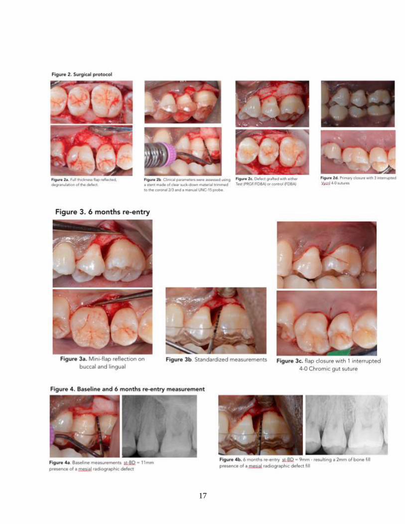
Surgical treatment of intrabony defects (Figure 2.): Intrasulcular incisions were made
including at least one tooth on either side of the tooth to be treated and full thickness gingival
flaps was elevated. The surgical sites were thoroughly debrided to remove granulation tissue
and calculus deposits using hand and ultrasonic instruments. The defects were measured
using a custom-made stent (St-BD) and the number of remaining walls were determined.
Defects were grafted randomly with the materials under study utilizing a split mouth design.
One side treated with PRGF+FDBA and the other side was treated with FDBA alone. Flaps
were sutured using 4-0 Vicryl interrupted sutures attempting to gain primary closure. Post-
operative instructions were given. Post-operative examinations were done to monitor healing
of the surgical sites at 1 week, 2 weeks and 3 months. All patients were prescribed amoxicillin
500mg three times daily for 7 days, ibuprofen 600mg every 6-8 hours and chlorhexidine
gluconate 0.12% rinse twice daily for 2 weeks. Sutures were removed 1 week postoperatively.
Recall appointment were scheduled 2 weeks post-operatively and 3 months for supragingival
debridement and reinforcement of oral hygiene.
Evaluation of treatment:
†† Plasmaterm H. - BTI Biotechnology Institute

10
Clinical and radiographic data were recorded at 6 months following surgery.
Clinical parameters were re-assessed by a re-entry procedure at 6 months, utilizing a
mini-flap that just reflected the interproximal buccal and lingual gingival papilla to
visualize the grafted site and to take measurements using the custom-made stent
(Figure 3). Probing depths, attachment level, surgical height of crest of alveolar bone,
and base of defect were measured. Standardized radiographs of the study sites were
taken at baseline and 6 months re-entry (Figure 4).
Statistical Analysis
Pre-treatment measurements were compared to the measurements recorded at 6 and
12 months. A one-sample t-test was used to determine if there is a significant clinical
attachment gain and defect fill in intrabony periodontal defects treated with PRGF/FBDA
at both time points. Significance was set at P=0.05.
RESULTS
Of 47 screened patients, 8 eligible and consenting participants were accepted.
2/8 patients were excluded due to shape of defect not meeting criteria at surgery
(intrabony defect <3mm). Thus, 6 patients completed the study with bilateral defects
(n=14). 1 out of the 6 patients had two bilateral defects, one in each arch. All defects
were located on the mesial of maxillary or mandibular first molars. Patient’s ages
ranged from 16 to 32 years, with a mean age of 19.5 year. They were all African
American, females, nonsmokers. Patient demographics and defect assignment are
reported on Table 1.

11
Baseline and 6 months parameters are reported in Table 2. At baseline, there
were no statistically significant differences between control and test groups for any of
the recorded parameters.
The changes in clinical parameters, comparing baseline to 6 months values, are
reported in (Figure 5a-f). At 6 months, both groups experienced improvements in terms
of probing depth reduction, clinical attachment level (CAL) gain, and bone level gain. In
addition, both resulted in similar level of radiographic bone fill.
The test (PRGF/FDBA) and control group (FDBA) experienced a mean clinical
attachment level gain of 1.93mm and 2.21mm, respectively (P=0.36) (Figure 5a). Mean
Probing depth decrease was 1.93mm for the test group and a 1.93mm for the control
group (P=0.36) (Figure 5b). Mean bone gain for the test group was 2.5mm, and a
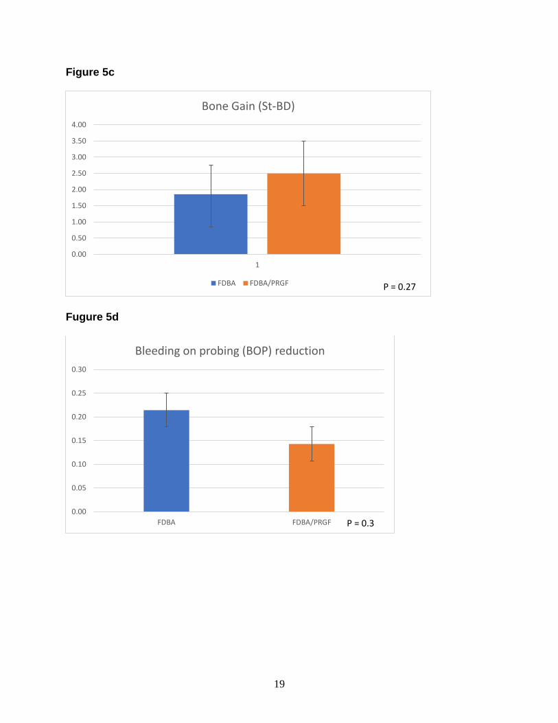
1.86mm for the control group (P=0.27) (Figure 5c). Clinical parameters such as BOP,
PI, GI had all improved from baseline to 6 month. Mean BOP reduction for the test
group was 0.14, and a 0.21 for the control group (P=0.3) (Figure 5d). Mean GI reduction
of 0.36 for the test group and a 0.5 reduction for the control group (P=0.18) (Figure 5e).
Mean PI reduction of 0.29 for the test group and 0.14 for the control group (P=0.09)
(Figure 5f).
CONCLUSION
In summary, all clinical parameters improved within the control and test groups at
6 months as compared to baseline measurement. No statistically significant difference
was found between the two groups. This study demonstrated that the addition of plasma
rich in growth factors (PRGF) in the treatment of intrabony defects did not enhance

12
defect fill and clinical attachment level gain in patients with aggressive periodontitis
(AP). Further long-term and large-sample size evaluation may be required to further
confirm this pilot study’s results.
DISCUSSION
The present study evaluated the additive effect of PRGF with FDBA in the
treatment of intrabony defects in aggressive periodontitis patients. To avoid the effect of
natural variations between different individuals, gender, race, smoking status and
location of defects were matched throughout the study. 14 defects in 6 female, African
American patients, non-smokers, with bilateral mesial of first molar comparable defects
were randomly assigned to either the test or the control group.
Patient’s compliance with dental appointment was a challenge for the recruited
patient’s population. Multiple failed appointments have occurred in the non-surgical
phase. According to Modin et al, they reported that the patient group with aggressive
periodontitis interrupted the periodontal treatment significantly more often (46%)
irrespective of background factors and risk factors, compared to patients with chronic
periodontitis (34%).(22) In addition, the non- compliant patients had significantly deeper
periodontal pockets at baseline as well as significantly more sites with bleeding at
probing (p<.01).(22) To improve compliance, educating and increasing awareness to
both patients and parents took a primary role.(23) In this study, verbal and written
instructions was provided to both the patients and their parents. The goal of the
provided instructions was to increase awareness of the severity of the disease and
stress the necessity to continue the surgical phase and the 6 months re-entry visit.
A similar study design by Ravi et al. was recently published to evaluate the effect

13
of PRGF with GTR as well as GTR alone in the treatment of intrabony defects in chronic
periodontitis patients.(24) At re-entry, all clinical parameters showed improvement but,
intergroup comparison did not show a statistical difference.(24) Thus, there was no
additional benefit of PRGF when used along with GTR in treatment of chronic
periodontitis patients.(24) Our findings are in agreement with the findings of that study.
There were some limitations to our study. Since it is a pilot study, the sample size
was small. Only 6 patients completed the study for a total of 14 defects using the split
mouth design.
Effort was made to identify comparable or symmetrical bilateral defects in the
same participant. However, due to the complex anatomy of each defect, it was difficult
to match the osseous defects in the test and control groups in all participants. The
randomization and the selection of only mesial first molar defect may have helped to
control this variability.
Furcation involvement was observed in more than half of the defects. Furcation
involvement in molar teeth render a less favorable prognosis.(25) In addition, evidence
shows that grafting in a furcation area results in about 50% improvement for grade II
furcation.(26) Of these, 20% were completely filled and 33% improved from grade II to
grade I.(26)
Standardized radiographs taking presented additional challenges. In some
cases, the measurements from the CEJ to the base of the defect was very difficult, due
to the CEJs being difficult to identify or new bone levels were not well defined. Even
though radiographs were standardized, data collection varied widely between
examiners. For this reason, radiographic analysis was excluded from the results of this

14
study.
Finally, defective restorations and margins were found in several molars where
old restorations should have been replaced prior to the surgical procedure for better
surgical prognosis.
The use of PRGF has several advantages in clinical practice in comparison with
other regenerative materials, as it is easier to handle in combination with grafting
materials, results in accelerated post-operative soft tissue healing and causes less
patient discomfort.(27) However, there is also a lack of evidence identifying the effect of
PRGF on postoperative symptoms like pain and swelling.
As the existing study exhibits methodological limitations, future studies with a long-
term evaluation and a larger sample size are necessary to confirm the clinical stability of
this therapeutic approach in addition to a larger sample size. There is a need for further
randomized controlled clinical trials to provide accurate information on the effect of
PRGF for periodontal intrabony defect treatment in patients with aggressive
periodontitis.

15
Table 1.
Demographics of study population
research code Age Race Gender FDBA FDBA+PRGF
X1 14 AA F 19,14 30,3
X2 15 AA F 30 19
X3 16 AA F 19 30
X4 40 AA F 14 3
X5 18 AA F 19 30
X6 14 AA F 14 3
Table 2.
Mean defect characteristic at baseline.
FDBA
(n=7)
PRGF/FDBA
(n=7) P value
PD 8.07 8.64 0.2
REC 0 0 n/a
BOP 0.64 0.64 0.55
GI 0 0 n/a
PI 0 0 n/a
St- BD 11.93 12.64 0.2

16
Figure 1. Study Design
17

18
Figure 5a
Figure 5b
0.00
0.50
1.00
1.50
2.00
2.50
3.00
3.50
4.00
4.50
1
Attachment level gain
FDBA FDBA/PRGFP = 0.36
0.00
0.50
1.00
1.50
2.00
2.50
3.00
3.50
4.00
4.50
1
Probing Depth Decrease
FDBA FDBA/PRGFP = 0.36
19
Figure 5c
Fugure 5d
0.00
0.50
1.00
1.50
2.00
2.50
3.00
3.50
4.00
1
Bone Gain (St-BD)
FDBA FDBA/PRGF P = 0.27
0.00
0.05
0.10
0.15
0.20
0.25
0.30
FDBA FDBA/PRGF
Bleeding on probing (BOP) reduction
P = 0.3

20
Figure 5e
Figure 5f
-0.60
-0.50
-0.40
-0.30
-0.20
-0.10
0.00
FDBA FDBA/PRGF
Gingival Index (GI) reduction
P = 0.18
-0.40
-0.35
-0.30
-0.25
-0.20
-0.15
-0.10
-0.05
0.00
FDBA FDBA/PRGF
Plaque Index (PI) reduction
P = 0.09

21
1. Califano JV, Research S, Therapy Committee American Academy of P. Position paper:
periodontal diseases of children and adolescents. J Periodontol. 2003;74(11):1696-704.
2. Albandar JM, Brown LJ, Loe H. Clinical features of early-onset periodontitis. J Am Dent
Assoc. 1997;128(10):1393-9.
3. Loe H, Brown LJ. Early onset periodontitis in the United States of America. J
Periodontol. 1991;62(10):608-16.
4. Parameter on aggressive periodontitis. American Academy of Periodontology. J
Periodontol. 2000;71(5 Suppl):867-9.
5. Teughels W, Dhondt R, Dekeyser C, Quirynen M. Treatment of aggressive periodontitis.
Periodontol 2000. 2014;65(1):107-33.
6. Lang NP. Focus on intrabony defects--conservative therapy. Periodontol 2000.
2000;22:51-8.
7. Mabry TW, Yukna RA, Sepe WW. Freeze-dried bone allografts combined with
tetracycline in the treatment of juvenile periodontitis. J Periodontol. 1985;56(2):74-81.
8. Yukna RA, Sepe WW. Clinical evaluation of localized periodontosis defects treated with
freeze-dried bone allografts combined with local and systemic tetracyclines. Int J Periodontics
Restorative Dent. 1982;2(5):8-21.
9. Sant'Ana AC, Passanezi E, Todescan SM, de Rezende ML, Greghi SL, Ribeiro MG. A
combined regenerative approach for the treatment of aggressive periodontitis: long-term follow-
up of a familial case. Int J Periodontics Restorative Dent. 2009;29(1):69-79.
10. Evans GH, Yukna RA, Sepe WW, Mabry TW, Mayer ET. Effect of various graft
materials with tetracycline in localized juvenile periodontitis. J Periodontol. 1989;60(9):491-7.
11. Anitua E, Tejero R, Zalduendo MM, Orive G. Plasma rich in growth factors promotes
bone tissue regeneration by stimulating proliferation, migration, and autocrine secretion in
primary human osteoblasts. J Periodontol. 2013;84(8):1180-90.
12. Anitua E, Troya M, Orive G. Plasma rich in growth factors promote gingival tissue
regeneration by stimulating fibroblast proliferation and migration and by blocking transforming
growth factor-beta1-induced myodifferentiation. J Periodontol. 2012;83(8):1028-37.
13. Anitua E, Andia I, Ardanza B, Nurden P, Nurden AT. Autologous platelets as a source of
proteins for healing and tissue regeneration. Thromb Haemost. 2004;91(1):4-15.
14. Schnabel LV, Mohammed HO, Miller BJ, McDermott WG, Jacobson MS, Santangelo
KS, et al. Platelet rich plasma (PRP) enhances anabolic gene expression patterns in flexor
digitorum superficialis tendons. J Orthop Res. 2007;25(2):230-40.
15. Anitua E, Murias-Freijo A, Alkhraisat MH, Orive G. Clinical, radiographical, and
histological outcomes of plasma rich in growth factors in extraction socket: a randomized
controlled clinical trial. Clin Oral Investig. 2015;19(3):589-600.
16. Del Fabbro M, Bortolin M, Taschieri S, Weinstein RL. Effect of autologous growth
factors in maxillary sinus augmentation: a systematic review. Clin Implant Dent Relat Res.
2013;15(2):205-16.
17. Khouly I, Pardinas Lopez S, Aliaga I, Froum SJ. Long-Term Implant Survival After 100
Maxillary Sinus Augmentations Using Plasma Rich in Growth Factors. Implant Dent.
2017;26(2):199-208.
18. Mauro S, Orlando L, Panzoni R, Orlando PF. Platelet gel biotechnology applied to
regenerative surgery of intrabony defects in patients with refractory generalized aggressive
peridontitis. Case report. Minerva Stomatol. 2003;52(7-8):401-12.

22
19. Armitage GC. Development of a classification system for periodontal diseases and
conditions. Ann Periodontol. 1999;4(1):1-6.
20. Loe H. The Gingival Index, the Plaque Index and the Retention Index Systems. J
Periodontol. 1967;38(6):Suppl:610-6.
21. Poyato-Ferrera M, Segura-Egea JJ, Bullon-Fernandez P. Comparison of modified Bass
technique with normal toothbrushing practices for efficacy in supragingival plaque removal. Int J
Dent Hyg. 2003;1(2):110-4.
22. Modin C, Abadji D, Adler L, Jansson L. Treatment compliance in patients with
aggressive periodontitis - a retrospective case-control study. Acta Odontol Scand. 2017;75(2):94-
9.
23. Wilson TG, Jr., Hale S, Temple R. The results of efforts to improve compliance with
supportive periodontal treatment in a private practice. J Periodontol. 1993;64(4):311-4.
24. Ravi S, Malaiappan S, Varghese S, Jayakumar ND, Prakasam G. Additive Effect of
Plasma Rich in Growth Factors With Guided Tissue Regeneration in Treatment of Intrabony
Defects in Patients With Chronic Periodontitis: A Split-Mouth Randomized Controlled Clinical
Trial. J Periodontol. 2017;88(9):839-45.
25. Miller PD, Jr., McEntire ML, Marlow NM, Gellin RG. An evidenced-based scoring index
to determine the periodontal prognosis on molars. J Periodontol. 2014;85(2):214-25.
26. Evans GH, Yukna RA, Gardiner DL, Cambre KM. Frequency of furcation closure with
regenerative periodontal therapy. J West Soc Periodontol Periodontal Abstr. 1996;44(4):101-9.
27. Mozzati M, Martinasso G, Pol R, Polastri C, Cristiano A, Muzio G, et al. The impact of
plasma rich in growth factors on clinical and biological factors involved in healing processes
after third molar extraction. J Biomed Mater Res A. 2010;95(3):741-6.