Embed Size (px)
Citation preview

612
AJCS 8(4):612-624 (2014) ISSN:1835-2707
Effect of nanoparticles on biological contamination of in vitro cultures and organogenic
regeneration of banana
Mohamed Naser Helaly, Mohamed Ahmed El-Metwally2, Hanan El-Hoseiny
3, Samar Abdelaziz
Omar4, Nabil Ibrahim El-Sheery
5*
1Agricultural Botany Department, Faculty of Agriculture, Mansoura University, El-Mansoura, Egypt 35516
2Mycological Research Department, Plant Pathology Research Institute, Agricultural Research Center, Giza,
Egypt 3Horticultural Research Institute, Agricultural Research Center, Giza, Egypt
4Genetic department, Faculty of Agriculture, Tanta University, Egypt
5Agricultural Botany Department, Faculty of Agriculture, Tanta University, Tanta, Egypt
*Corresponding author: [email protected]
Abstract
This study aimed to identify biological contaminants of banana in vitro cultures. We also tested the effect of Zn or ZnO nanoparticles
on elimination of some bacterial and fungal contaminants and their influence regeneration. Nine strains of bacterial contaminants
(Cellulomonas uda, Cellulomonas flarigena, Corynebacterium panrometabolum, Bacillus megaterium, Staphylococcus spp.,
Klebsiella spp., Erwinia cypripedii, Pseudomonas spp. and Proteus spp.) and four fungal (Fusarium spp., Aspergillus spp.,
Penicillium spp. and Candida spp.) contaminants were identified in nanoparticles-free media of banana in vitro cultures. They
eventually led the explants death. The contamination-free cultures of banana in vitro cultures were obtained as a result of application
of nano Zn and ZnO particles to the culture MS media, with no negative effect on regeneration. The callus growth decreased while
total proline as well as SOD, CAT and POX activities were increased significantly, when the nanoparticles doses increased. The
highest percent of somatic embryogenesis was observed in MS media supplemented with 100 mg/L nano Zn followed by nano ZnO.
Excellent shooting, rooting and regenerated plantlets were observed also in MS+100 mg/L nano Zn and ZnO. Regenerated plantlets
were successfully acclimatized with about 98% efficiency for the experimental period (one month). Nanoparticles treated somaclones
accumulated more proline, chlorophyll, antioxidant enzymes activity and developed more dry weight accumulation than the control.
In conclusion, the microbial contaminants in banana in vitro culture can effectively be eliminated by incorporation of nano Zn and
nano ZnO particles on growth media at different concentrations. However, 100 mg/L dose was preferable because it showed the best
effects on increasing the regeneration of plantlets with well-formed root systems. Further studies are needed to investigate the
mechanisms and the side effects of nanoparticles on genetic stability of banana in vitro cultures.
Keywords: Banana, Nanoparticles, Phytotoxicity, Microbial contaminates, Regeneration.
Abbreviations: Zn_Zinc; ZnO_Zinc Oxide, SOD_superoxide dismutase; POX_peroxidase and CAT_catalase.
Introduction
Plant cells growing in vitro are considered to be under some
degree of stress and may be predisposed to direct infection,
even by microbes which are not normally pathogenic to them.
Thus, microbial contaminants are the major challenges in
plant’s in vitro propagation during different stages of culture
processes such as initiation of callus and sub-culturing. It was
reported that, sub-culturing process is a major source of
contamination (5-15% for each sub-culture) due to
insufficient sterilization of explants, growth media, working
tools and operators (Omamor et al., 2007). Ödutanyo et al.
(2004) found that the major bacterial contaminants are
Pseudomonas syringae, Bacillus lichensformic, B. subtilis,
Comebacterium sp. and Erwinia spp. while Alternaria tenius,
Aspergillus niger, Aspergillus fumigates and Fusarium
culmorum have been reported to be the major fungal
contaminants frequently occur in plant tissue culture
(Msogoya et al., 2012). In banana (Mousa sapientum, L.),
huge number of explants are destroyed in the cultures due to
endogenous microbial contaminants. It was reported that
about 40-60% of banana in vitro cultures lost in spite of using
main aseptic reliable procedures (Msogoya et al., 2012).
Moreover, the growth media, in which the plant tissue is
cultivated, is also a source of nutrients for microbial growth,
both original constituents of the medium and exudates from
the plant cells. Thus, pathogens, endophytes, epiphytes and
incidental contaminants may all occur and interfere with
growth of the plant tissue culture and compete for nutrients.
In addition, some of them produce phytotoxins which result
in culture mortality, tissue necrosis and reduces shoot
proliferation and rooting (Khane 2003). For instance,
Aspergillus niger and A. flavus have reportedly produced
oxalate and aflatoxin poison, respectively, that kill plant
tissues (Obuekwe and Osagie 1989). To date, production of
banana plants through in vitro micropropagation has become
routine work in commercial planting in many private sectors

613
of Egypt. Most of these works have been hampered by
microbial contaminants. Generally, surface sterilization of the
explants eliminates most epiphytic contaminants except
endophytic ones (Habiba et al., 2002). An application of
systemic fungicides before the collection of plant materials
can suppress microbial contaminants in plant in vitro cultures
(Mngomba et al., 2007).
However, microbial contaminants at the base of the
explants and around them are a great problem. Alternatively,
an incorporation of antibiotics and antifungal into the growth
media of plant cultures has been suggested to eliminate
microbial contaminants (Habiba et al., 2002). Moreover,
toxicity of nanoparticles was also suggested for their
sensitivity, simplicity, low cost and suitability for unstable
chemicals or samples (Brunner et al., 2006). Nanotoxicology,
an emerging discipline is receiving increasing attraction and
has been the research focus of many publications (Wu 2003;
Nel et al., 2006). Engineered nanoparticles can be grouped
into four types. The metal based materials such as nanogold,
nanozinc (nano Zn) and nanoscale metal oxides like ZnO and
Al2O3 are among common nanoparticles (USEPA, 2005).
Some nanoparticles, such as Fullerene and TiO2 have been
widely used as test materials to reveal their nanotoxicity
mechanisms. However, available information on the topic is
too scarce to reach any consensus on nanotoxicity and its
mechanism, particularly for the nano Zn and nano ZnO
particles which used in this study. These nanoparticles
generally showed negative effects on the tested target
organisms or cell despite in different treatment methods used
(Yang and Watts 2005 and Wang et al., 2006) and have a
dose dependent response (USEPA, 2005). There are many
unresolved issues and challenges concerning the biological
effects of nanoparticles (<100 nm). Attentions to the
appropriate experimental design and interpretation are needed
to provide a defensible scientific understanding of the
biological effects of nanoparticles (Murachov, 2006).
Therefore, the objectives of this study were (1) to provide
new information about phytotoxicology of nano Zn and nano
ZnO particles incorporated into the growth media; (2)
identification of bacterial and fungal contaminants of banana
in vitro cultures; and (3) to evaluate the efficacy of these
nanoparticles on the explants regeneration.
Results and Discussion
Identification of microbial contaminants
Presence of bacterial and fungal contaminants was noticed
only in nanoparticles-free media (control) throughout the
developmental stages of banana shoot tip explants
regeneration (Fig. 1).
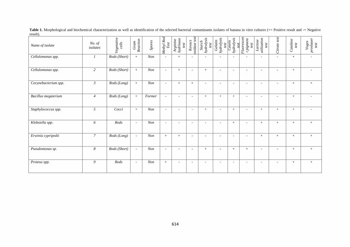
Nine bacterial strains were isolated from the contaminated
cultures. These contaminants of banana in vitro cultures and
the essential biochemical tests are presented in Table (1).
Four strains were gram positive, whereas the rest were gram
negative. The gram positive isolated were Cellulomonas uda,
Cellulomonas flarigena, Corynebacterium panrometabolum
and Bacillus megaterium. The gram negative isolates were
Staphylococcus spp., Klebsiella spp., Erwinia cypripedii,
Pseudomonas spp. and Proteus spp. All of the nine bacterial
strains were non spore former except Bacillus megaterium
strain. Moreover, three bacterial strains out of nine isolated
were exogenous bacteria (Proteus spp., Erwinia spp. and
Staphylococcus spp.) and the rest were endophytic. In banana
culture, Msogoya et al. (2012) identified only four exogenous
and endophytic bacterial strains from the isolated bacteria
contaminants. They isolated Proteus spp., Erwinia spp.,
Klebsiella spp. and Staphylococcus spp. The other isolated
strains in this study have been frequently reported in other
plant tissue cultures (Habiba et al., 2002 and Odutanyo et al.,
2007).
Several workers (Meghwal et al., 2000 and Khane, 2003)
indicated that exogenous bacteria are generally easy to
eliminate using normal surface sterilization techniques. Being
on plant surface, the occurrence of exogenous bacteria in
plant tissue cultures in the present investigation may be due
to an insufficient surface sterilization of explants, tools and
culture jars. Moreover, the growth media, in which the plant
tissues are cultivated, is a good source of nutrients for
microbial growth. The endophytical strains like Cellulomonas
and Klebsiella has been isolated in internal tissues of banana,
maize and wheat (Markins et al., 2003). Khane et al. (2003)
reported that endophytic bacteria are beneficial to host plants
as they enhance plant defense against diseases but become
problematic in tissue cultures when total asepsis is required.
In this investigation, the presence of endophytic bacteria was
noticed which might be due to the insufficient of surface
sterilization of the explants. The elimination of endophytic
bacteria may be effective when stronger and systemic
sterilants such as mercuric are used. Four fungal genera were
isolated from the contaminated culture throughout the
experimental period. These genera were Fusarium spp.,
Aspergillus spp., Penicillium spp. and Candida spp.
The isolated fungal contaminants found in this study have
been frequently reported in plant tissue cultures (Msogoya et
al., 2012). They are exogenously found in soil, water and
plant surfaces (Cassels, 1996) and also as endophytes in some
plant species (Suryanarayanan, et al., 2000). The later
authors, found Fusarium solani and F. oxysporum as
endophytic fungi in banana and pumpkin plants, while
Penicillium spp. as well as Aspergillus flavus and A. niger
were reported as exogenous fungus in internal tissues of
mallow parts (Ödutayo et al., 2007). On the other hand,
Hector and Domer (1983) reported that Candida is a genus of
yeasts that only occurs in animals and humans as a harmless
commensally or endosymbiont.
Therefore, it is expected that the incidence of Cassells spp.
in banana in vitro cultures in nanoparticles-free medium of
this study might be due to an insufficient asepsis among
workers during one or more stage of the developmental stage
of the explants micropropagation. Similar conclusion was
reported by Mosogoya et al. (2012).
Similarly, the occurrence of exogenous fungal
contaminants in banana in vitro cultures was possibly due to
an inadequate surface sterilization during tissue culture
operations. Several studies have also reported the incidence
of exogenous fungal contaminants in plant in vitro cultures
due to insufficient sterilization (Khane, 2003; Mosogoya et
al., 2012).
Culture susceptibility test of isolated microbial
contaminants
Data in Tables (3 a, b, c and d) of susceptibility test indicated
that nanoparticles of Zn and ZnO were effective in the
suppression of bacterial and fungal contaminants at all
concentrations. The Nano Zn and ZnO are broad-spectrum
anti-bacterial and fungal agent that suppresses their growth.
They are usually effective against a wide range of gram
negative and gram positive bacteria as well as fungal
contaminants. It could be mentioned that the effectiveness of
Zn and ZnO against identified bacteria and fungi in this study
was accompanied without having any advance effects on
explants regeneration and differentiation to plantlets
614
Table 1. Morphological and biochemical characterization as well as identification of the selected bacterial contaminants isolates of banana in vitro cultures (+= Positive result and -= Negative
result).
Name of isolate No. of
isolates
Veg
eta
tive
cell
s
Gra
m
Rea
ctio
n
Sp
ore
s
Met
hyl
Red
Tes
t
Arg
inin
e
hyd
rloa
se
test
Ko
vacs
oxi
da
se t
est
Sta
rch
hyd
roly
sis
test
Gel
ati
n
hyd
roly
sis
test
Ca
sein
hyd
roly
sis
test
Flu
ore
scen
t p
igm
ent
test
La
cto
se
uti
liza
tion
test
Cit
rate
tes
t
Ca
tala
se
test
Vo
ges
pro
slau
er
test
Cellulomonas spp. 1 Rods (Short) + Non - + - - - - - - - + -
Cellulomonas spp. 2 Rods (Short) + Non - + - + - - - - - + -
Corynebacterium spp. 3 Rods (Long) + Non - + + - - - - - - + +
Bacillus megaterium 4 Rods (Long) + Former - - - + + + - - - + -
Staphylococcus spp. 5 Cocci + Non - - - + - + - + + + -
Klebsiella spp. 6 Rods - Non - - - - - + - + + + +
Erwinia cypripedii 7 Rods (Long) - Non + + - - - - - + + + +
Pseudomonas sp. 8 Rods (Short) - Non - - - + - + + - - + +
Proteus spp. 9 Rods - Non + - - - - - - - - + +
615
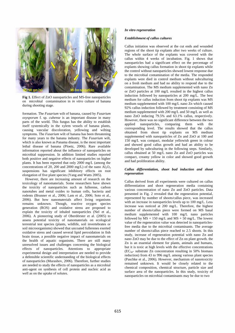
Fig 1. Effect of ZnO nanoparticles and MS-free nanoparticles
on microbial contamination in in vitro culture of banana
during shooting stage.
formation. The Fusarium wilt of banana, caused by Fusarium
oxysporum f. sp. cubense is an important disease in many
parts of the world. This fungus has the ability to establish
itself systemically in the xylem vessels of banana plants,
causing vascular discoloration, yellowing and wilting
symptoms. The Fusarium wilt of banana has been threatening
for many years to the banana industry. The Fusarium wilt,
which is also known as Panama disease, is the most important
lethal disease of banana (Ploetz, 2006). Rare available
information reported about the influence of nanoparticles on
microbial suppression. In addition limited studies reported
both positive and negative effects of nanoparticles on higher
plants. It has been reported that only 2000 mg/L (among the
concentrations of 20, 200 and 2000 mg/L) of the nano Al2O3
suspensions has significant inhibitory effects on root
elongation of five plant species (Yang and Watts 2005).
However, there are increasing amount of research on the
toxicology of nanomaterials. Some researchers have shown
the toxicity of nanoparticles such as fullerene, carbon
nanotubes and metal oxides to human cells, bacteria and
rodents (Brunner et al., 2006; Lam et al., 2006; Soto et al.,
2006). But how nanomaterials affect living organisms
remains unknown. Though, reactive oxygen species
generation (ROS) and oxidative stress are proposed to
explain the toxicity of inhaled nanoparticles (Nel et al.,
2006). A pioneering study of Oberdörster et al. (2005) to
assess potential toxicity of nanomaterials on ecological
terrestrial test species (plants, wildlife, soil invertebrates or
soil microorganisms) showed that uncoated fullerenes exerted
oxidative stress and caused several lipid peroxidation in fish
brain tissue, a possible negative impact of nanomaterials on
the health of aquatic organisms. There are still many
unresolved issues and challenges concerning the biological
effects of nanoparticles. Attentions to appropriate
experimental design and interpretation are needed to provide
a defensible scientific understanding of the biological effects
of nanoparticles (Murashov, 2006). Therefore, further studies
are needed to study the effects of nanoparticles as a microbial
anti-agent on synthesis of cell protein and nucleic acid as
well as on the uptake of solutes.
In vitro regeneration
Establishment of callus cultures
Callus initiation was observed at the cut ends and wounded
regions of the shoot tip explants after two weeks of culture.
The whole surface of the explants was covered with the
callus within 4 weeks of incubation. Fig. 1 shows that
nanoparticles had a significant effect on the percentage of
explants showing callus formation in shoot tip explants while
the control without nanoparticles showed lowest response due
to the microbial contamination of the media. The responded
explants were died in control medium without subculturing
on a fresh medium and had no ability to respond due to the
contamination. The MS medium supplemented with nano Zn
or ZnO particles at 100 mg/L resulted in the highest callus
induction followed by nanoparticles at 200 mg/L. The best
medium for callus induction from shoot tip explants was MS
medium supplemented with 100 mg/L nano Zn which caused
92% callus induction followed by treatment consisting of MS
medium supplemented with 200 mg/L and 50 mg/L as well as
nano ZnO inducing 79.5% and 63.1% callus, respectively.
However, there was no significant difference between the two
applied nanoparticles, comparing them with the
corresponding level. The results showed that the callus
obtained from shoot tip explants on MS medium
supplemented with nanoparticles of Zn and ZnO at 100 and
150 mg/L was compact, modular yellowish green in colour
and showed good callus growth and had an ability to be
developed by subculturing in the following steps. Similarly,
callus obtained at 50 mg/L nano Zn and ZnO particles was
compact, creamy yellow in color and showed good growth
and had proliferation ability.
Callus differentiation, shoot bud induction and shoot
regeneration
Callus derived from all experiments were cultured on callus
differentiation and shoot regeneration media containing
various concentration of nano Zn and ZnO particles. Data
presented in Fig. 2 revealed that the regeneration potential,
represented by number of shoots/callus piece, was increased
with an increase in nanoparticles levels up to 100 mg/L. Less
increase was noticed at 200 mg/L. Therefore, the highest
number of shoots/callus piece were formed on MS basal
medium supplemented with 100 mg/L nano particles
followed by MS + 150 mg/L and MS + 50 mg/L. The lowest
value of the regeneration value was detected in nanoparticles-
free media due to the microbial contaminants. The average
number of shoots/callus piece reached to 2.5 shoots. In this
study, increase of regeneration potential with nano Zn and
nano ZnO may be due to the effect of Zn on plant growth. the
Zn is an essential element for plants, animals and humans,
but it is toxic at high levels with the effective concentrations
(EC50- substrate Zn concentration resulting in 50% biomass
reduction) from 43 to 996 mg/L among various plant species
(Paschke et al., 2006). However, mechanism of nanotoxicity
remained unknown. It would be closely related to the
chemical composition, chemical structure, particle size and
surface area of the nanoparticles. In this study, toxicity of
nanoparticles on microbial contaminants may be due to two

616
Table 2. Isolates fungi from banana in vitro cultures.
Name of genus Name of species
Fusarium spp. Fusarium solani
Fusarium oxysporum
Aspergillus spp. Aspergillus flavus
Aspergillus niger
Penicillium spp.
Candida spp.
Fig 2. Mean percentage values of the explant forming callus (EFC), callus forming shoots (CFS) (Shooting %), shoot forming roots
(SFR), number of roots RN)/plantlets, root length (RL) as well as shoots and roots fresh weight/plant as affected by Zn and ZnO
particles.

617
different actions: (1) the chemical toxicity based on chemical
composition, e.g. release of toxic ion; and (2) stress or stimuli
caused by the surface, size and/or share of the particles. It
was confirmed that solubility of oxide nanoparticles greatly
affect the cell culture response (Brunner et al., 2006).
Rooting and plantlets hardening
Fig. 2 shows that the nanoparticles-free medium (control) did
not cause rooting. The maximum rooting (89.0%), average
number of roots per shoot (6.57%) and maximum average
root length of 2.93 cm was achieved on medium containing
nano Zn at 100 mg/L followed by 150 mg/L and nano ZnO at
100 mg/L and 150 mg/L, respectively. The minimum rooting
of 57%, average number of root per shoot 3.10 and lowest
average root length of 1.61 was achieved on medium
containing nano ZnO at 50 mg/L followed by nano Zn at the
same level. Roots could contact nanoparticles directly.
Therefore, rooting (number, length and weight) was affected
by Zn and ZnO nanoparticles more than shooting. Moreover,
rooting would have a dose-dependent response, whereas
shoots could grow to a certain degree even though rooting
was halted in the presence of nano Zn and nano ZnO in the
cultures. Thus S/R ratio was increased.
The same table also shows that the survival percent of
plantlets was about 90% after 6 weeks of transfer to earthen
pots containing soil and sand and remained constant
thereafter, indicating treatment with Zn and ZnO
nanoparticles caused successful establishment of tissue
culture, raised number of plantlets accompanied with
suppuration of microbial contaminants.
On the other hand, the results show that none of the
explants responded well to a nanoparticles of Zn and ZnO
free medium. Additional Zn and ZnO nanoparticles to the
medium suppressed the microbial contaminants without
affecting on shoot tip explants in vitro regeneration.
Furthermore, nanoparticles increased the regeneration
potential of banana. Callus formation (%) was much higher
compared to nanoparticles-free medium. Zn and ZnO
nanoparticles at 100 mg/L were found to be satisfactory on
prevention of microbial contaminants and produced healthy
plants. In the case of nanoparticles-free medium, the explants
did not produce healthy shoots and plantlets became yellow.
Falkiner (1990) mentioned that the agents who act
specifically on bacterial cell walls would be more suitable to
control infection in plant tissue cultures. A possible
explanation may be that Zn and ZnO nanoparticles inhibit
bacterial cell wall synthesis preventing contamination.
Consequently, the explants will be physiologically active
greatly. Available information on nanotoxcity is scarce to
reach any consensus on its mechanism, particularly for the
nanoparticles used in this study (Zn and ZnO). Previous
research data on the nanoparticles generally showed negative
effects on the tested organisms such as cells, despite different
test methods used (Wang et al., 2006). However,
nanoparticles are preferable than the bactericide or fungicide
due to their sensitivity, simplicity, low cost and suitability
over unstable chemicals (Wang et al., 2001).
Organogenesis is an outcome of the process of
dedifferentiation followed by re-differentiation of cells.
Dedifferentiation favors unorganized cell growth and the
resultant callus has randomly divided meristems (Deepike
and Komwar 2010).
Oberdörster et al. (2005) indicated that dose-response curve
of nano Zn and nano ZnO are “T” shaped. There was a
threshold, below which no obvious symptom appeared.
However, root length decreased with increasing dose after the
threshold. Paschke et al. (2006) showed the phytotoxicity of
nano Zn and nano ZnO on growth of radish, rape and
ryegrass (less than 50mg/L). This was lower than the
phytotoxic effect of Zn2+ on some plant species (65-156
mg/L).
Although, reports on phytotoxic effects of nanoparticles are
scanty, the antifungal agent has been reported to suppress
larval development in mussel in vitro culture (Owen et al.,
2010). However, both positive and negative effects of
nanoparticles on higher plants were reported. It was pointed
out that a mixture of nano scale SiO2 (nano SiO2) and TiO2
(nano - TiO2) could increase nitrate reductase in soybean
(Glycine max L.), enhance its abilities of absorbing and
utilizing water and fertilizer, stimulate its antioxidant system
and apparently hasten its germination and growth (Lu et al.,
2002). Nano-TiO2 was reported to promote photosynthesis
and nitrogen metabolism and then greatly improve growth of
spinach at a proper concentration (Hong et al., 2005 a, b;
Zheng et al., 2005; Yang et al., 2006 b). After investigating
the phytotoxicity of nano Zn and nano ZnO on microbial
contaminants of in vitro banana tissue, with no impact on
explants regeneration, it could be concluded that such
materials can generate adverse biological effects in living
cells. Particles in such a size (<100 nm) fall in the transitional
zone between individual atoms or molecules and the
corresponding bulk material, which can modify the
physiochemical properties of the material e.g. performing
exceptional feats of conductivity, reactivity and optical
sensitivity. The side effects of nanoparticles are hardly
known for it has not yet been used to suppress fungal
contaminants in plant tissue culture.
Biochemical characters
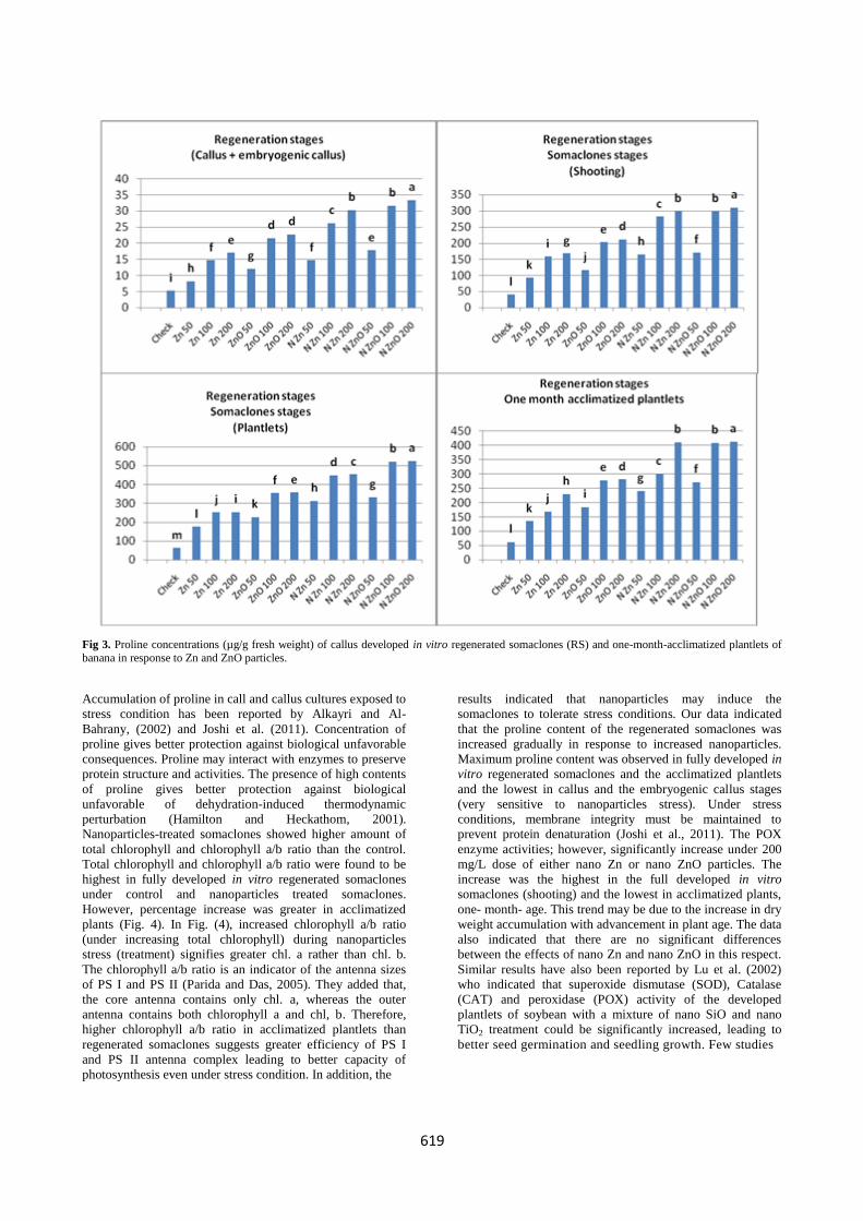
Fig. 3 shows that proline concentration in all in vitro
regeneration stages of banana explants increased gradually, in
response to increasing nano Zn and nano ZnO. Low dose of
nanoparticles (50 mg/L) caused about 4-fold increase in
proline concentration. However, significant highest increase
in proline concentration (about 8 fold) was observed at 200
mg/L nanoparticles. The increase was highest under nano
ZnO and the lowest under nano Zn. Moreover, proline
concentration was increased gradually with advancement of
the explants regeneration up to fully developed in vitro
regenerated somaclones and thereafter tended to decrease in
the acclimated one-month-old plantlets. The low proline
concentration was found in induced callus stage and the
highest in fully developed plantlets. Again, it could be
mentioned that addition of nanoparticles used to the cultures
media prevented bacterial and fungal contaminants and
favored growth of in vitro regenerated somaclones of banana.
Similar results were reported by Baskaran and Jayabalan
(2009) who found that BVN fungicide prevented fungal
contamination of the moist soil mixture. The use of
biochemical markers, such as proline can be used for
improving crop resistance to stress condition. Nanoparticles
play an important role as stress agent (USEPA, 2005).

618
Table 3. Percentage of detected microorganism contaminants of banana in vitro cultures throughout the various regeneration stages
in response to Zn and ZnO particles. (a) Callus initiation (b) Shooting (c) Rooting (d) Formed plantlets. a)
Particles
Level
mg/L
F. solani F.
oxysporum
A. niger A. flavus Penicillium
spp.
Candida
spp.
Bacteria
Check 22.80 a 27.93 a 14.25 a 12.75 a 7.81 a 3.28 a 26.05 a
Zn 50 21.20 b 25.97 b 13.25 ab 11.86 ab 7.26 ab 3.05 a 24.23 b
100 19.61 c 24.02 c 12.26 bc 10.97 bc 6.72 a-c 2.82 a 22.40 c
200 17.33 de 21.23 d 10.83 cd 9.69 d 5.93 b-d 2.49 a 19.80 d
ZnO 50 18.70 cd 22.9 c 11.69 cd 10.46 cd 6.40 bc 2.69 a 21.36 c
100 16.87 e 20.67 d 10.55 d 9.44 d 5.78 cd 2.43 a 19.28 d
200 14.14 f 17.32 e 8.84 e 7.91 e 4.84 d 2.03 ab 16.15 e
Nano Zn 50 5.24 g 6.42 f 3.28 f 2.93 f 1.80 e 0.75 bc 5.99 f
100 0.00 h 0.00 g 0.00 g 0.00 g 0.00 f 0.00 c 0.00 g
200 0.00 h 0.00 g 0.00 g 0.00 g 0.00 f 0.00 c 0.00 g
Nano ZnO 50 4.79 g 5.87 f 2.99 f 2.68 f 1.64 e 0.69 bc 5.47 f
100 0.00 h 0.00 g 0.00 g 0.00 g 0.00 f 0.00 c 0.00 g
200 0.00 h 0.00 g 0.00 g 0.00 g 0.00 f 0.00 c 0.00 g
b) Shooting
Particles
Level
mg/L
F. solani F.
oxysporum
A. niger A. flavus Penicillium
spp.
Candida
spp.
Bacteria
Check 24.80 a 30.38 a 15.50 a 13.87 a 8.49 a 3.57 a 28.33 a
Zn 50 23.06 b 28.25 b 14.42 ab 12.90 ab 7.90 ab 3.32 ab 26.35 b
100 21.33 c 26.13 c 13.33 bc 11.93 bc 7.30 a-c 3.07 ab 24.37 c
200 18.85 d 23.09 d 11.78 cd 10.54 d 6.46 cd 2.71 ab 21.53 d
ZnO 50 20.34 c 24.91 c 12.71 cd 11.38 cd 6.97 bc 2.93 ab 23.23 c
100 18.35 d 22.48 d 11.47 d 10.27 d 6.29 cd 2.64 ab 20.97 d
200 15.38 e 18.84 e 9.61 e 8.60 e 5.27 d 2.21 b 17.57 e
Nano Zn 50 5.70 f 6.99 f 3.57 f 3.19 f 1.95 e 0.00 c 6.52 f
100 0.00 g 0.00 g 0.00 g 0.00 g 0.00 f 0.00 c 0.00 g
200 0.00 g 0.00 g 0.00 g 0.00 g 0.00 f 0.00 c 0.00 g
Nano ZnO 50 5.21 f 6.38 f 3.26 f 2.91 f 1.78 e 0.00 c 5.95 f
100 0.00 g 0.00 g 0.00 g 0.00 g 0.00 f 0.00 c 0.00 g
200 0.00 g 0.00 g 0.00 g 0.00 g 0.00 f 0.00 c 0.00 g
c) Rooting
Particles
Level mg/L
F. solani F. oxysporum A. niger A. flavus Penicillium spp.
Candida spp.
Bacteria
Check 24.00 a 29.40 a 15.00 a 13.43 a 8.22 a 3.45 a 27.42 a
Zn 50 22.32 b 27.34 b 13.95 ab 12.49 ab 7.64 ab 3.21 ab 25.50 b
100 20.64 c 25.28 c 12.90 bc 11.55 bc 7.07 a-c 2.97 ab 23.58 c
200 18.24 d 22.34 d 11.40 cd 10.20 d 6.25 cd 2.62 ab 20.84 d
ZnO 50 19.68 c 24.11 c 12.30 cd 11.01 cd 6.74 bc 2.83 ab 22.48 c
100 17.76 d 21.76 d 11.10 d 9.93 d 6.08 cd 2.55 ab 20.29 d
200 14.88 e 18.23 e 9.30 e 8.32 e 5.10 d 2.14 b 17.00 e
Nano Zn 50 5.52 f 6.76 f 3.45 f 3.09 f 0.00 e 0.79 c 6.31 f
100 0.00 g 0.00 g 0.00 g 0.00 g 0.00 e 0.00 c 0.00 g
200 0.00 g 0.00 g 0.00 g 0.00 g 0.00 e 0.00 c 0.00 g
Nano ZnO 50 0.00 g 6.17 f 3.15 f 2.82 f 0.00 e 0.73 c 5.76 f
100 0.00 g 0.00 g 0.00 g 0.00 g 0.00 e 0.00 c 0.00 g
200 0.00 g 0.00 g 0.00 g 0.00 g 0.00 e 0.00 c 0.00 g
d) Formed plantlets
Particles
Level
mg/L
F. solani F. oxysporum A. niger A. flavus Penicillium
spp.
Candida
spp.
Bacteria
Check 22.36 a 27.39 a 13.98 a 12.51 a 7.66 a 3.22 a 25.55 a
Zn 50 20.79 b 25.47 b 13.00 ab 11.63 ab 7.12 ab 2.99 a 23.76 b
100 19.23 c 23.56 c 12.02 bc 10.76 bc 6.59 a-c 2.77 a 21.97 c
200 16.99 de 20.82 d 10.62 cd 9.51 d 5.82 b-d 2.44 a 19.42 d
ZnO 50 18.34 cd 22.46 c 11.46 b-d 10.26 cd 6.28 a-c 2.64 a 20.95 c
100 16.55 e 20.27 d 10.34 d 9.26 d 5.67 cd 2.38 a 18.90 d
200 13.86 f 16.98 e 8.66 e 7.75 e 4.75 d 1.99 a 15.84 e
Nano Zn 50 5.14 g 6.30 f 3.21 f 2.88 f 1.76 e 0.74 b 5.88 f
100 0.00 h 0.00 g 0.00 g 0.00 g 0.00 f 0.00 b 0.00 g
200 0.00 h 0.00 g 0.00 g 0.00 g 0.00 f 0.00 b 0.00 g
Nano ZnO 50 4.70 g 0.00 g 2.93 f 2.63 f 1.61 e 0.68 b 5.36 f
100 0.00 h 0.00 g 0.00 g 0.00 g 0.00 f 0.00 b 0.00 g
200 0.00 h 0.00 g 0.00 g 0.00 g 0.00 f 0.00 b 0.00 g
619
Fig 3. Proline concentrations (µg/g fresh weight) of callus developed in vitro regenerated somaclones (RS) and one-month-acclimatized plantlets of
banana in response to Zn and ZnO particles.
Accumulation of proline in call and callus cultures exposed to
stress condition has been reported by Alkayri and Al-
Bahrany, (2002) and Joshi et al. (2011). Concentration of
proline gives better protection against biological unfavorable
consequences. Proline may interact with enzymes to preserve
protein structure and activities. The presence of high contents
of proline gives better protection against biological
unfavorable of dehydration-induced thermodynamic
perturbation (Hamilton and Heckathom, 2001).
Nanoparticles-treated somaclones showed higher amount of
total chlorophyll and chlorophyll a/b ratio than the control.
Total chlorophyll and chlorophyll a/b ratio were found to be
highest in fully developed in vitro regenerated somaclones
under control and nanoparticles treated somaclones.
However, percentage increase was greater in acclimatized
plants (Fig. 4). In Fig. (4), increased chlorophyll a/b ratio
(under increasing total chlorophyll) during nanoparticles
stress (treatment) signifies greater chl. a rather than chl. b.
The chlorophyll a/b ratio is an indicator of the antenna sizes
of PS I and PS II (Parida and Das, 2005). They added that,
the core antenna contains only chl. a, whereas the outer
antenna contains both chlorophyll a and chl, b. Therefore,
higher chlorophyll a/b ratio in acclimatized plantlets than
regenerated somaclones suggests greater efficiency of PS I
and PS II antenna complex leading to better capacity of
photosynthesis even under stress condition. In addition, the
results indicated that nanoparticles may induce the
somaclones to tolerate stress conditions. Our data indicated
that the proline content of the regenerated somaclones was
increased gradually in response to increased nanoparticles.
Maximum proline content was observed in fully developed in
vitro regenerated somaclones and the acclimatized plantlets
and the lowest in callus and the embryogenic callus stages
(very sensitive to nanoparticles stress). Under stress
conditions, membrane integrity must be maintained to
prevent protein denaturation (Joshi et al., 2011). The POX
enzyme activities; however, significantly increase under 200
mg/L dose of either nano Zn or nano ZnO particles. The
increase was the highest in the full developed in vitro
somaclones (shooting) and the lowest in acclimatized plants,
one- month- age. This trend may be due to the increase in dry
weight accumulation with advancement in plant age. The data
also indicated that there are no significant differences
between the effects of nano Zn and nano ZnO in this respect.
Similar results have also been reported by Lu et al. (2002)
who indicated that superoxide dismutase (SOD), Catalase
(CAT) and peroxidase (POX) activity of the developed
plantlets of soybean with a mixture of nano SiO and nano
TiO2 treatment could be significantly increased, leading to
better seed germination and seedling growth. Few studies

620
Fig 4. Total chlorophyll concentrations (mg/g fresh weight)
and Chl. a/b ratio of fully developed in vitro regenerated
somaclones and one-month-acclimatized plantlets of banana
in response to Zn and ZnO particles.
have focused on the effects and mechanisms of nanoparticles
on plant growth and its relation with oxidative enzymes,
having a photo-catalyzed (Crabtree 1998) nano TiO2 under
light which could cause an oxidation-reduction reaction and
produce superoxide ion radical and hydroxide. These reactive
oxygen species (ROS) can go after organic substance and be
effective antimicrobial agents (Wang et al., 2001). Similar
results were reported by Helaly and Hanan El-Hoseiny
(2011) on stressed in vitro cultures of sweet orange. They
achieved higher enzyme activity of superoxide dismutase
(SOD), peroxidase (POX), ascorbic peroxidase (Apox),
Catalase (CAT) and glutathione reductase (GR). Wang et al.
(2001) confirmed that nano SiO2 treatment could be the cause
of increased strength, resistance to disease and; thus,
increased yield of rice (Tang and Cao, 2003). Zheng et al.
(2005) reported that nano TiO2 treatment could markedly
promote the vigor of aged seeds and chlorophyll formation of
spinach, by which the ribulose biphosphate carboxylase
activity and photosynthesis is obviously increased and the
growth and development of spinach promoted. Similarly, our
results indicated that addition of nano Zn and ZnO particles
in the in vitro media prevents microbial contaminants and
favored regenerating and plantlets growth. Moreover, the
regenerated plantlets were successfully established (98%) in
pots under field conditions. It is well known that
photosynthesis is consistent with photo-catalyzed chemical
reaction and enzyme catalyzed chemical reaction. As a photo-
catalyzed reaction, Hong et al. (2005 a and b) speculated that
photosynthetic rate enhancement by nano TiO2; catalysis
might be closely related to photochemical reaction activity
such as the absorption of light energy, transforming light
energy into electron energy, electron transport rate, oxygen
evolution rate and photophosphorylation efficiency and so
forth. In this study, it was observed that somaclones
developed from nanoparticles Zn and ZnO treated calli
attained greater growth and higher amount of proline, chl(s)
and a/b ratio, compared to control. Proline may interact with
enzymes to preserve protein structure (Joshi et al., 2011). A
higher proline content confers better protection against
biologically unfavorable of dehydration-induced
thermodynamic perturbation (Hamilton and Heckathom,
2001).
Materials and Methods
This study was carried out at the Plant Tissue Culture
Laboratory, Horticultural Research Institute, Ministry of
Agriculture, Agricultural Research Center (A. R. C). and the
Laboratories of Agric. Bot. Dept. and Plant Pathology Dept.,
Faculty of Agric., Mansoura Univ., Egypt during the period
from Jan. 2012 to March 2013.
Isolation, identification and characterization of microbial
contaminants
At the end of each culturing period, the developed bacterial
and fungal contaminants were isolated from banana cultures
and transferred to specific medium. Bacterial isolates were
aseptically streaked onto sterile nutrient agar medium and the
cultures were incubated at 25±1oC for 24 hr(s). Pure bacterial
isolates were obtained by repeated subculturing using a serial
dilution technique (Collins and Lyne, 1984).
Fungi were prepared for examinations under a stereoscopic
binocular microscope (6-50 X) to study their characters.
When necessary the compound microscope was used to
confirm the identification after having examined the
morphology of conidia and conidiophores. Fungi presented
on infested jars were identified by means of comparison with
the description sheets of Commonwealth Mycological
Institute, Kew, Surrey, England (CMI), Danish Government
Institute of Seed Pathology (DGISP) publications as well as
publications of Raper and Fennel, 1965; Ellis, 1971;
Moubasher, 1977; Booth, 1985; Burrges et al., 1988; and
Singh et al., 1991.
The purified bacterial isolates were observed under
microscope after staining for morphological characterization
based on vegetative cell chap, gram reaction and presence or
absence of spores. In addition, essential biochemical tests
were conducted per standard methods namely; methyl red,
arginine hydrolase, starch hydrolysis, casein hydrolysis,
fluorescent pigments, lactose, citrate and catalase productions
(Collins and Lyne, 1984; Krieg and Holt, 1984 and Sneath et
al., 1986).
For identification, characterized bacterial strains were
compared with the standard strains of Bergey's Manual
(Krieg and Holt, 1984 and Sneath et al., 1986). The key
proposed by Bradbury (1988) was also followed. Fungal
isolates were aseptically transferred onto Petri-dishes
containing potato dextrose agar (PDA) growth medium. The
cultures were incubated at 25±1oC for 5-15 days. The fungal
isolates were purified by repeated subcultures onto fresh
growth medium (PDA). Slides of pure fungal isolates were
prepared and stained with lactophenol cotton blue for
identification of the isolates based on microscopic
morphological appearance of conidiophores and conidia
(Barnett and Hunter, 1972).

621
Fig 5. Enzymes concentrations (units/g fresh weight) of developed in vitro regenerated explants throughout the various regenerated stages of banana in response to Zn and ZnO particles.

622
Mother plant sources and explant material
Mother plants, 3-years-old banana; Musa paradisiacal L. (M.
acuminate × M. balbisiana) cv. Grand Nain were used as an
experimental material. Small suckers of about 60-70 cm in
length were carefully removed from the mother plants grown
in Agricultural Research and taken immediately to the labs.
Shoot tips (explants) were prepared by removing the outer
layer tissues from the suckers. The pale white tissue blocks
containing the shoot tips and rhizomatous bases were surface
sterilized with three disinfectants; Sodium Hypochlorite
(NaOCl 80%), Ethanol 95% and Mercuric Chloride (HgCl2
0.1%) for 15 mins, respectively.
Nanoparticles examined and preparation
Two types of nanoparticles were examined denoted as nano
Zn and nano ZnO (MN6Z) at purity of 99.9%. Zn nanoscale
metal (diameter 35 nm) was purchased from Zunye
(Nanomaterials Co. Ltd. Shenzhea, China). Nanoscale ZnO
was from Hongchen (Material Sci. and Tech. Co. Ltd.
Zhejiang, China). The surface area provided by the producers
was 40±10 and 50±10 m2/g for nano Zn and nano ZnO,
respectively.
The nanoparticles used were suspended directly onto
deionized water and dispersed by ultrasonic vibration (100
W, 40 KHz) for 30 min. Small magnetic bars were placed in
the suspensions for stirring before use to avoid aggregation of
the particles. The nanoparticles suspensions after
centrifugation (3000 g for 1h) and filtration (0.7 µm glass
filter) were used for the treatments. Three levels of each
nanoparticled were used in addition to the control denoted 0
(control), 50, 100 and 200 mg/L.
Explants regeneration
Calls induction
To study the callogenic response and microbial contaminants
in the presence and absence of Zn and ZnO nanoparticles,
sterilized shoot tips explants were cultures on MS (Murashige
and Skoog, 1962) basal media supplemented with sucrose (30
g/L), 2, 4 D (2 mg/L) and one of each nanoparticles graded
levels. The final pH was adjusted to 5.8. Each treatment was
replicated 5 times (5 Jars), each jar with 3 explants and the
experiment was replicated three times. The media were
solidified with 7 g/L agar (Defco, India). Cultures containing
specific media were disperses in jars (325 ml) at the rate of
50 ml/jar, capped and autoclaved (at 121o C at 15 Ibs/insh2)
for 15 min and incubated at 25±1o C for 6 weeks with two
sub-culturing. At the end of callus induction period, bacterial
and fungal contaminants were characterized and identified
and the infection percentage calculated. Moreover, callus
induction frequency estimated as follows as the percentage of
explants that produce callus (Lee et al., 2009). Callus colors
and types proliferation were also recorded.
Callus proliferation and shooting
Calli obtained from each treatment were cut into small pieces
(about 0.8 cm3) and subsequently transformed on to free
auxin MS specific media (contained one of each
nanoparticles graded levels) supplemented with BA (3mg/L).
Similarity, each treatment consisted of five replicates (culture
jars) and the experimental unit cultured at the rate of five
callus pieces/jar. All cultures were maintained at the same
conditions used for callus proliferation. After shoot but
differentiation (4 weeks), the embryogenic calli pieces along
with the shoot buds were separated and transformed to the
subculture on same corresponding specific freshly media
prepared for shooting regeneration. Incubation was taken
place for 12 weeks with 3 subculturing. At the end of
shooting stage, the observations were taken for microbial
contaminants, percentage emberyoginic callus pieces
showing shoot bud induction and average number of
shoots/callus piece.
Rooting and plantlets acclimatization
Regenerated and developed shoots (3-4 cm in length) were
excised from the emberyogenic callus, transformed and
cultured on half strength MS media supplemented with one of
the different levels of nano Zn and nano ZnO examined and
IBA (3 mg/L), activated charcoal (0.5 g/L) and pH adjusted
to 5.8. The cultures were incubated at the growth room and
maintained under the same conditions previously mentioned.
Each treatment consisted of 5 replicates (experimental unit),
each with 3 shoots/jar. The experiment was replicated thrice
and the observation were recorded after two weeks of
incubation for microbial contaminants, percentage of rooting
based on number of shoots forming roots, number of roots
per shoot and length of roots. For acclimatization, the
obtained plantlets were removed from the media, gently
washed with tap water and selected based on their size.
Plantlets showed well-developed root system (4-5 cm in
length) and planted individually in plastic pots (5.5 x 6.5 cm;
tyrpido) containing sterilized peat moss + sand + perlite (1: 1:
1 v/v) and irrigated with one tenth MS solution devoid of
sucrose in a humid chamber at 26±1oC under 16/8 day/night
cycle. One month later, the potted plantlets were grown under
greenhouse conditions, covered with transparent plastic bags
to maintain the humidity. Acclimatization to the external
environmental was done by removing the transparent plastic
bags gradually to reduce humidity. After 4 weeks, the
observation for microbial contaminants, percentages of the
survival plants, fresh and dry weights of the plantlets were
recorded. In addition, total chlorophyll was isolated and
determined as given by Arnon (1949). Total proline was
detected as described by Bates et al. (1973). Antioxidant
enzyme activities were also determined.
Antioxidant enzymes activity
Antioxidant enzymes activity (unit/g F. Wt.) were extracted
from the plant material in a phosphate buffer (Inskeep and
Bloom, 1985) and assayed using the methods of Beauchamp
and Pridovich (1971), Herzog and Fahimi (1973) and
Bergmeyer (1970) for superoxide dismutase (SOD) (EC
1.15.1.1), peroxidase (POX) (EC 1.11.1.x) and catalase
(CAT) (EC 1.11.1.6) respectively.
Statistical analysis
The experimental design for each treatment was randomized
complete blocks system as each was replicated thrice and all
data recorded on different parameters were subjected to
analysis of variance (Gomez and Gomez, 1984). Each of the
experimental mean values was compared to its corresponding
control. Differences among treatment means were statistically
analyzed using the least significant differences test (LSD) at
P≤ 0.05 %, means separation using the CoStat program.

623
Conclusion
In this study, we tested phototoxicity of nano Zn and nano
ZnO on microbial contaminants in banana in vitro cultures
and examined the explants regeneration rate. It was found
that nano Zn and nano ZnO particles have significant
inhibitions on bacterial as well as fungal contaminants and
significantly prevent their growth. No marked negative
effects on explants regeneration was noticed even at 200
mg/L nanoparticles in the growth media. Nanoparticles
concentrations used were most likely lower than the threshold
values and did not show any negative effect on banana
regeneration. Further investigation should be directed to
phototoxicity mechanisms in microorganisms and higher
plant species. In addition, further studies are required to
investigate the adverse side-effects of nanoparticles on
genetic stability of banana in vitro cultures.
Acknowledgements
This work is supported by competitive project unit, Tanta
University, Egypt.
References
Al-Khayri JM, Al-Bahrany AM (2002) Callus growth and
proline accumulation in response to sorbitol and sucrose-
induced osmoptic stress in rice (Oryza sativa, L.). Biol
Plant. 45:609-611
Arnon DI (1949) Copper enzymes in isolated chloroplasts
phenoloxidase in Beta vulgaris. Plant Physiol 24: 1-15
Barnett HL, Hunter BB (1972) Illustrated genera of imperfect
fungi. Burgress Publishing Company, Minneapolis MN,
USA, pp. 241
Baskaran P, Jayabalan N (2009) In vitro propagation of
Psoralea carylifolia, L. by somatic embryogenesis in cell
suspention culture. Acta Physiol Plant. 31: 1119-1127.
Bates LS, Waldern RP, Teara ID (1973) Rapid determination
of free proline for water stress studies. Plant Soil. 39:205-
207
Beauchamp C, Pridovich I (1971) Superoxide dismutase.
Impressed assay and assay applicable to acrylamide gels.
Anal Biochem. 44:276-284
Bergmeyer HV (1970) Methods of enzymatic analysis.
Verlag Chemie. Weinheim, Bergstr, German.
Booth C (1985) The genus Fusarium. Commonwealth
Mycological Institute, Kew. Surrey, England 237 pp
Bradbury JF (1988) Identification of cultivable bacteria from
plants and plant tissue cultures by use of simple classical
methods. Acta Hort. 225: 27-37
Brunner TJ, Wick P, Manser P, Spohn P, Grass RN, Limbach
LK, Bruinink A, Stark WJ (2006) In vitro cyto-toxicity of
oxide nanoparticles comparison to asbestos, silica and the
effect of particle solubility. Environ Sci Technol. 40: 4374-
4381
Burrges LW, Liddell CM, and Summerell BA (1988)
Laboratory manual for Fusarium research. Incorporating a
Key and descriptions of common species found in Australia
(second edition). Fusarium research Laboratory
Department of Plant Pathology and Agricultural
Entomology the University of Sydney, 156 pp
Cassells AC (1996) Production of healthy plants. In
Proceeding of the Institute of Horticultural Symposium.
Micropropagation in Culture. Alerson PG. Dullforce. WM
(edition). Nothingham. University of Nothingham Trent
Print pp. 53-71
Collins CH, Lyne PM (1984) Microbiological methods (5th
ed.). Butterworths Co. Ltd. London, pp. 56-113
Crabtree RH (1998). A new type of hydrogen bond. Science.
282: 2000-2001
Danso KE, Azu E, Elegba W, Asumeng A, Amoatey HM,
Klu GYP (2011) Effective decontamination and subsequent
plantlet regeneration of sugarcane (Sacharum officinarum,
L.) in vitro. Inter J Integrative Biol. 11(2): 90-96.
Deepike R, Kamwar K (2010) In vitro regeneration of Punica
granatum L. plants from different juvenile expalnts. J Fruit
Ornamental Plant Res. 18(1):5-22
Ellis MB (1971) Dematiaceous Hyphomycetes. CMI, Kew,
Surrey, England, 608 pp
Falkiner FR (1990) The criteria for choosing an antibiotic for
control of bacteria in plant tissue culture. IAPTC Newslet.
60:13-22
Gomez KA, Gomez AA (1984) Statistical procedure for
Agricultural Research. 2nd ed. John Wiley and sons Inc.
New York, pp. 328-332.
Habiba U, Reza S, Saha ML, Khan R, Hadiuzzaman S (2002)
Endogenous bacterial contamination during in vitro culture
of table banana: Identification and prevention. Plant Tiss
Cult. 12(2):117-122
Hamilton EW, Heckathom SA (2001) Mitochondrial
adaptation to NaCl, complex I is protected by antioxidants
and small heat shock proteins, whereas complex II is
protected by proline and betaine. Plant Physiol. 126: 1266-
1274
Hecror RF, Domer JE (1983) Control of systemic spread of
Candida albicans with ketoconazole in the stomachs of
mice treated with cytarabine. J Infec Dis. 147(5):946-950
Helaly MN, El-Hoseiny H (2011) Combined effects between
genotypes and salinity on sweet orange during the
developments stages of its micropropagation. Res J Bot.
6(2): 38-67
Herzog V, Fahimi H (1973) Determination of the activity of
peroxidase. Anal Biochem. 55:554-562
Hong FS, Yang F, Liu C, Gao Q, Wan ZG, Gu FG, Wa C,
Ma Z, Zhou Z, Yang P (2005a). Influences of nano-TiO2 on
the chloroplast aging of spinash under light. Biol Trace
Elem Res. 104: 249-260
Hong FS, Zhou J, Liu C, Yang F, Wa C, Zheng L, Yang P
(2005b) Effect of nano-TiO2 on photochemical reaction of
chloroplast of spinash. Biol Trace Elem Res. 105: 269-279
Inskeep WP, Bloom PR (1985) Extinction coefficients of
chlorophyll a and b in N,N-dimethylformamide and 80%
acetone. Plant Physiol. 77:483-485
Joshi RA, Shukla E, Sairam RK (2011) In vitro screening of
rice genotypes for drought tolerance using polyethylene
glycol. Acta Physiol Plant. 33(6): 2209-2217
Khane M (2003) Bacterial and fungal indexing of tissue
cultures.http/www.hos.ufl.edu.moreweb/tissueculture/class
1/bacterial%20and%20fungal%20indexing%20of20tissue%
20cultures.doc
Krieg RN, Holt IG (1984) Bergey's annual of systematic
bacteriology Vol. 1. Williams and Wikins company,
Baltimore, USA pp. 308-429
Lam CW, James JT, Mccluskey R, Arepalli S, Hunter RI
(2006) A review of carbon nanotube toxicity and
assessment of potential occupational and environmental
health risks. Crit Rev Toxicol. 30: 189-217
Lee KW, Choi GJ, Kim KY, Hc L, Park HS, Yoon SH, Lee H
(2009) High frequency plant regeneration from mature seed
derived callus of Italian ryegrass (Lolium muliforum)
cultivars. Afr J Biotech. 8: 6828-6833

624
Lu CM, Zhang CY, Wen JQ, Wa GR, Tao MX (2002)
Research of the effect of nanometer materials on
germination and growth enhancement of Glycine max and
its mechanism. Soybean Sci. 21: 168-172
Markins M, Enreiro RT and Oliveira T (2003) Genetic
refatedness of Portuguese almond cultivars assessed by
RAPD and SSR markers. Plant Cell Rep. 22:71-78
Meghwal PR, Sharma HC, Singh SK (2000) Effect of surface
sterilizing agents on in vitro culture establishment of guava
(Psidium guajava, L.). J Applied Hort. 2(2): 94-95.
Mngomba SA, Toil D, Akinnifesi FK (2007) Effective
precondikoning methods for in vitro propagation of
Uapaca kirkiana Muell Arg. Tree species. Afr J Biotech.
6(14):1670
Moubasher AH, El-Kady IA, Farghally SM (1977) The
mycoflora of some Egyptian seeds and their potentialities
for production of aflatoxins. Zeszyty Problemowe
Postepow Nauk Rolniczych. 189: 141-147
Msogoya T, Kayagha H, Mutigitu J, Kulebelwa M, Mamiro
M (2012) Identification and management of microbial
contaminants of banana in vitro cultures. J Appl Biol.
55:3987-3994.
Murashige TA, Skoog F (1962) A revised medium for rapid
growth and bioassays with tobacco tissue cultures. Physiol
Plant. 15: 473-497
Murashov V (2006) Comments on “Particle surface
characteristic may play an important role in phytotoxicity
of alumina nanoparticles” by Yang, L.; Watts, D.J.
Toxicology letters, 2005, 185: 122-132. Toxicol Lett. 164:
185-187.
Nel A, Xio T, Mädler L, Li N (2006) Toxic potential of
materials at the nano level. Science. 311: 622-627
Oberdörster G, Oberdörster E, Oberdörster I (2005)
Nanotechnology: an emerging discipline evolving from
studies of ultrafine particles. Environ Healt Pers. 113: 823-
839
Obuckwe CO, Osagie II (1989) Morphological changes in
infected wilt-resistance and wilt susceptible oil palm
progenies and hydrolytic enzyme activities associated with
Fusarium oxysporum pathogens. Oeagureux. 44(11):8-9
Ödutanyo O I, Amusa NA, Okutade OO, Ogunsan YR (2007)
Sources of microbial contamination in tissue culture
laboratories in southern Nigeria. Afr J Agric Res. 2(3):67-
72
Ödutanyo OI, Oso RT, Akinyemi BO, Amusa NA (2004)
Microbial contaminants of cultured Hibiscus carnabinus
and Telfaria occidentalis tissues. Afr J of Biotech.
3(9):472-476
Omamor IB, Asemota AO, Eke CR, Eziashi EI (2007) Fungal
contaminants of the oil palm tissue culture in Nigerian
Institute for oil palm Research (NIFOR). Afr J Agric Res.
2(10):534-537
Owen CT, Alexander JE, McGregor M (2010) Control of
microbial contamination during in vitro culture of larval
anionid mussels. Invert Reprod Develop. 54(4):187-193
Parida A, Das B (2005) Salt tolerance and salinity effect on
plants; a review. Ecotoxicol Environ Safety. 60:324-349
Paschke MW, Perry LG, Redente EF (2006) Zinc toxicity
threshold for reclamation for species. Water Air Soil Polut.
170:317-330
Ploetz RC (2006) Fusarium-induced diseases of tropical,
perennial crops. Phytopath. 96:648–652
Raper KE, Fennel DI (1965) The genus Aspergillus. The
Williams and Wilkins Co., Baltimore 686 pp.
Sarkhosh A, Zamani Z, Fatahi R, Ebadi A (2006) RAPD
markers reveal polymorphism among some Iranian
pomegranate (Punica granatum L.) genotypes. Sci Hort.
111:24-29
Singh K, Frisvad JC, Thrance U, Mathur SB (1991) An
illustrated manual on identification of some seed-borne
Aspergilli, Fusaria, Penicillia and their Mycotoxins.
Danich Government Institute of Seed Pathology for
developing countries, Hellerup, Copenhagen, Denmark.
Sneath PH, Mair MS, Sharpe ME, Holt JG (1986) Bergey's
manual of systematic bacteriology. Vol.2. Williams and
Wilkins company, Baltimore, USA, pp. 1220.
Soto KF, Carrasco A, Powell TG, Murr LE, Garza KM
(2006) Biological effects of nano-particulate materials.
Mater Sci Eng. 26:1421-1427.
Suryanarayanan TS, Senthilarasu G, Muruganandam M
(2000) Endophytic fungi from Cuscuta reflexa and its host
plants. Fungal Diversity. 4:117-123
Tang SY, Cao YP (2003) Effects of spraying different forms
of silicon (Si) on growth as stress resistance of rice plants.
Sci Fertil. 2:16-22
USPEA (U.S. Environmental Protection Agency) (2005)
Nanotechnology white paper external review. Draft
Available from http://www.epa.gov/osa/pdfs.EPA_
nanotechnology_white_paper_external_review_draft_12-
02-2005
Wang BWY, Feng WY, Wang C, Jia M, Wang C, Shi W,
Zhang F, Zhao L, Chai F (2006) Acute toxicity of nano-and
micro scale zinc powder on healthy adult mice. Toxicol
Lett. 161:115-123
Wang XD, Sun C, Gao SX, Wang LS, Han K (2001)
Validation of germination rate and root elongation as
indicator to assess phytotoxicity with Cucmis sativus.
Chemosphere. 44:1711-1721
Wu WK (2003) Plant Physiology Science Press. Beijing pp.
117-143.
Yang F, Hong FS, You WJ, Liu C, Gao FQ, Wu C, Yang P
(2006) Influences of nanoanatase Ti O2 on the nitrogen
metabolism of growing spinach. Biol Trace Elem Res.
110:179-190
Yang L, Watts DJ (2005) Particle surface characteristics may
play an important role in phytotoxicity of alumina nano-
particles. Toxicol Leu. 158:122-132
Zheng L, Hong FS, Lu SP, Liu C (2005) Effects of nano Ti
O2 on strength of naturally aged seeds and growth of
spinach. Biol. Trace Elem Res. 104:83-91
![Characterizing self-assembled nanoparticles employed in ... · nanoparticles and the relationship between their structure and biological performance for DNA/ polysaccharide [1-3]](https://img.dokumen.tips/doc/110x75/601195bb0fb12178350a2dcf/characterizing-self-assembled-nanoparticles-employed-in-nanoparticles-and-the.jpg)