Embed Size (px)
Citation preview

BioMed Central
BMC Complementary and Alternative Medicine
ss
Open AcceResearch articleEffect of Lactobacillus acidophilus supernatants on body weight and leptin expression in ratsRenato Sousa1, Jaroslava Halper*1, Jian Zhang1, Stephen J Lewis2 and Wan-I O Li2Address: 1Department of Pathology, College of Veterinary Medicine, The University of Georgia, Athens, GA 30602, USA and 2Department of Physiology/Pharmacology, College of Veterinary Medicine, The University of Georgia, Athens, GA 30602, USA
Email: Renato Sousa - [email protected]; Jaroslava Halper* - [email protected]; Jian Zhang - [email protected]; Stephen J Lewis - [email protected]; Wan-I O Li - [email protected]
* Corresponding author
AbstractBackground: Lactobacillus extracts and supernatants have been used as probiotics in human andveterinary medicine for their ability to enhance wound healing and immunity. Previous data fromour laboratory demonstrated that Lactobacillus supernatant (LS) stimulated wound healing,angiogenesis and proliferation of embryonic cells after topical application. This current study showsthat LS after its administration into the cerebral ventricles of male rats exerts systemic effects.
Methods: The right lateral cerebral ventricle of young male rats was accessed throughintracerebroventricular cannulation (ICV) under anesthesia and aseptic conditions. One group ofcontrol rats received saline solution, a second control group received 0.8 M lactic acid solution (tocontrol for acidity of LS), and a third group received LS. The animals were sacrificed 12, 24, 48, 96and 120 hours after the injection. Selected tissues were collected, fixed in 10% buffered formalinand used for immunohistochemistry and in situ hybridization. Other tissues were frozen andextracted for immunoblotting
Results: LS-injected animals had a slight decrease in body weight when compared to their initialweight and to both control groups. Using immunohistochemistry and in situ hybridization leptinexpression was studied in multiple brain sections and peripheral adipose tissue of control and LS-injected rats. Strong cytoplasmic stain was observed by both techniques in neurons of the cerebralcortex, thalamus, hypothalamus, hippocampus and, to lesser degree, in the cells of the choroidplexus in the LS-injected rats. Control animals demonstrated much less intense staining in neuronslocated in the same regions using immunohistochemistry and almost no staining with in situhybridization technique. Adipose tissue exhibited slight presence of leptin in LS-treated animals. Incontrast no immunohistochemical staining for GM-CSF and TNFα was observed in brains fromcontrol and treated rats. Western blotting showed mild increase in leptin and leptin receptors inintestines and retroperitoneal adipose tissues of LS-injected rats.
Conclusion: This study demonstrates that direct administration of LS into rat CNS leads to adecrease in body weight of rats and an increase in the expression of leptin in specific areas of thebrain and retroperitoneal adipose tissue.
Published: 19 February 2008
BMC Complementary and Alternative Medicine 2008, 8:5 doi:10.1186/1472-6882-8-5
Received: 8 August 2007Accepted: 19 February 2008
This article is available from: http://www.biomedcentral.com/1472-6882/8/5
© 2008 Sousa et al; licensee BioMed Central Ltd. This is an Open Access article distributed under the terms of the Creative Commons Attribution License (http://creativecommons.org/licenses/by/2.0), which permits unrestricted use, distribution, and reproduction in any medium, provided the original work is properly cited.
Page 1 of 8(page number not for citation purposes)

BMC Complementary and Alternative Medicine 2008, 8:5 http://www.biomedcentral.com/1472-6882/8/5
BackgroundLactobacilli are non-pathogenic Gram-positive lactic acidbacteria found in the normal intestinal microflora of ani-mals and humans [1] and are classified as probioticagents. Lactobacillus derived products, including culturesupernatants have been used for their wound healing andantiviral properties as they are believed to boost energyand to be effective remedies for allergies, common cold,lactose intolerance, and have also been shown to reducecholesterol levels and the risk of colon cancer [2-4]. Super-natants of Lactobacillus acidophilus were also proved to beeffective against Helicobacter pylori in vitro and in vivo inpeople and were shown to possess antimicrobial activitiesagainst Bacillus anthracis and E. coli [3]. Our previous datahave shown that Lactobacillus supernatants (LS) promoteinflammatory response during tissue repair in rodents [5],stimulate proliferation of embryonic cells [6], and thatsubcutaneous injections of Lactobacillus supernatants intothe ears of rats lead to angiogenesis [5]. Using a cytokineantibody array, leptin and several other cytokines (e.g., IL-6, IL-8 and TGFβ) were detected in medium conditionedby bovine endothelial cells exposed to LS (data notshown).
This study represents a continuation of our previous work[5]. The purpose was to identify whether LS stimulatesangiogenesis in rodent CNS and/or expression of leptin inrodents. Leptin, the product of the obese gene (ob or lepob)is a 16 kDa non-glycosylated protein well known for itseffects on the reduction of body weight and involvementin certain aspects of wound healing, such as angiogenesis.Leptin acts in the central nervous system through bindingto leptin receptors located in the hypothalamus, particu-larly in the arcuate nucleus and by coordinating metabo-lism, feeding behavior, energy balance andneuroendocrine responses [7-9]. In addition to CNS lep-tin is also expressed in adipocytes, placenta [10], mam-mary gland [11], pituitary glands [12], and stomach [13].
In experiments designed to determine whether the super-natants elicit angiogenesis in the cerebral circulation, weinjected LS into the lateral cerebral ventricles of adult, nor-motensive Sprague-Dawley rats. A vital serendipitousfinding was that the injection led to reductions in bodyweight without changes in body temperature or intake offood and water. This was accompanied by increased leptinexpression in CNS, and intestinal and retroperitoneal adi-pose tissue.
MethodsPreparation of supernatants from Lactobacillus culturesBriefly, cultures of L. acidophilus (ATCC strains 4356 and43121) were grown in MRS broth (pH 5.5; Difco Labora-tories, Detroit, MI) at 37°C for 24 hr under microaer-ophilic conditions. This medium contains a rich nutrient
base as well as polysorbate, acetate, magnesium, andmanganese, which are known to promote the growth andproliferation of Lactobacilli. Overnight bacterial culturescontained 2.5 × 10⁸ colony-forming units, and these cul-tures were centrifuged at 10,000 g for 15 min at 4°C. Theresulting supernatants were filtered through a 0.2-µmmembrane filter to remove the remaining bacteria anddebris, lyophilized, and then stored at -20°C. At the timeof the experiments, the lyophilized LS was reconstitutedwith deionized water, filtered with puradiscs (0.22- µmpore size; Whatman Inc., Ann Arbor, MI) and termed LS.The pH of LS was 2.35 + 0.15, and osmolality was 320mOsm.
AnimalsThe use of animals and the animal experiments wasapproved by the Animal Use Committee at The Universityof Georgia. Thirty eight young male Sprague Dawley rats(Harlan, Sprague Dawley, Inc., Indianapolis, IN) rangingin weight from 250 to 350 g (or 6–8 weeks of age) at thebeginning of the experiment were used. The animals werehoused individually in a temperature-controlled room(22 ± 1°C) with a 12 h-12 h light-dark cycle. Food andwater were available ad libitum in their cages. Body param-eters such as rectal temperature and body weight werechecked daily, as was food and water consumption. At theend of each experiment rats were euthanized by CO2 over-dose.
Intra-cerebroventricular cannulationIntracerebroventricular (ICV) cannulations were per-formed under anesthesia and aseptic conditions. The rightlateral cerebral ventricle was accessed. The rats, weighingbetween 250 and 350 g, were anesthetized intra-muscu-larly with 0.2 ml/100 g of a mixture composed of acepro-mazine (5 mg (0.5 ml) of 10 mg/ml), ketamine (150 mg(1.5 ml) of 100 mg/ml and xylazine (30 mg (1.5 ml) of 20mg/ml). The dorsum of the head was shaved and rats wereplaced in a stereotaxic holding device. The skin was disin-fected with 70% isopropyl alcohol and a 22-gauge guidecannula (c313G – Single guide cannula, Plastics One, Inc.,Roanoke, VA), cut to a length of 15 mm, was implantedinto the right lateral ventricle. The stereotaxic coordinatesused were with respect to the bregma, AP = -0.8 mm, L = -1.4 mm and depth = 15 mm below the skull surface. Thecannula was held in place with three stainless-steel screwsand dental cement on the skull. A 32-gauge dummy can-nula (c313DC – Single guide cannula, Plastics One, Inc.)cut at 12 mm was inserted when the rat was not receivingan injection. Rats were allowed 1 week to recover, whenthey were expected to recover their initial body, presentedwhen the day the surgery was performed. The rats weredivided into 3 groups and sterile saline solution, LS solu-tion, and 0.8 M lactic acid solution were administered.
Page 2 of 8(page number not for citation purposes)
BMC Complementary and Alternative Medicine 2008, 8:5 http://www.biomedcentral.com/1472-6882/8/5
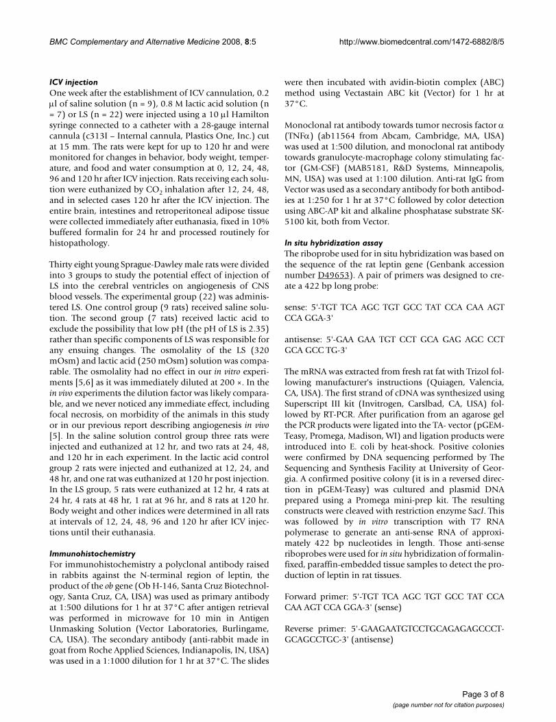
ICV injectionOne week after the establishment of ICV cannulation, 0.2µl of saline solution (n = 9), 0.8 M lactic acid solution (n= 7) or LS (n = 22) were injected using a 10 µl Hamiltonsyringe connected to a catheter with a 28-gauge internalcannula (c313I – Internal cannula, Plastics One, Inc.) cutat 15 mm. The rats were kept for up to 120 hr and weremonitored for changes in behavior, body weight, temper-ature, and food and water consumption at 0, 12, 24, 48,96 and 120 hr after ICV injection. Rats receiving each solu-tion were euthanized by CO2 inhalation after 12, 24, 48,and in selected cases 120 hr after the ICV injection. Theentire brain, intestines and retroperitoneal adipose tissuewere collected immediately after euthanasia, fixed in 10%buffered formalin for 24 hr and processed routinely forhistopathology.
Thirty eight young Sprague-Dawley male rats were dividedinto 3 groups to study the potential effect of injection ofLS into the cerebral ventricles on angiogenesis of CNSblood vessels. The experimental group (22) was adminis-tered LS. One control group (9 rats) received saline solu-tion. The second group (7 rats) received lactic acid toexclude the possibility that low pH (the pH of LS is 2.35)rather than specific components of LS was responsible forany ensuing changes. The osmolality of the LS (320mOsm) and lactic acid (250 mOsm) solution was compa-rable. The osmolality had no effect in our in vitro experi-ments [5,6] as it was immediately diluted at 200 ×. In thein vivo experiments the dilution factor was likely compara-ble, and we never noticed any immediate effect, includingfocal necrosis, on morbidity of the animals in this studyor in our previous report describing angiogenesis in vivo[5]. In the saline solution control group three rats wereinjected and euthanized at 12 hr, and two rats at 24, 48,and 120 hr in each experiment. In the lactic acid controlgroup 2 rats were injected and euthanized at 12, 24, and48 hr, and one rat was euthanized at 120 hr post injection.In the LS group, 5 rats were euthanized at 12 hr, 4 rats at24 hr, 4 rats at 48 hr, 1 rat at 96 hr, and 8 rats at 120 hr.Body weight and other indices were determined in all ratsat intervals of 12, 24, 48, 96 and 120 hr after ICV injec-tions until their euthanasia.
ImmunohistochemistryFor immunohistochemistry a polyclonal antibody raisedin rabbits against the N-terminal region of leptin, theproduct of the ob gene (Ob H-146, Santa Cruz Biotechnol-ogy, Santa Cruz, CA, USA) was used as primary antibodyat 1:500 dilutions for 1 hr at 37°C after antigen retrievalwas performed in microwave for 10 min in AntigenUnmasking Solution (Vector Laboratories, Burlingame,CA, USA). The secondary antibody (anti-rabbit made ingoat from Roche Applied Sciences, Indianapolis, IN, USA)was used in a 1:1000 dilution for 1 hr at 37°C. The slides
were then incubated with avidin-biotin complex (ABC)method using Vectastain ABC kit (Vector) for 1 hr at37°C.
Monoclonal rat antibody towards tumor necrosis factor α(TNFα) (ab11564 from Abcam, Cambridge, MA, USA)was used at 1:500 dilution, and monoclonal rat antibodytowards granulocyte-macrophage colony stimulating fac-tor (GM-CSF) (MAB5181, R&D Systems, Minneapolis,MN, USA) was used at 1:100 dilution. Anti-rat IgG fromVector was used as a secondary antibody for both antibod-ies at 1:250 for 1 hr at 37°C followed by color detectionusing ABC-AP kit and alkaline phosphatase substrate SK-5100 kit, both from Vector.
In situ hybridization assayThe riboprobe used for in situ hybridization was based onthe sequence of the rat leptin gene (Genbank accessionnumber D49653). A pair of primers was designed to cre-ate a 422 bp long probe:
sense: 5'-TGT TCA AGC TGT GCC TAT CCA CAA AGTCCA GGA-3'
antisense: 5'-GAA GAA TGT CCT GCA GAG AGC CCTGCA GCC TG-3'
The mRNA was extracted from fresh rat fat with Trizol fol-lowing manufacturer's instructions (Quiagen, Valencia,CA, USA). The first strand of cDNA was synthesized usingSuperscript III kit (Invitrogen, Carslbad, CA, USA) fol-lowed by RT-PCR. After purification from an agarose gelthe PCR products were ligated into the TA- vector (pGEM-Teasy, Promega, Madison, WI) and ligation products wereintroduced into E. coli by heat-shock. Positive colonieswere confirmed by DNA sequencing performed by TheSequencing and Synthesis Facility at University of Geor-gia. A confirmed positive colony (it is in a reversed direc-tion in pGEM-Teasy) was cultured and plasmid DNAprepared using a Promega mini-prep kit. The resultingconstructs were cleaved with restriction enzyme SacI. Thiswas followed by in vitro transcription with T7 RNApolymerase to generate an anti-sense RNA of approxi-mately 422 bp nucleotides in length. Those anti-senseriboprobes were used for in situ hybridization of formalin-fixed, paraffin-embedded tissue samples to detect the pro-duction of leptin in rat tissues.
Forward primer: 5'-TGT TCA AGC TGT GCC TAT CCACAA AGT CCA GGA-3' (sense)
Reverse primer: 5'-GAAGAATGTCCTGCAGAGAGCCCT-GCAGCCTGC-3' (antisense)
Page 3 of 8(page number not for citation purposes)

BMC Complementary and Alternative Medicine 2008, 8:5 http://www.biomedcentral.com/1472-6882/8/5
For in situ hybridization the slides were first heated at70°C for 10 min and deparafinized in Hemo-De. Tissuesections were allowed to dry and then were rehydrated inPBS + 5 mM MgCl2. Slides were incubated in Proteinase K(100 µl/ml) diluted in proteinase K buffer (10 mM TrispH 7.5 + 2 mM CaCl2). The enzymatic reaction wasstopped with 0.1 M glycine + 0.2 M Tris, pH 7.5. Prehy-bridization solution was added to sections for 1 hr(42°C). This was followed by the addition of the probe (3µl/slide) mixed with the prehybridization solution. Atotal of 70 µl of probe + prehybridization solution/slidewas used and applying direct onto a siliconized coverslip,which was sealed with nail hardener and kept in humidchamber overnight (42°C).
The next day, after several washes the slides were incu-bated with anti-dig AP diluted 1:300 in 2% normal serumsheep in buffer1(150 mM NaCl + 100 mM Tris, pH 7.5)for 2 hr. Slides were washed in buffers 1 and 3 (100 mMNaCl + 100 mM Tris+ 50 mM MgCl2, pH 9.5) for 5 min.Substrate (NBT + BCIP) was added and slides werechecked for staining. On average, the indicator color wasdetected in brain tissues after 40 min and in adipose tissueafter 3 hr.
Western blottingIntestines and retroperitoneal adipose tissues collectedfrom all three experimental groups were homogenized inbuffer composed of 20 mM Tris, 5 mM EDTA, 2 mMNa3NO4, 10% glycerol, 1% Triton X-100 and 1 mM PMSF.Aliquots of 10 µg protein were loaded per lane and sepa-rated in 8% SDS-polyacrylamide gels, then transferred onnitrocellulose membrane and with a polyclonal antibody
raised in rabbits against the N-terminal region of leptin,the product of the ob gene (Ob H-146) or a polyclonalantibody raised in rabbits against the leptin receptor (Ob-R H-300, both from Santa Cruz Biotechnology) at 1:1000dilutions. Antibody – antigen complexes were detected byDAB kit (Vector).
Statistical analysisOne-way analysis of variance test using the General LinearModels procedure of SAS was employed to test overall dif-ferences between initial weight and weight at euthanasiawithin LS-treated groups.
ResultsBecause our previous data have shown that topical admin-istration of LS led to influx of inflammatory cells into skinwound sites and to stimulation of angiogenesis in rodentskin and subcutaneous tissues [5], we tested LS for theability to induce angiogenesis in the rat brain. No angio-genesis was observed in brains of any of the rats, be it thetreatment or control groups. Neither was influx of inflam-matory cells noted previously in subcutaneous tissues [5]present anywhere in CNS. Instead the injection of LS elic-ited reductions in body-weight. In comparison with thecontrol groups, the animals that received Lactobacillussupernatants had a 24% decrease in body weight within48 hours, and were able to sustain it at least up to 120 hr(Table 1). A slight decrease in the body weight wasobserved in the animals that received lactic acid (4.4%).Because we did not have enough control rats for a statisti-cal analysis at later time points (at 96 and 120 hr) we didnot perform it for evaluation body weight loss among thethree groups. However, Table 1 reveals a clear trend of loss
Table 1: Comparison of weight between control and LS-treated rats.
Time 0 h 12 h 24 h 48 h 96 h 120 hRats ID BW%
C1-C9 (n = 9) 100 99C1-C6 (n = 6) 100 98.3 98.3C1-C4 (n = 4) 100 98.3 98.7 95.6C1-C2 (n = 2) 100 97.4 97.4 98.6 95.9 97.3LA1-LA7 (n = 7) 100 97.7LA1-LA5 (n = 5) 100 98.5 99.4LA1-LA3 (n = 3) 100 98.5 98.3 99.5LA1 (n = 1) 100 95.7 95.7 95.9 100.8 10.3.1LS1-LS22 (n = 22) 100 94.9LS1-LS17 (n = 17) 100 91.8 75.8LS1-LS13 (n = 13) 100 94.8 92.9 75.1LS1-LS8 (n = 9) 100 91.3 80.2 81.4 59.4LS1-LS8 (n = 8) 100 94.9 85.8 79.8 63.1 65
• Control rats labeled C1-C9 received 0.2 µl of saline solution, rats C7-C9 were euthanized at 12 hr; C5-C6 were euthanized at 24 hr; C3-C4 were euthanized at 48 hr; C1-C2 were euthanized at 120 hr.• Rats labeled LA1-LA7 received 0.2 µl of 0.8 M lactic acid solution; LA6- LA7 were euthanized at 12 hr; LA 4- LA 5 were euthanized at 24 hr; LA 2- LA 3 were euthanized at 48 hr; LA 1 was euthanized at 120 hr.• Rats labeled LS1-LS7 received 10 µl of LS, rats LS18- LS22 were euthanized at 12 hr; LS14- LS17 were euthanized at 24 hr; LS10- LS13 were euthanized at 48 hr; LS9 were euthanized at 96 hr; LS1-LS8 were euthanized at 120 hr.
Page 4 of 8(page number not for citation purposes)

BMC Complementary and Alternative Medicine 2008, 8:5 http://www.biomedcentral.com/1472-6882/8/5
of body weight in LS-treated rats, but not in the two con-trol groups. One-way analysis of variance was used toevaluate body weight loss within the LS-treated group(Fig. 1). In this analysis body weight was measured on allsurviving rats at time points before their euthanasia. Ourdata revealed significant difference in body weightbetween 0 and 48 hr in rats treated with LS at α = 0.05level (P-value = 0.084), between 0 and 96 hr (P-value =0.0025) and between 0 and 120 hr (P-value = 0.0064).
Because LS caused marked increase in leptin productionin bovine endothelial cells and because leptin is involvedin regulation of body weight we hypothesized that LSmight also regulate leptin expression in the brain. Weemployed both immunohistochemistry and in situhybridization to see whether LS injection leads to leptinoverexpression in the brain. Leptin expression was studiedin multiple brain sections of control and LS-injected rats.Strong cytoplasmic stain was observed by both techniquesin neurons of the cerebral cortex, thalamus (Fig. 2),hypothalamus and hippocampus (Fig. 3), and weakerstaining in the cells of the choroid plexus (data notshown). Control animals demonstrated much less intensestaining in neurons (and other cells) located in the sameregions and tissues using immunohistochemistry andalmost no staining with in situ hybridization technique.Peripheral adipocytes exhibited light immunostaining forleptin in LS-injected rats (Fig. 4). Immunoblotting of pro-tein extracts from intestines and from retroperitoneal adi-pose tissue from LS-treated and control rats showed onlymild increase in both leptin and leptin receptor (data notshown).
To see whether the effect on leptin synthesis was specificwe also performed immunohistochemistry for GM-CSFand TNFα in CNS sections. In contrast no immunohisto-
chemical staining for GM-CSF and TNFα was observed inbrains from control and treated rats (data not shown),though LS stimulated production of TNFα by several celllines [5]. Such results indicate that the effect of LS on lep-tin expression is specific.
DiscussionWe have shown that injection of LS into rat cerebral ven-tricles led to decrease in body weight. This reduction wasindependent of food and water intake and it occurredconcomitantly with a strong increase in leptin expressionin neurons of the cortical region, thalamus, hypothala-mus, and hippocampus. Mild increase in leptin was notedin the cells of the choroid plexus of the rat brain, andperipheral tissues such as intestines and fat. This findingwas in contrast with our previous studies where localadministration of LS led to angiogenesis and influx ofinflammatory cells [5]. Interestingly, in a separate series ofexperiments LS stimulated leptin secretion into mediumconditioned by bovine endothelial cells exposed to Lacto-bacillus supernatants. We hypothesize that the kind of bio-logical activity exerted by leptin depends on the tissue orcell type serving as an LS target. It is also possible that dif-ferential expression of other cytokines, induced by LS inCNS and not yet identified, play a crucial role in modulat-ing the effect of LS on body weight.
Several studies showed variable effects of Lactobacillusextracts on the production of leptin or its plasma level.The main reason for the discrepancy is the route of admin-istration (ours is the only study where a Lactobacillus prep-aration was administered directly into CNS), Lactobacillusstrain and/or animal species or breed used. Bleau et al. [1]described Lactobacillus-induced decrease in leptin releaseby adipocyte derived from SJL mice, whereas the sameLactobacillus preparation led to an increase in leptinrelease when used to treat C57BL/6 adipocytes. Theyascribed this to regulation of leptin synthesis by IL1β andTNFα produced by C57BL/6 macrophages upon stimula-tion by Lactobacillus preparation. In another report probi-otic capsules containing L. acidophilus and Bifidobacteriumlongum had no effect on plasma leptin in a group of meneven after 2 months of oral intake [14]. Oral administra-tion of L. plantarum to smokers led to decrease in plasmaconcentrations of leptin and other parameters which wasattributed to anti-inflammatory properties of L. plantarum[15]. In contrast, our preparations of L. acidophilus do pro-mote a variety of pro-inflammatory processes [5,6]. Someof these processes (e.g., angiogenesis), though not all,might be secondary to increased leptin expression. Ourprevious data have shown that LS stimulates TNFα pro-duction [5] which in turn would lead to increased leptinexpression [1]. This mechanism would be less likely to beoperational in CNS as immunohistochemistry did not
Average body weight of rats after intracerebroventricular injectionFigure 1Average body weight of rats after intracerebroven-tricular injection. ICV injection of LS led to statistically sig-nificant decrease in body weight 48 hr and later after a single ICV injection. The groups 1–4 represent four experiments. *time points significantly different from 0 h at α = 0.05.
Effect of LS on body weight in rats
0
20
40
60
80
100
120
1 2 3 4
BW
%
0h
12h
24h
48h
96h
120h
* **
**
Page 5 of 8(page number not for citation purposes)

BMC Complementary and Alternative Medicine 2008, 8:5 http://www.biomedcentral.com/1472-6882/8/5
Page 6 of 8(page number not for citation purposes)
Immunohistochemistry for leptin in brain tissueFigure 2Immunohistochemistry for leptin in brain tissue. Rats injected with saline solution, lactic acid or LS were euthanized 48 hr after injections and their brains were fixed in formalin immediately after euthanasia. Cytoplasmic immunostaining for leptin (arrows) was stronger in neurons of thalamus in LS-injected animals (B) than in corresponding cells of rats injected with saline (A). No counterstain. Magnification × 200.
In situ hybridization for leptinFigure 3In situ hybridization for leptin. Rats injected with saline solution, lactic acid or LS were euthanized 48 hr after injections and their brains were fixed in formalin immediately after euthanasia. Leptin mRNA was visualized using in situ hybridization in sections of the brains of rats injected with saline solution (A, C), and LS extract (C, D). Leptin expression is observed in the cytoplasm of neurons in the hypothalamus (A, B; arrows) and hippocampus (C, D; arrows). Stronger expression is noted in LS-injected animals. Light counterstain with hematoxylin. Magnification × 200.

BMC Complementary and Alternative Medicine 2008, 8:5 http://www.biomedcentral.com/1472-6882/8/5
identify any TNFα in CNS of rats after LS injection in ourexperiments.
We chose to look at the presence of GM-CSF and TNFα(neither cytokine was included in the angiogenesis anti-body array used by us) in the rat brain for several reasons.GM-CSF was described to suppress food intake and reduce
body weight in rodents after intraventricular injection[16]. In our experiments we found that food intake of LS-injected rats was comparable to food intake of control ani-mals and that no GM-CSF was present in rat CNS usingimmunohistochemistry. TNFα, also known as cachectin,leads to sometimes a significant weight loss. As men-tioned above LS stimulates the production of TNFα inperipheral macrophages [5] but not in CNS.
That leptin is primarily synthesized by adipocytes hasbeen known for many years [17]. Less well known butwell documented by many investigators has been the pro-duction of leptin in the brain. For example, Wiesner et al.have shown that leptin is released from the brain, particu-larly in obese men [18]. Similarly to our findings, immu-nostaining has localized leptin to many areas of the brain:cortex, cerebellum, hypothalamus and hippocampus[19]. In hypothalamus, leptin regulates energy intake andexpenditure [20], though some experiments point to lep-tin signaling in caudal brainstem neurons as transmittinggastrointestinal signals indicating satiety [21]. However,leptin during fetal and neonatal period also modulatesbrain development, especially in the hippocampus[22,23] and hypothalamus [17]. It is thought that leptinstimulates the proliferation and differentiation of neuralcell during early life as brain weight and DNA and proteincontent are reduced in ob/ob (leptin-deficient) mice [24].
ConclusionIn conclusion, this study shows that direct administrationof LS into the brains of rats resulted in weight loss withouta decrease in food consumption. This was accompaniedan increase in leptin expression in neurons and peripheraladipose tissues. The advantage (and a possible disadvan-tage because of potential side effects) of LS use over puri-fied leptin is the possibility that active components of LSmodulate the expression or release of other peptidesinvolved in metabolism and body weight control. Theidentification and large scale testing of these compoundsis currently under investigation in our laboratory.
Competing interestsJH, SJL and WOL are holding a provisional patent appli-cation describing the effects of LS. They have not receivedany financial reimbursement or fees. They have no othercompeting interests.
Authors' contributionsRS participated in experiment design, performed the ani-mal experiments, immunohistochemistry and in situhybridization and wrote the manuscript. JH, SJL and WOLconceived and designed the study, and supervised theexperiments. JZ performed the cell culture, angiogenesisassay, immunoblotting, statistical analysis and partici-
Immunohistochemistry for leptin in adipose tissueFigure 4Immunohistochemistry for leptin in adipose tissue. Rats injected with saline solution, lactic acid or LS were euth-anized 48 hr after injections and the retroperitoneal adipose tissue was fixed in formalin immediately after euthanasia. Cytoplasmic immunostaining for leptin (*) was stronger in adipocytes in LS-injected animals (B) than in corresponding adipocytes of rats injected with saline (A). Light counterstain with hematoxylin. Magnification × 400.
Page 7 of 8(page number not for citation purposes)

BMC Complementary and Alternative Medicine 2008, 8:5 http://www.biomedcentral.com/1472-6882/8/5
Publish with BioMed Central and every scientist can read your work free of charge
"BioMed Central will be the most significant development for disseminating the results of biomedical research in our lifetime."
Sir Paul Nurse, Cancer Research UK
Your research papers will be:
available free of charge to the entire biomedical community
peer reviewed and published immediately upon acceptance
cited in PubMed and archived on PubMed Central
yours — you keep the copyright
Submit your manuscript here:http://www.biomedcentral.com/info/publishing_adv.asp
BioMedcentral
pated in immunohistochemistry and in situ hybridization.All authors read and approved the final manuscript.
AcknowledgementsWe would like to thank Dr. Gaylen Edwards for helpful discussions and Ms. Kimberley Freeman for technical assistance.
References1. Bleau C, Lamontagne L, Savard R: New Lactobacillus acidophilus
isolates reduce the release of leptin by murine adipocytesleading to lower interferon-γ production. Clin Exper Immunol2005, 140:427-435.
2. Sanders ME, Klaenhammer TR: Invited review: the scientific basisof Lactobacillus acidophilus NCFM functionality as a probi-otic. J Dairy Sci 2001, 84:319-331.
3. Michetti P, Dorta G, Wiesel PH, Brassart D, Verdu E, Herranz M, Fel-ley C, Porta N, Rouvet M, Blum AL, Corthésy-Theulaz I: Effect ofWhey-Based Culture Supernatant of Lactobacillus acido-philus (johnsonii) La1 on Helicobacter pylori Infection inHumans. Digestion 1999, 60:203-209.
4. Wollowski I, Ji ST, Bakalinsky AT, Neudecker C, Pool-Zobel BL: Bac-teria used for the production of yogurt inactivate carcino-gens and prevent DNA damage in the colon of rats. J Nutr1999, 129:77-82.
5. Halper J, Leshin LS, Lewis SJ, Li WI: Wound healing and ang-iogenic properties of supernatants from Lactobacillus cul-tures. Exp Biol Med 2003, 228:1329-1337.
6. Li WI, Brackett BG, Halper J: Culture supernatant of Lactobacil-lus acidophilus stimulates proliferation of embryonic cells.Exp Biol Med 2005, 230:494-501.
7. Friedman JM, Halaas JL: Leptin and the regulation of bodyweight in mammals. Nature 1998, 395:763-770.
8. Casanueva FF, Dieguez C: Neuroendocrine regulation andactions of leptin. Front Neuroendocrinol 1999, 20:317-363.
9. Ahima RS, Saper CB, Flier JS, Elmquist JK: Leptin regulation ofneuroendocrine systems. Front Neuroendocrinol 2000,21:263-307.
10. Masuzaki H, Ogawa Y, Sagawa N, Hosoda K, Matsumoto T, Mise H,Nishimura H, Yoshimasa Y, Tanaka I, Mori T, Nakao K: Nonadiposetissue production of leptin: leptin as a novel placenta-derivedhormone in humans. Nature Med 1997, 3:1029-1033.
11. Smith-Kirwin SM, O'Connor DM, De Johnston J, Lancey ED, HassinkSG, Funanage VL: Leptin expression in human mammary epi-thelial cells and breast milk. J Clin Endocrinol Metab 1998,83:1810-1813.
12. Morash B, Li A, Murphy PR, Wilkinson M, Ur E: Leptin geneexpression in the brain and pituitary gland. Endocrinology 1999,140:5995-5998.
13. Bado A, Levasseur S, Attoub S, Kermorgant S, Laigneau J-P, BortoluzziM-N, Moizo L, Lehy T, Guerre-Millo M, Le Marchand-Brustel Y, LewinMJM: The stomach is a source of leptin. Nature 1998,394:790-793.
14. McMullen MH, Hamilton-Reeves JM, Bonorden MJL, Wangen KE,Phipps WR, Feirtag JM, Kurzer MS: Consumption of Lactobacillusacidophilus and Bifidobacterium longum does not alter phy-toestrogen metabolism and plasma hormones in men: apilot study. J Alter Compl Med 2006, 12:887-894.
15. Naruszewicz M, Johansson ML, Zapolska-Downar D, Bukowska H:Effect of Lactobacillus plantarum 299v on cardiovascular dis-ease risk factors in smokers. Am J Clin Nutr 2002, 76:1249-1255.
16. Reed JA, Clegg DJ, Smith KB, Tolod-Richter EG, Matter EK, Picard LS,Seeley RJ: GM-CSF action in the CNS decreases food intakeand body weight. J Clin Invest 2005, 115:3035-3044.
17. Carlo J, Sinha M, Kolaczynski J, Zhang L, Considine R: Leptin: thetale of an obesity gene. Diabetes 1996, 45:1455-1462.
18. Wiesner G, Vaz M, Collier G, Seals D, Kaye D, Jennings G, LambertG, Wilkinson D, Esler M: Leptin is released from the humanbrain: influence of adiposity and gender. J Clin Endocrinol Metab1999, 84:2270-2274.
19. Ur E, Wilkinson D, Morash BA, Wilkinson M: Leptin immunoreac-tivity is localized to neurons in rat brain. Neuroendocrinol 2002,75:264-272.
20. Halaas JL, Gajiwala KS, Maffei M, Cohen SL, Chait BT, Rabinowitz D,Lallone RL, Burley SK, Friedman JM: Weight-reducing effects of
the plasma protein encoded by the obese gene. Science 1995,269:543-546.
21. Huo L, Maeng L, Bjørbaek C, Grill HJ: Leptin and the control offood intake: neurons in the nucleus of the solitary tract areactivated by both gastric distension and leptin. Endocrinol2007, 148:2189-2197.
22. Walker CD, Long H, Williams S, Richard D: Long-lasting effects ofelevated neonatal leptin on rat hippocampal function, synap-tic proteins and NMDA receptor subunits. J Neurosci Res 2007,85:816-828.
23. Guo Z, Jiang H, Xu X, Duan W, Mattson MP: Leptin-mediated cellsurvival signaling in hippocampal neurons medicated by JAKSTAT3 and mitochondrial stabilization. J Biol Chem 2008,283:1754-1763.
24. Udagawa J, Hashimoto R, Suzuki H, Hatta T, Sotomaru Y, Hioki K,Kagohashi Y, Nomura T, Minami Y, Otani H: The role of leptin inthe development of the cerebral cortex in mouse embryos.Endocrinol 2006, 147:647-658.
Pre-publication historyThe pre-publication history for this paper can be accessedhere:
http://www.biomedcentral.com/1472-6882/8/5/prepub
Page 8 of 8(page number not for citation purposes)