Embed Size (px)
Citation preview

HISTOLOGIC, Vol. XLVI, No. 1
HP
CV
HC
Vol. XLVI, No. 1 June 2013
Managing Editor, Theresa Ford Scientific Editor, Vinnie Della Speranza,
MS, HTL(ASCP)HT, MT
Effect of Embalming Fluid on Histological Appearance of Organs From Embalmed West African Dwarf Goat CadaversUchenna Nlebedum, DVM; Ekele Ikpegbu, DVM; Okechukwu Nnadozie, DVM; Isaiah Agbakwuru, HNDDepartment of Veterinary AnatomyMichael Okpara University of AgricultureUmudike, Abia State, [email protected]
Managing Editor, Theresa Ford Scientific Editor, Vinnie Della Speranza,
MS, HTL(ASCP)HT, MT
IN THIS ISSUEEffect of Embalming Fluid on
Histological Appearance of Organs From Embalmed West African Dwarf Goat Cadavers . . . .1
Stereo-orientation of Mohs Surgery Frozen Section Specimens: A Technical Perspective . . . . . . . . . . . . . . . . . . . . .5
Third-hand Immobilization in Gross Sectioning of Pathology Specimens . . . . . . . . . . . .8
The Effectiveness of Even Ice Surface on Microtomy of Paraffin Blocks . . . . . . . . . . . . . . 12
Mark Your Calendar! . . . . . . . . . . . . 15
Abstract
The effects of a low-formalin embalming fluid on the histology of some organs from West African dwarf goat cadavers were investigated. This study was designed to ascertain the veracity of the claim believed by many histopathologists that the use of specimens from embalmed cadavers is good enough for investigative research and forensic medicine, especially in determining the cause of death at autopsy. The visceral organs examined include kidney, lung, spleen, and liver. The quality of slides from embalmed cadavers was graded using these criteria: general
Transverse section of liver from embalmed cadaver showing close to normal general organ microscopic architecture. Note the hepatocytes in the hepatic parenchyma (HP), central vein (CV), and hepatic capsule (HC). H&E, 100X.

HISTOLOGIC, Vol. XLVI, No. 1
organ microscopic architecture, cell morphology, and state of the epithelium. They were graded on a scale of 1 to 3, where 1 refers to a high degree of cell distortion, 2 is used to grade moderately good sections, and 3 is used for sections that are very close to normal. The slides from embalmed cadavers were seen to be moderately good, scoring 2 on the scale described above. Liver presented the least tissue distortion in this study, which may be attributed to its high degree of vascularization that allows for better perfusion by embalming fluid, resulting in proper fixation. This study suggests that tissue sections from embalmed cadavers with at least 4% formalin embalming fluid may be adequately fixed for histopathology.
Introduction
The objective of embalming is to achieve perfusion of fixative throughout all parts of the body. Immersion and direct injection into tissue cannot achieve this satisfactorily, so the injection of embalming fluid through the circulatory system is done for best results.1,2 The fluid used for embalming animals is designed to fix and preserve the tissue, preventing deterioration. It should also render the cadaver suitable for dissection, and prevent fungal and bacterial growth during the period of dissection.1,2 It is also important that tissue from embalmed cadavers be suitable for histological sectioning and observation.3-5
2
RCT
CT
SC
RC
RCT
RC
CT
Fig. 2. Transverse section of kidney from fresh sample showing renal cortex (RCT) containing renal corpuscle (RC) and convoluted tubules (CT). H&E, 100X.
Fig. 1. Transverse section of kidney from embalmed cadaver showing renal cortex (RCT) containing renal corpuscle (RC) and convoluted tubules (CT). Note the separation of the capsule from the cortex (SC). H&E, 100X.
EC
CT
EC
CC
Fig. 3. Transverse section of kidney from embalmed cadaver showing simple squamous cells of Bowman’s capsule (arrow), endothelial cells of the glomerulus (EC), and the simple cuboidal cells of the convoluted tubules (CC). Note the darkly stained nuclei. H&E, 400X.
Fig. 4. Transverse section of kidney from fresh sample showing simple squamous cells of Bowman’s capsule (arrow), endothelial cells of the glomerulus (EC), and the simple cuboidal cells of the convoluted tubules (CT). H&E, 400X.
HISTOLOGIC, Vol. XLVI, No. 1 3
A comparative study on the histological quality of specimens from cadavers embalmed with different embalming fluids was undertaken by Nicholson et al in 2005.5 A report by Kalanjati et al on the use of a low-formalin solution (5%-7.5% formaldehyde) for an effective, efficient, and safer embalming process was published in 2012.2 In Nigeria, the use of cadavers for forensic histopathology is a common practice, but there is a dearth of information in published literature on the suitability of these autopsy samples for histopathology, hence the need for this study. The aim of this work is to compare the histological appearance of samples processed from 4% formalin-embalmed animal cadavers with samples processed from freshly slaughtered animals fixed in 10% formalin. The information obtained from this study will fill the knowledge gap and help in ascertaining the extent of suitability of such cadaver samples for histopathology.
Materials and Methods
Tissue samples used in this study were obtained from West African dwarf goat (WADG) cadavers in the veterinary gross anatomy laboratory of Michael Okpara University of Agriculture, Umudike, Nigeria, and from apparently healthy freshly slaughtered WADGs from the goat farm at the same institution. The cadavers were embalmed with fluids containing formalin 4%, alcohol 33%, phenol 2.5%, glycerol 2.5%, and tap water 58%. The embalming fluid was infused into the cadaver through the common carotid artery. Slices of samples from the slaughtered goats were fixed in 10% formalin. The organs used for the study include kidney, lung, spleen, and liver.
RB
AD
RB
AP
C
Fig. 5. Transverse section of lung tissue from embalmed cadaver showing respiratory bronchiole (RB), alveolar duct (D), and alveolar sac (A). Note the distorted general organ morphology. H&E, 100X.
Fig. 6. Transverse section of lung tissue from fresh sample showing respiratory bronchiole (RB), pneumocytes (P), cartilage (C), and alveolar sac (A). Note the abundant pneumocytes in the organ parenchyma. H&E, 100X.
WP
RP
ST
RP
WPST
Fig. 7. Transverse section of spleen from embalmed cadaver showing organ microscopic architecture distortion. Red pulp (RP), white pulp (WP), and splenic trabeculae (ST). H&E, 100X.
Fig. 8. Transverse section of spleen from fresh sample showing normal organ microscopic architecture. Red pulp (RP), white pulp (WP), and splenic trabeculae (ST). H&E, 100X.

HISTOLOGIC, Vol. XLVI, No. 14
CV
HP
HC
Slices from these organs were dehydrated in graded alcohol, cleared in xylene, and embedded in paraffin wax. They were sectioned with a rotary microtome to 5 microns. Sections were routinely stained with hematoxylin and eosin (H&E).6 The stained sections were examined with an Olympus microscope and photographed with a Moticam camera attached to the microscope.
The qualities of the stained sections from embalmed cadavers were graded using these criteria: general organ microscopic architecture, cell morphology, and state of the epithelium. They were graded on a scale of 1 to 3, where 1 refers to a high degree of distortion, 2 is used to grade moderately good sections, and 3 is used for sections that are very close to normal.
Results
Kidney: Tissue sections from embalmed cadaver presented distorted microscopic architecture (Fig. 1) as seen in the separation of the capsule and the collapsed appearance of the convoluted tubules, in contrast to sections from fresh tissue (Fig. 2). At higher magnification, the nuclear morphology and epithelial covering of the convoluted tubules appeared moderately good (Fig. 3) when compared to the section from fresh tissue (Fig. 4), hence a grading of 2.
Lung: The architecture at low magnification for tissue sections from embalmed cadaver presented general organ morphology that appeared to be distorted, as demonstrated by irregular expanded open spaces (Fig. 5), unlike the sections from fresh tissue (Fig. 6). However, at higher magnification (not shown), the cellular morphology appeared normal as did the epithelial lining of the respiratory bronchioles, hence a grading of 2.
Spleen: General organ microscopic architecture of tissue sections from embalmed cadaver appears to be distorted (Fig. 7) as demonstrated by open spaces in the organ parenchyma, unlike the normal appearance of the section from fresh tissue (Fig. 8). However, cellular morphology of the cells of the white and red pulp, as well as the covering capsule and vascular endothelium, were normal, hence a grading of 2.
Liver: General organ microscopic architecture of tissue sections from embalmed cadaver appears to be moderately distorted (Fig. 9), but the cell morphology of the hepatocytes and Kupffer cells was normal. The central vein and portal triad were normal. The covering capsule and vasculature, especially the sinusoids, were normal, hence a grading of 3.
Discussion
Phenol was added to the embalming fluid because of its fungicidal and bactericidal properties. The alcohol serves as a fixative with a faster rate of penetration and better preservation of the nucleic acids while the glycerol helps the cadaver retain its natural color and also make it pliable during dissection.3,5
In this study, the loss of normal tissue architecture could be attributed to the inability of the 4% formalin embalming fluid to properly fix the connective tissue stroma supporting the cells. This may be due to its inadequate protein cross-linking in the connective tissue fibers by the low formalin content.6 Therefore, if the diagnosis is to be based strictly on cellular morphology, then samples from cadavers embalmed with fluids containing 4% formalin can be considered reliable.7 But if the tissue architecture is to be the determining factor, a slightly higher concentration of formalin—not more than 20% formalin—should be used in the preparation of the embalming fluid.2,5 The higher-quality preservation of the liver seen in cadaver liver samples may be attributed to the liver’s increased vascularization, since the vasculature was used as the route for the infusion of the embalming fluid.8
Conclusion
Results of tissue samples collected from 4% formalin-embalmed cadavers can be used reliably for histopathologic investigation if cellular morphology is the major criteria for diagnosis. This finding will encourage laboratory staff of morbid anatomy units to keep using a low percentage of formalin in preparing embalming fluids. When considering the health hazards associated with formaldehyde exposure, this will help increase safety for laboratory personnel who use formalin to fix and preserve cadavers for diagnosis and forensic investigation.
References1. Bajracharya S, Magar A. Embalming: an art of preserving human body.
Kathmandu Univ Med J (KUMJ). 2006;4(4):554-557.2. Kalanjati VP, Prasetiowati L, Alimsardjono H. The use of lower formalin-containing
embalming solution for anatomy cadaver preparation. Med J Indones. 2012;21(4):203-207.
3. Ihemelandu EC. Lecture monograph on VA 710: Anatomical techniques. Presented at: Department of Veterinary Anatomy at the University of Nigeria, Nsukka; 1985.
4. O’Connell HE, Anderson CR, Plenter RJ, Hutson JM. The clitoris: a unified structure: histology of the clitoral glans, body, crura and bulbs. Urodinamica. 2004;14:127-132.
5. Nicholson HD, Samalia L, Gould M, Hurst PR, Woodroffe M. A comparison of different embalming fluids on the quality of histological preservation in human cadavers. Eur J Morphol. 2005;42(4-5):178-184.
6. Bancroft JD, Stevens A. Theory and Practice of Histological Techniques. New York, NY: Churchill-Livingstone; 1977:16-28.
7. Fox CH, Johnson FB, Whiting J, Roller PP. Formaldehyde fixation. J Histochem Cytochem. 1985;33(8):845-853.
8. Sisson S, Grossman JD, Getty R. Sisson and Grossman’s the Anatomy of the Domestic Animals. Vol 1. 5th ed. Philadelphia, PA: W.B. Saunders; 1975:912-913.
Fig. 9. Transverse section of liver from embalmed cadaver showing close to normal general organ microscopic architecture. Note the hepatocytes in the hepatic parenchyma (HP), central vein (CV), and hepatic capsule (HC). H&E, 100X.

HISTOLOGIC, Vol. XLVI, No. 1 HISTOLOGIC, Vol. XLVI, No. 1 5
Stereo-orientation of Mohs Surgery Frozen Section Specimens: A Technical Perspective
Carlette M. Geddis, BS, HTL(ASCP); Jamie L. BenenhaleyTrident Dermatology Charleston, [email protected]
Abstract
Maintaining specimen orientation is of the utmost importance to Mohs surgeons and technologists who prepare specimens for cryotomy. Many techniques are employed to achieve correct tissue orientation, including placing nicks into the tissue sample or using marking dyes with a clockface orientation. Stereo-orientation of tissue sections offers useful advantages for maintaining the surgical sample’s orientation—from start to finish. This report offers detailed instructions for implementing this technique. Although there may be some reservations about or difficulties in making the transition to stereo-orientation, the series of illustrations presented in this paper will aid the reader in understanding how stereo-orientation is achieved in order to make the transition easier.
Introduction
Mohs micrographic surgery is a highly specialized treatment for the total surgical eradication of skin cancer. This technique was developed to conserve normal tissue while removing the malignancy, and is solely dependent on the microscopic examination of each surgical layer until clear margins are achieved. Proper orientation of Mohs specimens is vital to the successful surgical removal of the skin cancer.
This report discusses a technique first identified by Barbara Beck, HT(ASCP), a former Mohs histotechnologist, while working in Tallahassee, Florida, with Mohs surgeon Armand Cognetta Jr, MD, who quickly appreciated the value of the section-mounting technique Beck used in her work. As a result, Dr. Cognetta began to request that all of his slides be oriented in this manner; this is where the stereo-orientation technique was conceived.1
Laboratories use a number of different strategies when orienting tissue for Mohs micrographic surgery. In my own work at the same Mohs lab for 15 years, orienting and embedding tissue specimens became so routine that I never considered changing my technique. However, when I changed jobs, I was suddenly thrust into learning how to orient specimens using the stereo-orientation technique.
Before this technique was introduced, it was not uncommon for a tissue section to be placed onto a microscope slide randomly without consideration to its placement. Stereo-orientation requires mounting the frozen section specimens onto the microscope slide in such a way that the tissue placement precisely matches the orientation of the Mohs surgical map and the surgical wound (Fig. 1).2 When the Mohs surgeon views the slide through the microscope with the label to the left, the specimen is oriented exactly as it is on the surgical map. This strategy for specimen orientation can offer significant advantages for the accurate tracking of areas of malignancy.
Materials and Methods
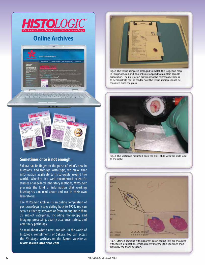
The Mohs surgeon delivers the specimen and map to the laboratory where the tissue is sectioned by the technologist. The sample is compared to the map to ensure accurate orientation and marking. The surgeon makes a nick in the specimen; this is where the marking dyes are applied. The map will reflect this color coding. To further relax the tissue, a nick can be placed opposite the original slit. Figure 2 demonstrates how the specimen is color coded and illustrates how its orientation is identical to the map and the surgical wound.
The tissue is embedded using Tissue-Tek® O.C.T.™ Compound (Sakura Finetek USA, Inc., Torrance, CA) and the slide embedding technique.3 Once the block is ready for sectioning, it is mounted in the block holder of the cryostat with the marked slit facing the blade. When the technician places the section onto the slide, the label should be to the right (Fig. 3); however, when the slide is viewed under the microscope, the slide should be rotated 180 degrees so that the label is to the left. This is to account for the optical inversion produced by the microscope. Now what the Mohs surgeon sees is an exact replica of the map and the excised Mohs tissue (Fig. 4).
If the specimen is bisected, both superior edges are dyed identically with one color, and the inferior edges are dyed identically to each other but with a different color. Each half of the bisected specimen is embedded separately and labeled as blocks
Fig. 1. (A) Tissue shown on the slides is not stereo-oriented; (B) tissue shown on the slide is stereo-oriented to the Mohs surgical map and the patient.
HISTOLOGIC, Vol. XLVI, No. 16
Sometimes once is not enough.Sakura has its finger on the pulse of what’s new in histology, and through HistoLogic, we make that information available to histologists around the world. Whether it’s well-documented scientific studies or anecdotal laboratory methods, HistoLogic presents the kind of information that working histologists can read about and use in their own laboratories.
The HistoLogic Archives is an online compilation of past HistoLogic issues dating back to 1971. You can search either by keyword or from among more than 25 subject categories, including microscopy and imaging, processing, quality assurance, safety, and veterinary pathology.
So read about what’s new–and old–in the world of histology, compliments of Sakura. You can access the HistoLogic Archives on the Sakura website at www.sakura-americas.com.
Online Archives
HISTOLOGIC, Vol. XLII, No. 1
1
HISTOLOGIC, Vol. XLII, No. 1
1
Vol. XLII, No. 1
June 2009
Managing Editor, Nancy Klemme
Scienti� c Editor, Vinnie Della Speranza,
MS, HTL(ASCP) HT, MT
IN THIS ISSUE
Recent Hematoxylin
Shortage and Evaluation
of Commercially Available
Substitutes . . . . . . . . . . . . . . . . .
. . . . . . 1
After 24 Years
in Formalin,
It Should Be Fixed . . . . . . . . . . . . . . . . .
6
Large Mount Trichrome
Method for Quanti� cation
of Ischemic Tissue in a
Porcine Cardiac Model . . . . . . . . . . . 10
Dyeing to Be Safe . . . . . . . . . . . . . . . . . 14
Picro-Sirius Red Dye,
Polarization, and
Collagenous Tissue . . . . . . . . . . . . . . 16
Letter From the
Editor . . . . . . . . . . . . . . . . . .
. . . . . . . . .20
Mark Your Calendar . . . . . . . . . . . . . . . 23
We evaluated different substitute stains
that were available through various
commercial sources, and in a blinded
study compared their staining results
with the Gill’s hematoxylin #2 solution
we routinely use in our laboratory.
Introduction
Hematoxylin, a derivative of the logwood
tree Haematoxylon campec
hianum, is
one of only a few dyes derived from
nature that is still in use in the modern
histology laboratory.1 Because the
tree is found in just a few regions of
the globe, supply of this natural
commodity may fl uctuate as a result of
climate, political, or economic forces.
Although there were once 4 logwood
Fig. 1. Basal cell carcinoma in human skin, Gill’s hematoxylin #2 stain. 100X
Abstract
In 2008, some laboratories reported
diffi culty obtaining hematoxylin stain
solutions or dye powder from their usual
commercial sources. Discussions on the
listserv Histonet quickly revealed rumors
of a hematoxylin dye shortage. Vendors
with a short supply of hematoxylin were
offering their customers substitute
nuclear stains as they were unable
to predict when hematoxylin would
once again be available. While some
predicted that the shortage would ease
“sometime in the fall,” the uncertainty
left laboratories scrambling for an
alternative nuclear stain.
Recent Hematoxylin Shortage
and Evaluation of Commercially
Available Substitutes
Ashley Groover, BS; Carlette Geddis, BS, HTL(ASCP);
Amanda Finney, BS
Medical University of South Carolina
Charleston, SC
SK5897_HistoNewsltr_JUNE_09_Release.indd 1
6/10/09 3:06 PM
HISTOLOGIC, Vol. XLIV, No. 1
HISTOLOGIC, Vol. XLIV, No. 1
1
HISTOLOGIC, Vol. XLIV, No. 1
1
Vol. XLIV, No. 1
June 2011Managing Editor, Nancy Klemme
Scienti� c Editor, Vinnie Della Speranza, MS, HTL(ASCP)HT, MT
IN THIS ISSUEExpedited Bone Throughput Using Microwave Decalci� cation . . . . . . . 1Cryopreservation Method
Optimization for Mouse Brain Tissues . . . . . . . . . . . . . . . . . . . . 5Is Rapid Tissue Processing the Right Choice forYour Lab? . . . . . . . . . . . . . . . . . . . . . . 10My Life-changing Opportunity
to Train Histotechs in Africa . . . . 14Histological and Histochemical Staining of Sections Using
an Inverted Vial Device . . . . . . . . 18 Optimization of a Fast Schi� Reaction for Tissue Staining . . . 20 Mark Your Calendar! . . . . . . . . . . . . 23
One potential method for decreasing
the overall processing time for
bone specimens involves the use of
microwave decalcification. The goal
of this study was to determine the
shortest microwave times that would
provide adequate decalcification
without compromising tissue quality.
Bones of various types (sternum, rib,
femur, femorotibial joint, and nasal
turbinate) from eight animal species
were decalcified in a microwave
processor for variable time periods.
Adequacy of bone decalcification was
evaluated on an hourly basis for small
animals (eg, mice and rats), and every
2 hours for larger animals (eg, dogs
and monkeys), until decalcification
was considered to be complete.
Depending on the size and type of
specimen, the time for complete
decalcification of small animal bones
was reduced from a period of 1-6 days
Fig. 1. Microwave decalci� ed dog femur. Stained with hematoxylin and eosin (H&E); 100X
AbstractHistology laboratories supporting toxicology testing are routinely tasked with
production of very large numbers of histologic specimens from standard laboratory
animal species. Because of this, minimizing slide preparation time is an ongoing
challenge. One step that is especially time consuming is the traditional decalci� cation of bones
by manual immersion in decalcifying agents at room temperature. This is particularly
true for large laboratory animals such as dogs and primates. Among other factors,
the rate of bone decalci� cation is dependent upon size of the specimen, age of the
animal, type of decalcifying agent, and methodology employed.
Expedited Bone Throughput Using Microwave Decalci� cation
Shelley L. Gruntz, HT(ASCP); Vivian English, HT(ASCP)
Experimental Pathology Laboratories Inc.
Sterling, [email protected]
HISTOLOGIC, Vol. XLV, No. 1
Vol. XLV, No. 1
June 2012
Managing Editor, Theresa Ford
Scienti� c Editor, Vinnie Della Speranza,
MS, HTL(ASCP)HT, MT
A Decemented, Decalci� ed Para� n
Processing Option for Resurfaced
Femoral Head Implant Specimens
Robert A. Skinner, BS, HTL(ASCP)*; Sandra G. McLaren, BS*;
Ginell R. Post, MD, PhD†; Larry J. Suva, PhD*
*Department of Orthopaedic Surgery, Center for Orthopaedic Research
† Department of Pathology
University of Arkansas for Medical Sciences
Little Rock, AR
HISTOLOGIC, Vol. XLV, No. 1
Managing Editor, Theresa Ford
Scienti� c Editor, Vinnie Della Speranza,
MS, HTL(ASCP)HT, MT
IN THIS ISSUE
A Decemented, Decalci� ed Para� n
Processing Option for Resurfaced
Femoral Head Implant Specimens . . 1
The E� ect of Temperature and
Agitation on Adipose
Tissue Fixation . . . . . . . . . . . . . . . . . .
. . 7
Mounting of Para� n Tissue
Sections on Filter Paper for
Storage, Mailing, and
Microdissection . . . . . . . . . . . . . . . . . .
11
Modi� ed Movat Stain Is E� cient
and Cost-e� ective . . . . . . . . . . . . . . . 14
Mark Your Calendar! . . . . . . . . . . . . . . 19
funding. The study of bone or
components of bone, which may be
optimally served by undecalcified
analysis in a methacrylate embedding
medium, could possibly be adequately
served by a decalci� ed para� n workup
that is almost always less expensive and
can provide a platform to support a
wider range of downstream analyses. A
particular dilemma arises when the bone
to be studied has been integrated with
biomaterials or orthopaedic appliances.
This report describes a method for
preparing para� n sections of decalci� ed
bone following removal of the cement
used to retain the prosthesis that allows
for the investigation of cellular changes
in metal cap resurfaced femoral heads.
Optimal safranin O-fast green (SOFG) staining of formalin-� xed, formic acid decalci� ed, and
para� n-processed articular cartilage in a pathologic femoral head. Crisp orange-red staining
of the proteoglycan clearly illustrates the di� erentiating chondrocytes (arrows). 100X
Abstract
The � eld of histotechnology in an orthopaedic research setting encompasses a wide
variety of procedures and instrumentation. Crossover versatility of both personnel and
equipment is key, not only to gathering information, but also for securing extramural
HISTOLOGIC, Vol. XLIV, No. 2
40 Years — A Remarkable MilestoneAs 2011 draws to a close, most of us are
likely distracted with deadlines coming
due or projects just beginning. It seems
that when one issue of this publication
is nearing completion, I am already preparing for the next. I was recently reminded that this year, 2011, marks the
40th year that HistoLogic® has been a
voice of the histotechnology community
through the generosity of Sakura Finetek
USA today, and what was formerly the Lab-Tek Products division of Miles
Laboratories, Inc. in years gone by.HistoLogic was the vision and dream of Lee G. Luna, whose name may be familiar to many of our readers. Perhaps
you have his textbooks on your shelf, likely tattered from many years of use
at the bench, or some of the 150+ published papers he authored. You may not know that he worked tirelessly
for histotechnologists everywhere, committed in his belief that the discipline and its practitioners needed a
vehicle with which to communicate and
share their knowledge with one another.
To that end, Luna founded and became
the � rst scienti� c editor of HistoLogic,
the � rst issue of which was published
in 1971.To put this in perspective, HistoLogic was founded before all but two of the state histotechnology societies in the United States and well before the creation of the National Society for Histotechnology (NSH). It was established at a time when there truly
was no other means for histotechs to communicate or conferences for them
to attend. As a result, Luna’s dream, in
the form of this bulletin, was realized.
He served as this publication’s � rst
editor for 21 years. I can only speculate
whether this publication contributed to the formation of the NSH, which was
incorporated in 1973. I do believe that
HistoLogic brought to histologists of the
time a glimpse of what could be in the
discipline. The coalition of histology practitioners that is now the NSH wasthe result of e� orts by Luna and many
others whose passion became the organization that serves our community
of professionals today. Visit www.nsh.org
to learn more about this organization
and what it can do for you.Over the years, HistoLogic has been received by more than one million readers from around the globe, largely
the result of the generous contributions
from so many authors who have o� ered
the wealth of information contained within the HistoLogic archives (http://www.sakura-americas.com/
histologic/index.html). I am humbled
by the distinguished editors who have
preceded me and by the privilege Sakura
has entrusted in me to continue the rich
legacy that was and is HistoLogic.
I sometimes wonder if Luna would approve of today’s version of his creation. I hope that he would. If you are reading these words, I hope
you will consider contributing to this publication’s rich history by sharing your work or knowledge with others who read these pages. Many aspiring
authors have had their � rst published
article appear here. I’m sure all would
tell you how gratifying it can be to see
your work in print and to know that the
information you share can help other
histotechs in their work. If you have news to share but are not quite sure how to go about it, I hope you will contact me. I’m sure I can help you.
How many of you have that very � rst issue of HistoLogic still in your library,
Vol. I, No.1 dated July 1971? If you do, I’d
love to hear from you. Let’s keep this rich
tradition and valuable resource going
strong for many more years to come!Vinnie Della SperanzaScienti� c [email protected]
Vol. XLIV, No. 2
December 2011Managing Editor, Theresa Ford
Scienti� c Editor, Vinnie Della Speranza, MS, HTL(ASCP)HT, MT
Fig. 4. Stained sections with apparent color-coding inks are mounted with stereo-orientation, which directly matches the specimen map drawn by the Mohs surgeon.
Fig. 2. The tissue sample is arranged to match the surgeon’s map. In this photo, red and blue inks are applied to maintain sample orientation. The illustration drawn onto the microscope slide is to demonstrate for the reader how the tissue section should be mounted onto the glass.
Fig. 3. The section is mounted onto the glass slide with the slide label to the right.

HISTOLOGIC, Vol. XLVI, No. 1 HISTOLOGIC, Vol. XLVI, No. 1 7
1 and 2. Figure 5 illustrates how the specimens are color coded and oriented in such a way that they replicate the map of the wound. Once the blocks are ready for sectioning, they are placed in the cryostat block holder with the marked superior edge of the specimen facing the blade. The sections are picked up with the slide label to the right and are oriented to be identical to the surgical map and the surgical wound (Fig. 6).
Once the slide is rotated 180 degrees and viewed under the microscope (with the label on the left), it will be an exact replica of the Mohs map and the surgical wound (Fig. 7).
Discussion
The premise behind this technique is that, when reading slides, the Mohs surgeon can be confident that the tissue mounted on the slide is an identical representation of the specimen map (as if the specimen were superimposed on the map) so that the location of residual tumor can be identified and tracked accurately. Adopting and consistently applying this strategy for mounting tissue sections onto glass slides can reduce any possibility of error while minimizing mental fatigue. It may even save time. In instances when the specimen is too large to fit onto a standard glass slide for stereo-orientation, the technologist should consult with the surgeon before proceeding.
Conclusion
Stereo-orientation is a novel technique for mounting tissue sections that offers important benefits to the Mohs surgeon for accurately tracking residual tumor foci. There are many ways to orient tissue specimens, each with its advantages and disadvantages. Stereo-orientation is one such method that may be unfamiliar to many Mohs surgeons and technologists. This article is meant to familiarize others with this technique and to allow them the opportunity to adapt it to their own practice.
References1. Barbara Beck (oral communication, 2013).
2. Cognetta AB Jr, Wolfe CM, Stewart-Eaves C, Edwards JL. Letter: stereo-orientation of Mohs surgical specimens: a novel histologic pearl to reduce mental fatigue. Dermatol Surg. 2012;38(11):1886-1887.
3. Geddis CM. A novel technique to embed tissues for frozen section cryotomy. HistoLogic. 2004;37(1):7-10.
Fig. 5. The sample is bisected, mounted, and color coded as indicated on the map.
Fig. 7. H&E stained slides that were stereo-oriented and labeled as specimens 1 and 2.
Fig. 6. Specimen is labeled as 2 on the map shown in Fig. 5.

HISTOLOGIC, Vol. XLVI, No. 18
Third-hand Immobilization in Gross Sectioning of Pathology Specimens
Izak B. Dimenstein, MD, PhD, HT(ASCP)Loyola University Chicago Medical Center (Ret.)Grand Rapids, [email protected]. grossing-technology.com
Abstract
This technical note proposes the “third-hand” principle for additional immobilization during gross sectioning of tissue specimens. It consists of pressing the specimen to be cut against a vertical surface (referred to here as the “third hand”). This method is especially beneficial for calcified specimens (bones and tumors) and soft tissue samples that require thin, uniform gross sections to be achieved. The third-hand immobilization principle is the underlying foundation for the design of the Biopsy Uniform Section Device, a concept developed by this author. Although the development of the device is still a work in progress, the testing of prototypes confirms the advantage of third-hand immobilization in achieving uniform and perpendicular sections of pathology specimens.
Introduction
There is no question that gross sectioning of surgical pathology specimens requires a firm surface beneath the specimen. The use of a cutting board surface (similar to that used in food preparation environments) is quite commonplace in the surgical pathology laboratory. The degree of firmness or pliability of the specimen dictates the need for such immobilization. In many cases, a skilled grossing technologist can achieve reasonably satisfactory sections freehand without much difficulty. However, some specimens require additional support from special techniques, devices, gadgets, and improvised at-hand materials. This technical note presents the third-hand principle for additional immobilization during gross sectioning of pathology specimens. This technique consists of pressing the area to be cut against a vertical surface (the “third hand”). The “first hand” holds and moves the specimen while the “second hand” operates the cutting instrument. The third-hand immobilization method is most beneficial when cutting calcified specimens and soft tissues that can otherwise pose significant challenges in achieving optimum sections.
Calcified Specimens
An acceptable section of femoral head with osteoarthritis can be easily achieved by using either a mechanical saw or handsaw because the relative firmness of the specimen is fairly consistent throughout. However, technical challenges can arise when sampling calcified tumors, fragile complicated bones, small fragments of bone, and samples requiring serial sectioning at the grossing table. In my experience, perfectly acceptable results can be achieved with a hacksaw when working with these specimens. There are certain particularities in sawing technique, but the main issue is preventing the specimen’s movement due to a snag between the saw and the calcified tissue. The employment of the third-hand principle for additional immobilization offers significant advantages when working with such samples.
It is important to find an appropriate vertical surface to press the bone specimen against during sawing. Depending on the size of the specimen, the vertical surface can be different in form, but must be more or less flat. A wooden holding tray, like the one used in the Davidson Marking System® (Bradley Products, Inc, Bloomington, MN), can be useful as an immobilization support gadget for some specimens. This wooden stand with round bottle holders and multiple different-shaped notches can be modified to accommodate many configurations of bone specimens. The pegs are very useful, especially if some of them are flattened so they can be utilized as the third hand during sectioning (Fig. 1).
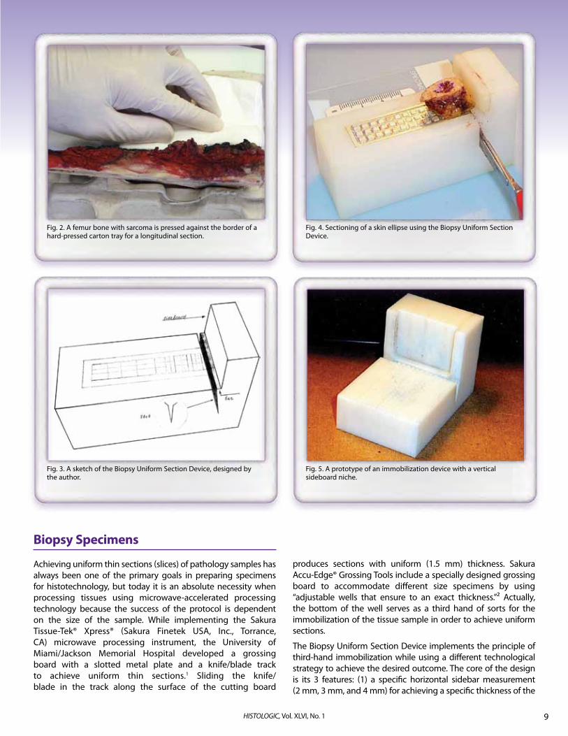
In the case of a fragile bone or calcified tumor, it is reasonable to fashion a hard-pressed packing carton into an immobilization support using this at-hand material (Fig. 2). In this situation, the wall serves as the third hand, preventing the fragile bone’s surface from crashing, while allowing the cutting instruments, such as a saw, to pass through the carton, maintaining the integrity of the specimen and the section.
Fig. 1. A small bone specimen can be pressed against a flattened wooden peg of a Davidson Marking System holding tray, which serves as the third hand, offering additional immobilization when cutting.
HISTOLOGIC, Vol. XLVI, No. 1 HISTOLOGIC, Vol. XLVI, No. 1 9
Biopsy Specimens
Achieving uniform thin sections (slices) of pathology samples has always been one of the primary goals in preparing specimens for histotechnology, but today it is an absolute necessity when processing tissues using microwave-accelerated processing technology because the success of the protocol is dependent on the size of the sample. While implementing the Sakura Tissue-Tek® Xpress® (Sakura Finetek USA, Inc., Torrance, CA) microwave processing instrument, the University of Miami/Jackson Memorial Hospital developed a grossing board with a slotted metal plate and a knife/blade track to achieve uniform thin sections.1 Sliding the knife/blade in the track along the surface of the cutting board
produces sections with uniform (1.5 mm) thickness. Sakura Accu-Edge® Grossing Tools include a specially designed grossing board to accommodate different size specimens by using “adjustable wells that ensure to an exact thickness.”2 Actually, the bottom of the well serves as a third hand of sorts for the immobilization of the tissue sample in order to achieve uniform sections.
The Biopsy Uniform Section Device implements the principle of third-hand immobilization while using a different technological strategy to achieve the desired outcome. The core of the design is its 3 features: (1) a specific horizontal sidebar measurement (2 mm, 3 mm, and 4 mm) for achieving a specific thickness of the
Fig. 2. A femur bone with sarcoma is pressed against the border of a hard-pressed carton tray for a longitudinal section.
Fig. 3. A sketch of the Biopsy Uniform Section Device, designed by the author.
Fig. 4. Sectioning of a skin ellipse using the Biopsy Uniform Section Device.
Fig. 5. A prototype of an immobilization device with a vertical sideboard niche.

HISTOLOGIC, Vol. XLVI, No. 110
section, (2) a vertical sideboard, and (3) a V-notched slot for the cutting blade. The sideboard provides immobilization for cutting the end of the tissue, ultimately functioning as the third hand (Fig. 3).
Figure 4 shows the principle of using the Biopsy Uniform Section Device. A skin ellipse is placed on the platform and moved toward the slot and the vertical sideboard using forceps. When the skin sample reaches the vertical sideboard, the part of the sample on the horizontal sidebar is the desired thickness of the section.
By pressing the specimen against the vertical sideboard, secure immobilization is achieved for a definitive uniform perpendicular cut. The slice is removed with forceps after each section has been made and placed into a processing cassette oriented as it is intended to be embedded. Milestone Medical has improved the design of the vertical sideboard by making a niche that provides additional immobilization for the section (Fig. 5). This is especially important in the case of large samples.
The Biopsy Uniform Section Device is designed for small specimens, but the vertical sideboard can be adjusted to accommodate larger specimens. However, as shown in Fig. 6, it is difficult to obtain uniform serial sections when the specimen is not adequately fixed. Third-hand immobilization is not sufficient in this situation.
Discussion
The principle of precise cutting against a sideboard is used in a variety of applications, such as the cutting of lumber, for example. This principle is also used in grocery stores for the uniform slicing of cheese and ham. Although the cutting tools differ from each other in their specific uses, the immobilization platform is a common element that contributes to a precise cut. However, the goal in surgical pathology is to obtain a diagnostically valuable section. This often requires additional devices and materials in the preparation of optimum specimens, including some that can be used in the practical application of the third-hand immobilization principle.
Variants of gadgets and at-hand improvised materials that might serve as additional third-hand immobilization aids are endless.
The aforementioned contrivances reflect personal experience, which is always limited.
Numerous experiments I have undertaken in search of optimal third-hand support materials have led me to conclude that hard-pressed packing cartons appear to be optimal for the type of support needed for bone sampling.3 They provide the necessary firmness, but not too much. The relative roughness of the carton’s surface gently grasps the specimen’s inevitably uneven surface. The saw can cut through the specimen and carton without crashing, which can occur at the end of the cut. With a collection of disposable immobilizing materials, it would be appropriate to point out that every calcified specimen can be cut without preliminary decalcification, including bones from facial-maxillary surgery and calcified tumor, providing valuable sections for evaluating surgical margins or to answer other diagnostic questions.
The holding trays from the Davidson Marking System are permanent fixtures in every surgical pathology laboratory.4 The configuration provides endless options for third-hand immobilization of small bone or calcified tissue. Saw-friendly wood makes it possible to cut a fragile or small fragment without fear of a crash toward the end of sawing. Even some excisional biopsies can be cut in this way, although a special gadget like the Biopsy Uniform Section Device would be a better option.
The Biopsy Uniform Section Device not only provides sections of predetermined thickness, depending on the horizontal bar’s size and the size of the vertical sideboard, but its third-hand immobilization capability facilitates a strictly perpendicular gross section that is very important in dermatopathology, gastroenterology, and gynecology samples, as well as in the evaluation of surgical resection margins.
The development of the Biopsy Uniform Section Device is still a work in progress. Much work remains before a final design will be ready.
At this time, there have been no reports of studies with comparable devices, perhaps because sectioning strategies differ. The main, and perhaps most important, variable is in the ability to visualize the sample to be sectioned while it is immobilized, which is a large advantage and the primary focus of the concepts proposed in this report. A blind, indiscriminate cut is not acceptable in excisional biopsy slicing.
The desire for one universal tool that can be used to achieve thin, uniform gross sections for all types of pathology specimens is unrealistic. The diversity of surgical pathology specimens will make a variety of devices necessary to achieve the desired outcome in each instance. The principle of third-hand immobilization lays the foundation for the design of such devices.
References1. Romaguera R, Nassiri M, Morales AR. Tools to facilitate and standardize grossing.
HistoLogic. 2003;36(1):17-21.
2. Accu-Edge® Grossing Tools. Sakura Finetek USA, Inc. Web site. http://www.sakura-americas.com/products/grossingtools.html. Accessed May 15, 2013.
3. Equipment instruments gadgets. Grossing Technology in Surgical Pathology Web site. http://www.grossing-technology.com/newsite/home/equipment-instruments-gadgets/. Accessed February 20, 2013.
4. Dimenstein IB. Grossing biopsies: an introduction to general principles and techniques. Ann Diagn Pathol. 2009;13(2):106-113.
When you’re in sync with your work, it shows.
Fig. 6. Sectioning of an unfixed tissue (placenta).

HISTOLOGIC, Vol. XLVI, No. 1 HISTOLOGIC, Vol. XLVI, No. 1 11
Same day results with Tissue-Tek® Xpress® x120.
Sakura Finetek USA, Inc. • 1750 West 214th St., Torrance, CA 90501 USA • Phone: 800-725-8723 ©2013 Sakura Finetek USA, Inc. All rights reserved.
Most people want to become better at what they do. With Sakura instrumentation, becoming better comes naturally.
Tissue-Tek® Automation is enhancing productivity for people around the world.
Because better efficiency and a never-ending desire to improve are what drive us. Shouldn’t your lab equipment be a reflection of you?
Learn more at www.sakura-americas.com.
When you’re in sync with your work, it shows.

12 HISTOLOGIC, Vol. XLVI, No. 1
The Effectiveness of Even Ice Surface on Microtomy of Paraffin Blocks
Peter Fabian, BS, HTL(ASCP)QIHC; Orlando MoralesAmeriPath Northeast, GI DivisionShelton, [email protected]
Abstract
Our high-volume histology lab requires that a histotechnologist be able to cut 50 paraffin blocks per hour, an increase from the previous requirement of 30 blocks per hour. In order to ensure consistent, high-quality outcomes, the histotechnologist must develop a routine that reduces the variables that can slow output or diminish quality since there is no time for adjustments during cutting.
In our laboratory, we established a consistent output of slides using a methodology that makes blocks easy to cut with quick output and fewer cutting artifacts. The technique consists of chilling the blocks on an even, flat surface of ice. The even cooling of the blocks allows for the temperature and soaking of the blocks to be uniform, which helps achieve sections that are free of chatter and folds.
Introduction
Chatter, which is the term for artifact that results from microscopic vibration during microtomy, can be observed in tissue sections when viewed with the microscope (Fig. 1). Thompson and Luna attribute this artifact primarily to overdehydration or removal of protein-bound water, which can occur when tissues are overexposed to dehydrating solutions during tissue processing.1 Soaking paraffin blocks prior to sectioning can restore the necessary moisture to tissues, which facilitates sectioning (Fig. 2). Microscopic chatter can also be caused by a dull blade or an incorrect blade angle (knife tilt), which causes the section to be scraped rather than sliced. Chatter can also occur by cutting too rapidly.2
The fact is, in today’s laboratories, the mass production of slides and rapid turnaround time expectations have raised the bar for production from technicians. The use of a flat, even ice surface for the cooling of blocks allows more blocks to be sectioned per minute. Even though rapid cutting is required to achieve desired output, the uniform ice surface reduces artifacts that can result from rapid sectioning and ensures that section quality is acceptable.
Materials and Methods
Achieving an even ice surface for block cooling is easy to accomplish by placing clean water on a tray and keeping the tray level in the freezer until frozen. The goal is to obtain a surface of ice similar to the ice of an ice skating rink. The ice should be flat and even throughout the entire surface area without bumps or cracks (Fig. 3).
Fig. 1. Chatter in section of gastric antrum. H&E, 200X.
Fig. 2. Uniform cooling and soaking of paraffin blocks yield a section of gastric antrum that is free from artifacts. H&E, 200X.
Fig. 3. An even ice surface allows for uniform cooling of paraffin blocks.
Fig. 4. A rough or uneven ice surface provides inconsistent cooling of blocks, leading to sectioning artifacts.
13HISTOLOGIC, Vol. XLVI, No. 1
A flat ice surface can also be achieved by scraping the ice. The extra time needed to prepare the ice properly is worth the effort when your cutting output achieves both the desired quality and quantity. In our lab, we use large blue thermal trays that allow the ice to remain frozen longer (Fig. 3).
We process our gastrointestinal (GI) biopsies with a 2-hour processing protocol on a conventional closed-system processor. GI tissue is prone to overdehydrating if longer processing times are used. If overdehydration is a problem, this can be overcome by soaking the faced paraffin block in water directly over ice, which is imperative in order to achieve good sectioning outcomes. This is easier to accomplish when water is added onto a flat, even ice surface before placing your block face down on the surface with or without a paper towel. If the ice surface is not level, you can use wet paper towel directly in contact with the ice surface to compensate.
Blocks are soaked for about 20 minutes to allow sufficient moisture to reenter the blocks to soften the tissues. You may need to determine the best time for your tissues through trial and error. Each laboratory should optimize soaking times based on the tissue types and processing schedules you use. Soaking time can vary between different tissue types. Exercise caution not to oversoak your blocks, which can cause other artifacts that are undesirable at cutting. With optimal soaking time, artifact-free sections can be achieved more rapidly during sectioning.
Results
The chatter artifact is often invisible to the eye while sectioning tissue on a microtome. More dramatic artifacts, like the “Venetian blind” effect, can be evident when sections are placed onto the water bath surface. Chatter identified in microscopic sections is often the result of tissue that has not been well soaked; it may also occur when sectioning (cutting) with the microtome is done too rapidly.
On an uneven ice surface, the blocks cool at uneven temperatures due to bumps on the ice and uneven exposure to ice surface (Fig. 4). Well-soaked blocks on a smooth, even ice surface will chill at even temperatures and produce ribbons of good quality that reduce chatter and can be cut with ease.
References1. Thompson SW, Luna LG. An Atlas of Artifacts Encountered in
the Preparation of Microscopic Tissue Sections. Springfield, IL: Charles C. Thomas; 1978:102.
2. Carson FL. Histotechnology: A Self-Instructional Text. 2nd ed. Chicago, IL: American Society for Clinical Pathology; 1997:48-58.
Acknowledgment The author would like to acknowledge Dr. Gerald P. Bailey, MD, PhD, for his collaboration in preparing the micrographics for this article.
Quick-Start.Quick-Punch.Quick-Ray.TM
Generate superior quality TMAs in a fraction of the time than with traditional methods.• Preformed ready-to-use paraffin recipient blocks• Multiple samples on one slide
For more information, please visit our website at www.sakuraus.com.
Trusted InnovationTM
Sakura Finetek USA, Inc. • 1750 West 214th St., Torrance, CA 90501 USA • Phone: 800-725-8723 ©2013 Sakura Finetek USA, Inc. All rights reserved.

HISTOLOGIC, Vol. XLVI, No. 114
Same day results withTissue-Tek® Xpress® x50.
I’ve never been so scared in my whole life... I need to know now.
Sakura Finetek USA, Inc. • 1750 West 214th St., Torrance, CA 90501 USA • Phone: 800-725-8723 ©2013 Sakura Finetek USA, Inc. All rights reserved.
Waiting for biopsy results can cause enormous anxiety for patients. But now, Tissue-Tek® Automation is helping to reduce waiting time—in some cases, down to as little as 1 day.
That means patients can get answers sooner and physicians can begin treatment faster.
Making every moment count is what Tissue-Tek® Automation is all about.
Learn more at www.sakura-americas.com.
For patients, waiting is the hardest part.

HISTOLOGIC, Vol. XLVI, No. 1 HISTOLOGIC, Vol. XLVI, No. 1 1515
✔ Mark Your Calendar!Educational Opportunities in 2013
19 University of Texas Health Sciences Ctr/San Antonio Teleconference 12:00 pm Central Time (800) 982-8868 Title: Green Histology Speaker: Lawrence Patton Leica Biosystems Buffalo Grove, IL
24 NSH Teleconference 1:00 pm Eastern Time Title: How Did This Slide Contamination Happen? Speaker: Violet Swazer Detroit Medical Center Detroit, MI Phone: (443) 535-4060 or register online at www.nsh.org
16 University of Texas Health Sciences Ctr/San Antonio Teleconference 12:00 pm Central Time (800) 982-8868 Title: Effects of Pre-Analytical Factors on IHC Speaker: Damien Matusiak Leica Biosystems Buffalo Grove, IL
28 NSH Teleconference 1:00 pm Eastern Time Title: What Should a Good H&E Look Like? Speaker: Robert L. Lott Skin Pathology Associates Birmingham, AL Phone: (443) 535-4060 or register online at www.nsh.org
21 University of Texas Health Sciences Ctr/San Antonio Teleconference 12:00 pm Central Time (800) 982-8868 Title: Digital Evolution: Taking the Pathologist From the Basement to the Bedside Speaker: Damien Matusiak Leica Biosystems Buffalo Grove, IL
26 NSH Teleconference 1:00 pm Eastern Time Title: Identifying Histology Look-Alikes Speaker: Amy Aulthouse, PhD Ohio Northern University Ada, OH Phone: (443) 535-4060 or register online at www.nsh.org
JUNE
AUGUST
18 NSH Teleconference 1:00 pm Eastern Time Title: Immunohistochemistry, Antibodies, and Pathology Speaker: George Yang, MD Cell Marque Rocklin, CA Phone: (443) 535-4060 or register online at www.nsh.org
20 University of Texas Health Sciences Ctr/San Antonio Teleconference 12:00 pm Central Time (800) 982-8868 Title: Histology Process Improvement: Tissue Processing From Conventional Overnight to Continuous Speaker: William DeSalvo Collaborative Advantage Consulting Tempe, AZ
20-25 National Society for Histotechnology Symposium/Convention Site: Rhode Island Convention Center Providence, RI Contact: Aubrey Wanner Phone: (443) 535-4060 or register online at www.nsh.org Fax: (443) 535-4055 Email: [email protected]
SEPTEMBER
18 University of Texas Health Sciences Ctr/San Antonio Teleconference 12:00 pm Central Time (800) 982-8868 Title: Safety in the Histology Laboratory Speaker: Terri L. Braud Holy Redeemer Hospital Rockledge, PA
23 NSH Teleconference 1:00 pm Eastern Time Title: Building Effective Teams Speaker: Louis Anderson Johns Hopkins Medical Institutions Baltimore, MD Phone: (443) 535-4060 or register online at www.nsh.org
18 NSH Teleconference 1:00 pm Eastern Time Title: Double IHC Staining Speaker: Charlie Dorner Dako North America Inc. Carpinteria, CA Phone: (443) 535-4060 or register online at www.nsh.org
OCTOBER
NOVEMBER
DECEMBER
15 University of Texas Health Sciences Ctr/San Antonio Teleconference 12:00 pm Central Time (800) 982-8868 Title: Role of Pathologist in Colorectal Cancer Diagnosis and Management Speaker: Prashant A. Jani, MD, FCAP, FRCPC Thunder Bay Regional Health Sciences Centre Thunder Bay, Ontario, Canada 20 NSH Teleconference 1:00 pm Eastern Time Title: An Introduction to Commonly Used Immunohistochemical (IHC) Stains in Dermatopathology Speaker: Alison Uzeblo, MD William Beaumont Hospital Royal Oak, MI Phone: (443) 535-4060 or register online at www.nsh.org
JULY

Presorted StandardUS Postage
PaidSakura Finetek USA, Inc. 1750 West 214th Street Torrance, CA 90501
To receive your own copy of HistoLogic® or to have someone added to the mailing list, submit your home address to: Sakura Finetek USA, Inc., 1750 West 214th Street, Torrance, CA 90501.
The editor wishes to solicit information, questions, and articles relating to histotechnology. Submit these to: Vinnie Della Speranza, HistoLogic® Scientific Editor, 165 Ashley Avenue, Suite 309, Charleston, SC 29425. Articles, photographs, etc, will not be returned unless requested in writing when they are submitted.
Uniform Results With Accu-Edge® Grossing Tools• Grossing board has two adjustable wells for varying size and thickness• Grossing forks feature two sets of sharp tines, separated by a space
of 1.5, 2.0, or 2.5 mm for precise grossing
• Tissue tampers hold specimens firmly in the wells for easier trimming• Scalpel and trimming blades provide unsurpassed sharpness
for maximum cutting efficiency
Designed for Specimen Standardization
The foundation for uniform grossing and optimal results.
Visit our web site at www.sakuraus.com
Sakura Finetek USA, Inc. • 1750 West 214th St., Torrance, CA 90501 USA • Phone: 800-725-8723 ©2013 Sakura Finetek USA, Inc. All rights reserved.
No varnish in this area.