Embed Size (px)
Citation preview
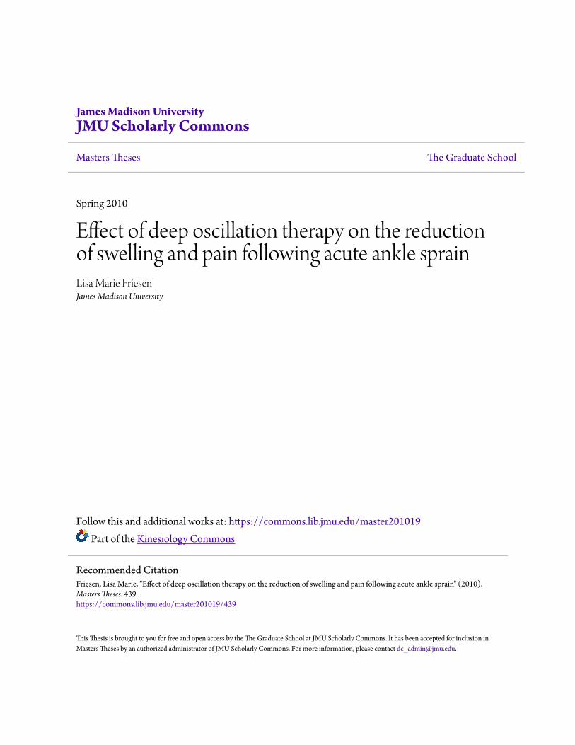
James Madison UniversityJMU Scholarly Commons
Masters Theses The Graduate School
Spring 2010
Effect of deep oscillation therapy on the reductionof swelling and pain following acute ankle sprainLisa Marie FriesenJames Madison University
Follow this and additional works at: https://commons.lib.jmu.edu/master201019Part of the Kinesiology Commons
This Thesis is brought to you for free and open access by the The Graduate School at JMU Scholarly Commons. It has been accepted for inclusion inMasters Theses by an authorized administrator of JMU Scholarly Commons. For more information, please contact [email protected].
Recommended CitationFriesen, Lisa Marie, "Effect of deep oscillation therapy on the reduction of swelling and pain following acute ankle sprain" (2010).Masters Theses. 439.https://commons.lib.jmu.edu/master201019/439
Effect of Deep Oscillation Therapy on the Reduction of Swelling and Pain following
Acute Ankle Sprain
Lisa Marie Friesen
A thesis submitted to the Graduate Faculty of
JAMES MADISON UNIVERSITY
In
Partial Fulfillment of the Requirements
For the degree of
Masters of Science
Kinesiology
May 2010

ii
Dedication
This project is dedicated to my Dad and Mom, Drs. Paul and Virginia Friesen.
Thank you for always teaching me to do my best in all things and to persevere especially
when it gets tough.
I love you both so much and pray that I will honor God as much with my life as you do
with yours.

iii
Acknowledgements
I would like to thank my thesis committee for their patients and commitment to
this project and myself in the undertaking of this thesis. Dr. Frye, thank you for seeing
potential in me. Thank you for being willing to walk me through the relatively
unknown world of research. There is no question that I could not have done it without
your help. Dr. Womack, thank you for your enthusiasm and encouragement along the
way. My graduate career at JMU has been wonderful and I am blessed that I have been
able to learn, grow, and mature under your teaching and advising. Grace Weniger,
thank you for your attention to detail and support. Thank you for believing in me and
allowing me to learn from you. Thank you for being a support both on an academic and
spiritual level. Dr. Saunders, thank you for challenging me to think like a researcher
and for strengthening this project with your research experience. I have learned so
much from sitting under your teaching these last two years.
I would like to thank Tom Kuster and the James Madison University Sports
Medicine Department for allowing the use of their facilities and modalities. Tom, I can
not think of a better supervisor. Thank you for investing in me over the last two years
and giving me to the freedom and guidance to grow. A special thank you to the Sports
Medicine Staff for your support of my project by allowing me to work with your
athletes. You guys are the best and I am blessed to work with each and every one of
you!
I would like to thank Jen Stollery and Chris Boop for their willingness to help me
collect data and to support me in so many ways. I could not have completed this
project without your help and support.

iv
I would like to thank my fellow clinical GA’s, Stephanie Demers, Erin Moore,
Leah Morrison, and Rachel Ondeck for helping to make this a great two years! I am
thankful for friendship with each of you!
I would like to thank the University Health Center for their support through
referrals. Your communication and willingness to participate in this study was much
appreciated.
I want to thank my family for their continuous love and support! I know I could
not have made it through these last two years without your prayers and care. You have
encouraged me when I needed encouragement, listened when I needed to process, and
pushed me when I needed to be pushed. I love you so much!

v
Table of Contents
Dedication.......................................................................................................................ii
Acknowledgments .........................................................................................................iii
List of Tables ................................................................................................................vi
Abstract.........................................................................................................................vii
I. Introduction .........................................................................................................1
II. Review of Literature ............................................................................................8
Ankle Sprains ......................................................................................................8
Phases of Healing................................................................................... 10
Acute Inflammatory Response .................................................... 10
Proliferation Phase...................................................................... 12
Maturation Phase ........................................................................ 13
Common Treatment of Ankle Sprains..................................................... 14
Deep Oscillation Therapy....................................................................... 17
Girth Measurements ............................................................................... 19
Pain Measurement .................................................................................. 22
III. Methods............................................................................................................. 24
IV. Results .............................................................................................................. 35
Table 1............................................................................................................... 36
Table 2............................................................................................................... 36
V. Discussion ......................................................................................................... 37
VI. Appendix ........................................................................................................... 45
A-IRB .................................................................................................... 45
B- Informed Consent .............................................................................. 65
C- Data Collection Sheets....................................................................... 72
D-Volunteer take home instructions ....................................................... 80
E-Additional methods............................................................................. 82
VII. References ......................................................................................................... 91
vi
List of Tables
Table 1
Change in Pain Scores
Change in pre to post scores on treatment 1
Verbal Pain scale of 0-10
Table 2
Change in Girth Measurements
Change in pre to post scores on treatment 1
Centimeters

vii
Abstract
Context: Deep oscillation therapy is a novel form of therapy that claims to reduce the
amount of pain and swelling in acute orthopedic injuries. However, these claims are
based on anecdotal evidence and there have not been any published studies on the
efficacy of deep oscillation therapy in reduction of pain and swelling. Due to the
prevalence of ankle sprains in physically active individuals, this study compared the
effect of deep oscillation therapy in reduction of pain and swelling to conventional
treatment.
Objective: To compare the effect of deep oscillation therapy on pain and swelling
resulting from acute ankle sprains to conventional therapy consisting of cryotherapy and
compression.
Methodology: Volunteers were healthy, physically active, college students with acute
ankle sprains, ranging in age from 18-25. Volunteers were randomly assigned to a
control group (11 volunteers) or a treatment group (10 volunteers). The control group
was treated with conventional cryotherapy and compression therapy and a placebo deep
oscillation therapy treatment. The treatment group was treated with conventional
treatment and deep oscillation therapy. Each group was treated two times a day for a
five-day treatment period.
Measurements: Objective data for swelling measurements was obtained through the use
of the figure-of-eight girth measurement reported in centimeters. Subjective data for pain
measurements was obtained utilizing a verbal numeric pain scale (0-10). Measurements
were taken before and after every treatment administered.

viii
Findings: There was not a significant difference in girth measurements or perceived
pain between the two groups over the five-day treatment period.
Future Recommendations: 1) Utilize a more sensitive measurement tool for swelling.
2) Investigate injuries that have more measurable swelling.

Chapter 1
Introduction
Ankle sprains are one of the most common injuries in America resulting in 25% of time
lost in many sports 1-3
. It is estimated that 23,000 ankle sprains occur per day in the
United States representing 4.7-24.4 percent of all musculoskeletal injuries 4,5
. Ankle
sprains can vary in healing time and treatment is often used to reduce injury time and
promote tissue healing.
Traditional treatment of ankle sprains consists of cryotherapy, compression, and
elevation. Therapeutic modalities have been used to control swelling, increase tissue
mobility, and decrease pain. Research has shown that cryotherapy reduces healing time
significantly by lowering cellular metabolism that in turn decreases secondary ischemic
injury6,7
. Secondary ischemic injury is the result of diminished oxygen supply, decreased
fuel, and an inability to remove waste from the site of injury 8. The more the swelling is
controlled, the less secondary ischemic injury occurs and the faster the injury can
progress and heal. Compression and elevation help reduce excess fluids at the site of
injury by aiding the vascular and lymphatic systems 9.
Clinically, swelling can be measured by volumetric measurements or girth
measurements. Both methods have been found to be valid and reliable 10-12
. Girth
measurements are commonly used in clinical settings to measure swelling, and are often
preferable over volumetric measurements due to lower cost and ease of use. The figure-
of-eight method, the most common way to measure girth at the ankle joint, was first

2
published by Esterson in 1979 13
.
Nociceptors feedback to the central nervous system appears to be associated with
the degree of soft tissue damage 14
, thus pain level assessments can indicate soft tissue
healing and are often used clinically. Although there are varying ways to measure pain,
the numeric pain scale is preferable to other pain assessments due to the ease of use 15
.
The numeric pain scale ranges from 0-10 and allows the individual to assign a number to
the amount of pain they are experiencing 1.
While traditional therapy, including rest and protection, ice, compression, and
elevation continue to be a gold standard of ankle therapy treatment; new therapies are
often utilized to help improve the healing environment of the injured tissue. The
HIVAMAT, a relatively new modality created in Germany, claims to reduce swelling
through the utilization of a pulsed electrostatic field. The pulsed electrostatic field causes
vibrations in the tissue referred to as deep oscillations. These oscillations increase the
micro-circulation in the affected connective tissue, helping to reduce swelling, and thus
minimize secondary ischemic injury. It also aids the affected tissue by helping to flush
out waste products and increase lymphatic flow. Two studies have shown that deep
oscillation therapy can reduce swelling and pain as well as improve wound healing 17,18
.
Anecdotal evidence and some published literature supports that deep oscillation therapy
aides in the reduction of pain and swelling, but to date no clinical studies have been
performed documenting reduced swelling and pain in acute orthopedic injuries.
The purpose of this study is to compare the effectiveness of deep oscillation
therapy on the reduction of pain and swelling in patients following acute ankle sprains. It

3
is hypothesized that there will be a greater reduction in swelling and a decrease in pain in
the ankle with the deep oscillation group when compared to the control group.
Statement of Problem
The purpose of this study is to study the efficacy of deep oscillation therapy to
reduce swelling and pain in acute ankle sprains. To date there has not been any published
research to support the claims made regarding the effectiveness of deep oscillation
therapy in acute orthopedic injuries. This study will compare conventional treatment of
ankle sprains to conventional treatment with the deep oscillation treatment in the
reduction of measured swelling and reported pain. A group of twenty subjects will
randomly be assigned to either a control group or the treatment group. It is hypothesized
that there will be greater reduction in swelling and a decrease in pain in the ankle with the
deep oscillation group compared to the control group. The findings of this study could
greatly affect the future treatments of one of the most common injuries in athletics.
This study will address the following questions:
1) Does the deep oscillation therapy in conjunction with conventional
treatment allow greater reduction in swelling in acute ankle sprains?
2) Does the deep oscillation therapy in conjunction with conventional
treatment allow greater reduction in pain following acute ankle
sprains?

4
Significance of Study
Ankle sprains are the most common injuries reported in athletics. Lingering
swelling often slows healing time and therefore increases time loss due to injury. If this
study shows significance in the reduction of swelling and pain with the therapeutic affect
of deep oscillations, treatment of ankle sprains could be improved by allowing symptoms
to decrease more rapidly. The benefit to the patients would be appreciable in reducing
injury time and helping to return to play more expediently.
Clinically speaking, athletic trainers and other allied health professionals are
continually faced with new products on the market. This study will help to assess the
efficacy of a modality that currently has no research to support its claims on orthopedic
injuries. This study will help clinicians assess whether this therapeutic modality is worth
buying and utilizing in their clinics.
Assumptions of the Study
The study assumes that the volunteers will comply with the guidelines set forth in
the study. The volunteers are asked to ice three times a day for twenty minutes in
addition to the treatments applied by the examiner. Volunteers are also expected to keep
the injured extremity wrapped continuously in a compression wrap and horseshoe pad
and to have it elevated above their heart as much as possible. In addition, they will be
expected to follow a home exercise program that will be given to them each day.

5
Delimitations of the Study
Application of our findings are limited to patients with lateral ankle sprains who
are experiencing pain and swelling. We will not know the effect of deep oscillation
therapy on other orthopedic injuries.
Limitations of the Study
There are no known limitations to the study at this point.
Operational Definitions
Lateral ankle sprain- injury to the lateral ligamentous complex of the ankle. The injury
can be classified in three grades (I-III).
Grade I lateral ankle sprain- minimal stretching or tearing of the lateral ligamentous
complex. Classified by minimal swelling, minimal pain, and minimal/no loss of
function.
Grade II lateral ankle sprain- moderate stretching or tearing of the lateral ligamentous
complex as noted by a positive anterior drawer test, moderate swelling, point tenderness
over the lateral ligaments.
Grade III lateral ankle sprain- complete tear of the lateral ligament complex resulting in
instability. Classified by extreme swelling and point tenderness on the lateral aspect of
the ankle.
Secondary Ischemic Injury- After the primary injury, the chemical and vascular response

6
can cause further cell death because of a decreased amount of oxygen and fuel to the site
of injury.
(P.) R.I.C.E.- (Protection), Rest, Ice, Compression, and Elevation- the acronym for
conventional treatment of lateral ankle sprains.
Rehabilitation- Exercises and stretches done to regain strength and full range of motion
following an injury.
Electrostatic Field- The charge that is created between two different electric charges.
Johnsen-Rahbek Effect-The force created by passing voltage between two metal plates
and a semiconductive material such as a porous stone. The voltage is passed through the
metal plate and the semiconductive material and creates an electric attraction between the
two materials.
Deep Oscillation- The repeated lifting and dropping of tissue due to the electrostatic
field.
Figure-of-Eight Girth Measurement- A method to measure swelling of the ankle by
measuring the distance around the ankle using a figure eight pattern.
Numeric Pain Scale - A pain assessment system in which patients are asked to rate their
pain on a scale from 0 to 10, with 0 representing no pain and 10 representing the worst
pain they have experienced or could imagine.
GameReady! System - An advanced cold and intermittent compression regimen that can
7
help reduce pain, swelling, tissue damage and muscle spasms.
Histamine- A hormone which aides in healing by increasing the vascular permeability
allowing other necessary healing agents to enter site of injury.
Leukocytes- White blood cells which aide in the removal of waste from the site of injury.
Macrophages- aid in regulation during injury repair by removal of waste from the site of
injury. Also aides in healing by keeping inflammation localized.
Prostaglandin- Necessary to maintain vascular permeability, regulates the levels of
serotonin and histamine.
Neutrophil- Type of white blood cell which aides in the healing process by attracting
other needed chemical to the site of injury and by increasing phagocytois at the site of
injury.
Summary
Ankle sprains are the most common orthopedic injury in America. To date there
has not been any research on the effectiveness of deep oscillation therapy in the reduction
of pain and swelling during the healing process following a lateral ankle sprain. This
study will look at the efficacy of deep oscillation therapy in reducing pain and swelling in
acute ankle sprain management.

Chapter Two
Review of Literature
The purpose of this literature review is to discuss: ankle sprains, soft tissue healing
progression, traditional therapeutic treatment (including rest, ice, compression, and
elevation), deep oscillation therapy, and swelling measurements.
Ankle Sprains
Ankle sprains are the most common injury in sports 1,2,19
. Sprains represent 4.7-24.4
percent of all musculoskeletal injuries 5 and are responsible for 25% of time lost in
basketball, cross country, and football 20
. There are an estimated 23,000 ankle sprains
per day in the United States of America 4. One definition of a lateral ankle sprain is a
lesion resulting from an inversion injury, with effusion and pain at the lateral ankle
joint with no fractures 1. Sprains can also be defined as an injury that stretches the
fibers of a ligament; with the more severe the injury the more damage of the fibers 19
.
The most often reported mechanism of injury is inversion and plantar flexion of the
ankle joint 1.
Anatomically, there are three ligaments that compose the lateral ligamentous complex;
the anterior talofibuar ligament (ATFL), the calcaneofibular ligament (CFL), and the
posterior talofibular ligament (PTFL). The ATFL is the most often injured ligament in
the lateral ligamentous complex due to its anatomical position 19,22
. Injury to the ATFL

9
is an isolated injury 66% of the time 23
. These three ligaments provide three major
functions for the ankle. First, they aid in proprioception. Secondly, the ligaments
provide static stability to the ankle joint 3. Lastly, the ligaments act as a guide for ankle
motion: inversion, eversion, plantar flexion, and dorsiflexion 19
.
Sprains can be classified by degree of injury to the soft tissue. Three grades of sprains
are commonly used to distinguish the severity of the sprain. One system of
classification defines grade one ankle sprains as a partial tear of the lateral ligaments24
.
Grade one sprains or mild sprains can also be classified as mild stretching of the
ligaments resulting in no loss of function or loss of strength 25
. Grade one sprains have
been defined by minimal tenderness and swelling, minimal impairment, and
microscopic tearing of collagen fibers 26
.
Grade two, or moderate sprains, include decreased range of motion and loss of strength.
Moderate swelling is seen as well as moderate tenderness with palpation. An injury
that presents with a positive anterior drawer test signifying instability and complete
tears of some but not all of the collagen fibers in the ligament are classified as grade
two sprains 3,24,26
.
Grade three sprains present with complete loss of function, swelling, and extreme point
tenderness. Ligamentous instability indicates capsular disruption and complete tears or
rupture of the lateral ligamentous complex. Special tests that indicate a grade three
injury include a positive talar tilt and positive anterior drawer test 26,27
.
10
Phases of Healing
Regardless of the severity of the injury, all tissue healing will progress through the
same stages of healing 19
. The more damaged the soft tissue, the longer each phase will
take. Traditionally the phases of healing are; 1) inflammation, 2) proliferation, 3) and
maturation or remodeling 9. While each of these phases are defined by distinct roles in
healing, they occur on a continuum and overlap. The following section will describe
the general time frame and the events that occur in each phase.
Acute Inflammatory Response -Phase I
Phase one in healing is the inflammatory response phase that occurs from the onset of
injury to as many as four 28,29
to five days 9depending on the extent of injury. This
phase is characterized by pain, active effusion, and increased temperature at the sight of
the injury 3.
Physiologically, as injury occurs, there is degeneration of the sarcolema (the outer
membrane of the muscle fiber). The disruption of the sarcolema causes a change in
membrane permeability that allows larger amounts of calcium to enter into the
myofiber at a greater rate. The influx of calcium starts a cascade of chemical reactions
resulting in myofiber death and creating a gap in the myofibers 30
. In addition, the
influx of calcium is linked with the change in mitochondrial membrane depolarization
and osmotic swelling which leads to outer membrane rupture. This chain of events is
linked to cell death associated with what is known as secondary hypoxic injury 8. The
disruption of the myofiber causes ruptures of capillaries in the muscle. The increased

11
blood from the disrupted capillaries settles in the gap of the myofiber and a hematoma
is formed.
Two main categories of response exist in the inflammatory response phase: the vascular
response phase and the chemical response. They occur simultaneously and are
interlinked on a number of levels 29
.
Vascular Response:
The vascular response begins as soon as an injury occurs. First, immediate
vasoconstriction occurs in the first five to ten minutes following injury 9,28
. The
purpose of the vasoconstriction is to create a local anemia 28
. The local anemia is
followed by vasodilation that allows increased blood flow to the site of injury 9. The
vasodilation is important for increasing the blood flow to the area allowing for the
chemical response. In addition to vasodilation, there is a corresponding vascular
permeability that is essential for the healing process to occur. The increased vascular
permeability allows for plasma proteins and white blood cells to enter into the area of
injury 29
.
Chemical Response:
The primary function of the chemical response is to aide in debris removal and to
prepare the site for repair 29
. Histamine is one of the most important chemicals
released at the site of injury by platelets and mast cells 9, and is crucial for the initiation
of inflammation. Histamine has a short half-life and is followed by prostaglandins,
which promote attraction of leukocytes, and perpetuates vascular permeability 9,31
.

12
The chemical response causes the influx of certain elements that aide in healing.
Neutrophils are the first cell to enter the site of injury and peak at 24 hours post injury.
Macrophages, which aide in the removal of dead cells and help stimulate repair, are
attracted to the cytokines and enter into the site of injury 30
. Platelets, which enter due
to the vascular permeability, begin binding to collagen and release phospholipids to
aide in clotting. Fibrin and fibronectin begin constructing a “fibrin lattice” which gives
minimum structural support 9. Neovascularization begins within 24 hours of injury and
continues for the next 72 hours 30
. Lastly, fibroblasts enter the area and begin to lay
down collagen that will later form the scar tissue 9,29
.
Secondary Ischemic Injury
Although inflammation is necessary for healing, controlling the edema is vital for the
injured person to heal expediently. Controlling edema decreases the effects of
secondary hypoxic injury, defined as cell death that occurs due to local ischemia 8.
Secondary ischemic injury explains the two levels of cellular injury. The primary injury
is the cell death that occurs because of a mechanical, chemical, thermal, metabolic, or
biological mechanism 8. The chemical and vascular reaction to the primary injury can
lead to further cell death due to decreased oxygen, decreased fuel, and an inability to
remove waste from the site of injury 8. The more the edema is controlled, the less
secondary ischemic injury occurs and the faster the injury can progress and heal.
Proliferation Phase-Phase II
The proliferation phase begins during the inflammatory response phase as early as 24-
13
48 hours post injury 29
. The proliferation phase is needed for the repair and production
of collagen 9,29
. The beginning of this phase is marked by the increase of fibroblast,
myofibroblasts, and endothelial cells 9,29
. Fibroblasts from the inflammatory stage are
responsible for producing collagen during the proliferation phase 9,29
. At the same time
the angiogenic response is responsible for the formation of new blood vessels that are
needed to provide oxygen in environments where fibrolasts are limited by the
availability of oxygen 29
. These two systems together create granulation tissue, which
replaces the fibrin clot from the inflammatory stage. The granulation tissue has more
strength compared to the fibrin clot 9. Tissue at this stage is continually being replaced
and strengthened. By day seven the granulation tissue, replaced by type III collagen, is
considerably stronger and by day twelve the collagen is replaced by type one collagen,
a more mature and stronger tissue 9. At this point the maturation phase and the
proliferation phase are difficult to differentiate due to the cross over of these two phases
in healing.
Maturation-Remodeling Phase-Phase III
The remodeling phase is believed to begin as early as a week after the original injury 30
and can last for months to over a year 29,32
. During the maturation phase the collagen
continues to get stronger as more fibers switch from type three (immature) to type one
(mature) collagen 9. According to the SAID principle (specific adaptations to imposed
demands) and Wolfs Law, the collagen fibers align based on the stresses that are
applied to it during this phase 9,33
.
14
Common Treatment of Acute Ankle Sprains
Treatment for ankle sprains has been studied extensively. Treatment often includes
some combination of ice, compression, non-steroidal anti-inflamatory drugs (NSAIDS),
elevation, taping and bracing 2. The acronym PRICE (protection, rest, ice, compression,
elevation) or RICE (rest, ice, compression, and elevation) are used to help injured
athletes remember how to properly treat sprains.
Treatment of acute sprains (0-72 hrs following injury) during the first hours of injury
often will apply the principles of PRICE: Protection, Rest, Ice, Compression, and
Elevation 19,34
. The goal of this stage is to control edema and inflammation, which can
be controlled in a number of ways 3,35
.
Protection and Rest
Protection and rest are usually the first treatments applied to ankle sprains. It is
important to protect the injured ligaments from further damage. This can be done with
the use of a cast, air cast, or a walking boot. There is controversy as to the helpfulness
of immobilizing the injured limb during the early stages of injury. Lamb et al proposed
that casting below the knee for ten days as the best care to reduce further injury and
complication for an acute ankle sprain36
. Yet, classification for conventional treatment
according to van Rijn et al included early mobilization with support of some type of
support (brace, tape or compression bandage)37
. Rest is the key in this stage. It is vital
for the injured person to be non-weight bearing as long as there is pain and altered gait
with ambulation. This protects the ankle structures from further damage and injury.

15
Stress placed upon injured structures can lead to a delay in the healing process and a
longer recovery 9.
Cryotherapy
Ice is one of the most commonly used modalities in controlling inflammation and
reducing pain. Ice has been cited for decreasing pain, slowing metabolic rates and
muscle spasm, and controlling inflammation 38,39
. The use of ice is most important for
the reduction of cellular metabolic activity in the area of injury immediately after the
injury occurs 38
. The decrease in cellular metabolic activity reduces the oxidative
requirements and the inflammatory chemicals released at the sight of injury 9. Ice is
used in acute situations to decrease secondary ischemic injury by reducing metabolism
and further soft tissue injury 38,39
.
The sooner ice is initiated, the more effective it is in controlling inflammation 40
.
Standard ice application includes placing ice on the site of injury for twenty minutes
and repeating every one to two hours 38
.
Compression
Compression wraps, such as ACE wraps, have been shown to be very effective in
limiting and controlling edema 27,39
. Compression helps reduce hydrostatic pressure at
the site of injury by aiding the vascular and lymphatic systems 9. Research comparing
the time of return to pain-free walking between the common ace wrap, the Aircast ankle
brace, and the combination of the two found that for mild to moderate ankle sprains, the
combination of the ace wrap and the Aircast created the best environment for the return

16
to functional activity 27
. Though a reduction in healing time was noted, the reduction
was not statistically significant between the three groups. Similarly, a comparative
study between the traditional ace wrap compression and the Aircast ankle brace in
controlling moderate to severe ankle injury found no statistical difference between the
two treatments. They did not, however, test a third group with both the compression
wrap and the Aircast 41
. Compression is vital in controlling edema and allowing for
faster recovery.
Another study looked at the effects of an elastic bandage alone to the effects of an
elastic bandage combined with intermittent compression in subjects with ankle sprains.
The study compared two groups of 22 subjects who had sustained ankle sprains. The
control group was treated with a compression wrap. The treatment group was treated
with one treatment a day of intermittent compression for thirty minutes. After the
treatment was performed the subject was then wrapped in an elastic wrap until the next
treatment. Pain was recorded as less for the treatment group compared to the control
group. Edema was measured by water plethysmography. A statistically significant
decrease in edema was observed in the treatment group. It can be concluded from this
study that intermittent compression is more effective in reducing edema and pain in
patients with ankle sprains compared to compression wraps alone 1.
Elevation
Elevation is essential to assist in reducing and controlling swelling. Elevation reduces
the amount of blood pooling in the injured extremity by using gravity to bring the blood
back to central circulation 28
. Elevation helps aide the vascular and lymphatic systems

17
to reduce fluids in the area of injury 9. The injured site should be elevated above the
heart to maximize effects. The longer the injury is elevated the more effective the
elevation is in reducing swelling 28
.
Rehabilitation
Rehabilitation goals include controlling inflammation, gaining full range of motion
(ROM), and increasing strength through exercises 28,42
. During Phase I, rehabilitation is
focused on controlling the swelling and resting the injury. Swelling is controlled with
the above-mentioned PRICE principle. Gentle ROM may be initiated at this time
within a pain free range of motion. During phase II of the healing process, it is
important to restore full ROM, begin to regain strength and regain neuromuscular
strength. Lastly, phase III rehabilitation is focused on returning to full sports activity.
For the purposes of this paper, rehabilitation will focus on exercises that increase range
of motion. Exercises to increase ROM can include, but are not limited to, alphabet
exercises, ankle circles, plantar flexion, dorsiflexion, inversion, and eversion 27,35
.
Deep Oscillation Therapy
The HIVAMAT is a relatively new therapeutic modality that uses an intermittent
electrostatic field to provide deep oscillations to the treated area. In order to understand
the theory, it is important to understand the fundamentals of electricity.
Electricity is the force created by an imbalance in the number of electrons at two points.
The force created is known as an electromagnetic force. An electrical current is created

18
when the electromagnetic force causes the electrons to flow in an attempt to equalize
charges. Electromagnetic fields are produced by an electric current passing through a
wire and can be manipulated by changing the current 43
. An electrostatic field is defined
as the charge that is created between two different electric charges. Deep oscillation
therapy utilizes the Johnsen-Rahbeck effect to produce and deliver energy to the tissues.
The Johnsen-Rahbeck effect is developed when two electrodes are separated by a barrier
layer (porous stone) creates a high magnetic force between the two electrodes (manual).
More specifically, the Johnsen-Rahbek Effect is the force created by passing voltage
between two metal plates and a semi conductive material such as a porous stone 44,45
.
The voltage is passed through the metal plate and the semi conductive material and
creates an electric attraction between the two materials. The HIVAMAT uses the
Johnsen-Rahbek effect to create an electrostatic charge that is delivered to the body. The
force of the electrostatic field is delivered intermittently providing deep oscillations to the
tissues through the lifting and dropping of the tissue being treated. The lifting and
dropping of the tissue increases mobility and flexibility of the tissue being treated
(Manual).
While much anecdotal evidence supports the claims of deep oscillation therapy, little
empirical research has been done to document its effects. One study found that deep
oscillation therapy was effective in improving wound healing, reducing inflammation,
and increasing antioxidants in rat tissue 18
. In a separate study, deep oscillation therapy
also reduced pain and swelling in women experiencing lymphoedema after surgery.
Twenty-one subjects had all undergone breast sparing surgery and radiation and were all
experiencing reduced motion and increased pain due to swelling before deep oscillation

19
treatments 17
. Therefore, while there is limited research on deep oscillation therapy, these
limited studies have documented positive effects on tissue healing.
Girth Measurements
Common ways of measuring change in effusion are circumferential girth measurement
and volumetric measurements. Volumetric measurements are based on Archimedes
Principle, which states that the water volume displaced is equal to the volume of the
object immersed in the water 46
.
A study of water displacement found that volumetrics have a standard error of less than
1% in measuring hands and upper extremities in subjects without edema 46
.
Volumetric measurements are considered the gold standard for measuring limb volume,
yet these measurements are often too difficult to perform in the clinical setting due to
their complexity 10
.
Girth measurements are easier to use in a clinical setting and have been validated by
numerous studies to be comparable to the volumetric measurements 11,47
. Girth
measurements are used to obtain data regarding the change in size of a limb. These
measurements are taken by identifying fixed points of the injured limb and recording
the distance around that limb.
The validity of both of these techniques was studied by conducting a literature search of
all the studies comparing the two techniques 10
. Comparisons of measurements of the
leg using both girth measurements and volumetric measurements have also been
studied and shown strong correlation (r=0.80). An even higher correlation coefficient
20
has been found between girth measurements and volumetric measurements of the leg
minus the foot (r =0.98 and 0.99) 48,49
.
Differences in measured volume between the girth measurements and the volumetric
measurements for the upper extremity have also yielded strong correlation 10
. A
correlation between the calculated volume (girth measurement) and the upper extremity
water displacement volume had an r-value of 0.99.
The figure-of-eight method of girth measurement was published by Esterson in 1979 13
.
This method was developed so that the measurement would include many of the
common sites of swelling during acute ankle sprains. The technique measures ankle
swelling using a tape measure beginning between the tibialis anterior and the lateral
maleolus and wrapping in a figure eight pattern around the foot. The foot is in a neutral
position while the tape measure is wrapped around. This measurement is easier to
obtain in a clinical setting and significantly less expensive than volumetric
measurements. A study performed by Tato-Adams et al 50
showed significant interrater
and intrarater reliability (ICC=0.99) when testing 50 non-injured participants.
The interrater and intrarater reliability of water volumetry and the figure-of-eight
method has also been studied in subjects with ankle swelling 47
. Twenty-nine subjects
with ankle swelling volunteered to have their ankles measured both by volumetry and
by the figure-of-eight method. Each tester performed three measurements using both
types of volume measures. The interrater reliability for water volumetry was 0.99 and
the interrater reliability for the figure-of-eight method was 0.98. There was a positive
correlation between the two types of measurements with a Pearson coefficient r=0.95

21
and the coefficient of determination r^2=0.91.
Another study examined the validity of the figure-of-eight method compared to the
volumetric measurement 11
. Fifteen subjects with ankle swelling due to ankle sprains or
another musculoskeletal injury were included in the study. The participants were
measured three times using the figure-of-eight method and then by water volumetry
measurement. Out of the three measurements the third figure-of-eight measure and the
volumetry measurement had the highest Pearson Product moment correlation
coefficient (r=0.92).
Similarly, the correlation between the figure-of-eight method and volumetry was
studied in males with no injury 51
. Twenty males with no previous trauma between the
age of 15 and 30 volunteered for the study. Each measurement was taken three times
and recorded. The statistical analysis showed no significant difference between the
figure-of-eight method and the volumetry measurements and a high Pearson product-
moment correlation coefficient of r= 0.91, 0.95 and 0.96 (variation between examiners).
Based on these past studies, either method for measuring volume of the ankle is valid
and reliable 10,46,51-53
. The protocol for the figure-of-eight girth measurement based on
the original protocol by Esterson was used by both Mawdsley and Rios is as follows.
Protocol:
1) Place ankle in a neutral position.
2) Place the zero point of the tape between the tibialis anterior tendon and

22
the lateral malleolus.
3) Draw tape measure was medially across the instep and distal to the
navicular tuberosity.
4) Pull tape measure across the Achilles tendon, continue to the distal edge
of the lateral malleolus.
5) End the figure eight at the zero point of the tape measure.
6)Pull the tape measure snugly and release slightly to prevent compression of
soft tissue (Mawdsley; Rios).
Pain Measurement
Pain is a difficult aspect of injury to measure based on the subjective nature of
reporting. Many different scales can be used to assess pain including but not limited to:
Visual analog scale (VAS), McGill Pain Questionnaire (MPQ), Faces Pain Scale (FPS-
11), and Numeric rating scale. Finding an effective way to rate pain is essential in
many health care practices. A study was conducted to find which pain scale was the
most sensitive and easy to use for older adults and younger adults 15
. The results of this
study showed that younger adults preferred to use the numeric rating scale to assess and
express pain. Similar findings were reported after an extensive Medline review
comparing three popular pain rating scales; verbal rating scale, numeric rating scale,
and the visual analogue scale. All three scales were found to be valid and reliable. For

23
sensitivity and ease of use the numeric rating scale was recommended 15,16
. The
numeric rating scale ranges from 0-10 and allows the individual to assign a number to
the amount of pain they are experiencing 16
. For the purposes of this study the numeric
rating scale was chosen based on the validity, accuracy, and ease of use.
Chapter Three
Methodology
Experimental Design
A repeated measures design was used for this study. The independent variable was
treatment group (control and deep oscillation group). The dependent variables were girth
and pain measurements.
Subjects
Volunteers with inversion ankle injuries resulting in swelling and point tenderness were
used in this study. Volunteers were randomly assigned to each of the two groups. The
control group had 11 subjects (Height =175.67 ± 10.81 cm, Weight= 75.83±13.03 kg,
Age=20.08±1.56 years) and the treatment group had 10 (Height=173.97±13.78 cm,
Weight= 80.67±26.92 kg, Age=19.86±1.79 years) subjects. All groups received
treatment for 5 days.
Subjects were excluded for any of the following contraindications: acute infection, active
tuberculosis, infectious skin disease, untreated malignant disease, untreated thromboses
or vascular disorders, erysipelas, heart complaints, heart disease, pregnancy, cardiac
pacemakers, implanted stimulators, or sensitivities to electrical fields. If the volunteer

25
answered positively to any of these contraindications for deep oscillation therapy, they
were not allowed to participate in the study. Contraindications were applied to both the
volunteer and person delivering the treatment.
Instrumentation
The deep oscillation therapy was performed using a HIVAMAT 200 Deep Oscillation
Therapy Unit (Physiomed North America, Farmerville, LA). Intermittent compression
and cryotherapy was administered using the GameReady! (CoolSystems Alameda, CA).
Measurements were taken using unmarked white, ! inch athletic tape. The tape was then
measured using a generic tape measure. The pain scale was assessed using a verbal
numerical rating scale of 0-10. The injured extremity was elevated using a standard foam
elevation bolster with a height of 20.32cm.
Study Entrance
Volunteers were informed about the study and were given an opportunity to ask
questions. Once all questions were answered, the volunteer signed an informed consent
document. Baseline measurements, including height, weight, bilateral figure-of-eight
girth measurement, and pain level (taken by a verbal numerical rating scale), were taken
before beginning the study.
Volunteers were randomly assigned to one of two treatment groups. One group was
assigned as the control group and the second group was assigned as the deep oscillation
therapy group.

26
Group 1 = Control Group
Individuals in each group received conventional treatment for a sprained ankle. They
first received a 20 minute cold/intermittent compression treatment with a Game Ready®
cold/compression unit. The Game Ready® was filled with cold water to 50-55°C. The
injured extremity was elevated 20.32 cm during conventional treatment.
The subject was instructed to lay supine on a treatment table with their affected leg
elevated using a standard elevation bolster with a height of 20.32cm. An ankle
compression sleeve was placed on their lower limb. The parameters on the unit were set
to place moderate intermittent compression (5 mm Hg- lowest pressure to 50 mm Hg-
highest pressure) approximately 2 to 3 minutes of inflation to one minute of deflation,
on their limb for 20 minutes. The pressure was first set to medium and then adjusted as
necessary to maintain patient comfort. This treatment resulted in comfortable pressure
aimed to decrease swelling.
Following the cold/compression treatment, volunteers in Group 1 received a placebo deep
oscillation treatment. The volunteer remained supine on the table but bolster was
removed. The volunteer was given a bar electrode to hold in their hand. The second
electrode was placed on the therapists arm. (This completes the circuit between the
volunteer and therapist.) The therapist applied the placebo deep oscillation treatment
through their hands. The therapist wore non-latex gloves covered in baby powder (to
maintain a dry surface) to apply the treatment. The machine was not turned on for group
1, thus no therapeutic treatment was administered.

27
Lymph Node Progression Treatment:
The placebo deep oscillation treatment was given according to the treatment guidelines of
the owner’s manual. Deep oscillation treatment begins by treating major lymph nodes
within the lymph system to increase circulatory abilities of the system. This progression
began all placebo deep oscillation treatments.
The researcher set the HIVAMAT frequency to 150 Hz but did not turn on the machine,
thus no therapeutic treatment was administered, and began with 12 revolutions of their
index and middle fingers over the patient’s cervical lymph nodes. This was followed by
12 revolutions over the subclavian trunk lymph node. Twelve revolutions were then done
over the mediastinal lymph node, located in the center of the sternum. The examiner then
did approximately 30 seconds of clockwise circular revolutions over the cysternia chyle
lymph node, which is halfway between the xyphoid process and the navel. This was
followed by 12 revolutions over the inguinal lymph node. The therapist continued the
progression into the lower extremity; the examiner spent about 1 minute of circular
revolutions on the popliteal lymph node. All of the above revolutions were done with the
machine turned off and in a circular motion, with the cysternia chyle lymph node being
only done in a clockwise direction. This was all done with the athlete lying supine,
without their ankle elevated.
Acute Deep Oscillation protocol (Days 1 and 2):
Following the Lymph Node Progression, the volunteer was instructed to lay prone with a
small bolster under their lower leg, with the knee was flexed and the ankle elevated. The

28
HIVAMAT remained off. The placebo deep oscillation was then applied for 10 minutes.
This treatment was administered with a circular pattern moving distal to proximal and
vice versa. This was followed by 5 minutes of a specific distal to proximal motion. The
examiner applied movement only distal to proximal during this last 5 minutes. This
treatment was performed for a total of 15 minutes.
Outcome Measurements:
Pain Measurement: A numeric pain scale was used to assess the subject’s pain before and
after each treatment. The subject described their pain on a scale of 0 to 10 (0 being no
pain at all, 10 being excruciating pain).
Swelling Measurements: Assessment of the subject’s swelling was taken by the figure-
of-eight girth measurement. The girth measurement involved wrapping ! inch athletic
tape around the ankle and measuring the ankle girth. This was done three times. Each
measurement was recorded and the average was recorded. The figure-of-eight girth
measurement began on the anterior tibial tendon then passed over the navicular bone and
under the medial arch. The tape then came over at the base of the fifth metatarsal and on
to the dorsal aspect of the foot. The tape was pulled over the medial maleolus and around
the calcaneous. Lastly, the tape was pulled over the lateral maleolus to the zero point of
the tape. Minimal tension was applied to the tape.
Rehabilitation Exercises
On day 1, the subject was given basic exercises following their treatment and girth
measurements to help increase their range of motion. Subjects were instructed to do the

29
following exercises:
Alphabet: The subject moved the injured foot at the ankle joint through the air as if
he/she was writing the lower case alphabet. The subject went through the
alphabet two times.
Circles: The subject moved their injured foot through the air in a circular fashion.
The subject was instructed to do 15 clockwise circles and 15 counterclockwise
circles.
The examiner used their discretion to limit number of exercises based on the
subject’s pain feedback.
On day 2 of the treatment, the subjects received the same modality treatments and
continued with the exercises listed above. They were also given active range of motion
exercises. These exercises continued to be performed post treatment and measurements.
Subjects were instructed to do the following exercises:
1. 4-way ankle range of motion exercises: The subject was seated with the knee
extended and a rolled towel was placed under their injured ankle. The subject was
instructed to move their ankle in four directions. He/she was instructed to pull the
foot up towards the body, down towards the table, towards the midline of their
body, and away from the midline of their body. The subject performed this
exercise 30 times, with a rest after each repetition of 10.
The examiner used their discretion to limit number of exercises based off of the

30
subject’s pain feedback. Resistance was added on days 3 -5 if these exercises
became easy for the subject and the examiner felt it was advisable.
Following all treatment sessions, the subjects injured ankle was placed in a compresssion
wrap from the base of their toes to the bottom third of their lower leg. A horseshoe felt
pad was placed around the lateral malleolus before the compression wrap was put in
place. The volunteer was taught how to remove and re-wrap his/her ankle for when they
were at home.
Instructions for the rest of the day:
Volunteers were instructed to elevate their ankle above their heart throughout the day
whenever possible and keep their ankle wrapped in a compression wrap at all times
(unless bathing). In the event the volunteer felt their ankle, foot, or leg throbbing, they
were instructed to remove the compression wrap for approximately 20 minutes and then
re-apply the wrap. The volunteer was instructed to sleep with their compression wrap on
unless it became uncomfortable or was throbbing. In that case, the volunteer was
instructed to remove the wrap for the remainder of the night. Throughout the day, the
volunteer was instructed to ice their ankle for 20 minutes at 3 different times while at
home. The volunteer was instructed to leave ice off for at least an hour in between icing
sessions.
Subacute Placebo Deep Oscillation Protocol (Days 3-5):
As stated above, all deep oscillation treatments and placebo deep oscillation treatments
began with the same lymph node progression.

31
Following the Lymph Node Progression, the volunteer was instructed to lay prone with a
small bolster under their lower leg, with the knee was flexed and the ankle elevated. The
HIVAMAT again was not turned on. The placebo deep oscillation therapy was applied
for 20 minutes in a circular pattern moving distal to proximal and vice versa. This was
followed by 5 minutes in a specific distal to proximal motion. The examiner applied only
distal to proximal treatment during the last 5 minutes of all treatments. These parameters
were chosen due to manufacturer recommendation and lasted a total of 25 minutes.
Outcome measures were taken and recorded in the same fashion described above.
Instructions for the rest of the day follow the same guidelines as described above.
Group 2: Deep Oscillation Group
Following the 20 minute cold/compression treatment with the GameReady!, volunteers
in Group 2 received a deep oscillation treatment.
The volunteer remained in supine position that they were in during their
cold/compression treatment with the bolster removed. The volunteer was given a bar
electrode to hold in their hand. The second electrode was placed on the therapists arm.
(This completed the circuit between the volunteer and therapist.) The therapist applied
the therapeutic vibrations through their hands. The therapist wore non-latex gloves
covered in baby powder (to maintain a dry surface) to apply the treatment.
Lymph Node Progression Treatment:
The deep oscillation treatment was given according to the treatment guidelines of the

32
owner’s manual. Deep oscillation treatments began by treating major lymph nodes
within the lymph system to increase the circulatory abilities of the system. This
progression was administered at the start of all deep oscillation treatments. Lymph node
progression was performed in an identical fashion to the control group with the
HIVAMAT turned on.
Acute Deep Oscillation protocol (Days 1 and 2):
As stated above, all deep oscillation treatments began with the same lymph node
progression.
Following the Lymph Node Progression, the volunteer was placed prone with a small
bolster under their lower leg, with the knee flexed and the ankle elevated. The
HIVAMAT mode setting was 1:1 and at 85% intensity. The deep oscillation was applied
for 10 minutes at 150 Hz. Treatment was administered in a circular pattern moving
distal to proximal and vice versa. This was followed by 5 minutes at 20 Hz with a
specific distal to proximal motion. The examiner applied hand movement only in a distal
to proximal during this last 5 minutes. This treatment was performed for a total of 15
minutes.
Outcome measures were obtained in the same fashion for group 2 as for group 1.
Volunteers in group 2 performed the same exercises as those in group 1. Volunteers in
the group 2 were instructed to elevate their ankle and to use a compression wrap just as
group 1. Group 2 was instructed to follow the same exercise protocol as the volunteers in

33
group 1.
Subacute Deep Oscillation Protocol (Days 3-5):
As stated above, all deep oscillation treatments began with the same lymph node
progression.
Following the Lymph Node Progression, the volunteer was instructed to lay prone with a
small bolster under their lower leg, with the knee was flexed and the ankle elevated. The
HIVAMAT mode setting was set at 1:1 and at 85% intensity. The deep oscillation
therapy was applied for 20 minutes at 150 Hz with a circular pattern moving distal to
proximal and vice versa. This was followed by 5 minutes at 20 Hz with a specific distal
to proximal motion. The examiner administered only distal to proximal during this last 5
minutes. These parameters were chosen due to manufacturer recommendation and were
applied for a total of 25 minutes.
Outcome measures were taken and recorded in the same fashion as group 1. Instructions
for the rest of the day followed the same guidelines as group 1.
Post-treatment Protocol:
Following treatment, the volunteers were instructed to drink " of their body weight in
fluid ounces through out the day. This is encouraged by the HIVAMAT manufacturer to
aid in flushing the system and removing toxins that were introduced back into their
bloodstream due to the treatment.

34
Independent Variables:
1. Control group receiving conventional treatment and the placebo deep
oscillation treatment.
2. Deep Oscillation group (receiving conventional treatment + Deep
Oscillation treatment)
Dependent Variables:
1. Ankle girth measurement
2. Pain measurement
Statistical Analysis
Independent T-tests were used to analyze the change score between injured and uninjured
extremities.
A 2 x 5 repeated measures ANOVA (group and day) was used to determine statistical
significance on the change score of the first treatment of the day (pre-measurement –
post-measurement) over the five days for both in girth measurements and pain
measurements.

Chapter 4
Results
Ten subjects completed the full five-day trial in the treatment group and eleven
subjects in the control group. There were five subjects who participated in the study
but for various reasons did not finish the entire five day treatment.
Table 1 displays means and standard deviations of perceived pain in both the
control and treatment groups. A significant difference between groups was found on
day one of the treatment for pain reduction (F (1,24) = 4.477, p=0.045), although no
significant differences were found for on days 2 through 5. Mean pain measurements
for the control group were 1.58 ± 1.38. Mean pain measurements for the HIVAMAT
group were 1.06 ± 1.25.
Table 2 displays means and standard deviations of girth measurements in both
the control and treatment groups. A significant difference between groups was found
on day four of the treatment for girth reduction (F(1,20) = 4.951, p = 0.038). Mean
girth measurements for the control group were -0.11 ± 0.46. Mean girth measurements
for the HIVAMAT group were 0.11 ± 0.46.

36
Table 1
Change in Pain Scores
Change in pre to post scores on treatment 1
Verbal Pain scale of 0-10
Treatment Day 1* Day 2 Day 3 Day 4 Day 5
Deep
Oscillation
Therapy
0.6071 ±
0.964
0.392 ±
0.487
0.136 ±
0.762
0.136 ±
0.762
0.107 ±
0.289
Conventional
Treatment
1.5 ± 1.389 1.08 ± 1.56 0.136 ±
0.762
0.136 ±
0.762
0.25 ±
0.622
*=P<0.05
Table 2
Girth Measurements
Change in pre to post scores on treatment 1
Centimeters
Treatment Day 1 Day 2 Day 3 Day 4* Day 5
Deep
Oscillation
Therapy
0.089 ±
0.47
-0.0236 ±
0.401
0.16 ±
0.443
0.292 ±
0.375
0.013 ±
0.33
Conventional
Treatment
-0.148 ±
0.473
-0.132 ±
0.319
0.095 ±
0.447
-0.107 ±
0.466
0.084 ±
0.369
*=P<0.05

Chapter 5
SUMMARY, CONCLUSIONS, DISCUSSION, AND
RECOMMENDATIONS FOR FUTURE STUDY
Summary
Only two studies to date have evaluated the effectiveness of deep oscillation
therapy on wound healing and the reduction of pain and swelling. Unfortunately, these
studies did not involve musculoskeletal injuries. One study measured the reduction of
swelling in lymphodema patients to see if deep oscillation therapy reduced girth
measurements more than manual therapy 17
. This study showed a reduction in swelling
with the use of deep oscillation therapy compared to manual therapy treatments.
Swelling was measured using a three-dimensional measuring system. There was no
difference in pain recorded between the two groups. There was, however, a statistical
decrease in swelling post deep oscillation therapy treatment 17
.
The second study researched deep oscillation therapy and wound healing in rats
18. The wounds were full thickness excision wounds inflicted by the researcher. This
study found a significant improvement in wound size reduction with deep oscillation
therapy 18
. Additionally, deep oscillation therapy reduced myeloperoxidase, a highly
reactive oxidizing enzyme released by neutrophils at the site of initial injury 54,55
. A
decrease in this enzyme demonstrates the anti-inflammatory affect of deep oscillation
therapy in wounds 18
.
The present study was conducted to assess the efficacy of deep oscillation

38
therapy to reduce swelling and pain in acute ankle sprains in addition to cryotherapy
and compression treatments. Numerous studies have shown the effectiveness of
reducing both swelling and pain through the combination of cryotherapy and
compression 7,38-40,56,57
although limited research exists on the effects of deep oscillation
therapy on acute orthopedic injuries. While the manufacturers of deep oscillation
therapy make multiple claims on its uses, this study focused on the possible ability to
reduce swelling and pain.
Our primary goal was to assess the effectiveness of deep oscillation therapy to
decrease swelling following ankle sprains. Our data did not demonstrate that deep
oscillation therapy decreased swelling, as measured by a standard figure-of-eight girth
measurement, more than standard cryotherapy/compression treatment. Therefore,
swelling went down at the same rate for both the control and deep oscillation therapy
groups. It should be noted that the study did not include a true control group (i.e. a
group that received no treatment). Ethically, it was determined to be wrong to not
provide treatment that has been shown to decrease pain and swelling in acute
orthopedic injuries.
Injured volunteers were randomly assigned to the control and treatment group in
an attempt to attain two similar groups to compare. The control group had seven grade
I, four grade II, and no grade III sprains. The treatment group reported five grade I,
four grade II, and one grade III sprains. When evaluating the means of the girth
measurements for each group, it was noted that the means for the control group were
slightly higher than our treatment group (Control, mean±SD, 6.8 mm± 7.7 mm;
Treatment, mean±SD, 5.8 mm ± 8.1 mm). Differences in girth between groups may be

39
due to the amount of injury swelling and the body type of the individuals. During the
initial evaluation baseline figure-of-eight measurements were taken of the injured ankle
and the uninjured ankle. To determine if the groups differed in the amount of initial
swelling, we ran an independent t-test on the changes in girth (calculated as: injured
initial girth measurement – uninjured girth measurement). No significant differences
(F(1,23) = 0.449, p = 0.509) were observed between the control group (mean ± SD, 6.8
mm ± 7.7 mm) and the treatment group (mean ±SD, 5.8 mm ± 8.1 mm). Therefore, the
two groups tested were statistically similar at the start of treatment. These
measurements support the findings of the study, similar groups of ankle sprains
responded in similar ways to two different treatments. Therefore, the deep oscillation
therapy does not reduce swelling faster than cryotherapy and compression over a five-
day treatment period.
There is little published data reporting the difference of girth between injured
ankles and uninjured ankles, thus it is difficult to determine if the measured swelling in
subjects in the current study are representative of normal Grade I and II ankle sprains.
The severity of ankle sprains reported in our study appear to be consistent with the
severity of sprains found in other studies. The majority of sprains included in other
studies were also classified as either grade I or grade II58
, or with mild to moderate
swelling47
. The variation of severity of injury in our subject pool is consistent with
other studies.
Future studies should be performed on injuries that cause a greater amount of
swelling when compared to the uninjured extremity. Greater girth differences between
injured and uninjured subjects have been reported when studying ankle swelling in

40
malleolar fracture post-operative patients 59
. Differences between injured and uninjured
ankles ranged from 27.4 ±11.3 mm for mild edema and 41.7 ± 10.2 mm for severe
edema59
. These differences are much larger than the differences reported in our study.
The reported differences in the control group (6.8 mm ± 7.7 mm) and the treatment
group (5.8 mm ± 8.1 mm) are comparatively smaller than the swelling associated with
post-operative swelling. Future studies should therefore consider including injuries that
produce more severe swelling than ankle sprains.
In order to measure swelling, we choose to utilize a figure-of-eight measurement,
as it is a common clinical tool used to assess swelling clinically. While it has been
shown to be a reliable tool, it may not be sensitive enough to assess small differences in
girth. Current literature has shown the reliability and validity of the figure-of-eight
method11,47,51,59
but to our knowledge there has not been an investigation conducted to
validate its sensitivity in measuring swelling. One reliability study which examined
the reliability of the “figure-of-eight -20” method (a modification to the figure of eight
method where the foot is braced in 20 degrees of plantar flexion) limited the inclusion
of the study to malleolar fracture post-operative patients59
. The study supported other
research regarding the reliability of the figure-of-eight-20 method but concluded that
the figure-of-eight-20 method was most reliable in measuring localized ankle edema in
patients with severe ankle trauma and thus severe swelling 59
. It can be hypothesized
that if the current study had obtained data on more significant swelling the effect of the
therapy may have been more conclusive. Further investigation using either a more
sensitive measuring device for swelling or injuries with greater swelling should be
considered. After the conclusion of the study the statistical power was calculated
41
(0.12). This indicates that 144 subjects would be needed to sufficiently power this
study. These findings support the inclusion of more sensitive measures to strengthen
the study.
Upon working with the subjects, it was noted that some subjects retained
swelling in their ankle while some retained swelling in their foot during the course of
the study. While girth measurements are a reliable and quick tool for clinicians to use,
volumetric measurements may be a better tool to measure girth following acute ankle
sprains as means to encompass swelling found throughout the low leg, ankle, and foot.
This consideration was addressed by Man, 60
noting that the figure-of-eight girth
measurement does not take into account swelling in the toes and forefoot. Peterson et
al 52
made a similar point, noting that though the coefficient of determination between
the figure-of-eight and the volumetric measurement was high (r^2=0.91), 9% of
variance could be attributed to the swelling above the malleoli and in the foot 52
. This
would most likely not change the findings between the two groups, but could add
statistical power to the study. Thus, attempting to capture swelling in the foot, ankle,
and low leg may be a better choice for future studies.
Our secondary goal was to assess the effectiveness of deep oscillation therapy to
decrease pain following ankle sprains. The present study utilized a numeric pain scale
to objectively assess pain during the five-day treatment period both before and after
each treatment. A significant decrease in pain both pre and post treatment and over the
five-day period was observed in both the control and deep oscillation groups. This
finding is consistent with prior tissue healing literature 9. However, no differences were
found between groups over the five-day period. Swelling is caused by the cellular and

42
vascular response to injury. After the acute inflammatory phase there is a reduction of
chemical responses that increase edema and pain at the site of the injury. The pain and
swelling should both decrease as the inflammatory phase transitions to the proliferation
phase. This is estimated to occur approximately between the third and fifth day post
injury 9. Chemical mediators such as histamine, prostoglandins, and bradykinin are
directly related to pain 9.
Previous studies have documented the analgesic effects of cryotherapy 6,7,40,57
.
Cryotherapy has shown to decrease nerve conduction velocity, cellular metabolism, and
blood flow to the site of injury 61
. It is likely that some, and possibly all of the decrease
in pain could be attributed to the analgesic effect of the cryotherapy treatment. Again,
this brings up the issue of not having a true control group receiving no treatment.
Conventional treatment including the GameReady! (cryotherapy and intermittent
compression), has already been shown to decrease pain and swelling in acute
orthopedic injuries, thus it is difficult to assess if the deep oscillation therapy would
have reduced pain and swelling to a greater extent if the comparison had been to no
treatment at all.
Delimitations
The protocol requested two treatments per day but allowed for flexibility of
when those treatments occurred to accommodate the volunteers’ schedule. Activity
performed during the day can either increase or decrease the volunteers’ swelling
measurement, thus it may be advisable to regulate treatments times to standardized to
similar times during each 24-hour period. However, it is unlikely in a clinical setting to
treat all of your patients at the same time of day. Therefore, we do not believe
43
variations in treatment times should influence the results of the study.
Subjects were accepted into the study with varying levels of ankle sprains.
Therefore, some volunteers participating in the study who suffered mild ankle sprains
who were able to return to functional activity while still receiving their 5-day treatment.
Six volunteers in the deep oscillation group and seven in the control group returned to
activity before the treatment was over. This could indicate that the injury was not
severe enough to warrant extensive treatment.
Future studies should consider if swelling and pain are the most accurate
measures of comparison or if a more functional comparison should be made to assess
greater changes. Other studies have included criteria such as range of motion, motor
activity score, and self-reported athletic ability58
. This multifaceted approach may be
more of a functional and clinically helpful determinant to the efficacy of deep
oscillation therapy rather than limiting change scores to pain and swelling.
Conclusion
Based on the findings of the current study, it can be concluded that the addition
of deep oscillation therapy to cryotherapy and compression is not more effective at
reducing pain and swelling over a five-day period in acute ankle sprains.
After evaluating the study, we believe future research should utilize more precise
measurements of swelling and assessing more severe injuries that cause larger amounts
of swelling. This may mean including a different type of injury such as swelling in
post-operative cases or soft tissue injuries involving larger surface area such as injuries
to the knee.
The measurement of swelling should be more precise and be sure to encompass

44
all areas of swelling. It would also be beneficial to consider other means of swelling
measurements such as three-dimensional measurements or a specific measure of one of
the chemical mediators such as myeloperoxidase to measure the anti-inflammatory
affect of deep oscillation therapy.
45
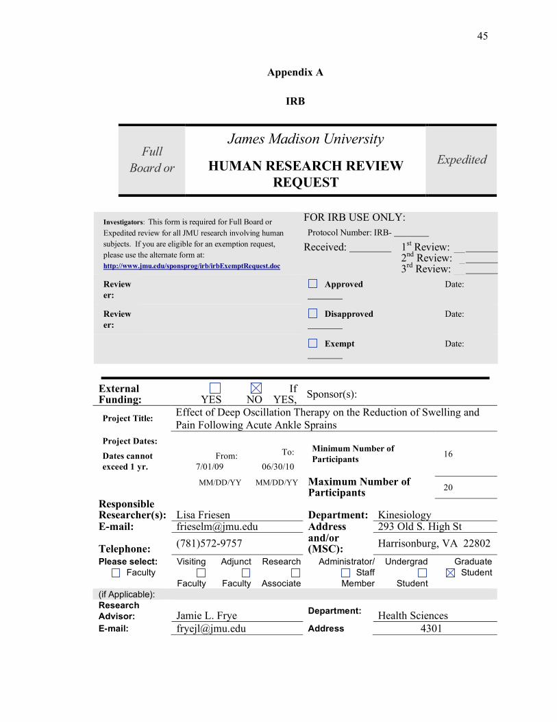
Appendix A
IRB
James Madison University Full
Board or HUMAN RESEARCH REVIEW
REQUEST
Expedited
Investigators: This form is required for Full Board or
Expedited review for all JMU research involving human
subjects. If you are eligible for an exemption request,
please use the alternate form at:
http://www.jmu.edu/sponsprog/irb/irbExemptRequest.doc
FOR IRB USE ONLY:
Protocol Number: IRB-
Received: 1st Review: __
2nd
Review: _ 3
rd Review: __
Review
er:
Approved Date:
Review
er:
Disapproved Date:
Exempt Date:
External Funding:
YES
NO
If YES,
Sponsor(s):
Project Title: Effect of Deep Oscillation Therapy on the Reduction of Swelling and
Pain Following Acute Ankle Sprains
Project Dates:
Dates cannot
exceed 1 yr.
From:
7/01/09
To:
06/30/10
Minimum Number of
Participants 16
MM/DD/YY MM/DD/YY Maximum Number of Participants
20
Responsible Researcher(s): Lisa Friesen Department: Kinesiology E-mail: [email protected] Address 293 Old S. High St
Telephone: (781)572-9757
and/or (MSC):
Harrisonburg, VA 22802
Please select: Visiting Adjunct Research Administrator/ Undergrad Graduate
Faculty
Faculty
Faculty
Associate
Staff
Member
Student
Student
(if Applicable):
Research
Advisor: Jamie L. Frye Department:
Health Sciences
E-mail: [email protected] Address 4301

46
Telephone: (419) 902-3760 (cell) 8-
8836 (office) and/or (MSC):
Investigator: Please respond to the questions below. The IRB will utilize your responses to evaluate
your protocol submission.
1. YES NO Does the James Madison University Institutional Review Board define the project
as research?
The James Madison University IRB defines "research" as a "systematic investigation designed to develop or contribute to
generalizable knowledge.”
All research involving human participants conducted by James Madison University faculty, staff, and students is subject to IRB
review.
Some, but not all, studies that involve human participants are considered research and are subject to full or expedited IRB
review, including those:
• intended to satisfy the academic requirements for Independent Study, Bachelor’s Essay, Honors/Senior Thesis,
or the Master’s Thesis;
• intended or expected to result in publication, presentation outside the classroom, or public dissemination in some
other form;
• conducted outside the classroom and/or departmental research participant pool if they involve
-- external funding
-- minors (i.e., persons under the age of 18),
-- a targeted population of adults whose ability to freely give informed consent may be compromised (i.e., persons who are
socio-economically, educationally, or linguistically disadvantaged, cognitively impaired, elderly, terminally ill, or
incarcerated),
-- pregnant women and/or fetuses who may be put at risk of physical harm,
-- a topic of a sensitive or personal nature, the examination or reporting of which may place the research
participant at more than minimal risk, or
-- any type of activity that places research participants at more than minimal risk.
Other studies are eligible to request exemption from IRB review, including those
• conducted solely within the confines of the classroom or within a departmental research participant pool if they
-- are a general requirement of a course,
-- have the sole purpose of developing the student's research skills, and
-- will be overseen by a faculty member;
• conducted outside the classroom and outside departmental research participant pools, provided they do not
involve minors, do not target special adult populations, do not pose a risk of physical harm to pregnant
women and fetuses, do not deal with a topic of sensitive or personal nature, or do not involve any type of activity that places the participants at more than minimal risk (see details above); and provided the investigator does not intend to publish the results or share them with others in a public forum (i.e. conference
presentations, senior theses). • that are part of a larger research project that has current James Madison University IRB approval; or
• that are part of a larger research project that has current approval of a registered IRB at another institution,
provided that, if research participants are to be recruited at James Madison University, the University’s IRB
has given permission for such on-campus recruitment.
2. YES NO Are the human participants in your study living individuals?
3. YES NO Will you obtain data through intervention or interaction with these individuals?
“Intervention” includes both physical procedures by which data are gathered (e.g., measurement of heart rate or venipuncture) and
manipulations of the participant or the participant's environment that are performed for research purposes. “Interaction” includes
communication or interpersonal contact between the investigator and participant (e.g., surveying or interviewing).
4. YES NO Will you obtain identifiable private information about these individuals?
"Private information" includes information about behavior that occurs in a context in which an individual can reasonably expect

47
that no observation or recording is taking place, or information provided for specific purposes which the individual can reasonably
expect will not be made public (e.g., a medical record or student record). "Identifiable" means that the identity of the participant
may be ascertained by the investigator or associated with the information (e.g., by name, code number, pattern of answers, etc.).
5. YES NO Does the study present more than minimal risk to the participants?
"Minimal risk" means that the risks of harm or discomfort anticipated in the proposed research are not greater, considering
probability and magnitude, than those ordinarily encountered in daily life or during performance of routine physical or
psychological examinations or tests. Note that the concept of risk goes beyond physical risk and includes psychological,
emotional, or behavioral risk as well as risks to employability, economic well being, social standing, and risks of civil and
criminal liability.
CERTIFICATIONS:
For James Madison University to obtain a Federal Wide Assurance (FWA) with the Office of Human Research Protection
(OHRP), U.S. Department of Health & Human Services, all research staff working with human participants must sign this
form and receive training in ethical guidelines and regulations. "Research staff" is defined as persons who have direct and
substantive involvement in proposing, performing, reviewing, or reporting research and includes students fulfilling these roles
as well as their faculty advisors. The Office of Sponsored Programs maintains a roster of all researchers who have
completed training within the past three years.
By signing below, the Responsible Researcher(s), and the Faculty Advisor (if applicable), certifies that he/she is familiar with the
ethical guidelines and regulations regarding the protection of human research participants from research risks. In addition, he/she
agrees to abide by all sponsor and university policies and procedures in conducting the research. He/she further certifies that he/she
has completed training regarding human participant research ethics within the last three years.
Test module at OSP website http://www.jmu.edu/sponsprog/irb/irbtraining.html
Name of Researcher(s) Signature of Researcher(s)
and Faculty Advisor (if applicable) Date
Training
Complete
d
Lisa Friesen 6/24/09
Grace Weniger 6/24/09
Dr. Christopher Womack 6/24/09
Dr. Michael Saunders 6/24/09
Signature of Faculty Advisor also
required (if Student protocol) Jamie Frye 6/24/09
For additional training interests visit the National Institutes of Health Web Tutorial at:
http://cme.nci.nih.gov/
To Submit a Complete protocol, this document should include the following:
• Human Research Review Request form (i.e. the questions above)
• IRB Checklist (included on this form)
• Research Narrative (use the categories indicated below. 10 pages maximum, do not include your literature
review)
• Additional relevant research materials (i.e. letter of consent, questionnaire, survey, where used)
PLEASE SUBMIT AN ELECTRONIC VERSION OF YOUR ENTIRE PROTOCOL TO [email protected]
PLEASE PROVIDE A SIGNED HARD COPY OF THE RESEARCH REVIEW REQUEST FORM TO:
OFFICE OF SPONSORED PROGRAMS, MSC 5728, JAMES MADISON ADMINISTRATIVE COMPLEX, BLDG #6,
SUITE 26
48
Research Proposal Checklist
for Submission to the Institutional Review Board on the Use of Human Subjects in Research
Title of Study: Effect of Deep Oscillation Therapy on the Reduction of Swelling and Pain Following Acute Ankle
Sprains Name of Investigator(s): Lisa Friesen Phone
: (781)572-9757
Campus
Address: Godwin 128 MSC: 2301
Email Address: [email protected]
Research Advisor (if applicable): Dr. Jamie Frye Phon
e:
419-902-3760 (cell)or 8-
8836
Email Address: [email protected] MSC: 4301
(Investigator - Please Organize Material on the following page using the Topics Below)
PURPOSE OR OBJECTIVE(S)
Limited to one page
PROCEDURES (Included are:)
Research design and sampling
Method of collecting data (emphasize possible risks, and protection of subjects)
Time frame of study
DATA ANALYSIS
Discussed how confidentiality of subjects and their responses will be maintained
Discussed how data will be stored to ensure confidentiality of subjects
REPORTING PROCEDURES
Identified audience to be reached in the report of the study
Identified the presentation method(s) to be used
Discussed how feedback will be provided to subjects
EXPERIENCE OF THE RESEARCHER
Prior relevant experience of the researcher, supervisor, and/or consultants
ADDITIONAL ATTACHMENTS (if applicable:)
Consent forms
Letters of permission
Cover letter(s)

49
Questionnaire
Tests
Additional attachments relevant to the study
NOTIFY OSP OF INTENT TO SUBMIT FOR EXTERNAL FUNDING
Project will be submitted for External Funding
If yes, submit proposal to Sponsored Programs: MSC 5728
Funding
Agency
Program
*SUBMIT PROPOSAL AND CHECKLIST ELECTRONICALLY TO: [email protected]
TRAINING, TESTING AND FORM COMPLETION REQUIREMENTS
Completed IRB training on (5/24/2009) at http://www.jmu.edu/sponsprog/irb.html
50
Purpose and Objectives:
The purpose of this study is to assess the effectiveness of deep oscillation therapy provided by the
Physiomed Hivamat System® (Hivamat®) on decreasing acute swelling and pain following ankle sprain
injuries.
Deep oscillation therapy is a therapeutic modality that uses a magnetic force to create a minimal
electrostatic field. It was developed in Germany and was patented in 1987 and since has been used by
numerous clinicians, including clinicians in the Athletic Training Facility at James Madison University.
When applied to human tissue the electrostatic field is designed to create deep oscillations that produce
tiny vibrations in the treated tissue. This type of therapy makes numerous claims regarding its
therapeutic benefits including the ability to reduce edema (i.e. swelling) following injury. It is also
marketed to relieve pain once edema is reduced. While this modality is marketed to decrease edema and
pain and has gotten promising testimonial results, there has been no orthopedic research done on this
modality.
Deep oscillation therapy has been used in the Athletic Training Facility at James Madison University for
the past year with no adverse events on their patients. Their own anecdotal benefits of the therapy have
supported the use of the device enough to warrant purchasing a second Hivamat® Device for the
department.
Procedures/Research Design/Methodology/Timeframe:
The data in this study will be collected over the next two years.
Volunteers will be recruited from James Madison University athletics, university recreational center
athletes, and the student population. Individuals entered into the study will have sustained an ankle
sprain resulting in moderate swelling and pain. The advantage to all participants enrolled in the study is
to receive free treatment for their ankle sprain for 5 days.
Twenty volunteers will be recruited and will be randomly assigned to one of two groups. Participants in
both groups will receive standard therapeutic care for ankle sprains (rest, cold therapy, compression,
elevation, and light exercises). All groups will receive treatment 2 times per day for 5 days.
The independent variables will be group (control vs. treatment) and time (days 1-5). The dependent
variables will be swelling and pain. A separate 2 x 5 repeated measures ANOVA will used to analyze the
data on swelling and pain.
Volunteers will be asked if any of the following contraindications can be applied to their current physical
state: acute infection, active tuberculosis, infectious skin disease, untreated malignant disease, untreated
thromboses or vascular disorders, erysipelas (a skin infection), heart complaints, heart disease,
pregnancy, cardiac pacemakers, implanted stimulators, or sensitivities to electrical fields. These
conditions are considered to be contraindications for deep oscillation therapy by the manufacturer of the
Physiomed Hivamat System®. If the volunteer has any of the above contraindications they will not be
allowed to participate in the study.

51
Volunteers will be informed about the study and will be given an opportunity to ask
questions. Once all questions have been answered to the volunteer’s satisfaction,
they will sign an informed consent document. Baseline measurements including
height, weight, bilateral figure-8 girth measurement (taken with a measuring tape),
and pain level (taken by a numeric pain scale) will be taken before they begin the
study.
Control: Group 1
Ten volunteers will randomly be placed into the control group. This group will receive traditional
treatment for a sprained ankle. This will include 2 treatments per day of 20 minutes of cold/compression,
followed by light range of motion exercises, and having a compression wrap applied to their ankle.
Additionally, they will receive a sham deep oscillation therapy. During this treatment, no therapeutic
energy will be emitted into their tissues. This group will be used to evaluate the effectiveness of
traditional ankle therapy plus a sham treatment to decrease pain and swelling when compared to the
treatment group.
Experimental Treatment: Group 2
Ten volunteers will receive traditional treatment (20 minutes cold/compression and compression wrap
application) plus deep oscillation treatment. The deep oscillation treatment will be 15 minutes for the
first 2 days following injury and 25 minutes for days 3-5. The deep oscillation treatment will be
followed by light range of motion exercises and having a compression wrap applied to their ankle.
Treatment: Group 1 and 2
All volunteers will have their ankle girth taken to measure their ankle size. A measuring tape will be
placed around their ankle in a figure 8 pattern. Three measurements will be taken and the three
measurements will be averaged. This will serve as a reference for the amount of ankle swelling in their
injured ankle. This measurement will be repeated on their unaffected ankle so we can individually
compare the amount of swelling found in the volunteer’s affected and unaffected ankles.
The volunteer’s pain level will be assessed using the Numeric Pain Scale. The patient will be asked to
move their ankle and stand on it as they are able. They will then be asked to assess their pain on a scale
of 1-10. Ten will be the worst pain they could imagine; one will be little to no pain. This measurement
will serve as a reference of the volunteer’s pain prior to treatment.
All volunteers in each group will receive traditional treatment for a sprained ankle. The subject will lie
on their back on a treatment table and will have their affected ankle elevated with an elevation foam pad.
A lower leg compression sleeve will be placed on their lower limb. First, they will receive a 20 minute
cold/compression treatment with a Game Ready® cold/compression unit. In this therapy, a compression
sleeve is placed over the ankle. When turned on, the sleeve fills with cold water (50-55 degrees) thus
applying both cold and compressive therapy designed to decrease swelling. The parameters on the unit
will be set to place moderate compression (beginning at 5 mmHg and increasing to 50 mm Hg) on their
limb for 20 minutes. The pressure will first be set so that the volunteer feels comfortable compression
and will be adjusted as necessary to maintain patient comfort. When the treatment begins, the sleeve will
provide intermittent compression by inflating and deflating to encourage swelling to be pressed out of the
swollen ankle.

52
Volunteers in Group 1 will now receive their sham deep oscillation treatment. The machine will be
turned on so that volunteers believe they are getting a treatment but the treatment will not be started. A
towel will be used to cover the face of the machine anytime the Hivamat® unit is used. The researcher
will go through the motions of the lymph node progression and the treatment (described below in the
Group 2 treatment). The patient will feel the clinician’s hands on their skin and the light massage that
accompanies the normal deep oscillation treatment.
Following all volunteers therapeutic treatment (following the sham and deep oscillation therapy), the
volunteers will perform light ankle exercises to help the patients regain their ankle range of motion.
Their ankle exercises will be followed by placing a compression wrap similar to an ACE bandage on
their ankle to decrease swelling.
On day 1, the subject will be given basic exercises following their treatment to help
increase their ankle range of motion. Volunteers will be instructed to do the
following exercises within their comfort level:
! Alphabet: The subject will move the injured foot at the ankle through
the air as if he/she is writing the lower case alphabet. The subject will go
through the alphabet two times.
! Circles: The subject will move their injured foot through the air in a
circular fashion. The subject will do 15 clockwise circles and 15
counterclockwise circles.
The examiner will use their discretion to limit number of exercises based on
the subject’s pain feedback.
On day 2 of the treatment, the volunteers will receive the same modality treatments
and will continue with the exercises listed above and will also be given the following
active range of motion exercises. These exercises will continue to be performed post
conventional treatment and measurements for the rest of the volunteers’ time in the
study. Volunteers will be instructed to do the following exercises within their
comfort level:
! 4-way ankle range of motion exercises: The subject will be seated with
the knee extended and a foam roll placed under their injured ankle. The
subject will move their ankle four directions. He/she will pull the foot up
towards the body, down towards the table, in towards the midline, and
out away from the midline. The subject will perform this exercise 30
times, with a rest after each repetition of 10.
Treatment: Group 2
Following the traditional treatment described above, the treatment group (Group 2) will receive deep
oscillation therapy as recommended by the manufacturer. When applying the deep oscillation therapy,
the patient feels a mild vibration sensation similar to the vibrations felt from an inexpensive back
massager. These vibrations are supposed to help get the excess fluid in a joint (or other swollen body
part) moving back into the lymph system so that it may return to the circulatory system and be removed.

53
The treatment is not painful and is relatively pleasant for the patient. The treatment begins with therapy
over the lymph nodes to stimulate their ability to help drain the fluid from the swollen body part
(manufacturer’s claim). The lymph node treatment is followed by treatment over the injured ankle.
Lymph Node Progression Treatment:
The deep oscillation treatment will be given according to the treatment guidelines of
the owner’s manual. The therapy will begin by treating major lymph nodes within
the lymph system to increase their circulatory abilities of the system. This
progression will begin both the acute and subacute deep oscillation treatments and
takes approximately 1.5-2 minutes to complete.
The researcher will set the HIVAMAT® frequency to 150 Hz and will provide
treatment to the following lymph nodes:
1. Twelve revolutions over the patients cervical lymph nodes (found in
their neck)
2. Twelve revolutions over the subclavian trunk lymph node (found
near their collar bone)
3. Twelve revolutions over the mediastinal lymph node (found in the
center of the sternum)
4. 30 seconds of revolutions over the cysternia chyle lymph node
(found halfway between the base of the sternum and the navel)
5. Twelve revolutions over the inguinal lymph node (found where the
thigh bends at the hip)
6. The therapist’s revolutions will continue by working down into the
lower extremity. The examiner will do about 1 minute of circular
revolutions on the popliteal lymph node (the back of the knee).
All of the above revolutions will be done at a frequency of 150 Hz and in a circular
motion, with the cysternia chyle lymph node being only done in a clockwise
direction. This should all be done with the athlete lying supine, without their ankle
elevated.
Acute deep oscillation protocol (Days 1 and 2):
The deep oscillation treatment will be applied for 10 minutes using a high frequency (150 Hz) followed
by 5 minutes of a low frequency (20 Hz; total of 15 minutes) based on the manufacturer
recommendations. The volunteer will be placed prone with a small bolster under their lower leg, so the
knee is flexed but the ankle is elevated. The HIVAMAT® will be applied for 10 minutes at 150 Hz
treatment with a circular pattern moving from the toes toward the shin and back down toward the toes.
This will be followed by 5 minutes at 20 Hz moving specifically from the toes toward the shin. This

54
treatment will be performed for a total of 15 minutes.
Subacute deep oscillation Protocol (Days 3-5):
The deep oscillation therapy will be applied for 20 minutes using a high frequency (150 Hz) and 5
minutes using a low frequency (20 Hz; total of 25 minutes).
While reactions to the treatment are rare, a few possible reactions may occur. Volunteers will be advised
of these possible reactions to treatment in the informed consent document. Patients will be reminded of
these possible reactions upon completing each treatment. If the volunteer does have a reaction, they will
be instructed to contact the researchers.
The following are possible reactions to the treatment as described by the manufacturer:
! Pain sensations in the area undergoing treatment that develop approximately 3-4 hours
following treatment. These sensations may go away following urination.
! Color and smell of urination may be altered from increased lymph drainage.
! Increased sensitivity of the skin treated. This reaction is most likely redness that may last
for 1-2 days.
! Tiredness following treatment.
! Rise in local temperature (rare).
! Reduction in blood pressure.
Following Group 1 and Group 2’s treatment, their ankle girth will be taken as a measurement of their
ankle size. A measuring tape will be placed around their ankle in a figure 8 pattern. Three measurements
will be taken and the three measurements will be averaged. This measurement will serve as a reference
to see if the treatments decreased ankle swelling. Pain will again be assessed using the Numeric Rating
Scale (scale from 1-10) to determine if the volunteer’s pain level decreased as a result of the treatment.
The same therapeutic exercises described above will be done now for group 2.
Data Analysis:
Data gathered from our volunteers will never be referred to by name. Each volunteer will be randomly
assigned to a number that will be the same throughout the study. Findings will be stored in a locked
office on the campus of JMU. No names will be used in future presentations or publications of this data.
Reporting Procedures:
The findings of this study will be used as material for the thesis of Lisa M. Friesen. Following
completion, it will be submitted for publication in a relevant athletic training journal.
The volunteers in the study will be given an option to know the results at the end of the study. Once the
data is completed, the results will be e-mailed to the volunteer.
Experience of the researcher (and advisor, if student):

55
Lisa Friesen is a master’s student at JMU working to complete her thesis. This is her first research
experiment; however, she has completed basic research classes in her Kinesiology Masters Program. The
primary advisors for the conduct of this study will be by Jamie L. Frye, PhD, ATC and Grace T.
Weniger, ATC. Jamie L. Frye is professor in Health Sciences who has been involved with a number of
studies involving human volunteers while a master’s student at Indiana State University, a doctoral
student at the University of Virginia, a faculty member at the University of Toledo, and a faculty member
at James Madison University. Additionally, Dr. Frye is a certified athletic trainer. Grace T. Weniger is a
certified athletic trainer employed by athletics at James Madison University. She is experienced in the
use of deep oscillation therapy as it has been used in the sports medicine department at JMU for the past
year. During this time, there have been no documented side effects from the therapy.
Dr. Chris J. Womack and Dr. Michael J. Saunders, from the Kinesiology Department at JMU, will
primarily be providing research design input. Each has conducted numerous studies at JMU with human
volunteers.

56
Additional Attachments as applicable: Volunteer Advertisement
Research Volunteers Needed
Clinical Investigator: Lisa Friesen, ATC
Purpose:
This research project will study the effect of deep oscillation
treatment on ankle pain and swelling in new ankle sprains. Deep
oscillation treatment causes deep massage and a pumping effect at the site of an injury to remove swelling.
Eligibility:
• All participants must be between 18 and 60 years of age
• All subjects must have a new ankle sprain with noticeable swelling
You may NOT participate if:
• You currently have a broken bone in your ankle
• You have an acute infection
• active tuberculosis
• infectious skin disease
• untreated malignant disease
• untreated thromboses or vascular disorders
• erysipelas
• heart complaints
• heart disease
• pregnancy
• cardiac pacemakers
• implanted stimulators
• sensitivities to electrical fields
Benefits:
• Initial evaluation of an ankle sprain by a certified athletic trainer.
• Treatment for an ankle sprain by a certified athletic trainer.
• Free compression wrap, exercise band, and home exercise program.
Time:
• 2 visits a day for 5 days each lasting approximately 1 hour.

57
Study Location:
Godwin Athletic Training Room
If interested in participating, please contact:
Lisa Friesen [email protected] 781-572-9757

58
Consent to Participate in Research
Identification of Investigators & Purpose of Study
You are being asked to participate in a research study conducted by Lisa Friesen, a master’s student at
James Madison University. Her research will be supported by her research committee Jamie Frye, Chris
Womack, Michael Saunders, and Grace Weniger. This study will contribute to the student’s completion
of her master’s thesis.
The purpose of this study is to test treatments used to reduce pain and swelling following ankle sprains.
In addition to ice and compression, a piece of equipment called the Hivamat® unit will be used to treat
your ankle sprain. The Hivamat® is a machine that will use electrostatic principles to help decrease the
swelling and pain caused by your ankle sprain. It will use the machine to help “push and pull” the
swelling from your ankle. This is a massage type of treatment in which the examiner will lightly rub
their hands over your ankle joint.
Research Procedures
Should you decide to participate in this research study, you will be asked to sign this consent form once
all your questions have been answered to your satisfaction. This study consists of receiving treatments
for your ankle sprain two times a day for five days. Treatments will be administered on the JMU campus
in the Godwin Hall Athletic Training Room. When you arrive, you will first receive a basic evaluation of
your ankle injury. We will evaluate how much pain you have when certain parts of your ankle are
touched, how much you can move your ankle, and how much strength you have in your ankle. We will
also perform some special examination tests to see how bad you have injured your ankle and what
structures may be injured.
Once we have determined that we agree you have an ankle sprain, you will be asked to rate your pain on
a scale of one to ten based on the amount of pain you are feeling when moving your ankle and placing
weight on your ankle. On this scale, one will be no pain at all and ten will be excruciating pain. After
your answers are recorded, measurements of the swelling around your ankle will also be taken. We will
gently wrap a tape measure around your ankle joint to measure how big it is. We will also do this on
your non-injured ankle so we can compare the sizes of your ankles. Lastly, we will take your height and
weight. We will record these measurements.
Once these initial measurements have been taken, volunteers will be randomly assigned to one of two
groups. These groups will be called the control group and the treatment group.
Group A
If you are placed in Group A, you will be asked to report to the Athletic Training Room two times a day.
You will lie down on your back on a treatment table with your shoe and sock off. Your injury leg will be
elevated with a foam cushion. Your ankle will be wrapped with a sleeve that is attached to an electric
unit called the Game ReadyTM
. This unit will provide cold therapy and comfortable compression to the
ankle joint. This is applied to help reduce swelling and pain. You will feel a cold sensation and mild
pressure while you are attached to the Game Ready. The sleeve will fill and deflate to create a pumping
motion to help remove swelling. If you feel uncomfortable at any point, please tell the clinician so they
can change the settings in order to make your more comfortable. You will receive this treatment for 20
minutes.
Following the Game ReadyTM
treatment, you will receive a treatment with the Hivamat® unit. The

59
Hivamat® is a machine that will use basic electrostatic principles to help decrease the swelling and pain
caused by your ankle sprain. It will use the machine to help “push and pull” the swelling from your
ankle. This is a massage type of treatment in which the examiner will lightly rub their hands over your
ankle joint. You will lie on your stomach with your knee slightly bent. You will be given a metal bar
electrode to hold in your hand, which will help complete the electric circuit needed for the Hivamat® to
properly work. During this treatment you will feel the clinician’s hands as they move over your skin but
should not expect to feel anything else during the treatment. This treatment includes a series of circular
movements around the body.
The Hivamat® treatment will start with a treatment to the major lymph nodes of the body. This will help
to move the swelling into the lymphatic system and help it drain. This part will take approximately 1.5-2
minutes to complete. Once this is completed the following treatment will be performed:
With two fingers, they will move clockwise over the front portion of your neck,. The will do 12
revolutions.
They will move over the middle of the collar bone and with two fingers they will move clockwise 12
times.
The will continue to move down to the center of your chest bone where they will move clockwise 12
times.
They will then move halfway between the base of the chest bone and belly button (top of the
stomach) and will perform a clockwise motion for 30 seconds.
The examiner will move to the front area where your thigh bends at your hip. 12 revolutions will be
done in a clockwise motion.
The examiner will move to the back of the knee and will do about 1 minute of clockwise revolutions
at this location.
This progression will be performed every time before receiving treatment with the Hivamat®.
On Days 1 and 2, the Hivamat® will be applied for 10 minutes with a circular pattern moving from the
toes toward the shin and back down toward the toes. This will be followed by 5 minutes moving
specifically from the toes toward the shin. This treatment will be performed for a total of 15 minutes.
On Days 3-5, the Hivamat® will be applied for 20 minutes with a circular pattern moving from the toes
toward the shin and back down toward the toes. This will be followed by 5 minutes moving specifically
from the toes toward the shin. This treatment will be performed for a total of 15 minutes.
Following the Hivamat® treatment, you will once again be asked to rate your pain on a scale of one to
ten based on the amount of pain you are feeling due to your ankle sprain. After your answers are
recorded, measurements of the swelling around your ankle will be taken with the tape measure again.
We will take this measurement and compare it to your non-injured ankle to see how much swelling you
still have in your injured ankle.
You will complete your therapy by performing light ankle exercises that will help increase your ankle
movement and increase your ankle strength. These exercises should be mostly pain-free. You may feel
slight discomfort, but should report anything that is painful to the clinician so they can modify or
discontinue the exercise.
On Day One, these exercises will include:

60
! Writing the alphabet with your ankle: You will move your injured foot
at the ankle joint through the air as if you are writing the lower case
alphabet. You will go through the alphabet two times if not too painful.
! Making circles with your ankle: You will move your injured foot
through the air in a circular fashion. You will do 15 clockwise circles and
15 counterclockwise circles if not too painful.
The clinician will use their discretion to limit the number of exercises based
on how much pain you are experiencing. Therefore, it is important that you
tell the clinician if you are experiencing pain with the exercises.
On day 2 -5 of the treatment, you will continue with the exercises listed above and
will also be given the following exercises. These exercises will continue to be
performed after you have received treatment and measurements have been taken.
You will add the following exercise to the ones listed above:
! 4-way ankle movement exercises: You will be seated with your knee
straight and a foam roll placed under your injured ankle. You will move
your ankle in four directions. You will pull the foot up towards the body,
down towards the table, in towards the midline, and out away from the
midline. You will perform this exercise 30 times, with a rest after each
repetition of 10.
Once the measurements have been performed, you will have a foam pad in the shape of a horseshoe
placed around the outside of your ankle bone. The ankle will be wrapped with a compression wrap,
similar to an ACE bandage. The examiner will start the wrap at the toes and end at the mid calf. The
horseshoe pad and compression wrap will help push swelling out of the ankle. The examiner will teach
you how to place the wrap on your ankle in case you need to remove it and replace it later.
You will be instructed to wear the wrap for the remainder of the day. You may take the wrap off to take
a shower or bath or if it becomes uncomfortable. If you take it off, please put the wrap back on after
about 20 minutes. You will be given ice bags and will be instructed to ice three more times during the
day while at home. Please keep the ice on for 20 minutes. You will also be given the exercises above to
perform at home. These exercises are very basic and should not take more than 15 minutes. The purpose
of the exercises is to increase the range of motion in your ankle.
Group B
If you are assigned to Group B, you will be asked to report to the Athletic Training Room two times a
day. You will lie down on your back on a treatment table with your shoe and sock off. Your injury leg
will be elevated with a foam cushion. Your ankle will be wrapped with a sleeve that is attached to an
electric unit called the Game ReadyTM
. This unit will provide cold therapy and comfortable compression
to the ankle joint. This is applied to help reduce swelling and pain. You will feel a cold sensation and
mild pressure while you are attached to the Game ReadyTM
. The sleeve will fill and deflate to create a
pumping motion to help remove swelling. If you feel uncomfortable at any point, please tell the clinician
so they can change the settings in order to make your more comfortable. You will receive this treatment
for 20 minutes.

61
Once the Game ReadyTM
treatment is completed, you will receive the Hivamat® treatment. You will lie
on your stomach with your knee slightly bent. You will be given a metal bar electrode to hold in your
hand, which will help complete the electric circuit needed for the Hivamat® to properly work. The
treatment should not be painful. You will feel the examiner’s hands as they are rubbing over the injured
ankle. You may feel a light tapping or vibrating sensation caused by the Hivamat® treatment. This
sensation should not be painful.
The Hivamat® Treatment will start with a treatment to the major lymph nodes of the body. This will
help to move the swelling into the lymphatic system and help it drain. This part will take approximately
1.5-2 minutes to complete. After setting the machine to a frequency of 150 Hz, the following progression
will be performed with the examiner’s index and middle finger:
1. With two fingers, they will move clockwise over the front portion of your neck,. The will do 12
revolutions.
2. They will move over the middle of the collar bone and with two fingers they will move
clockwise 12 times.
3. The will continue to move down to the center of your chest bone where they will move
clockwise 12 times.
4. They will then move halfway between the base of the chest bone and belly button (top of the
stomach) and will perform a clockwise motion for 30 seconds.
5. The examiner will move to the front area where your thigh bends at your hip. 12 revolutions
will be done in a clockwise motion.
6. The examiner will move to the back of the knee and will do about 1 minute of clockwise
revolutions at this location.
This progression will be performed every time before receiving treatment with the Hivamat®.
On Days 1 and 2, the Hivamat® will be applied for 10 minutes at a high frequency (150 Hz) with a
circular pattern moving from the toes toward the shin and back down toward the toes. This will be
followed by 5 minutes at a low frequency (20 Hz) moving specifically from the toes toward the shin.
This treatment will be performed for a total of 15 minutes.
On Days 3-5, the Hivamat® will be applied for 20 minutes at a high frequency (150 Hz) with a circular
pattern moving from the toes toward the shin and back down toward the toes. This will be followed by 5
minutes at a low frequency (20 Hz) moving specifically from the toes toward the shin. This treatment
will be performed for a total of 15 minutes.
Following the Hivamat® treatment, you will once again be asked to rate your pain on a scale of one to
ten based on the amount of pain you are feeling due to your ankle sprain. After your answers are
recorded, measurements of the swelling around your ankle will be taken with the tape measure again.
We will take this measurement and compare it to your non-injured ankle to see how much swelling you
still have in your injured ankle.
You will complete your therapy by performing light ankle exercises that will help increase your ankle
movement and increase your ankle strength. These exercises should be mostly pain-free. You may feel
slight discomfort, but should report anything that is painful to the clinician so they can modify or
discontinue the exercise.
On Day One, these exercises will include:
62
! Writing the alphabet with your ankle: You will move your injured foot
at the ankle joint through the air as if you are writing the lower case
alphabet. You will go through the alphabet two times if not too painful.
! Making circles with your ankle: You will move your injured foot
through the air in a circular fashion. You will do 15 clockwise circles and
15 counterclockwise circles if not too painful.
The clinician will use their discretion to limit the number of exercises based
on how much pain you are experiencing. Therefore, it is important that you
tell the clinician if you are experiencing pain with the exercises.
On day 2 -5 of the treatment, you will continue with the exercises listed above and
will also be given the following exercises. These exercises will continue to be
performed after you have received treatment and measurements have been taken.
You will add the following exercise to the ones listed above:
! 4-way ankle movement exercises: You will be seated with your knee
straight and a foam roll placed under your injured ankle. You will move
your ankle in four directions. You will pull the foot up towards the body,
down towards the table, in towards the midline, and out away from the
midline. You will perform this exercise 30 times, with a rest after each
repetition of 10.
Once the measurements have been performed, you will have a foam pad in the shape of a horseshoe
placed around the outside of your ankle bone. The ankle will be wrapped with a compression wrap,
similar to an ACE bandage. The examiner will start the wrap at the toes and end at the mid calf. The
horseshoe pad and compression wrap will help push swelling out of the ankle. The examiner will teach
you how to place the wrap on your ankle in case you need to remove it and replace it later.
You will be instructed to wear the wrap for the remainder of the day. You may take the wrap off to take
a shower or bath or if it becomes uncomfortable. If you take it off, please put the wrap back on after
about 20 minutes. You will be given ice bags and will be instructed to ice three more times during the
day while at home. Please keep the ice on for 20 minutes. You will also be given the exercises above to
perform at home. These exercises are very basic and should not take more than 15 minutes. The purpose
of the exercises is to increase the range of motion in your ankle.
Time Required
Participation in this study will require approximately two hours a day of your time. Each treatment will
last approximately one hour and you will be asked to receive treatment two times a day for five days.
Exclusions
If you have any of the following conditions, you will be asked to not participate in the study: acute
infection, active tuberculosis, infectious skin disease, untreated malignant disease, untreated thromboses

63
or vascular disorders, erysipelas, heart complaints, heart disease, pregnancy, cardiac pacemakers,
implanted stimulators, or sensitivities to electrical fields. Please let the clinician know if any of these
contraindications apply to you.
Risks
The investigator does not perceive more than minimal risks from your involvement in this study.
Following the ice treatment, your skin may appear red. Occasionally people may have a cold reaction
where the iced area is itchy and the area becomes raised. This usually goes away within 24 hours. If
this occurs, you will be dismissed from the study because you may have a cold allergy.
Following the Hivamat® treatment, we do not expect more than minimal risks. The makers of the
Hivamat® report that you could possibly have the following reactions to the treatment:
! You could experience pain sensations in the area undergoing treatment that develop
approximately 3-4 hours following treatment. These sensations may go away following
urination. (described by manufacturer)
! You could experience an increased desire to urinate. The color and smell of urination
may be altered from normal due to the removal of the swelling in your ankle. This is not
dangerous but a product of your body trying to remove the increased fluid in your system
from the swelling.
! Your skin in the area treated could be more sensitive following treatment. This reaction will
most likely be redness that may last for 1-2 days.
! You could feel tiredness following treatment.
! You could have a rise in local skin temperature (rare).
! You could have a reduction in blood pressure.
Benefits
Potential benefits from participation in this study include treatment of your ankle sprain by a certified
athletic trainer. This will aide in the reduction of swelling and healing of your injury.
Would like to have the results of the study sent to you by e-mail when the study is completed? Yes
No
E-mail Address: _____________________________
Confidentiality
Your name and identity will not be disclosed at any point during or after this study. You will be
identified by a number to keep records separate and to maintain your privacy.
The results of this research will be presented at James Madison University to Lisa Friesen’s Thesis
committee. The results of this project will be coded in such a way that the respondent’s identity will not
be attached to the final form of this study. The researcher retains the right to use and publish non-
identifiable data. While individual responses are confidential, aggregate data
will be presented representing averages or generalizations about the responses as a whole. All data will
be stored in a secure location accessible only to the researcher.
Participation & Withdrawal
Your participation is entirely voluntary. You are free to choose not to participate. Should you choose to
participate, you can withdraw at any time without consequences of any kind.

64
Questions about the Study
If you have questions or concerns during the time of your participation in this study, or after its
completion or you would like to receive a copy of the final aggregate results of this study, please contact:
Lisa Friesen Dr. Jamie L. Frye Dr. Christopher J. Womack
Kinesiology Health Sciences Advisor Kinesiology Advisor
James Madison University James Madison University James Madison
University
(781) 572-9757 (540) 568-8836 (540) 568-6145
[email protected] [email protected] [email protected]
Questions about Your Rights as a Research Subject
Dr. David Cockley
Chair, Institutional Review Board
James Madison University
(540) 568-2834
Giving of Consent
I have read this consent form and I understand what is being requested of me as a participant in this
study. I have been given satisfactory answers to my questions. I freely consent to participate and
understand that I can withdraw from the study at any time without consequence. The investigator has
provided me with a copy of this form. I certify that I am at least 18 years of age.
______________________________________
Name of Participant (Printed)
______________________________________ ______________
Name of Participant (Signed) Date
______________________________________ ______________
Name of Researcher (Signed) Date

65
Appendix B
Informed Consent
Consent to Participate in Research
Identification of Investigators & Purpose of Study
You are being asked to participate in a research study conducted by Lisa Friesen, a
master’s student at James Madison University. Her research will be supported by her
research committee Jamie Frye, Chris Womack, Michael Saunders, and Grace Weniger.
This study will contribute to the student’s completion of her master’s thesis.
The purpose of this study is to test treatments used to reduce pain and swelling
following ankle sprains. In addition to ice and compression, a piece of equipment
called the Hivamat® unit will be used to treat your ankle sprain. The Hivamat® is a
machine that will use electrostatic principles to help decrease the swelling and pain
caused by your ankle sprain. It will use the machine to help “push and pull” the
swelling from your ankle. This is a massage type of treatment in which the examiner
will lightly rub their hands over your ankle joint.
Research Procedures
Should you decide to participate in this research study, you will be asked to sign this
consent form once all your questions have been answered to your satisfaction. This
study consists of receiving treatments for your ankle sprain two times a day for five
days. Treatments will be administered on the JMU campus in the Godwin Hall Athletic
Training Room. When you arrive, you will first receive a basic evaluation of your
ankle injury. We will evaluate how much pain you have when certain parts of your
ankle are touched, how much you can move your ankle, and how much strength you
have in your ankle. We will also perform some special examination tests to see how
bad you have injured your ankle and what structures may be injured.
Once we have determined that we agree you have an ankle sprain, you will be asked to
rate your pain on a scale of one to ten based on the amount of pain you are feeling when
moving your ankle and placing weight on your ankle. On this scale, one will be no pain
at all and ten will be excruciating pain. After your answers are recorded, measurements
of the swelling around your ankle will also be taken. We will gently wrap a tape
measure around your ankle joint to measure how big it is. We will also do this on your
non-injured ankle so we can compare the sizes of your ankles. Lastly, we will take

66
your height and weight. We will record these measurements.
Once these initial measurements have been taken, volunteers will be randomly assigned
to one of two groups. These groups will be called the control group and the treatment
group.
Group A
If you are placed in Group A, you will be asked to report to the Athletic Training Room
two times a day. You will lie down on your back on a treatment table with your shoe
and sock off. Your injury leg will be elevated with a foam cushion. Your ankle will be
wrapped with a sleeve that is attached to an electric unit called the GameReadyTM
. This
unit will provide cold therapy and comfortable compression to the ankle joint. This is
applied to help reduce swelling and pain. You will feel a cold sensation and mild
pressure while you are attached to the Game Ready. The sleeve will fill and deflate to
create a pumping motion to help remove swelling. If you feel uncomfortable at any
point, please tell the clinician so they can change the settings in order to make your
more comfortable. You will receive this treatment for 20 minutes.
Following the GameReadyTM
treatment, you will receive a treatment with the
Hivamat® unit. The Hivamat® is a machine that will use basic electrostatic principles
to help decrease the swelling and pain caused by your ankle sprain. It will use the
machine to help “push and pull” the swelling from your ankle. This is a massage type
of treatment in which the examiner will lightly rub their hands over your ankle joint.
You will lie on your stomach with your knee slightly bent. You will be given a metal
bar electrode to hold in your hand, which will help complete the electric circuit needed
for the Hivamat® to properly work. During this treatment you will feel the clinician’s
hands as they move over your skin but should not expect to feel anything else during
the treatment. This treatment includes a series of circular movements around the body.
The Hivamat® treatment will start with a treatment to the major lymph nodes of the
body. This will help to move the swelling into the lymphatic system and help it drain.
This part will take approximately 1.5-2 minutes to complete. Once this is completed
the following treatment will be performed:
1) With two fingers, they will move clockwise over the lower front portion of
your neck. They will do 12 revolutions.
2) They will move about 4 inches down and with two fingers to the middle
part of your collar bone, they will move clockwise 12 times..
3) The will continue to move down to the center of your chest bone where
they will move clockwise 12 times.
4) They will then move to the next lymph node, found halfway between the
base of the chest bone and belly button (at the top of your stomach). They
will do a clockwise motion for 30 seconds.

67
5) The examiner will move to the area where your thigh bends at your hip.
12 revolutions will be done in a clockwise motion.
6) The examiner will move to the back of the knee and will do about 1
minute of revolutions at this location.
This progression will be performed every time before receiving treatment with the
Hivamat®.
On Days 1 and 2, the Hivamat® will be applied for 10 minutes with a circular pattern
moving from the toes toward the shin and back down toward the toes. This will be
followed by 5 minutes moving specifically from the toes toward the shin. This
treatment will be performed for a total of 15 minutes.
On Days 3-5, the Hivamat® will be applied for 20 minutes with a circular pattern
moving from the toes toward the shin and back down toward the toes. This will be
followed by 5 minutes moving specifically from the toes toward the shin. This
treatment will be performed for a total of 15 minutes.
Following the Hivamat® treatment, you will once again be asked to rate your pain on a
scale of one to ten based on the amount of pain you are feeling due to your ankle sprain.
After your answers are recorded, measurements of the swelling around your ankle will
be taken with the tape measure again. We will take this measurement and compare it to
your non-injured ankle to see how much swelling you still have in your injured ankle.
You will complete your therapy by performing light ankle exercises that will help
increase your ankle movement and increase your ankle strength. These exercises
should be mostly pain-free. You may feel slight discomfort, but should report anything
that is painful to the clinician so they can modify or discontinue the exercise.
On Day One, these exercises will include:
! Writing the alphabet with your ankle: You will move your injured foot at the
ankle joint through the air as if you are writing the lower case alphabet. You
will go through the alphabet two times if not too painful.
! Making circles with your ankle: You will move your injured foot through the
air in a circular fashion. You will do 15 clockwise circles and 15
counterclockwise circles if not too painful.
The clinician will use their discretion to limit the number of exercises based on
how much pain you are experiencing. Therefore, it is important that you tell the
clinician if you are experiencing pain with the exercises.

68
On day 2 -5 of the treatment, you will continue with the exercises listed above and will
also be given the following exercises. These exercises will continue to be performed after
you have received treatment and measurements have been taken. You will add the
following exercise to the ones listed above:
! 4-way ankle movement exercises: You will be seated with your knee straight
and a foam roll placed under your injured ankle. You will move your ankle in
four directions. You will pull the foot up towards the body, down towards the
table, in towards the midline, and out away from the midline. You will
perform this exercise 30 times, with a rest after each repetition of 10.
Once the measurements have been performed, you will have a foam pad in the shape of
a horseshoe placed around the outside of your ankle bone. The ankle will be wrapped
with a compression wrap, similar to an ACE bandage. The examiner will start the wrap
at the toes and end at the mid calf. The horseshoe pad and compression wrap will help
push swelling out of the ankle. The examiner will teach you how to place the wrap on
your ankle in case you need to remove it and replace it later.
You will be instructed to wear the wrap for the remainder of the day. You may take
the wrap off to take a shower or bath or if it becomes uncomfortable. If you take it off,
please put the wrap back on after about 20 minutes. You will be given ice bags and
will be instructed to ice three more times during the day while at home. Please keep the
ice on for 20 minutes. You will also be given the exercises above to perform at home.
These exercises are very basic and should not take more than 15 minutes. The purpose
of the exercises is to increase the range of motion in your ankle.
Group B
If you are assigned to Group B, you will be asked to report to the Athletic Training
Room two times a day. You will lie down on your back on a treatment table with your
shoe and sock off. Your injury leg will be elevated with a foam cushion. Your ankle
will be wrapped with a sleeve that is attached to an electric unit called the
GameReadyTM
. This unit will provide cold therapy and comfortable compression to the
ankle joint. This is applied to help reduce swelling and pain. You will feel a cold
sensation and mild pressure while you are attached to the GameReadyTM
. The sleeve
will fill and deflate to create a pumping motion to help remove swelling. If you feel
uncomfortable at any point, please tell the clinician so they can change the settings in
order to make your more comfortable. You will receive this treatment for 20 minutes.
Once the GameReadyTM
treatment is completed, you will receive the Hivamat®
treatment. You will lie on your stomach with your knee slightly bent. You will be
given a metal bar electrode to hold in your hand, which will help complete the electric
circuit needed for the Hivamat® to properly work. The treatment should not be painful.
You will feel the examiner’s hands as they are rubbing over the injured ankle. You
may feel a light tapping or vibrating sensation caused by the Hivamat® treatment. This

69
sensation should not be painful.
The Hivamat® Treatment will start with a treatment to the major lymph nodes of the
body. This will help to move the swelling into the lymphatic system and help it drain.
This part will take approximately 1.5-2 minutes to complete. After setting the machine
to a frequency of 150 Hz, the following progression will be performed with the
examiner’s index and middle finger:
With two fingers, they will move clockwise over the mid front portion of your
neck,. The will do 12 revolutions.
They will move about 4 inches down and with two fingers over the middle of the
collar bone, they will move clockwise 12 times.
The will continue to move down to the center of your chest bone where they will
move clockwise 12 times.
They will then move to the next lymph node, found halfway between the base of the
chest bone and belly button (top of the stomach). They will do a clockwise
motion for 30 seconds.
The examiner will move to the inguinal lymph node, which is located where your
thigh bends at your hip. 12 revolutions will be done in a clockwise motion.
The examiner will move to the back of the knee and will do about 1 minute of
revolutions at this location.
This progression will be performed every time before receiving treatment with the
Hivamat®.
On Days 1 and 2, the Hivamat® will be applied for 10 minutes at a high frequency (150
Hz) with a circular pattern moving from the toes toward the shin and back down toward
the toes. This will be followed by 5 minutes at a low frequency (20 Hz) moving
specifically from the toes toward the shin. This treatment will be performed for a total
of 15 minutes.
On Days 3-5, the Hivamat® will be applied for 20 minutes at a high frequency (150
Hz) with a circular pattern moving from the toes toward the shin and back down toward
the toes. This will be followed by 5 minutes at a low frequency (20 Hz) moving
specifically from the toes toward the shin. This treatment will be performed for a total
of 15 minutes.
Following the Hivamat® treatment, you will once again be asked to rate your pain on a
scale of one to ten based on the amount of pain you are feeling due to your ankle sprain.
After your answers are recorded, measurements of the swelling around your ankle will
be taken with the tape measure again. We will take this measurement and compare it to
your non-injured ankle to see how much swelling you still have in your injured ankle.
You will complete your therapy by performing light ankle exercises that will help

70
increase your ankle movement and increase your ankle strength. These exercises
should be mostly pain-free. You may feel slight discomfort, but should report anything
that is painful to the clinician so they can modify or discontinue the exercise.
On Day One, these exercises will include:
! Writing the alphabet with your ankle: You will move your injured foot at the
ankle joint through the air as if you are writing the lower case alphabet. You
will go through the alphabet two times if not too painful.
! Making circles with your ankle: You will move your injured foot through the
air in a circular fashion. You will do 15 clockwise circles and 15
counterclockwise circles if not too painful.
The clinician will use their discretion to limit the number of exercises based on
how much pain you are experiencing. Therefore, it is important that you tell the
clinician if you are experiencing pain with the exercises.
On day 2 -5 of the treatment, you will continue with the exercises listed above and will
also be given the following exercises. These exercises will continue to be performed after
you have received treatment and measurements have been taken. You will add the
following exercise to the ones listed above:
! 4-way ankle movement exercises: You will be seated with your knee straight
and a foam roll placed under your injured ankle. You will move your ankle in
four directions. You will pull the foot up towards the body, down towards the
table, in towards the midline, and out away from the midline. You will
perform this exercise 30 times, with a rest after each repetition of 10.
Once the measurements have been performed, you will have a foam pad in the shape of
a horseshoe placed around the outside of your ankle bone. The ankle will be wrapped
with a compression wrap, similar to an ACE bandage. The examiner will start the wrap
at the toes and end at the mid calf. The horseshoe pad and compression wrap will help
push swelling out of the ankle. The examiner will teach you how to place the wrap on
your ankle in case you need to remove it and replace it later.
You will be instructed to wear the wrap for the remainder of the day. You may take the
wrap off to take a shower or bath or if it becomes uncomfortable. If you take it off,
please put the wrap back on after about 20 minutes. You will be given ice bags and
will be instructed to ice three more times during the day while at home. Please keep the
ice on for 20 minutes. You will also be given the exercises above to perform at home.
These exercises are very basic and should not take more than 15 minutes. The purpose
of the exercises is to increase the range of motion in your ankle.

71
Time Required
Participation in this study will require approximately two hours a day of your time.
Each treatment will last approximately one hour and you will be asked to receive
treatment two times a day for five days.
Exclusions
If you have any of the following conditions, you will be asked to not participate in the
study: acute infection, active tuberculosis, infectious skin disease, untreated malignant
disease, untreated thromboses or vascular disorders, erysipelas, heart complaints, heart
disease, pregnancy, cardiac pacemakers, implanted stimulators, or sensitivities to
electrical fields. Please let the clinician know if any of these contraindications apply to
you.
Risks
The investigator does not perceive more than minimal risks from your involvement in
this study. Following the ice treatment, your skin may appear red. Occasionally people
may have a cold reaction where the iced area is itchy and the area becomes raised. This
usually goes away within 24 hours. If this occurs, you will be dismissed from the study
because you may have a cold allergy.
Following the Hivamat® treatment, we do not expect more than minimal risks. The
makers of the Hivamat® report that you could possibly have the following reactions to
the treatment:
! You could experience pain sensations in the area undergoing treatment that
develop approximately 3-4 hours following treatment. These sensations may
go away following urination. (described by manufacturer)
! You could experience an increased desire to urinate. The color and smell of
urination may be altered from normal due to the removal of the swelling in
your ankle. This is not dangerous but a product of your body trying to
remove the increased fluid in your system from the swelling.
! Your skin in the area treated could be more sensitive following treatment.
This reaction will most likely be redness that may last for 1-2 days.
! You could feel tiredness following treatment.
! You could have a rise in local skin temperature (rare).
! You could have a reduction in blood pressure.
Benefits
Potential benefits from participation in this study include treatment of your ankle sprain
by a certified athletic trainer. This will aide in the reduction of swelling and healing of
your injury.
72
Confidentiality
Your name and identity will not be disclosed at any point during or after this study.
You will be identified by a number to keep records separate and to maintain your
privacy.
The results of this research will be presented at James Madison University to Lisa
Friesen’s Thesis committee. The results of this project will be coded in such a way that
the respondent’s identity will not be attached to the final form of this study. The
researcher retains the right to use and publish non-identifiable data. While individual
responses are confidential, aggregate data will be presented representing averages or
generalizations about the responses as a whole. All data will be stored in a secure
location accessible only to the researcher.
Participation & Withdrawal
Your participation is entirely voluntary. You are free to choose not to participate.
Should you choose to participate, you can withdraw at any time without consequences
of any kind.
Questions about the Study
If you have questions or concerns during the time of your participation in this study, or
after its completion or you would like to receive a copy of the final aggregate results of
this study, please contact:
Lisa Friesen Dr. Jamie L. Frye Dr. Christopher J.
Womack
Kinesiology Health Sciences Advisor Kinesiology Advisor
James Madison University James Madison University James Madison
University
(781) 572-9757 (540) 568-8836 (540) 568-6145
[email protected] [email protected] [email protected]
Questions about Your Rights as a Research Subject
Dr. David Cockley
Chair, Institutional Review Board
James Madison University
(540) 568-2834

73
Giving of Consent
I have read this consent form and I understand what is being requested of me as a
participant in this study. I have been given satisfactory answers to my questions. I
freely consent to participate and understand that I can withdraw from the study at any
time without consequence. The investigator has provided me with a copy of this form.
I certify that I am at least 18 years of age.
______________________________________
Name of Participant (Printed)
______________________________________ ______________
Name of Participant (Signed) Date
______________________________________ ______________
Name of Researcher (Signed) Date

74
Appendix C
Data Collection Sheets
Initial Evaluation
Screen for the following contraindications: acute infection, active tuberculosis,
infectious skin disease, untreated malignant disease, untreated thromboses or vascular
disorders, erysipelas, heart complaints, heart disease, pregnancy, cardiac pacemakers,
implanted stimulators, allergy to ice, or sensitivities to electrical fields. Any
contraindications are cause for removal from study.
Date: _____________________
Treatment Group: A/B
Researcher: __________________________________________________
Subject Identification Number: ____________________________ M/F
Ht (cm): _______ Wt (kg): ______ Pain Scale: 0 1 2 3 4 5 6 7 8 9 10
Girth Measurement:
Pre-treatment
Right Injured/Uninjured
1)______+2)______+3)_____=_______/3=__________ (cm)
Left Injured/Uninjured
1)______+2)______+3)_______=_______/3=__________ (cm)
History:
Mechanism:
____________________________________________________________
______________________________________________________________________
______________________________________________________________________
____
Observation:
____________________________________________________________
Previous Treatment to current injury:
______________________________________
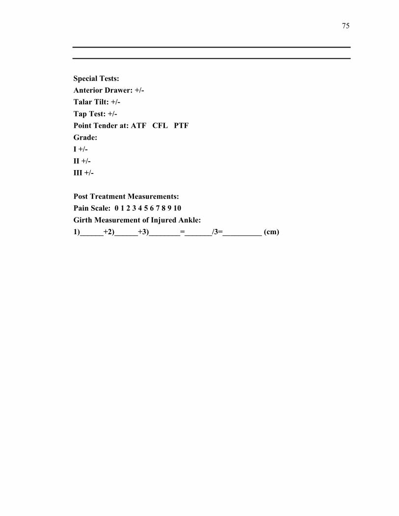
75
Special Tests:
Anterior Drawer: +/-
Talar Tilt: +/-
Tap Test: +/-
Point Tender at: ATF CFL PTF
Grade:
I +/-
II +/-
III +/-
Post Treatment Measurements:
Pain Scale: 0 1 2 3 4 5 6 7 8 9 10
Girth Measurement of Injured Ankle:
1)______+2)______+3)________=_______/3=__________ (cm)

76
Researcher Log
*Make sure pain is assessed before the treatment is given*
Name of Researcher: ___________________________________________
Subject Identification Number:_________________________________
Date: __________ Time: __________
Treatment: A B
Day of Treatment: 1 2 3 4 5
Subject Notes: (Please take note of any changes from the day before, reported
increased pain, activities that increased pain, amount of time off feet)
Observation: (ecchymosis) Y/ N? If yes, where ?_____________________
______________________________________________________________________
______________________________________________________________________
______________________________________________________________________
______________________________________________________________________
________
Pre treatment:
Pain Scale 0 1 2 3 4 5 6 7 8 9 10
Girth Measurement: 1)________ 2)_________ 3)________
Post Treatment:
Pain Scale 01 2 3 4 5 6 7 8 9 10
Girth Measurement: 1)________ 2)_________ 3)________
Name of Researcher: ___________________________________________
Subject Identification Number:_________________________________
Date: __________ Time: __________
Treatment: A B
Day of Treatment: 1 2 3 4 5
Subject Notes: (Please take note of any changes from the day before, reported
increased pain, activities that increased pain, amount of time off feet)
Observation: (ecchymosis) Y/ N? If yes, where ?_____________________
______________________________________________________________________
______________________________________________________________________
______________________________________________________________________

77
______________________________________________________________________
______________________________________________________________________
______________________________________________________________________
____________
Pre treatment:
Pain Scale 0 1 2 3 4 5 6 7 8 9 10
Girth Measurement: 1)________ 2)_________ 3)________
Post Treatment:
Pain Scale 0 1 2 3 4 5 6 7 8 9 10
Girth Measurement: 1)________ 2)_________ 3)________
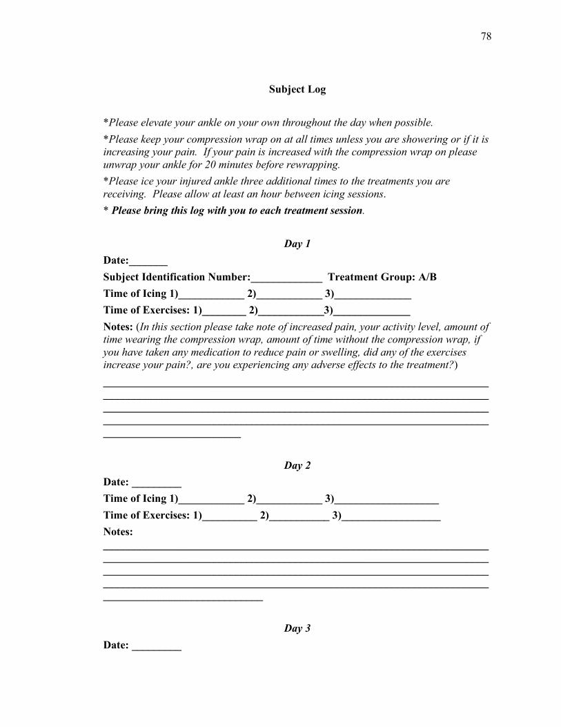
78
Subject Log
*Please elevate your ankle on your own throughout the day when possible.
*Please keep your compression wrap on at all times unless you are showering or if it is
increasing your pain. If your pain is increased with the compression wrap on please
unwrap your ankle for 20 minutes before rewrapping.
*Please ice your injured ankle three additional times to the treatments you are
receiving. Please allow at least an hour between icing sessions.
* Please bring this log with you to each treatment session.
Day 1
Date:_______
Subject Identification Number:_____________ Treatment Group: A/B
Time of Icing 1)____________ 2)____________ 3)______________
Time of Exercises: 1)________ 2)____________3)______________
Notes: (In this section please take note of increased pain, your activity level, amount of
time wearing the compression wrap, amount of time without the compression wrap, if
you have taken any medication to reduce pain or swelling, did any of the exercises
increase your pain?, are you experiencing any adverse effects to the treatment?)
______________________________________________________________________
______________________________________________________________________
______________________________________________________________________
______________________________________________________________________
_________________________
Day 2
Date: _________
Time of Icing 1)____________ 2)____________ 3)___________________
Time of Exercises: 1)__________ 2)___________ 3)__________________
Notes:
______________________________________________________________________
______________________________________________________________________
______________________________________________________________________
______________________________________________________________________
_____________________________
Day 3
Date: _________

79
Time of Icing 1)____________ 2)____________ 3)___________________
Time of Exercises: 1)__________ 2)___________ 3)__________________
Did you add resistance to the four way ankle exercises? Y/N
Notes:
______________________________________________________________________
______________________________________________________________________
______________________________________________________________________
______________________________________________________________________
_____________________________
______________________________________________________________________
_________________________________
Day 4
Date: _________
Time of Icing 1)____________ 2)____________ 3)___________________
Time of Exercises: 1)__________ 2)___________ 3)__________________
Notes:
______________________________________________________________________
______________________________________________________________________
______________________________________________________________________
______________________________________________________________________
_____________________________
Day 5
Date: _________
Time of Icing 1)____________ 2)____________ 3)___________________
Time of Exercises: 1)__________ 2)___________ 3)__________________
Notes:
______________________________________________________________________
______________________________________________________________________
______________________________________________________________________
______________________________________________________________________
_________________________
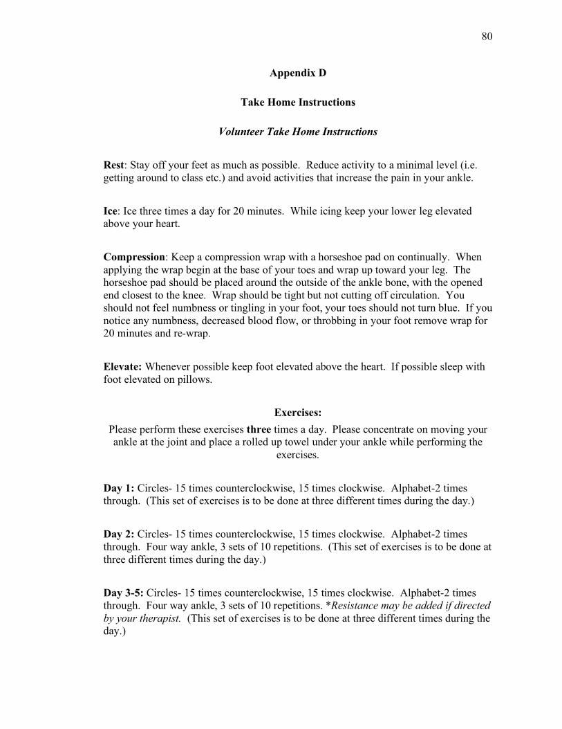
80
Appendix D
Take Home Instructions
Volunteer Take Home Instructions
Rest: Stay off your feet as much as possible. Reduce activity to a minimal level (i.e.
getting around to class etc.) and avoid activities that increase the pain in your ankle.
Ice: Ice three times a day for 20 minutes. While icing keep your lower leg elevated
above your heart.
Compression: Keep a compression wrap with a horseshoe pad on continually. When
applying the wrap begin at the base of your toes and wrap up toward your leg. The
horseshoe pad should be placed around the outside of the ankle bone, with the opened
end closest to the knee. Wrap should be tight but not cutting off circulation. You
should not feel numbness or tingling in your foot, your toes should not turn blue. If you
notice any numbness, decreased blood flow, or throbbing in your foot remove wrap for
20 minutes and re-wrap.
Elevate: Whenever possible keep foot elevated above the heart. If possible sleep with
foot elevated on pillows.
Exercises:
Please perform these exercises three times a day. Please concentrate on moving your
ankle at the joint and place a rolled up towel under your ankle while performing the
exercises.
Day 1: Circles- 15 times counterclockwise, 15 times clockwise. Alphabet-2 times
through. (This set of exercises is to be done at three different times during the day.)
Day 2: Circles- 15 times counterclockwise, 15 times clockwise. Alphabet-2 times
through. Four way ankle, 3 sets of 10 repetitions. (This set of exercises is to be done at
three different times during the day.)
Day 3-5: Circles- 15 times counterclockwise, 15 times clockwise. Alphabet-2 times
through. Four way ankle, 3 sets of 10 repetitions. *Resistance may be added if directed
by your therapist. (This set of exercises is to be done at three different times during the
day.)

81
Explanation of Exercises
Please perform all exercises seated with leg straight and towel rolled under the ankle
joint.
Circles: move ankle joint in a circular motion gently both clockwise and
counterclockwise.
Alphabet:
With ankle supported on a rolled up towel move foot as if writing the alphabet in the
air.
Four Way Ankles:
With ankle supported on a rolled up towel:
Dorsiflexion and plantarflexion- point toes toward the floor and then pull toes up
toward the body.
Inversion: pull toes in toward the midline of the body; try to move just the foot and
not the leg.
Eversion: pull toes away from the midline of the body. Again try to keep movement
at ankle joint.

82
Appendix E
Additional Methods
Swelling and Pain Measurements:
Swelling: Assessment of the subject’s edema will be taken a girth measurement. The
girth measurement involves wrapping " inch generic athletic tape around the ankle.
1) Begin by asking volunteer to sit down with leg extended on a table. The ankle should
extend off the table from mid calf.
2) Have volunteer keep ankle in a neutral position.
3) Begin measurement on the anterior tibial tendon.
4) Pass over the navicular bone and under the medial arch.
5) Come over at the base of the fifth metatarsal and onto the dorsal aspect of the foot.
6) Pull tape over the medial maleolus and around over the calcaneous.
7) Pull the tape measure over the lateral maleolus to the zero point of the tape measure.
8) This measure should be taken three times.
9) Measure tape with a generic tape measure.
10) Record measurements and average the three measurements.
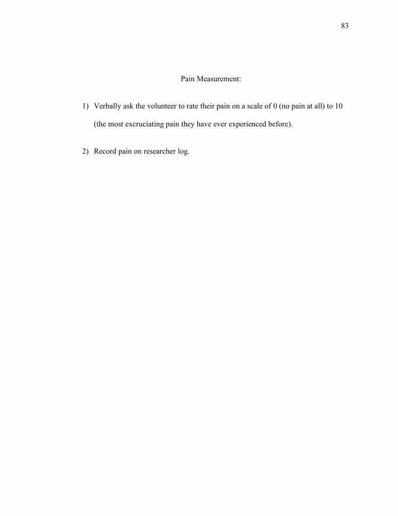
83
Pain Measurement:
1) Verbally ask the volunteer to rate their pain on a scale of 0 (no pain at all) to 10
(the most excruciating pain they have ever experienced before).
2) Record pain on researcher log.

84
GameReady! Set up:
1) Fill bladder of GameReady! with ice and then water.
2) Bring GameReady! to treatment table.
3) Plug GameReady! into power source.
4) Wrap GameReady! ankle boot comfortably around injured ankle.
5) Make sure ankle boot is secure.
6) Turn GameReady! on
7) Increase pressure to medium for 20 minutes.
8) Place foam bolster under injured ankle to keep elevated for entire 20 minute
treatment.

85
Lymph Node Progression
This progression should be done before all HIVAMAT treatments (both placebo and
real) are performed.
1) Start with athlete lying supine, without their ankle elevated.
2) Set HIVAMAT to 150 Hz, 85% dosis (intensity) for desired time (day 1 and 2 for 18
minutes, days 3-5 28 minutes).
3) Set mode at 1:1.
4) Give volunteer bar electrode and ask them to hold it in their hand.
5) Stick the circular electrode on the therapists arm. If electrode has lost some of its
adhesiveness it is recommended to secure it to arm with an elastic wrap.
6) Put on non-latex gloves and pour a small amount of baby powder in hand to help
keep surface dry.
7) With the HIVAMAT frequency set to 150 Hz and begin with 12 revolutions of their
fingers over the patient’s cervical lymph nodes. Use the index and middle finger for
the revolutions.
8) Progress to 12 revolutions over the subclavian trunk lymph node.
9) Then do 12 revolutions over the mediastinal lymph node, located in the center of the
sternum.

86
10) The sternal revolutions will be followed by 30 seconds of clockwise circular
revolutions over the cysternia chyle lymph node, which is halfway between the xyphoid
process and the navel.
11) Move to the inguinal lymph node and do 12 more revolutions.
12) Lastly, do 1 minute of circular revolutions on the popliteal lymph node.
13) All of the above revolutions will be done at a frequency of 150 Hz and in a circular
motion, with the cysternia chyle lymph node being only done in a clockwise direction.
*All lymph node revolutions should be done on skin when possible to maximize
treatment.

87
Deep Oscillation Therapy Treatment
After the lymph node progression have volunteer lay in a prone position and place a
bolster under the injured ankle, with knee in slight flexion and injured ankle elevated.
For days 1 and 2:
1) the deep oscillation will be applied for 10 minutes at 150 Hz Treatment with a
circular pattern moving distal to proximal and vice versa.
2) Apply light pressure and move hands in an effleurage pattern.
3) This will be followed by 5 minutes at 20 Hz with a specific distal to proximal
motion. The examiner will move only distal to proximal during this last 5 minutes.
4) This treatment will be performed for a total of 15 minutes.
For days 3-5:
1) Begin with same lymphatic progression as above.
2) After lymphatic progression ask volunteer to lay on stomach (prone) with a small
bolster under their lower leg, so the knee is flexed and the ankle is elevated.
3) The HIVAMAT mode setting will be 1:1 (this may be adjusted for the patients
comfort) and at 85% intensity.
4) Set time for 28 minutes.
5) The deep oscillation therapy will be applied for 20 minutes at 150 Hz with a
circular pattern moving distal to proximal and vice versa.

88
6) This will be followed by 5 minutes at 20 Hz with a specific distal to proximal
motion. The examiner will move only distal to proximal during this last 5
minutes. These parameters were chosen due to manufacturer recommendation and
will be for a total of 25 minutes.
After all deep oscillation treatments advise volunteer to drink plenty of water throughout
the day. They will be instructed to drink " of their body weight in fluid ounces. This is
encouraged by the HIVAMAT manufacturer to aid in flushing their system and removing toxins that were
introduced back into their bloodstream due to the treatment.
At the end of each treatment take three girth measurements. Average these three girth
measurements and record. Record verbal pain quotient.
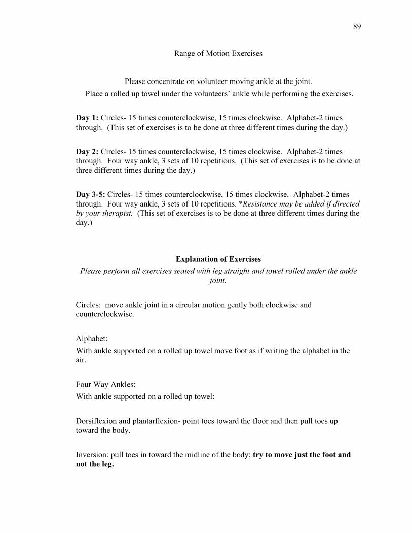
89
Range of Motion Exercises
Please concentrate on volunteer moving ankle at the joint.
Place a rolled up towel under the volunteers’ ankle while performing the exercises.
Day 1: Circles- 15 times counterclockwise, 15 times clockwise. Alphabet-2 times
through. (This set of exercises is to be done at three different times during the day.)
Day 2: Circles- 15 times counterclockwise, 15 times clockwise. Alphabet-2 times
through. Four way ankle, 3 sets of 10 repetitions. (This set of exercises is to be done at
three different times during the day.)
Day 3-5: Circles- 15 times counterclockwise, 15 times clockwise. Alphabet-2 times
through. Four way ankle, 3 sets of 10 repetitions. *Resistance may be added if directed
by your therapist. (This set of exercises is to be done at three different times during the
day.)
Explanation of Exercises
Please perform all exercises seated with leg straight and towel rolled under the ankle
joint.
Circles: move ankle joint in a circular motion gently both clockwise and
counterclockwise.
Alphabet:
With ankle supported on a rolled up towel move foot as if writing the alphabet in the
air.
Four Way Ankles:
With ankle supported on a rolled up towel:
Dorsiflexion and plantarflexion- point toes toward the floor and then pull toes up
toward the body.
Inversion: pull toes in toward the midline of the body; try to move just the foot and
not the leg.

90
Eversion: pull toes away from the midline of the body. Again try to keep movement
at ankle joint.
After exercises re-wrap volunteers ankle with the compression wrap and horseshoe pad.

91
References
1. Airaksinen O. Elastic bandages and intermittent pneumatic compression for
treatment of acute ankle sprains. Arch Phys Med Rehabil. 1990;71(6):380-383.
2. Osborne MD. Prevention and Treatment of Ankle Sprain in Athletes. Sports
medicine (Auckland). 2003;33(15):1145-1150.
3. Safran MR. Lateral ankle sprains: A comprehensive review part 1: Etiology,
pathoanatomy, histopathogenesis, and diagnosis. Med Sci Sports Exerc. 1999;31(7
suppl.):S429-S437.
4. Hubbard TJ. Ankle ligament healing after an acute ankle sprain: An evidence-based
approach. Journal of athletic training. 2008;43(5):523-529.
5. Garrick JG. The frequency of injury, mechanism of injury, and epidemiology of
ankle sprains. Am J Sports Med. 1977;5(6):241-242.
6. Bleakley CM. The PRICE study (Protection Rest Ice Compression Elevation):
Design of a randomised controlled trial comparing standard versus cryokinetic ice
applications in the management of acute ankle sprain [ISRCTN13903946]. BMC
musculoskeletal disorders. 2007;8:125.
7. Bleakley CM. Cryotherapy for acute ankle sprains: A randomised controlled study of
two different icing protocols. Br J Sports Med. 2006;40(8):700-705.
8. Merrick MA. Secondary injury after musculoskeletal trauma: a review and update.
Journal of athletic training. 2002;37(2):209-217.
9. Houglum PA. Soft tissue healing and its impact on rehabilitation. J Sport Rehab.
1992;1(1):19-39.

92
10. Karges JR. Concurrent validity of upper-extremity volume estimates: Comparison
of calculated volume derived from girth measurements and water displacement volume.
Phys Ther. 2003;83(2):134-145.
11. Mawdsley RH. Criterion-related validity of the figure-of-eight method of measuring
ankle edema. J Orthop Sports Phys Ther. 2000;30(3):149-153.
12. Pugia ML. Comparison of acute swelling and function in subjects with lateral ankle
injury. J Orthop Sports Phys Ther. 2001;31(7):384-388.
13. Esterson PS. Measurement of ankle joint swelling using a figure of 8. J Orthop
Sports Phys Ther. 1979;1(1):51-52.
14. Julius D. Molecular mechanisms of nociception. Nature (London).
2001;413(6852):203-210.
15. Herr KA. Pain intensity assessment in older adults: Use of experimental pain to
compare psychometric properties and usability of selected pain scales with younger
adults. Clin J Pain. 2004;20(4):207-219.
16. Williamson A. Pain: A review of three commonly used pain rating scales. J Clin
Nurs. 2005;14(7):798-804.
17. Jahr S. Effect of treatment with low-intensity and extremely low-frequency
electrostatic fields (deep oscillation®) on breast tissue and pain in patients with
secondary breast lymphoedema. J Rehabil Med. 2008;40(8):645-650.
18. Mikhalchik E., Titkova S., Anurov. M., Suprun M., Ivanova A., Trakhtman I.,
Reinhold J. Wound Healing Effects of DEEP OSCILLATION. . 2005.
19. Safran MR. Lateral ankle sprains: A comprehensive review part 2: Treatment and
93
rehabilitation with an emphasis on the athlete. Med Sci Sports Exerc. 1999;31(7
suppl.):S438-S447.
20. Brand RL. Ligamentous injuries to the lateral aspect of the ankle: the ankle sprain. J
Med Assoc Ga. 1992;81(6):293-295.
21. Lephart SM. The Role of Proprioception in the Management and Rehabilitation of
Athletic Injuries. Am J Sports Med. 1997;25(1):130-137.
22. Hertel J. Immobilisation for acute severe ankle sprain. The Lancet (British edition).
2009;373(9663):524-526.
23. Hubbard TJ. Mechanical contributions to chronic lateral ankle instability. Sports
medicine (Auckland). 2006;36(3):263-277.
24. Bergfeld JA. In ABC's of sports medicine, Malabar, Florida, Robert E. Krieger
Pub. Co., c1982. ; 1982.
25. O'Donoghue DH. Treatment of injuries to athletes. Treatment of injuries to athletes.
1962.
26. Bernstein J, ed. Musculoskeletal Medicine. American Academy of Orthopedic
Surgeons; 2003.
27. Beynnon BD. A prospective, randomized clinical investigation of the treatment of
first-time ankle sprains. Am J Sports Med. 2006;34(9):1401-1412.
28. Prentice WE, ed. Therapeutic Modalities. Fifth ed. Boston: McGraw-Hill; 2003.
29. Watson T. TISSUE REPAIR: THE CURRENT STATE OF THE ART. Sportex
medicine. 2006(28):8-12.
30. Gates C. Management of skeletal muscle injuries in military personnel. Operative

94
techniques in sports medicine. 2005;13(4):247-256.
31. Salter, R.B., Simmons, D. F., Malcom, B.W., Rumble, E.J., Macmicael, D. and
N.D. Clements. The biological effet of continuous passive motion on the healing of
full-thickness defects in articular cartilage. Journal of Bone and Joint Surgery.
1980;62A:1232-1251.
32. Hildebrand KA. The basics of soft tissue healing and general factors that influence
such healing. Sports medicine and arthroscopy review. 2005;13(3):136.
33. Woo SL. Injury and repair of ligaments and tendons. Annu Rev Biomed Eng.
2000;2(2000):83-118.
34. Prentice W, ed. Rehabilitation Techniques. Third ed. Boston: McGraw-Hill; 1999.
35. Bassett SF. Home-based physical therapy intervention with adherence-enhancing
strategies versus clinic-based management for patients with ankle sprains. Phys Ther.
2007;87(9):1132-1143.
36. Lamb SE. Mechanical supports for acute, severe ankle sprain: a pragmatic,
multicentre, randomised controlled trial. The Lancet (British edition).
2009;373(9663):575-581.
37. van Rijn RM. What Is the Clinical Course of Acute Ankle Sprains? A Systematic
Literature Review. Am J Med. 2008;121(4):324; 324-331.e6; 331.e6.
38. Bleakley C. The Use of Ice in the Treatment of Acute Soft-Tissue Injury: A
Systematic Review of Randomized Controlled Trials. Am J Sports Med.
2004;32(1):251-261.
39. Bleakley CM. The PRICE study (Protection Rest Ice Compression Elevation):
95
Design of a randomised controlled trial comparing standard versus cryokinetic ice
applications in the management of acute ankle sprain [ISRCTN13903946]. BMC
musculoskeletal disorders. 2007;8:125.
40. Knight KL. Ice for immediate care of injuries. The Physician and sportsmedicine.
1982;10(2):137.
41. Boyce SH. Management of ankle sprains: A randomised controlled trial of the
treatment of inversion injuries using an elastic support bandage or an aircast ankle
brace. Br J Sports Med. 2005;39(2):91-96.
42. SAFRAN MR. Lateral ankle sprains: a comprehensive review Part 1: etiology,
pathoanatomy, histopathogenesis, and diagnosis. Med Sci Sports Exerc.
1999;31(7):S429.
43. Rumbaut RE. Magnetic therapy for edema in inflammation: A physiological
assessment. American journal of physiology.Heart and circulatory physiology.
2008;294(1):H19-H20.
44. Fitch R. Modes of operation of an electrostatic charged particle oscillator. Vacuum.
1970;20(10):445-446.
45. Atkinson R. A simple theory of the Johnsen-Rahbek effect. Journal of physics.D,
Applied physics. 1969;2(3):325; 303-332.
46. DeVore GL. Volume measuring of the severely injured hand. The American journal
of occupational therapy. 1968;22(1):16-18.
47. Petersen EJ. Reliability of water volumetry and the figure of eight method on
subjects with ankle joint swelling. J Orthop Sports Phys Ther. 1999;29(10):609-615.

96
48. Kaulesar Sukul DMKS. Direct and indirect methods for the quantification of leg
volume: Comparison between water displacement volumetry, the disk model method
and the frustum sign model method, using the correlation coefficient and the limits of
agreement. J Biomed Eng. 1993;15(6):477-480.
49. Stranden E. A comparison between surface measurements and water displacement
volumetry for the quantification of leg edema. J Oslo City Hosp. 1981;31(12):153-155.
50. Tatro-Adams D. Reliability of the figure-of-eight method of ankle measurement. J
Orthop Sports Phys Ther. 1995;22(4):161-163.
51. Abdalla dos Reis F. Analysis of the Figure-of-Eight method and volumetry
reliability for ankle edema measurement | Análise da confiabilidade do método Figura
Oito e da volumetria para mensuração do edema de tornozelo. Revista brasileira de
medicina do esporte. 2004;10(6):468-474.
52. Petersen EJ. Reliability of water volumetry and the figure of eight method on
subjects with ankle joint swelling. J Orthop Sports Phys Ther. 1999;29(10):609-615.
53. Man IOW. Effect of neuromuscular electrical stimulation on ankle swelling in the
early period after ankle sprain. Phys Ther. 2007;87(1):53-65.
54. Bradley P. Cellular and extracellular myeloperoxidase in pyogenic inflammation.
Blood. 1982;60(3):618.
55. Toumi H. The inflammatory response: friend or enemy for muscle injury? Br J
Sports Med. 2003;37(4):284-286.
56. Hocutt JE. Cryotherapy in ankle sprains. Am J Sports Med. 1982;10(5):316-319.
57. Hubbard TJ. Does cryotherapy improve outcomes with soft tissue injury? Journal of
97
athletic training. 2004;39(3):278-279.
58. Wilson RW. Reliability and responsiveness of disablement measures following
acute ankle sprains among athletes. J Orthop Sports Phys Ther. 1998;27(5):348-355.
59. Rohner-Spengler M. Reliability and minimal detectable change for the figure-of-
eight-20 method of measurement of ankle edema. J Orthop Sports Phys Ther.
2007;37(4):199-205.
60. Man IOW. Effect of neuromuscular electrical stimulation on ankle swelling in the
early period after ankle sprain. Phys Ther. 2007;87(1):53-65.
61. Dykstra JH. Comparisons of cubed ice, crushed ice, and wetted ice on intramuscular
and surface temperature changes. Journal of athletic training. 2009;44(2):136-141.